Proc. Natl. Acad. Sci. USA Vol. 88, pp. 946-950, February 1991 Medical Sciences Free radical-derived quinone methide mediates skin tumor promotion by butylated hydroxytoluene hydroperoxide: Expanded role for electrophiles in multistage carcinogenesis (chemical carcinogenesis/phenoxyl radicals/reactive intermediates/metabolic switching/ornithine decarboxylase) KATHRYN Z. GUYTON*, PURSHOTAM BHANt, PERIANNAN KUPPUSAMYt, JAY L. ZWEIER , MICHAEL A. TRUSH*, AND THOMAS W. KENSLER*§ *Division of Toxicological Sciences, Department of Environmental Health Sciences, tDepartment of Biochemistry, and tElectron Paramagnetic Resonance Laboratories, Department of Medicine, Division of Cardiology, Johns Hopkins Medical Institutions, Baltimore, MD 21205 Communicated by Paul Talalay, October 23, 1990 (received for review September 25, 1990) ABSTRACT Free radical derivatives of peroxides, hydrop- eroxides, and anthrones are thought to mediate tumor promo- tion by these compounds. Further, the promoting activity of phorbol esters is attributed, in part, to their ability'to stimulate the cellular generation of oxygen radicals. A hydroperoxide metabolite of butylated hydroxytoluene, 2,6-di-tert-butyl4- hydroperoxyl4-methyl-2,5-cyclohexadienone (BHTOOH), has previously been shown to be a tumor promoter in mouse skin. BHTOOH is extensively metabolized by murine keratinocytes to several radical species. The primary radical generated from BHTOOH is a phenoxyl radical that can disproportionate to form butylated hydroxytoluene quinone methide, a reactive electrophile. Since electrophilic species have not been previously postulated to mediate tumor promotion, the present study was undertaken to examine the role of this electrophile in the promoting activity of BHTOOH. The biological activities of two chemicalanalogs of BHTOOH, 4-trideuteromethyl-BHTOOH and 4-terl-butyl-BHTOOH, were compared with that of the parent compound. 4-Trideuteromethyl-BHTOOH and 4-tert- butyl-BHTOOH have a reduced ability or inability, respectively, to form a quinone methide; however, like the parent compound, they both generate a phenoxyl radical when incubated with keratinocyte cytosol. The potency of BHTOOH, 4-trideutero- methyl-BHTOOH, and 4-tert-butyl-BHTOOH as inducers of ornithine decarboxyiase, a marker of tumor promotion, was commensurate with their capacity for generating butylated hydroxytoluene quinone metbide. These initial results were confirmed in a two-stage tumor promotion protocol in female SENCAR mice. Together, these data indicate that a quinone methide is mediating tumor promotion by BHTOOH, providing direct evidence that an electrophilic intermediate can elicit this stage of carcinogenesis. Butylated hydroxytoluene (BHT; 2,6-di-tert-butyl4methyl- phenol) has found wide commercial application because of its excellent antioxidant properties. Although BHT has attained generally regarded as safe (GRAS) status as afood additive, this phenolic antioxidant has toxic as well as carcinogenic properties (1, 2). For example, BHT is toxic in both liver and lung and has been reported to increase tumor formation in the progeny of rats that had high lifetime feeding of BHT (3). BHT is also a weak hepatocarcinogen in male mice (4). The most notable carcino- genic property of BHT, however, lies in its ability to act as a tumor promoter in a variety of tissues, including the liver, lung, colon, bladder, and thyroid (5). BHT is known to be extensively metabolized in its target tissues, and the toxic as well as tumor-promoting activities of BHT are thought to be mediated by metabolites of the parent compound. The role of the me- tabolism of BHT in its actions as a promoter and toxin is highlighted by the ability of several inhibitors of cytochrome P450 to suppress BHT toxicity (6, 7) and the observation that a hydroxylated metabolite is more effective than BHT as either a tumor promoter or toxin in mouse lung (8, 9). Glutathione- depleting agents enhance liver and lung damage and elevate the covalent binding of BHT in these tissues, suggesting that toxicity may be mediated through an electrophilic intermediate (10, 11). Structure-activity studies of the 4-methyl position of BHT indicate that a quinone methide intermediate may be the toxic electrophile (12). In a study of acute toxicities 2,6-di-tert-butyl-4-hydroper- oxyl-4-methyl-2,5-cyclohexadienone (BHTOOH), a hydro- peroxide metabolite of BHT, was demonstrated to be 18-fold more potent than the parent compound (13). Further, this metabolite, in contrast with the parent compound, is a tumor promoter in mouse skin (14), a tissue that apparently does not generate BHTOOH from BHT (unpublished observations). BHTOOH is of particular interest because it undergoes extensive metabolism in murine keratinocytes to form sev- eral free radical intermediates, including phenoxyl, peroxyl, alkoxyl, and alkyl radical derivatives (14). Considerable evidence suggests that free radicals and free radical-mediated processes are involved in the biochemical and biological events of tumor promotion (15, 16), and the propensity of BHTOOH to generate free radicals may, therefore, be related to its enhanced potency as a toxin and tumor promoter. The primary radical species generated from BHTOOH is the BHT phenoxyl radical; this radical metabolite will readily undergo disproportionation to regenerate the parent com- pound BHT with the concomitant production of 2,6-di-tert- butyl-4-methylene-2,5-cyclohexadienone (BHT-QM), a reac- tive electrophile (Fig. 1). Electrophiles have long been rec- ognized to play preeminent roles in the initiation of chemical carcinogenesis through the covalent modification of nucleic aci-ds (18). On the other hand, electrophilic species have not previously been linked with tumor promotion. The present study was undertaken to probe the possible role of BHT-QM, a model electrophile, in this biological process. MATERIALS AND METHODS Chemicals and Syntheses. BHT, 3,5-di-tert-butyl-hydroxy- benzoic acid, 2,4,6-tri-tert-butylphenol, LiAI[2Hh4, deuter- Abbreviations: BHT, butylated hydroxytoluene (2,6-di-tert-butyl4 methylphenol); [2 H3]BHT, 2,6-di-tert-butyl-4-[aaa- 2 H3]methyl- phenol; BHTOOH, 2,6-di-tert-butyl-4-hydroperoxyl-4-methyl-2,5- cyclohexadienone; [2H3]BHTOOH, 2,6-di-tert-butyl-44[a,aa-2H31- methyl-2,5-cyclohexadienone; t-Bu-BHTOOH, 2,4,6-tri-tert-butyl4 hydroperoxyl-2,5-cyclohexadienone; BHT-QM, 2,6,-di-tert-butyl4 methylene-2,5-cyclohexadienone; PMA, phorbol 12-myristate 13- acetate; ODC, ornithine decarboxylase; EPR, electron paramagnetic resonance. §To whom reprint requests should be addressed. 946 The publication costs of this article were defrayed in part by page charge payment. This article must therefore be hereby marked "advertisement" in accordance with 18 U.S.C. §1734 solely to indicate this fact.
Transcript
Proc. Natl. Acad. Sci. USAVol. 88, pp. 946-950, February 1991Medical Sciences
Free radical-derived quinone methide mediates skin tumorpromotion by butylated hydroxytoluene hydroperoxide: Expandedrole for electrophiles in multistage carcinogenesis
KATHRYN Z. GUYTON*, PURSHOTAM BHANt, PERIANNAN KUPPUSAMYt, JAY L. ZWEIER ,MICHAEL A. TRUSH*, AND THOMAS W. KENSLER*§*Division of Toxicological Sciences, Department of Environmental Health Sciences, tDepartment of Biochemistry, and tElectron Paramagnetic ResonanceLaboratories, Department of Medicine, Division of Cardiology, Johns Hopkins Medical Institutions, Baltimore, MD 21205
Communicated by Paul Talalay, October 23, 1990 (received for review September 25, 1990)
ABSTRACT Free radical derivatives of peroxides, hydrop-eroxides, and anthrones are thought to mediate tumor promo-tion by these compounds. Further, the promoting activity ofphorbol esters is attributed, in part, to their ability'to stimulatethe cellular generation of oxygen radicals. A hydroperoxidemetabolite of butylated hydroxytoluene, 2,6-di-tert-butyl4-hydroperoxyl4-methyl-2,5-cyclohexadienone (BHTOOH), haspreviously been shown to be a tumor promoter in mouse skin.BHTOOH is extensively metabolized by murine keratinocytes toseveral radical species. The primary radical generated fromBHTOOH is a phenoxyl radical that can disproportionate toform butylated hydroxytoluene quinone methide, a reactiveelectrophile. Since electrophilic species have not been previouslypostulated to mediate tumor promotion, the present study wasundertaken to examine the role of this electrophile in thepromoting activity ofBHTOOH. The biological activities of twochemicalanalogs of BHTOOH, 4-trideuteromethyl-BHTOOHand 4-terl-butyl-BHTOOH, were compared with that of theparent compound. 4-Trideuteromethyl-BHTOOH and 4-tert-butyl-BHTOOH have a reduced ability or inability, respectively,to form a quinone methide; however, like the parent compound,they both generate a phenoxyl radical when incubated withkeratinocyte cytosol. The potency of BHTOOH, 4-trideutero-methyl-BHTOOH, and 4-tert-butyl-BHTOOH as inducers ofornithine decarboxyiase, a marker of tumor promotion, wascommensurate with their capacity for generating butylatedhydroxytoluene quinone metbide. These initial results wereconfirmed in a two-stage tumor promotion protocol in femaleSENCAR mice. Together, these data indicate that a quinonemethide is mediating tumor promotion by BHTOOH, providingdirect evidence that an electrophilic intermediate can elicit thisstage of carcinogenesis.
Butylated hydroxytoluene (BHT; 2,6-di-tert-butyl4methyl-phenol) has found wide commercial application because of itsexcellent antioxidant properties. Although BHT has attainedgenerally regarded as safe (GRAS) status as afood additive, thisphenolic antioxidant has toxic as well as carcinogenic properties(1, 2). For example, BHT is toxic in both liver and lung and hasbeen reported to increase tumorformation in the progeny ofratsthat had high lifetime feeding ofBHT (3). BHT is also a weakhepatocarcinogen in male mice (4). The most notable carcino-genic property of BHT, however, lies in its ability to act as atumor promoter in a variety oftissues, including the liver, lung,colon, bladder, and thyroid (5). BHT is known to be extensivelymetabolized in its target tissues, and the toxic as well astumor-promoting activities ofBHT are thought to be mediatedby metabolites of the parent compound. The role of the me-
tabolism of BHT in its actions as a promoter and toxin ishighlighted by the ability of several inhibitors of cytochromeP450 to suppress BHT toxicity (6, 7) and the observation thata hydroxylated metabolite is more effective than BHT as eithera tumor promoter or toxin in mouse lung (8, 9). Glutathione-depleting agents enhance liver and lung damage and elevate thecovalent binding of BHT in these tissues, suggesting thattoxicity may be mediated through an electrophilic intermediate(10, 11). Structure-activity studies of the 4-methyl position ofBHT indicate that a quinone methide intermediate may be thetoxic electrophile (12).
In a study of acute toxicities 2,6-di-tert-butyl-4-hydroper-oxyl-4-methyl-2,5-cyclohexadienone (BHTOOH), a hydro-peroxide metabolite of BHT, was demonstrated to be 18-foldmore potent than the parent compound (13). Further, thismetabolite, in contrast with the parent compound, is a tumorpromoter in mouse skin (14), a tissue that apparently does notgenerate BHTOOH from BHT (unpublished observations).BHTOOH is of particular interest because it undergoesextensive metabolism in murine keratinocytes to form sev-eral free radical intermediates, including phenoxyl, peroxyl,alkoxyl, and alkyl radical derivatives (14). Considerableevidence suggests that free radicals and free radical-mediatedprocesses are involved in the biochemical and biologicalevents of tumor promotion (15, 16), and the propensity ofBHTOOH to generate free radicals may, therefore, be relatedto its enhanced potency as a toxin and tumor promoter.The primary radical species generated from BHTOOH is
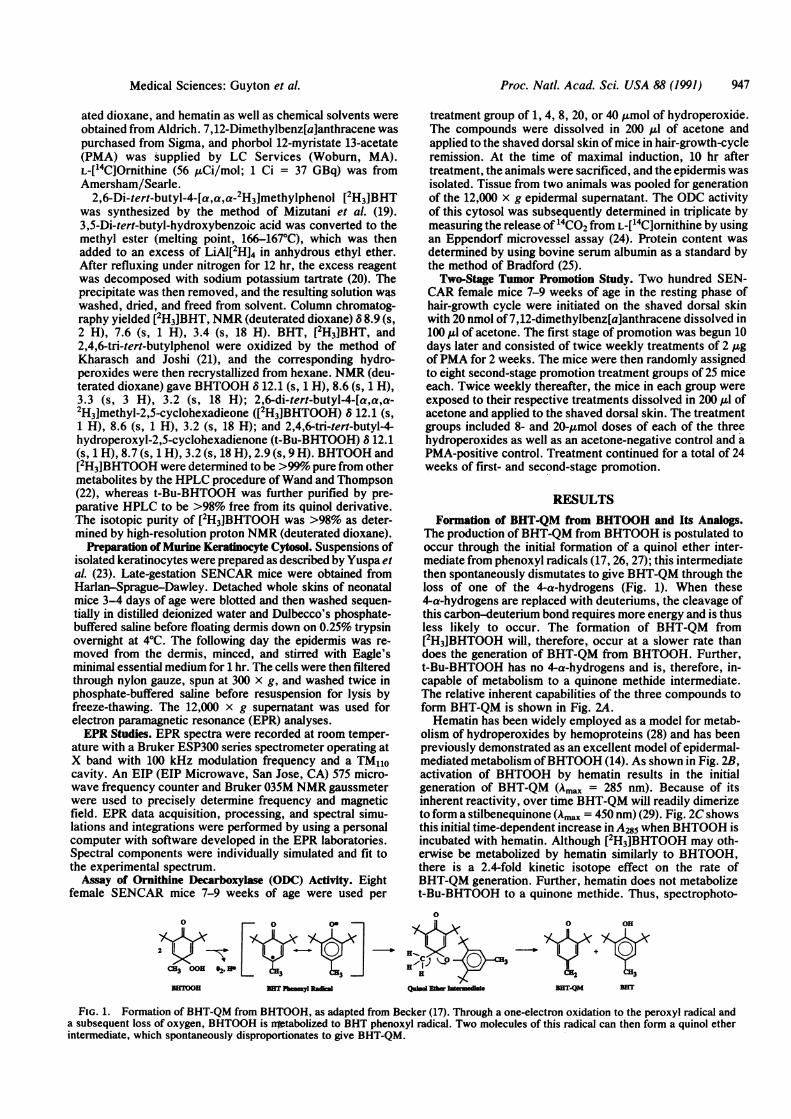
the BHT phenoxyl radical; this radical metabolite will readilyundergo disproportionation to regenerate the parent com-pound BHT with the concomitant production of 2,6-di-tert-butyl-4-methylene-2,5-cyclohexadienone (BHT-QM), a reac-tive electrophile (Fig. 1). Electrophiles have long been rec-ognized to play preeminent roles in the initiation of chemicalcarcinogenesis through the covalent modification of nucleicaci-ds (18). On the other hand, electrophilic species have notpreviously been linked with tumor promotion. The presentstudy was undertaken to probe the possible role ofBHT-QM,a model electrophile, in this biological process.
MATERIALS AND METHODSChemicals and Syntheses. BHT, 3,5-di-tert-butyl-hydroxy-
The publication costs of this article were defrayed in part by page chargepayment. This article must therefore be hereby marked "advertisement"in accordance with 18 U.S.C. §1734 solely to indicate this fact.
Proc. Natl. Acad. Sci. USA 88 (1991) 947
ated dioxane, and hematin as well as chemical solvents wereobtained from Aldrich. 7,12-Dimethylbenz[a]anthracene waspurchased from Sigma, and phorbol 12-myristate 13-acetate(PMA) was supplied by LC Services (Woburn, MA).L-[4CjOrnithine (56 uCi/mol; 1 Ci = 37 GBq) was fromAmersham/Searle.
2,6-Di-tert-butyl-4-[a,a,a-2H3]methylphenol [2H3JBHTwas synthesized by the method of Mizutani et al. (19).3,5-Di-tert-butyl-hydroxybenzoic acid was converted to themethyl ester (melting point, 166-1670C), which was thenadded to an excess of LiAl[2H14 in anhydrous ethyl ether.After refluxing under nitrogen for 12 hr, the excess reagentwas decomposed with sodium potassium tartrate (20). Theprecipitate was then removed, and the resulting solution w~swashed, dried, and freed from solvent. Column chromatog-raphy yielded [2H3]BHT, NMR (deuterated dioxane) 8 8.9 (s,2 H), 7.6 (s, 1 H), 3.4 (s, 18 H). BHT, [2H3]BHT, and2,4,6-tri-tert-butylphenol were oxidized by the method ofKharasch and Joshi (21), and the corresponding hydro-peroxides were then recrystallized from hexane. NMR (deu-terated dioxane) gave BHTOOH 6 12.1 (s, 1 H), 8.6 (s, 1 H),3.3 (s, 3 H), 3.2 (s, 18 H); 2,6-di-tert-butyl-4-[a,a,a-2H3]methyl-2,5-cyclohexadieone ([2H3]BHTOOH) 6 12.1 (s,1 H), 8.6 (s, 1 H), 3.2 (s, 18 H); and 2,4,6-tri-tert-butyl-4-hydroperoxyl-2,5-cyclohexadienone (t-Bu-BHTOOH) 812.1(s, 1 H), 8.7 (s, 1 H), 3.2 (s, 18 H), 2.9 (s, 9 H). BHTOOH and[2H3]BHTOOH were determined to be >99% pure from othermetabolites by the HPLC procedure ofWand and Thompson(22), whereas t-Bu-BHTOOH was further purified by pre-parative HPLC to be >98% free from its quinol derivative.The isotopic purity of [2H3]BHTOOH was >98% as deter-mined by high-resolution proton NMR (deuterated dioxane).
Preparation of Murine Keratinocyte Cytosol. Suspensions ofisolated keratinocytes were prepared as described by Yuspa etal. (23). Late-gestation SENCAR mice were obtained fromHarlan-Sprague-Dawley. Detached whole skins of neonatalmice 3-4 days of age were blotted and then washed -sequen-tially in distilled deionized water and Dulbecco's phosphate-buffered saline before floating dermis down on 0.25% trypsinovernight at 4°C. The following day the epidermis was re-moved from the dermis, minced, and stirred with Eagle'sminimal essential medium for 1 hr. The cells were then filteredthrough nylon gauze, spun at 300 x g, and washed twice inphosphate-buffered saline before resuspension for lysis byfreeze-thawing. The 12,000 x g supernatant was used forelectron paramagnetic resonance (EPR) analyses.EPR Studies. EPR spectra were recorded at room temper-
ature with a Bruker ESP300 series spectrometer operating atX band with 100 kHz modulation frequency and a TM110cavity. An EIP (EIP Microwave, San Jose, CA) 575 micro-wave frequency counter and Bruker 035M NMR gaussmeterwere used to precisely determine frequency and magneticfield. EPR data acquisition, processing, and spectral simu-lations and integrations were performed by using a personalcomputer with software developed in the EPR laboratories.Spectral components were individually simulated and fit tothe experimental spectrum.Assay of Ornithine Decarboxylase (ODC) Activity. Eight
female SENCAR mice 7-9 weeks of age were used per
treatment group of 1, 4, 8, 20, or 40 ,umol of hydroperoxide.The compounds were dissolved in 200 ,Al of acetone andapplied to the shaved dorsal skin ofmice in hair-growth-cycleremission. At the time of maximal induction, 10 hr aftertreatment, the animals were sacrificed, and the epidermis wasisolated. Tissue from two animals was pooled for generationof the 12,000 x g epidermal supernatant. The ODC activityof this cytosol was subsequently determined in triplicate bymeasuring the release of 14CO2 from L-[14C]ornithine by usingan Eppendorf microvessel assay (24). Protein content wasdetermined by using bovine serum albumin as a standard bythe method of Bradford (25).Two-Stage Tumor Promotion Study. Two hundred SEN-
CAR female mice 7-9 weeks of age in the resting phase ofhair-growth cycle were initiated on the shaved dorsal skinwith 20 nmol of 7,12-dimethylbenz[a]anthracene dissolved in100 Al of acetone. The first stage of promotion was begun 10days later and consisted of twice weekly treatments of 2 ,gofPMA for 2 weeks. The mice were then randomly assignedto eight second-stage promotion treatment groups of 25 miceeach. Twice weekly thereafter, the mice in each group wereexposed to their respective treatments dissolved in 200/4 ofacetone and applied to the shaved dorsal skin. The treatmentgroups included 8- and 20-Amol doses of each of the threehydroperoxides as well as an acetone-negative control and aPMA-positive control. Treatment continued for a total of 24weeks of first- and second-stage promotion.
RESULTSFormation of BHT-QM from BHTOOH and Its Analogs.
The production ofBHT-QM from BHTOOH is postulated tooccur through the initial formation of a quinol ether inter-mediate from phenoxyl radicals (17, 26, 27); this intermediatethen spontaneously dismutates to give BHT-QM through theloss of one of the 4-a-hydrogens (Fig. 1). When these4-a-hydrogens are replaced with deuteriums, the cleavage ofthis carbon-deuterium bond requires more energy and is thusless likely to occur. The formation of BHT-QM fromI2H3JBHTOOH will, therefore, occur at a slower rate thandoes the generation of BHT-QM from BHTOOH. Further,t-Bu-BHTOOH has no 4-a-hydrogens and is, therefore, in-capable of metabolism to a quinone methide intermediate.The relative inherent capabilities of the three compounds toform BHT-QM is shown in Fig. 2A.Hematin has been widely employed as a model for metab-
olism of hydroperoxides by hemoproteins (28) and has beenpreviously demonstrated as an excellent model of epidermal-mediated metabolism ofBHTOOH (14). As shown in Fig. 2B,activation of BHTOOH by hematin results in the initialgeneration of BHT-QM (Amax = 285 nm). Because of itsinherent reactivity, over time BHT-QM will readily dimerizeto form a stilbenequinone (Ama = 450 nm) (29). Fig. 2C showsthis initial time-dependent increase in A285 when BHTOOH isincubated with hematin. Although [2H3]BHTOOH may oth-erwise be metabolized by hematin similarly to BHTOOH,there is a 2.4-fold kinetic isotope effect on the rate ofBHT-QM generation. Further, hematin does not metabolizet-Bu-BHTOOH to a quinone methide. Thus, spectrophoto-
0
H ° +H
Q-d Ew _me
*tx+BUT.QM BET
FIG. 1. Formation of BHT-QM from BHTOOH, as adapted from Becker (17). Through a one-electron oxidation to the peroxyl radical anda subsequent loss of oxygen, BHTOOH is nrtabolized to BHT phenoxyl radical. Two molecules of this radical can then form a quinol etherintermediate, which spontaneously disproportionates to give BHT-QM.
0 0 0
CH3 OOE 62. El
DETOO} NET Pam~yl Rak
Medical Sciences: Guyton et al.
948 Medical Sciences: Guyton et al.
'u
z
0
250
0a
0000
500 0
WAVELENGTH (nm) TIME (seconds)
FIG. 2. (A) Relative inherent capabilities of the three hydroperoxides to form BHT-QM. (B) Spectrophotometric measurement of theformation of BHT-QM (A02) and, subsequently, the dimer stilbenequinone (A45o) over time from BHTOOH (2.5 AuM) incubated with hematin(0.75 AtM) in phosphate-buffered saline. The spectrophotometer was "blanked" against this mixture, and then successive scans were made'at1 and 10 min. (C) Actual kinetics of BHT-QM formation, as measured by A2A5, from the three hydroperoxides incubated with hematin.4-CD3-BIiTOOH, [2H3]BHTOOI.
metric determination of the relative rates of BHT-QM gen-eration from in vitro metabolism of the three hydroperoxidesconfirms their intrinsic abilities to form this intermediate.
Phenoxyl Radical Formation from BHTOOH and Its Ana-ogs by Keraginocytes. An alkaline ethanol chemical systemwas initillly used to evaluate the activation of either BHT-OOH, [zIH3]BHTOOH, or t-Bu-BHTOOH to' an EPR-detectable phenoxyl radical (Fig. 3 scans q, e, and i). Thesespectra were then simulated for characterization and deriva-tion of the coupling constants (Fig. 3 scans b, f, and j). TheEPR signal for the BHT phenoxyl radical shown in Fig. 3 scana is comprised of a quartet structure of a 1:3:3:1 signalintensity consistent with electron coupling with the three
BHTOOH 4-CD3-BHTOOH
equivalent methyl hydrogens. The splitting ofeach ofthe fourlines into a triplet of 1:2:1 intensity concurs with the inter-action of the electron with the 2 equivalent meta hydrogens.Coupling constants derived from computer simulations ofthespectrum (Fig. 3 scan b) were identical with those reported byMacomber (30) for the BHT phenoxyl radical (aiHP3 = 11.3 Gand alcta = 1.5 G). The [2H3]BHT phenoxyl radical gives riseto a 9-line EPR spectrum of a 1:5:12:24:26:24:12:5:1 signalintensity (Fig. 3 scan e), which is consistent with the com-bined interactions of the electron with the two meta hydro-gens and the three deuteriums at the 4-methyl position.Simulation of this EPR signal '(Fig. 3 scan f) gave approxi-mately equal hyperfine coupling constants for the deuterium
t-Bu-BHTOOH
alkaline ethanol
simulation
keratinocytecytosol
hoat-inactivatedcytosol
25 SMM--
d'VFIG. 3. Formation of phenoxyl radical signals from BHTOOH, [2H31BHTOOH (4-CD3-BHTOOH), and t-Bu-BHTOOH. EPR spectra were
recorded with the following instrument conditions: frequency, 9.77 GHz; microwave power, 20 mW; time constant, 0.16 s; and scan time, 60s. The modulation amplitude was 0.5 G for scans a-h and 0.25 G for scans i-l. The gain was 1 x 10⁶ for scans a, c, d, g, and h; for scan e itwas 1 x 10 and for scans i, k, and I it was 1 x 10⁴. All spectra repiesent averaged EPR signals of 10 scans except for scan g (one scan). Allincubations were-bubbled with nitrogen before aspiration into the EPR flat cell. Scans a, e, and i show the chemical generation ofphenoxyl radicalfrom 25 mg of compound dissolved in 0.4 ml of alkaline ethanol. Scans b, f, and j show the computer simulations of these signals; all spectraare centered at a g value of 2.004, and the coupling constants used were as follows: for the parent phenoxyl radical, am'et = 1.5 G, a = 11.3G; for the'deuterated analog, aWeu = aD = 1.6 G; and for the tert-butyl analog, a~ea = 1.6 G. Scans c, g, and k are the spectra generated fromincubations of keratinocyte cytosol and 20 mg of hydroperoxide in 70%' ethanol. Spectra d, h, and I are the EPR signals seen when 20 mg ofcompound is incubated in 70%o ethanol with cytosol that was heated in vacuo for 30 min.
A 0<1)
CH3 OOH
BHTOOH
CD3O3 H
OOHt-Bu-BHTOOH
60
Proc. Natl. Acad. Sci. USA 88 (1991)
R-Hor D
b
Proc. Natl. Acad. Sci. USA 88 (1991) 949
and hydrogen interactions, with aeta = aD = 1.6 G. Alkalineethanol activates t-Bu-BHTOOH to a phenoxyl radicalwhose EPR signal is shown in Fig. 3 scan i. The broad linespectrum is consistent with the couplings of the electron withthe two equivalent meta hydrogens (aHeta = 1.6 G) aspreviously reported by Valoti et al. (31).
EPR-detectable phenoxyl radicals were also producedupon incubation of the three compounds with cytosol pre-pared from keratinocytes (Fig. 3 scans c, g, and k). Theradicals generated from both BHTOOH and [2H3]BHTOOHhad hyperfine structure identical to the corresponding phe-noxyl radicals produced with alkaline ethanol. However, thespectra generated from the incubation of t-Bu-BHTOOHwith cytosol appears to consist of two separate radicalsignals; in addition to a signal identical to the chemicallygenerated phenoxyl radical, the spectrum contains a finetriplet structure identified by subtraction of the first signalfrom the cytosol-mediated spectrum. Although the identity ofthis second radical remains tentative, it is likely to be thesemiquinone radical. It is well established that 4-alkyl hy-droperoxide analogs of BHTOOH can undergo univalentreduction to produce alkoxyl radicals (32). These alkoxylradicals subsequently undergo fragmentation through P-scis-sion by which alkyl radicals and quinone intermediates aregenerated. The BHT quinone can then be readily reduced tothe semiquinone radical, whose EPR signal is consistent withthe fine triplet structure seen when t-Bu-BHTOOH is incu-bated with cytosol. Double integration of the keratinocyte-mediated spectra (corrected for differences in gain, numberof scans, modulation amplitude, and sweep width) indicateda 2-fold increase in the amount of phenoxyl radical producedfrom [2H3]BHTOOH as compared with BHTOOH, whereast-Bu-BHTOOH generated 270-fold more of this radical thandid an equivalent amount of the parent compound. Finally,no phenoxyl radicals were generated when the cytosol washeat inactivated (Fig. 3 scans d, h, and 1).ODC Activity. The induction of ODC activity has been
widely used as a marker of tumor promotion, and althoughnot sufficient, it represents a characteristic effect of tumorpromoters (33). BHTOOH has been previously demonstratedto cause a rapid, transient, and dose-dependent induction ofepidermal ODC activity (34). Fig. 4 shows the results of acomparative dose-response study of BHTOOH and its4-methyl-deuterated and 4-tert-butyl analogs. At correspond-ing doses, [2H3]BHTOOH caused a lesser induction ofODCactivity as compared to the parent hydroperoxide, whereast-Bu-BHTOOH did not increase the basal activity of thisenzyme. From this study, the 8- and 20-I.mol doses of each
o e 1.2 *-* BHTOOHa. A-A 4-CD3-BHTOOHW al 06 *-* t-Bu-BHTOOH
o'.9~:0.6
T T
z 0 0.3
0.04- ~1 10 100
DOSE (jsmoles)
FIG. 4. Dose-response for the induction of ODC activity byBHTOOH, [2H3]BHTOOH (4-CD3-BHTOOH), and t-Bu-BHT-OOH. Each point represents the mean ± SEM of triplicate deter-minations of ODC activity from four epidermal cytosolic prepara-tions, each of which consisted of tissue pooled from two separateanimals. Enzyme activity was determined as described.
FIG. 5. Time course of tumor induction by BHTOOH, [2H3]-BHTOOH (4-CD3-BHTOOH), and t-Bu-BHTOOH as expressed byincidence and multiplicity of papillomas. Mice were initiated andsubsequently treated as described. At end of the study, .92% of themice in each group was alive. Not shown is data for the controlgroups: first-stage tumor promotion followed by acetone treatmentfor 22 weeks did not induce tumor formation, whereas treatment with2 ,ug ofPMA for 24 weeks produced a 100%o incidence and an averageof 5.5 papillomas per mouse.
compound were chosen for examination in a long-term pro-motion study.Tumor-Promotion Study. The results of a 24-week, two-
stage tumor-promotion study are shown in Fig. 5. Consistentwith a previous study (34), repetitive treatments with 20 ,umolof BHTOOH caused a 64% incidence of papillomas with anaverage tumor burden of 1.9 tumors per mouse at the end ofthe study. The use of a two-stage promotion protocol,however, seemed to significantly reduce the tumor-latencyperiod as compared with the application of BHTOOH as acomplete promoter. By contrast to BHTOOH, treatmentwith an equimolar dose of [2H3]BHTOOH resulted in only a28% incidence of papillomas and a tumor burden of <1 tumorper mouse. The 8-,umol dose of the parent compound pro-duced a tumor incidence and multiplicity that was remarkablysimilar to those of a 2.5-fold higher dose of the deuteratedanalog. The low dose of [2H3]BHTOOH was inactive as atumor promoter in this study, as were both doses of t-Bu-BHTOOH. No tumors formed in mice that were initiated butreceived only first-stage promoter treatment.
DISCUSSIONInterest in the peroxide, hydroperoxide, and anthrone tumorpromoters has centered on their capacity for generating freeradicals (16, 35, 36). Much indirect evidence suggests thatradicals may play a prominent role in mediating tumorpromotion (15, 16). However, the intracellular targets ofthese species are not known, and the actual free radicalspecies formed from promoters in target tissues have rarelybeen characterized. The primary radical known to be gener-ated from BHTOOH in keratinocytes is the phenoxyl radical(14). Because this radical also serves as an intermediate in theformation of BHT-QM, the production of the phenoxylradical was investigated using chemical analogs of BHT-OOH. Integration of the keratinocyte-mediated EPR signalsindicated that, from the same amount of hydroperoxide,
Medical Sciences: Guyton et al.
950 Medical Sciences: Guyton et al.
[2H3]BHTOOH and t-Bu-BHTOOH generate =2 and 270times as much phenoxyl radical, respectively, as does BHT-OOH. These increases in phenoxyl radical concentrationscorrelate with the reduced ability, and inability, respectively,of [2H3]BHTOOH and t-Bu-BHTOOH to eliminate this rad-ical through quinone methide formation (Fig. 2A). Throughthe process of metabolic switching, these two analogs ofBHTOOH may also preferentially undergo alternative routesof metabolism, including other free radical pathways. Aprinciple alternative pathway involves reduction and 1-scis-sion of the hydroperoxide to form alkoxyl, alkyl, and semi-quinone radicals. Metabolism of t-Bu-BHTOOH by keratin-ocyte cytosol yielded another directly detectable free radicalin addition to the phenoxyl radical, which may be thesemiquinone radical generated through j-scission. Thus,[2H3JBHTOOH and t-Bu-BHTOOH may also generate higheryields of other free radical species than does the parenthydroperoxide in addition to producing increased steady-state concentrations of phenoxyl radicals.The results of the tumor promotion study indicate, how-
ever, that tumor formation by BHTOOH was not solelydependent on phenoxyl or other radical generation. Theanalog that produces the highest level of phenoxyl radical(t-Bu-BHTOOH) did not act as a tumor promoter in thisstudy. Instead, the ability of the hydroperoxides to act astumor promoters was directly related to their ability to formBHT-QM. Thus, while the production of phenoxyl radicalrepresents an obligatory step in the generation of the ultimatereactive metabolite, it is not itself sufficient for tumor pro-motion. The 2.4-fold kinetic isotope effect on BHT-QMgeneration of the deuterated analog determined in vitro withhematin was borne out in the tumor promotion study. In thisexperiment, the tumor response of the high dose of[2H3JBHTOOH was equivalent to a 2.5-fold lower dose oftheparent compound; these doses presumably generate equimo-lar amounts ofBHT-QM in the epidermis. Collectively, theseresults provide explicit evidence that reactive intermediates,in general, and electrophiles, in particular, are directly in-volved in tumor promotion. This observation broadens thepostulated roles for electrophiles in carcinogenesis fromparticipants in initiation and progression to include all stagesof carcinogenesis.Tumor promotion involves the selection and clonal expan-
sion ofinitiated but preneoplastic cells from their surroundingnormal counterparts. This effect on initiated cells may occurthrough the direct stimulation of mitogenesis or by theindirect induction of compensatory proliferation (37). Nor-mal cells have been shown to be more sensitive to thecytotoxic effects of tumor promoters than initiated cells (38).Further, by contrast with initiated cells, normal keratin-ocytes will undergo terminal differentiation and migrate outof the basal layer of the skin in response to a proliferativestimulus. By either mechanism, the removal of growth inhi-bition provided by contact with normal cells will stimulateinitiated cells to undergo compensatory proliferation andexpansion.The mechanism through which electrophiles mediate ini-
tiation and progression involves a direct genetic effectthrough covalent interactions with DNA. The changes ingene expression characteristic oftumor promotion, however,appear epigenetic in origin. Molecular targets subject tocovalent modification that can mediate this stage of carcino-genesis and, thus, a mechanism through which electrophilesact in tumor promotion remain to be elucidated. The widelystudied phorbol ester tumor promoters are thought to actthrough a receptor-mediated stimulation of second-messenger pathways, which ultimately leads to altered cellproliferation and differentiation patterns in both initiated andnoninitiated cells (39). Although an electrophile may alsomediate tumor promotion through the alteration of intracel-
lular signaling pathways, the initial mechanism is probablydifferent. Covalent interaction of the quinone methide withsulfhydryl groups or other nucleophiles in its target cell, thekeratinocyte, must somehow transmit the molecular signalfor division and replication. Modification of proteins func-tionally involved in the control of second-messenger signals(i.e., calcium ions) may provide an enhanced growth stimulusto initiated cells; a coordinate possibility provides that alteredion fluxes trigger a cytotoxic response in normal but notinitiated cells.
We thank Chang-Sing Lee and Lou-Sing Kan for analyses per-formed at the nuclear magnetic resonance facility of Johns HopkinsUniversity (established by National Institutes of Health GrantGM27512). The electron paramagnetic resonance laboratories aresupported by National Institutes of Health Grants HL17655-13 andHL38324. This work was supported by grants CA44530, ES07141,and ES05131 from the National Institutes of Health. T.W. Kensler isrecipient of National Institutes of Health Research Career Develop-ment Award CA01230.
1. Kahl, R. (1984) Toxicology 33, 185-228.2. Witschi, H., Malkinson, A. M. & Thompson, J. A. (1989) Pharmacol.
Ther. 42, 89-113.3. Olsen, P., Meyer, O., Bille, N. & Wfrzten, G. (1986) Food Chem.
Toxicol. 24, 1-12.4. Inai, K., Kobuke, T., Nambu, S., Takemoto, T., Kou, E., Nishina, H.,
Fujihara, M., Yonehara, S., Suehiro, S., Tsuya, T., Horiuchi, K. &Tokuoka, S. (1988) Jpn. J. Cancer Res. 79, 49-58.
5. Ito, N., Fukushima, S. & Tsuda, H. (1985) CRCRev. Toxicol. 15, 109-150.6. Malkinson, A. M. & Beer, D. S. (1984) J. Nat. Cancer Inst. 73, 925-933.7. Masuda, Y. & Nakayama, N. (1984) Toxicol. Appl. Pharmacol. 75,81-90.8. Malkinson, A. M., Thaete, L. G., Blumenthal, E. J. & Thompson, J. A.
(1989) Toxicol. Appl. Pharmacol. 101, 196-204.9. Thompson, J. A., Schullek, K. M., Fernandez, C. A. & Malkinson,
A. M. (1989) Carcinogenesis 10, 773-775.10. Mizutani, T., Nomura, H., Yamamoto, K. & Tajima, K. (1984) Toxicol.
Lett. 23, 327-331.11. Mizutani, T., Nomura, H., Nakanishi, K. & Fujita, S. (1987) Toxicol.
Appl. Pharmacol. 87, 166-176.12. Mizutani, T., Yamamoto, K. & Tajima, K. (1983) Toxicol. Appl. Phar-
macol. 69, 283-290.13. Yamamoto, K., Tajima, K. & Mizutani, T. (1980) Toxicol. Lett. 6,173-175.14. Taffe, B. G., Zweier, J. L., Pannell, L. K. & Kensler, T. W. (1989)
Carcinogenesis 10, 1261-1268.15. Cerutti, P. (1985) Science 227, 375-381.16. Kensler, T. W. & Taffe, B. G. (1986) Adv. Free Radical Biol. Med. 2,
347-387.17. Becker, H.-D. (1969) J. Org. Chem. 34, 1203-1209.18. Miller, J. A. (1970) Cancer Res. 30, 559-576.19. Mizutani,T., Yamamoto, K. & Tajima, K. (1983) Toxicol. Appl. Phar-
macol. 69, 283-290.20. Bhan, P., Soman, R. & Dev, S. (1980) Agric. Biol. Chem. 44, 1483-1488.21. Kharasch, M. S. & Joshi, B. S. (1957) J. Org. Chem. 22, 1439-1443.22. Wand, M. D. & Thompson, J. A. (1986) J. Biol. Chem. 261,14049-14056.23. Yuspa, S. H., Hawley-Nelson, P., Stanley, J. R. & Hennings, H. (1976)
Transplant. Proc. 12S, 114-122.24. Kozumbo, W. J., Seed, J. L. & Kensler, T. W. (1983) Cancer Res. 43,
2555-2559.25. Bradford, M. M. (1976) Anal. Biochem. 72, 248-254.26. Rilegge, D. & Fischer, H. (1988) J. Chem. Soc. Faraday Trans. 1 84,
3187-3205.27. Omura, K. (1984) J. Org. Chem. 49, 3046-3050.28. Kalyanaraman, B., Mottley, C. & Mason, R. P. (1983) J. Biol. Chem.
258, 3855-3858.29. Filar, L. J. & Weinstein, S. (1960) Tetrahedron Lett. 25, 9-16.30. Macomber, R. S. (1982) J. Org. Chem. 47, 2481-2483.31. Valoti, M., Sipe, H. J., Jr., Sgaragli, G. & Mason, R. P. (1989) Arch.
Biochem. Biophys. 269, 423-432.32. Yumibe, N. P. & Thompson, J. A. (1988) Chem. Res. Toxicol. 1, 385-390.33. O'Brien, T. G., Simsiman, R. C. & Boutwell, R. K. (1975) Cancer Res.
35, 2426-2433.34. Taffe, B. G. & Kensler, T. W. (1988) Res. Commun. Chem. Pathol.
Pharmacol. 61, 291-303.35. Taffe, B. G., Takahashi, N., Kensler, T. W. & Mason, R. (1987) J. Biol.
Chem. 262, 12143-12149.36. DiGiovanni, J., Kruszewski, F. H., Coombs, M. M., Bhatt, T. S. &
Pezechk, A. (1988) Carcinogenesis 9, 1437-1443.37. Cohen, S. M. & Eliwein, L. B. (1990) Science 249, 1007-1011.38. Farber, E. (1984) Cancer Res. 44, 4217-4223.39. Perchellet, J. P. & Perchellet, E. M. (1988) ISI Atlas of Science: Phar-


![Construction of pillar[4]arene[1]quinone–1,10 ... · 2954 Construction of pillar[4]arene[1]quinone–1,10-dibromodecane pseudorotaxanes in solution and in the solid state Xinru€Sheng‡1,](https://static.documents.pub/doc/80x56/60d5135454ce13149c70c500/construction-of-pillar4arene1quinonea110-2954-construction-of-pillar4arene1quinonea110-dibromodecane.jpg)

