FREEDOM OF INFORMATION SUMMARY IMPORT TOLERANCE I. GENERAL INFORMATION: A. Established Name: teflubenzuron B. Food-Animal Species: Atlantic salmon C. Tissues: muscle and skin D. Tolerance: 0.5 parts per million (ppm) E. File Number: VMF 005-958 F. Petitioner: Skretting Agricultural Research Center, P.O. Box 48, 4001 Stavanger, Norway G. U.S. agent: Center for Regulatory Services, Inc. 5200 Wolf Run Shoals Rd. Woodbridge, VA 22192-5755
Transcript
FREEDOM OF INFORMATION SUMMARY
IMPORT TOLERANCE
I. GENERAL INFORMATION:
A. Established Name: teflubenzuron
B. Food-Animal Species: Atlantic salmon
C. Tissues: muscle and skin
D. Tolerance: 0.5 parts per million (ppm)
E. File Number: VMF 005-958
F. Petitioner: Skretting Agricultural Research Center,
P.O. Box 48, 4001 Stavanger, Norway
G. U.S. agent: Center for Regulatory Services, Inc.
5200 Wolf Run Shoals Rd. Woodbridge, VA 22192-5755
2
II. HUMAN FOOD SAFETY:
A. Microbial Food Safety (Antibiotic Resistance):
The feeding of teflubenzuron to Atlantic salmon is not thought or been reported to impact antimicrobial resistance among bacteria of public health concern in or on treated animals. The Office of Surveillance and Compliance, Division of Animal Feeds, determined that an assessment of the microbial food safety (antimicrobial resistance) associated with this use of teflubenzuron in Atlantic salmon was not necessary at this time.
B. Impact of Residues on Human Intestinal Flora:
Residues and metabolites of teflubenzuron in or on the edible tissues of treated Atlantic salmon are not thought or been reported to impact the intestinal flora of human consumers. The Office of Surveillance and Compliance, Division of Animal Feeds, determined that an assessment of the impact of residues or metabolites of teflubenzuron in the edible tissues of treated Atlantic salmon on human intestinal flora was not necessary at this time.
C. Toxicology:
1. Summary of Toxicology Studies
a. 13-Week Oral Toxicity Study in Rats
Study Title: 13-Week Subchronic Toxicity (Feeding) Study in Rats
Project Number: 018843
Report Date: December 1, 1987
Study Director: Dr. P. Suter
Study Location: Research and Consulting Company AG, Itingen, Switzerland
Experimental Design: Wistar rats (15/sex/group) received teflubenzuron in the diet at concentrations of 0, 100, 1,000, or 10,000 ppm for 13 weeks. Ten rats/sex/group were necropsied on Week 13. Additional rats in the control and 10,000 ppm groups (5/sex) were maintained on control diet for 4 more weeks after the 13-week treatment, and necropsied on Week 17. Food consumption and body weights were recorded weekly. Ophthalmoscopic examination was performed prior to dosing and at Weeks 13 and 17. Clinical observations were conducted twice daily. Clinical chemistry, urinalyses and hematology parameters were recorded for all animals on Weeks 6 and 13 or 17. Gross examination and organ weight determinations were performed on all animals. Animals in 0 and 10,000 ppm groups were examined histopathologically. Only heart, liver, kidneys, lungs, and gross lesions were examined microscopically from animals at 100 and 1,000 ppm.
3
Results Conclusion: Liver was the target organ for toxicity in this study. There were treatment-related increases in liver enzyme activities (aspartate aminotransferase (ASAT), alanine aminotransferase (ALAT), alkaline phosphatase (ALP), ornithine carbamoyl transferase (OCT)) in males and females at 1,000 and 10,000 ppm at Weeks 6 and 13. Absolute and relative (organ/body and organ/brain) weights of liver in females at 10,000 ppm and testes of males at 10,000 ppm were increased significantly at Week 13. No changes in liver enzymes or organ weights were observed at Week 17 (the recovery period). Based on effects on liver enzymes, a no-observed-effect- level (NOEL) of 100 ppm (equivalent to 8 mg/kg BW/day for males, and 9 mg/kg BW/day for females) was established.
b. 13-Week Oral Toxicity Study in Mice
Title of Study: 13-Week Subacute Toxicity Study with Teflubenzuron in Mice
Report Number: T-15
Report Date: January 1987
Study Director: K. Takahashi
Study Location: Institute of Environmental Toxicology, Kodaira Laboratory, Japan
Experimental Design: CD-1 mice (12/sex/group) received teflubenzuron in the diet at concentrations of 0, 100, 1,000, or 10,000 ppm (equivalent to 0, 12, 115, and 1210 mg/kg BW/day in males and 0, 14, 143, and 1,450 mg/kg BW/day in females) for 13 weeks. Food consumption and body weight were determined weekly. Ophthalmologic examination was made on all animals of the control and 10,000 ppm group prior to dosing and at study termination. Clinical chemistry parameters were measured on all surviving animals at study termination. Urinalyses were performed on all surviving animals during the last week of the study. Hematological examinations were made on all surviving animals at study termination. Gross examination, organ weight determinations, and histopathological examination were made on all animals.
Results and Conclusions: Liver was the target organ for toxicity in this study. The findings included enlargement and increased absolute and relative liver weight in males and females at 1,000 and 10,000 ppm. Microgranuloma was seen in males at 10,000 ppm and dark liver color in males at 1,000 and 10,000 ppm. Centrilobular hepatocellular swelling was found in almost all males and females at 1,000 and 10,000 ppm. Centrilobular hepatocellular fatty change was observed in males at 1,000 and 10,000 ppm. Clinical chemistry parameters revealed alterations in liver enzymes (increased ALAT activity in animals of both sexes, increased ALP activity in females at 10,000 ppm, and elevated cholesterol concentrations in females at 1,000 and 10,000 ppm). Based on liver effects, a NOEL of 100 ppm (equivalent to the test article consumption of 12 mg/kg BW/day for males, and 14 mg/kg BW/day for females) was established.
4
c. 13-Week Oral Toxicity Study in Beagle Dogs
Study 1:
Study Title: 13-Week Oral (Feeding) Toxicity Study with CME 134 in Beagle Dogs
Project Numbers: 018865
Report Date: 1984
Study Director: R. Bathe
Study Location: Research and Consulting Company AG, Itingen, Switzerland
Experimental Design: Beagle dogs (4/sex/group) received teflubenzuron in the diet at concentrations of 0, 100, 1,000 or 10,000 ppm for 13 weeks. Evaluated parameters included clinical observations, food consumption, body weight, auditory perception, ophthalmoscopic observations, hematology (including methemoglobin concentrations), clinical chemistry, urinalysis, gross necropsy and histopathology.
Results and Conclusion: Treatment-related findings at 10,000 ppm included increases in hepatic enzyme activities (such ALAT, ALT, ALP and OCT) and liver weights, and histopathological findings in stomach and liver (such as increased incidence of nodular foci in the pyloric or fundic area, focal gastritis, isolated dark-red foci in the pyloric area of the stomach, follicular hyperplasia of the pyloric mucosa in the stomach). Isolated dark-red foci in the pyloric area of the stomach as well as moderate focal gastritis were noted in animals at 1,000 ppm. The NOEL was set at 100 ppm (equivalent to the test article consumption of 4.1 mg/kg BW/day) based on pathological findings in the stomach at 1,000 ppm.
Study 2 (Supplementary Study):
Study Title: 13-Week Oral (Feeding) Toxicity Study with CME 134 in Beagle Dogs
Project Number: 040702
Report Date: 1985
Study Director: R. Bathe
Study Location: Research and Consulting Company AG, Itingen, Switzerland
Experimental Design: This is a supplementary study to Study 1 designed to establish a clear NOEL. Beagle dogs (4/sex/group) were fed diets containing teflubenzuron at 0, 30, or 100 ppm (equivalent to 0, 0.75 or 2.5 mg/kg BW/day) for 13 weeks. The same parameters were assessed as in Study 1.
5
Results and Conclusion: No toxicologically significant effects were observed in either treated groups.
The combined NOEL for these two 13-week studies in dogs was determined to be 100 ppm (equal to 4.1 mg/kg BW/day), on the basis of pathological findings in the stomach of dogs treated at 1,000 ppm in the first study.
d. 52-Week Oral Toxicity Study in Beagle Dogs
Study Title: 52-Week Oral (Feeding) Toxicity Study with CME 134 in Beagle Dogs
Study Number: 034828
Report Date: 1986
Study Director: K Sachsse
Study Location: Research and Consulting Company AG, Itingen, Switzerland
Experimental Design: Beagle dogs (4/sex/group) received teflubenzuron in the diet at concentrations of 0, 30, 100, or 500 ppm for 52 weeks. Investigated parameters included clinical observations, food consumption, body weight, auditory perception, ophthalmoscopic observations, hematology (including methemoglobin concentrations), clinical chemistry, urinalysis, gross necropsy and histopathology.
Results and Conclusion: Liver weights were increased in males at 500 ppm. No other treatment-related effects were noted. The NOEL of 100 ppm (equivalent to 3.2 mg/kg BW/day) was determined based on increased liver weights seen at the next highest dose tested.
e. Oral Embryotoxicity Toxicity Study in Rats
Study Title: Embryotoxicity Study in Rats After Oral Administration
Report Number: T 9216
Report Date: April 29, 1984
Study Director: J. Gleich
Study Location: Toxicology Institute of E. Merck, Darmstadt, Germany
Experimental Design: Female Wistar rats (25/group) were dosed orally by gavage with teflubenzuron at dose levels of 10, 50 and 250 mg/kg BW/day from Days 6 to 15 of gestation. A control group received the vehicle (0.5% carboxymethylcellulose mucilage) only. Clinical observations, body weight, and food consumption were recorded. On Day 20 of gestation, dams were sacrificed, ovaries and uteri were removed, and the following data were collected: number of corpora lutea, number of dead and live fetuses, fetal resorptions, abortions, individual fetal weight, and sex of fetuses. Fetuses from
6
one third of the dams were examined for soft tissue malformations, and the remaining for skeletal malformations or anomalies.
Results and Conclusion: Teflubenzuron was embryotoxic in rats, as evidenced by the increased number of spontaneous abortions at both 10 and 250 mg/kg BW/day. There also was a dose-related decrease in the number of live fetuses. A NOEL could not be determined in this study.
f. Oral Embryotoxicity Toxicity Study in Rats
Title of Study: Teratological Study in Rats Treated Orally with Teflubenzuron
Report Number: R-128
Report Date: January 30, 1987
Study Director: Ishida Shigeru
Study Location: Gotenba Laboratory, Bozo Research Centre Ltd., Japan
Experimental Design: Pregnant Wistar rats (25/group) were dosed orally by gavage with teflubenzuron at dose levels of 100, 300 or 1,000 mg/kg BW/day from Days 7 to 17 of gestation. A control group received the vehicle (0.5% carboxymethylcellulose mucilage) only. Information on clinical observations, body weight, food consumption and gross examination were collected. On Day 20 of gestation, dams were sacrificed. The organs were examined by eye, the ovaries and uteri were removed, and an investigation was made of the number of corpora lutea, number of implantations, number of live fetuses, number of dead and resorbed fetuses and their categories (resorbed embryo, placental remnant, early macerated fetus, late macerated fetus, dead fetus). The placentas were removed and their weights were measured. The live fetuses obtained were measured for body weight and examined for the presence of any external abnormalities including the oral cavity.
Results and Conclusion: No abnormalities in general condition, body weight, food consumption and necropsy findings were observed in any treatment group. Nor were there any deaths in the dams. No changes attributable to the test compound were observed in any treatment group with respect to the condition of pregnancy, including number of corpora lutea and implantations, death or resorbed fetuses, number of live fetuses, and placenta weight. Sex ratio and fetal body weight showed no statistically significant differences when compared with those of the control group, nor were any external abnormalities, visceral or skeletal anomalies. NOELs of higher than 1,000 mg/kg BW/day for maternal and developmental toxicity were established.
g. Oral Teratogenicity Study in Rabbits
Title of Study: Oral (Gavage) Teratogenicity Limit Test in the Rabbit
Report Number: 695-460/13
Report Date: March 20, 1987
7
Study Director: Ingrid Osterburg
Study Location: Hazleton laboratories, Deutschland GmbH, Mϋnster, Germany
Experimental Design: Female New Zealand White rabbits (22 for the treated group, 16 for the control group) were dosed orally by gavage with teflubenzuron (1,000 mg/kg BW/day) or the vehicle (0.5% carboxymethylcellulose mucilage) from Days 6 to 8 post-coitum. Clinical observations, body weight, and food consumption were recorded. Dams were sacrificed on Day 29 post-coitum. Ovaries and uteri were removed and the following data were recorded: number of corpora lutea, number and position of implantations, live and dead fetuses, early and late resorptions, individual fetal weights, and sex of fetuses. All fetuses were examined for external, skeletal, and visceral malformations.
Results and Conclusions: Liver findings, mainly grossly granulated cut surface of the liver, were found in approximately 39% of the treated females. Teflubenzuron was maternally toxic at 1,000 mg/kg BW/day; therefore, a NOEL for maternal toxicity could not be determined from this study. The NOEL of 1,000 mg/kg BW/day was established for fetotoxicity and teratogenicity.
h. Oral Embryotoxicity Study in Rabbits
Title of Study: Embryotoxicity Study in Rabbits After Oral Administration
Report Number: 4/63/84
Report Date: September 12, 1984
Study Director: J. Gleich
Study Location: Institute of Toxicology, E. Merck, Darmstadt, Federal Republic of Germany
Experimental Design: Female Himalaya rabbits (15/group) were dosed orally by gavage with teflubenzuron (10, 50, or 250 mg/kg BW/day) or the vehicle (0.5% carboxymethylcellulose mucilage) from Days 6 to 18 post-coitum. Clinical observations, body weight, and food consumption were recorded. On Day 29 post-coitum, animals were sacrificed and examined macroscopically. Ovaries and uteri were removed and the following data were recorded: number of corpora lutea, number and position of implantations, live and dead fetuses, early and late resorptions, individual fetal weights, and sex of fetuses. All fetuses were examined for malformations using X-ray photographs. Fetuses with unclear macroscopic changes were examined for skeletal malformations.
Results and Conclusion: Teflubenzuron was embryotoxic based on decreased survival within 24 hours, and was possibly teratogenic based on increased number of congenital runts and increased number of malformations at a dose level of 250 mg/kg BW/day. The NOEL for this study was 50 mg/kg BW/day.
8
i. Two-Generation Oral Reproductive Toxicity Study in Rats
Title of Study: Two Generation Oral (Dietary Administration) Reproduction Toxicity Study in the Rat
Study Number: 460/1 (Report Number 449-460/1)
Report Date: August 31, 1989
Study Director: Gabriele Suren
Study Location: Hazleton Laboratories, Deutschland GmbH, Kesselfed 29, 4400 Mϋnster, Germany
Experiment Design: Sprague Dawley rats (25/sex/group) received teflubenzuron in the diet at concentrations of 0, 20, 100, and 500 ppm (corresponding to 0, 1.5 to 1.9, 7.4 to 9.6 or 36.9 to 48.2 mg/kg BW/day in males and 0, 1.6 to 3.6, 7.9 to 18.5, or 39.5 to 89.3 mg/kg BW/day in females). A minimum of 25 F1a male and female pups were raised to produce F2a litters. Parental animals were sacrificed on lactation day 21. Mating, insemination, and fertility parameters were studied in parental animals. Litter parameters such as viability, weight, physical development, and functional tests were studied in F1a and F2a litters. Gross examination was performed in all animals, and histopathological examination was performed in all high dose and control parental animals.
Results and conclusion: There was no evidence of toxicity or effects on reproductive performance in either of the two generations. A dose-related increase in incidence of the dilation of renal pelvis in F1a pups was observed. A NOEL could not be established due to the kidney findings.
j. Bacterial Reverse Mutation Assay Using Salmonella typhimurium. Plate Incorporation Test with and without metabolic activation (Ames Test)
Title of Study: In vitro Assessment for Mutagenic Potential in Bacteria With and Without Addition of a Metabolizing System
Study No.: Experiment T 12 568 (Project PN-GN.94/03)
Report Date: November 10, 1982
Study Director: P.J. Kramer
Study Location: Institute of Toxicology, E. Merck, Darmstadt, Germany
Experiment Design: The objective of this study was to evaluate the potential of CME 134 (teflubenzuron) to induce reverse mutation in specific bacterial tester strains of Salmonella typhimurium. The study was conducted using the direct plate incorporation method with and without the addition of a metabolic activation system (S9 mix). Five histidine dependent auxotrophic mutant strains of S. typhimurium, i.e., TA1535, TA1537, TA1538, TA98, TA100, were used in the assay. The test substance was dissolved in dimethylsulfoxide
9
(DMSO). Strain specific, direct acting mutagens and a mutagen that requires metabolic activation were used in the test. A dose-range finding study was not conducted. Six concentrations ranging from 125 mcg to 5000 mcg CME 134 per plate were tested in the presence and absence of S9 mix. The test was conducted once, using 4 plates per dose level in the presence and absence of S9 mix.
Result and Conclusion: Teflubenzuron did not show mutagenic activity in the concentrations tested. The test substance precipitated at doses above 500 mcg/plate in the top agar layer. Teflubenzuron did not induce significant increases in the numbers of revertants in the tester strains used in this assay. However, more information is needed in order to establish a final conclusion that the test substance is not mutagenic to S. typhimurium strains used in the assay.
k. Unscheduled DNA Synthesis Test
Title of Study: Unscheduled DNA Synthesis (UDS) in Hepatocytes of Male Rats In vitro (UDS Test)
Study No.: LMP 135A (Report No.)
Report Date: January 13, 1986
Study Director: Ewald Muller
Study Location: Laboratory for Mutagenicity Testing, Technische Hochschule, Darmstadt, Germany
Experimental Design: This study was conducted to assess the ability of CME 134 (teflubenzuron) to induce unscheduled DNA synthesis (UDS) in primary hepatocytes of male Wistar rats. Six doses of CME 134 ranging from 0.4 to 100 mcg/mL were used in the study. Higher concentrations of the test substance precipitated in the medium. The relative percent survival of hepatocytes after 3 hours of treatment with CME 134 ranged from 56 to 88. Based on the results of the range-finding study, five doses of CME 134 ranging from 1.0 to 100 mcg/mL were used in the main study. The hepatocytes were incubated at 37° C in the culture medium supplemented with hydroxyurea for 1 hour and then treated with the test material, solvent (acetone) or positive control (7,12-dimethylbenzanthracene), and (3H)-thymidine [(3H)-TdR], and the incubation was continued for another 3 hours. After the incubation period, the cells were lysed and nuclei were collected by pelleting. The DNA was isolated from the nuclei by the trichloroacetic acid (TCA) precipitate method for determining the radioactivity by the liquid scintillation counting. The (3H)-TdR incorporation was given as dpm/mcg DNA for each experimental group (six replicate cultures/group) and the mean dpm/mcg DNA was used to describe the distribution of radioactivity. The (3H)-TdR incorporation was not enhanced in the experimental groups treated with the test substance. The values for dpm/mcg DNA in the treated groups were comparable to that of the negative control.
10
Results and Conclusion: The positive control (DMBA) induced a 2.1-fold increase in (3H)-TdR incorporation per mcg/DNA as compared to the negative control. A positive response was described as that in which a clear increase in the (3H)-TdR incorporation was observed and the increase was above the level of the negative controls. This increase should be demonstrated in at least one concentration of the test substance. Based on this evaluation criterion, teflubenzuron did not induce UDS under the conditions of the assay as conducted.
l. 78-Week Oral Carcinogenicity Study in Mice
Title of Study: 18-Month Oncogenicity (Feeding) Study with CME 134 in Mice
Study No.: 027810
Report Date: August 27, 1987
Study Director: Dr. P. Suter
Study Location: Research and Consulting Company AG, Itingen, Switzerland
Experiment Design: SPF-bred KFM:NORM mice (60/sex/group) received CME 134 (teflubenzuron) in the diet at concentrations of 0, 15, 75, or 375 ppm (equivalent to 2.1, 10.5, or 53.6 mg/kg BW/day for males, and 3.1, 15.4, or 71.7 mg/kg BW/day for females) for 18 months. Clinical signs, food consumption and body weights were recorded periodically. Ophthalmoscopic examination was performed at Months 6, 12 and 18. At Weeks 52 and 78, blood samples (10/sex/group) were taken for clinical chemistry and hematology analyses. Animals were sacrificed at Weeks 52 (10/sex/group) or 78, necropsied, and examined post-mortem macroscopically and microscopically.
Results and Conclusion: There were no treatment-related effects on mortality or signs of toxicity, ophthalmoscopy, food consumption, and hematology. Body weights and body weight gains were consistently reduced in the male high dose group relative to the controls throughout the study. Hepatotoxicity of the test substance was demonstrated in all treatment groups by multiple test parameters such as increases in liver weights, elevations of hepatic enzymes, and/or increased incidence and severity of hepatic degenerative and proliferative lesions. Males were consistently more severely affected than females and were more sensitive to toxic effects at lower dose levels. A NOEL was not determined for this study due to treatment-related effects in the low dose groups of both sexes. A lowest-observed-effect-level (LOEL) was determined at 15 ppm (equivalent to 2.1 mg/kg BW/day for males and 3.1 mg/kg BW/day for females). It was concluded that chronic consumption of teflubenzuron was tumorigenic in males at the higher doses but not carcinogenic in either sex under the conditions of the test.
11
m. Combined Chronic Oral Toxicity and Carcinogenicity Study in rats
Study 1
Title of Study: 120-Week Chronic Toxicity and Oncogenicity Study with CME 134 in the Rat
Study No.: 027472
Report Date: September 2, 1987
Study Director: Dr. P. Suter
Study Location: Research and Consulting Company AG, Itingen, Switzerland
Experiment Design: Wistar rats (70/sex/group) received CME 134 (teflubenzuron) in the diet at concentrations of 0, 20, 100 or 500 ppm (equivalent to approximately 0, 1, 5 or 25 mg/kg BW/day) for 53 or 107 weeks for chronic toxicity and 120 weeks for oncogenicity. Clinical signs, food consumption and body weight were recorded periodically. Ophthalmoscopic examination and hearing tests were performed periodically. During the treatment period and at the terminal sacrifice, blood samples (10/sex/group) were taken for clinical chemistry and hematology analyses. Animals were sacrificed at Weeks 53, 105 or 120, necropsied, and examined macroscopically and microscopically post-mortem.
Results and Conclusion: The chronic component of this study indicated that teflubenzuron was mildly hepatotoxic in the high dose (500 ppm) males based primarily on the elevated leakage-enzymes (ASAT, ALAT and OCT). This conclusion was based on the evaluation of the data expressed as treatment means, but examination of individual data within the mid dose groups (100 ppm) revealed a few rats with similar types and degrees of changes to those observed at 500 ppm. The NOEL of 100 ppm (approximately 5 mg/kg BW/day) was established from this study. The oncogenicity component of this study indicated that the type and number of neoplastic lesions in the selected tissues were similar to the corresponding control groups at each of the three sampling periods (Weeks 53, 107, & 120), and similar to historical control data.
Study 2
Title of Study: Chronic Toxicity and Oncogenicity (Feeding) Study with CME 134 in the Rat
Project Number: 064192
Report Date: December 6, 1989
Study Director: Dr. H. A. Tennekes
Study Location: Research and Consulting Company AG, Itingen, Switzerland
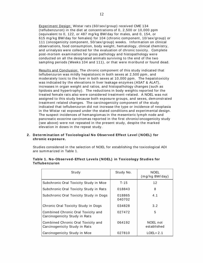
12
Experiment Design: Wistar rats (60/sex/group) received CME 134 (teflubenzuron) in the diet at concentrations of 0, 2,500 or 10,000 ppm (equivalent to 0, 122, or 487 mg/kg BW/day for males, and 0, 154, or 615 mg/kg BW/day for females) for 104 (chronic component, 10/sex/group) or 111 (oncogenicity component, 50/sex/group) weeks. Information on clinical observations, food consumption, body weight, hematology, clinical chemistry, and urinalysis were collected for the evaluation of chronic toxicity. Complete post-mortem examination for gross pathology and histopathology were conducted on all the designated animals surviving to the end of the two sampling periods (Weeks 104 and 111), or that were moribund or found dead.
Results and Conclusion: The chronic component of this study indicated that teflubenzuron was mildly hepatotoxic in both sexes at 2,500 ppm, and moderately toxic to the liver in both sexes at 10,000 ppm. The hepatotoxicity was indicated by the elevations in liver leakage enzymes (ASAT & ALAT), increases in organ weight and ratios, and histopathology changes (such as lipidosis and hypertrophy). The reductions in body weights reported for the treated female rats also were considered treatment-related. A NOEL was not assigned to this study because both exposure groups, and sexes, demonstrated treatment related changes. The carcinogenicity component of the study indicated that teflubenzuron did not increase the type or incidence of neoplasia in the Wistar rat exposed under the stated conditions and experimental design. The suspect incidences of hemangiomas in the mesenteric lymph node and pancreatic exocrine carcinomas reported in the first chronic/oncogenicity study (see above) were not repeated in the present study, despite the marked elevation in doses in the repeat study.
2. Determination of Toxicological No Observed Effect Level (NOEL) for chronic exposure.
Studies considered in the selection of NOEL for establishing the toxicological ADI are summarized in Table 1.
Table 1. No-Observed-Effect Levels (NOEL) in Toxicology Studies for Teflubenzuron
Study Study No. NOEL (mg/kg BW/day)
Subchronic Oral Toxicity Study in Mice T-15 12
Subchronic Oral Toxicity Study in Rats 018843 8
Subchronic Oral Toxicity Study in Dogs 018865 040702
4.1
Chronic Oral Toxicity Study in Dogs 034828 3.2
Combined Chronic Oral Toxicity and Carcinogenicity Study in Rats
027472 5
Combined Chronic Oral Toxicity and Carcinogenicity Study in Rats
064192 NOEL not established
Carcinogenicity Study in Mice 027810 LOEL=2.1
13
Study Study No. NOEL (mg/kg BW/day)
Teratogenicity Study in Rats R-128 >1000 (maternal and developmental)
Embryotoxicity Study in Rabbits 4/63/84 50 (embryotoxicity)
Teratogenicity Study in Rabbits 695-460/13 1000 (fetotoxicity and Teratogenicity)
Two-Generation Reproductive Study in Rats
460/1 NOEL not established
(kidney effects at lowest dose of 1.5)
Based on these toxicology studies, the chronic oral toxicity study in mice with an established LOEL of 2.1 mg/kg BW/day was determined to be the most appropriate study to determine the toxicological ADI.
FDA concluded that further genotoxicity information is not needed for the purpose of addressing the potential carcinogenic potential of teflubenzuron for establishing an import tolerance due to lack of evidence of carcinogenicity demonstrated in the three carcinogenicity studies.
3. Determination of Toxicological ADI
The toxicological ADI is calculated using the following formula based on the LOEL of 2.1 mg/kg BW/day from the mice study and a safety factor of 200. A safety factor of 200 was used taking into consideration an additional 2-fold factor for LOEL to NOEL extrapolation, a 10-fold factor for animal-to-human variability and a 10-fold factor for human-to-human variability.
(Equation 1: Toxicological Acceptable Daily Intake (ADI) equals the lowest NOEL divided by the Safety Factor, which equals 2.1 mg/kg BW/day divided by 200, which equals 10 µg/kg BW/day.)
The toxicological ADI for teflubenzuron is 10 µg/kg BW/day.
14
D. Assignment of the Final ADI:
Because teflubenzuron is not an antimicrobial agent and has no/negligible antibacterial activity, a microbiological ADI was not needed. Therefore, we assign the toxicological ADI (0.01 mg/kg BW/day) as the final ADI for total teflubenzuron residues.
E. Safe Concentrations for Total Residues:
The calculation of the tissue safe concentrations is based on the General Principles for Evaluating the Safety of Compounds used in Food-Producing Animals (FDA/CVM, revised July 2006). The daily consumption value of the edible tissue of fish (muscle and skin in natural proportion) is approximated as 300 g. Therefore, the safe concentration total teflubenzuron residues (ppm) in the edible tissue of fish (muscle with skin in natural proportion) is calculated as the following:
(Equation 2: Safe Concentration (SC) equals Acceptable Daily Intake (ADI) times Average Human Body Weight divided by Food Consumption Value. The Safe Concentration (muscle) equals 10 µg/kg BW/day times 60 kg divided by 300 g/day, which equals 10 µg/g or 2 ppm.) Therefore, the safe concentration for total teflubenzuron residues is determined to be 2.0 ppm in fish muscle and skin in natural proportions.
F. Residue Chemistry:
1. Summary of Residue Chemistry Studies
a. Total Residue and Metabolism Studies
Study Title – “Determination of the metabolism and radioactive depletion of [14C]-CME 134 in the target species Atlantic salmon at 10 ºC following a single oral administration” - Study NTO/007
Study Director: Martin Auger, MS, Huntingdon Life Sciences, Suffolk, England
Study Report Date: June 22, 1995
Study Location: In-Life Facility – Atlantic Fish Health Inc. (AFHI), Prince Edward Island, Canada Analytical Facility – Pharmaco LSR Ltd., Suffolk, England
15
Animals: Forty-three Atlantic salmon (Salmo salar) held at 10 ºC
Test Substance: Teflubenzuron (CME 134) with 97.4% purity was used. CME 134 was radiolabeled with carbon-14 to form two radiolabeled test materials: [14C]-Benzoyl-CME 134 with a purity of 99.9% (specific activity of 64.1 mCi/mg) and [14C]-Aniline-CME 134 with a purity of 98.7% (specific activity of 38.7 mCi/mg). The radiochemical purity of both forms of [14C]-CME 134 was checked by HPLC prior to the study. The test material was prepared with equal amounts of the two radioactive forms of CME 134 and non-radiolabeled CME 134 in tetrahydrofuran (THF). THF was then removed. The day before dosing, the test material was re-suspended in dimethyl sulphoxide (DMSO). This formulation was added to fish food.
Treatment Groups and Study Design: There were two study groups - a preliminary experiment and the main study. The preliminary study was conducted to check for regurgitation after dosing and to provide control tissue. The fish in the preliminary study were dosed with DMSO spiked onto control diet. In the main study, fish were treated with a single dose of 10 mg/kg [14C]- CME 134 by oral gavage.
The preliminary study was conducted with 3 fish collected 3 hours after dosing. In the single dose main study, groups of five fish were sampled at 9 hours, 1, 2, 3, 4, 6, 13, and 18 days post-dose. The following tissues were collected: mucus, liver, kidney, muscle, skin, and gall bladder.
Tissue Residue Analysis: Total residues were determined by sample combustion, followed by liquid scintillation counting (LSC). Parent teflubenzuron concentrations in liver, muscle and skin samples were determined by validated HPLC and LC-MS methods. Metabolic profiles in pooled samples were examined on Days 1 and 8 post-dose.
Metabolism: The major component detected in liver, kidney, muscle and skin was parent teflubenzuron. Parent drug concentrations in muscle were 398 ppb on Day 1 and 74 ppb on Day 8 post-dose. Parent drug concentrations in skin were 749 ppb on Day 1 and 108 ppb on Day 8 post-dose. The presence of parent drug was confirmed by LC-MS.
Acidic hydrolysis of the unextracted residues from liver and kidney released very little of the bound material, whereas basic hydrolysis of the unextracted residues released 44% and 58% of the activity in the liver residues on 1 and 8 days, respectively and 97% in the kidney residue at 8 days. Two metabolites were found in liver (3’-hydroxy-teflubenzuron and 3,5-dichloro-2,4-difluoro aniline). Two metabolites were found in kidney (3’-hydroxy-teflubenzuron and 2’-hydroxy-teflubenzuron). Four other metabolites were found (3 in kidney, 1 in liver) but these were not identified.
Results: The overall mean percent dose delivered to the fish was 94.54 + 1.46%. For the Day 1 muscle sample, 98.58% of total radioactivity was extracted by the acetonitrile while 8.48% of total radioactivity was unextracted (22 ppb). For the Day 8 muscle sample, 83.9% of total radioactivity was extracted and 22.81% was not extracted (22 ppb). For Day 1 skin samples, 103.8% of total radioactivity was extracted with the acetonitrile while 11.11% of total radioactivity was unextracted. For Day 8 skin samples,
16
77.11% of total radioactivity was extracted with the acetonitrile while 11.29% of total radioactivity was unextracted.
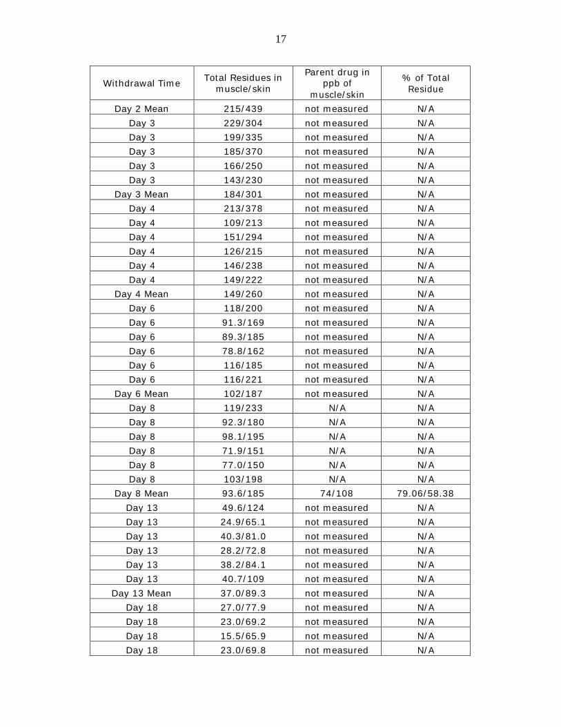
The highest concentration of radioactivity in the muscle was at 24 hours (2.2% of the administered dose or 410 ng eq/g). Total residues in muscle decreased to 20.9 ng eq/g by 18 days post-dose. The maximum level of radioactivity in skin was 0.2% of the administered dose or 753 ng eq/g at 1 day post-dose. Total residues in skin decreased to 73.0 ng eq/g by 18 days post-dose. At all timepoints, total residue levels were < 0.2% of the administered dose in liver, < 0.1% of the administered dose in kidney, and < 0.05% of the administered dose in mucus. The gall bladder retained the most radioactivity with 0.3% of the administered dose remaining at 18 days post-dose. The report states that the high total percentage of the administered dose present in skeletal muscle is due to the large mass of skeletal muscle relative to the other tissues and that, because of the smaller mass of the other tissues sampled (skin, liver, kidney, gall bladder and mucus), the maximum concentration of radioactivity accounted for a lower percentage of the administered dose, even though a higher concentration of radioactivity was observed in the tissue sample. The total residues (ng equivalents/g) and parent teflubenzuron (ppb) in the muscle and skin of Atlantic salmon dosed by oral gavage one time with 10 mg [14C]-CME 134/kg body weight are shown in Table 2.
Table 2. Total Residues (ng equivalents/g) and parent teflubenzuron (ppb) in muscle and skin of Atlantic salmon after a single oral administration of 10 mg [14C]-CME 134/kg body weight.
Withdrawal Time Total Residues in muscle/skin
Parent drug in ppb of
muscle/skin
% of Total Residue
9 hours 319/413 not measured N/A 9 hours 197/320 not measured N/A 9 hours 279/371 not measured N/A 9 hours 213/464 not measured N/A 9 hours 352/508 not measured N/A 9 hours 238/295 not measured N/A
9 hours Mean 266/395 not measured N/A Day 1 292/833 N/A N/A Day 1 471/1030 N/A N/A Day 1 303/435 N/A N/A Day 1 493/950 N/A N/A Day 1 437/652 N/A N/A Day 1 464/618 N/A N/A
Day 1 Mean 410/753 398/749 97.07/99.47 Day 2 204/458 not measured N/A Day 2 203/537 not measured N/A Day 2 266/503 not measured N/A Day 2 203/417 not measured N/A Day 2 228/80 not measured N/A
17
Withdrawal Time Total Residues in muscle/skin
Parent drug in ppb of
muscle/skin
% of Total Residue
Day 2 Mean 215/439 not measured N/A Day 3 229/304 not measured N/A Day 3 199/335 not measured N/A Day 3 185/370 not measured N/A Day 3 166/250 not measured N/A Day 3 143/230 not measured N/A
Day 3 Mean 184/301 not measured N/A Day 4 213/378 not measured N/A Day 4 109/213 not measured N/A Day 4 151/294 not measured N/A Day 4 126/215 not measured N/A Day 4 146/238 not measured N/A Day 4 149/222 not measured N/A
Day 4 Mean 149/260 not measured N/A Day 6 118/200 not measured N/A Day 6 91.3/169 not measured N/A Day 6 89.3/185 not measured N/A Day 6 78.8/162 not measured N/A Day 6 116/185 not measured N/A Day 6 116/221 not measured N/A
Day 6 Mean 102/187 not measured N/A Day 8 119/233 N/A N/A Day 8 92.3/180 N/A N/A Day 8 98.1/195 N/A N/A Day 8 71.9/151 N/A N/A Day 8 77.0/150 N/A N/A Day 8 103/198 N/A N/A
Day 8 Mean 93.6/185 74/108 79.06/58.38 Day 13 49.6/124 not measured N/A Day 13 24.9/65.1 not measured N/A Day 13 40.3/81.0 not measured N/A Day 13 28.2/72.8 not measured N/A Day 13 38.2/84.1 not measured N/A Day 13 40.7/109 not measured N/A
Day 13 Mean 37.0/89.3 not measured N/A Day 18 27.0/77.9 not measured N/A Day 18 23.0/69.2 not measured N/A Day 18 15.5/65.9 not measured N/A Day 18 23.0/69.8 not measured N/A
18
Withdrawal Time Total Residues in muscle/skin
Parent drug in ppb of
muscle/skin
% of Total Residue
Day 18 17.7/74.5 not measured N/A Day 18 Mean 20.9/73.0 not measured N/A
The data show that total residues are composed primarily of parent drug with more than 97% and 58% at Day 1 and Day 8, respectively. The fish in this study were dosed only one time compared to the repeated dosing scheme proposed for teflubenzuron administration in medicated feed (10 mg/kg BW for 7 consecutive days via medicated feed). Plasma pharmacokinetic data for teflubenzuron show that steady state is not reached until the third day of dosing. Data from fish dosed a single time likely underestimate the residue values that would result from the labeled dosing treatment regime.
Study Title – “[14C]-CME 134: Determination of the metabolism and radioactive depletion of [14C]-CME 134 (a benzoyl urea insecticide) in the target species Atlantic Salmon (Salmo salar) at 10 ºC following repeated oral administration” - Study NTO/009
Study Director: Martin Auger, MS, Huntingdon Life Sciences, Suffolk, England
Study Report Date: July 1, 1996
Study Location: In-Life Facility – Atlantic Fish Health Inc. (AFHI), Prince Edward Island, Canada Analytical Facility – Huntingdon Life Sciences, Eye, Suffolk, England
Animals: Fifty-four Atlantic salmon (Salmo salar) held at 10 ºC
Test Substance: Teflubenzuron (CME 134) with 97.4% purity was used. CME 134 was radiolabeled with carbon-14 to form two radiolabeled test materials: [14C]-Benzoyl-CME 134 with a purity of 99.9% (specific activity of 64.1 mCi/mg) and [14C]-Aniline-CME 134 with a purity of 98.7% (specific activity of 38.7 mCi/mg). The radiochemical purity of both forms of [14C]-CME 134 was checked prior to the study by HPLC. The test material was prepared with equal amounts of the two radioactive forms of CME 134 and non-radiolabeled CME 134 in tetrahydrofuran (THF). THF was then removed. The day before dosing, the test material was re-suspended in dimethyl sulphoxide (DMSO). This formulation was added to fish food and administered directly into the fish by intra-oesophageal intubation.
Treatment Groups and Study Design: Teflubenzuron was administered via medicated feed at the target rate of 10 mg CME 134/kg body weight for 5 consecutive days. On the sixth day, 10 mg [14C]-CME 134/kg body weight was administered directly into the fish by intra-oesophageal intubation. Groups of six fish were sampled each day on days 1, 4, 8, 12, 18, 24, 35, 50, and 120 post-dose. The following tissues were collected: mucus, liver, kidney, muscle, skin, gall bladder and gastro-intestinal contents (Day 1 group only). The entire muscle, skin and mucus for each fish were collected. Tank environment sub-samples were also collected.
19
Tissue Residue Analysis: Total residues were determined by liquid scintillation counting (LSC) either by direct addition of the sample to a suitable scintillation fluid or after pre-treatment of the samples by either of three methods: (1) solubilization of tissue samples using Soluene-350, followed by decolorization using hydrogen peroxide in propanol and mixing with scintillation fluid before analysis; (2) solubilization of the skin sample using 2 M KOH in methanol:water (1:1 by volume); (3) combustion of the gastro-intestinal contents and tank effluent and mixing with scintillation fluid. Parent teflubenzuron concentrations in liver, muscle and skin samples were determined by validated HPLC and LC-MS methods. Metabolic profiles in pooled samples were examined at Day 1 and 8 post-dose.
Metabolism: The major component detected in liver, kidney, muscle and skin at Day 1 was parent drug. Parent drug concentrations in muscle were 241 ppb at Day 1 and 2.7 ppb at Day 8 post-dose. Parent drug concentrations in skin were 467 ppb at Day 1 and 7.7 ppb at Day 8 post-dose. The presence of parent drug was confirmed by LC-MS. Two minor metabolites, other than CME 134, were found in liver (3’-hydroxy-teflubenzuron and 3,5-dichloro-2,4- difluoro aniline). Three other metabolites were found (1 in liver, 2 in muscle) but did not chromatograph with any of the authentic reference standards.
Results: The overall mean percent dose delivered to the fish was 101.8 ± 1.79%. For the Day 1 muscle sample, 95.63% of total radioactivity was extracted by the acetonitrile. For the Day 8 muscle sample, 128.62% of total radioactivity was extracted. For Day 1 skin samples, 83.43% of total radioactivity was extracted with the acetonitrile while 3.05% of total radioactivity was unextracted. For Day 8 skin samples, the study report stated the entire amount of radioactivity was extracted (81.86% of total radioactivity was extracted with the acetonitrile). The highest concentration of radioactivity in the muscle was at 1 day post-dose (1.5% of the administered dose or 310 ± 124 ng eq/g). Total residues in muscle had decreased to less than 0.3% and 0.1% of the administered dose at 4 and 8 days following treatment, respectively. By 18 days post-dose, total residues in muscle had decreased to 2.4 ng eq/g. The highest concentration of radioactivity in the skin was at 1 day post-dose (554 ± 178 ng eq/g). Total residues in skin decreased to 3.3 ng eq/g by 18 days post-dose.
At all timepoints, total residue concentrations were 0.2% of the administered dose in liver, 0.1% of the administered dose in kidney, and 0.0001% of the administered dose in mucus. Parent teflubenzuron concentrations (ppb) in combined muscle and skin were determined arithmetically by the following calculation.
20
(Equation 3: Total Parent Teflubenzuron in Combined Muscle and Skin equals Concentration of Teflubenzuron in Muscle times Weight of Muscle plus Concentration of Teflubenzuron in Skin times Weight of Skin divided by Total Weight of Muscle and Skin.)
The concentration of parent teflubenzuron in combined muscle and skin decreased from a maximum of 332 ± 130 ppb at 1 day post-dose to 3.6 ± 2.2 ppb at 24 days post-dose. This was followed by a rise to 6.3 ± 2.1 ppb at 35 days and a steady decrease to 1.3 ± 1.1 ppb at 120 days post-dose. The total residues (ng equivalents/g) and parent diflubenzuron (ppb) in fillet (muscle and skin) of Atlantic salmon after oral administration of 10 mg CME 134/kg body weight for six consecutive days are shown in Table 3.
Table 3. Total Residues (ng equivalents/g) and parent teflubenzuron (ppb) in muscle and skin of Atlantic salmon after a single oral
administration of 10 mg [14C]-CME 134/kg body weight following a 5-day feeding regime of CME 134 (13 days total) at 10ºC.
Withdrawal
Time
Total Residues in muscle/skin
Parent drug in ppb in
muscle/skin
Calculated parent drug in
ppb of combined
muscle/skin
% of Total Residue
Day 1 408/662 N/A N/A N/A Day 1 226/412 N/A N/A N/A Day 1 508/837 N/A N/A N/A Day 1 288/541 N/A N/A N/A Day 1 179/339 N/A N/A N/A Day 1 249/535 N/A N/A N/A
Day 1 Mean 310/554 241/467 332 77.74/84.30 Day 4 68.3/108 not measured N/A N/A Day 4 30.7/83.1 not measured N/A N/A Day 4 62.2/138 not measured N/A N/A Day 4 84.7/170 not measured N/A N/A Day 4 46.2/101 not measured N/A N/A Day 4 54.6/130 not measured N/A N/A
Day 4 Mean 57.8/122 not measured 63.2 N/A Day 8 16.3/37.0 N/A N/A N/A Day 8 5.1/36.0 N/A N/A N/A Day 8 9.2/35.3 N/A N/A N/A Day 8 23.8/58.0 N/A N/A N/A Day 8 9.4/23.7 N/A N/A N/A Day 8 23.7/54.8 N/A N/A N/A
Day 8 Mean 14.6/40.8 2.7/7.7 17.3 18.49/18.87 Day 12 7.2/28.2 not measured N/A N/A Day 12 8.7/29.5 not measured N/A N/A
21
Withdrawal Time
Total Residues in muscle/skin
Parent drug in ppb in
muscle/skin
Calculated parent drug in
ppb of combined
muscle/skin
% of Total Residue
Day 12 21.2/42.8 not measured N/A N/A Day 12 10.7/32.4 not measured N/A N/A Day 12 14.6/35.2 not measured N/A N/A
Day 12 Mean 12.0/33.7 not measured 14.2 N/A Day 18 ND/13.9 not measured N/A N/A Day 18 6.1/17.2 not measured N/A N/A Day 18 ND/18.5 not measured N/A N/A Day 18 ND/14.7 not measured N/A N/A Day 18 3.4/13.4 not measured N/A N/A Day 18 5.0/19.8 not measured N/A N/A
Day 18 Mean 2.4/16.3 not measured 4.1 N/A Day 24 4.2/12.3 not measured N/A N/A Day 24 ND/18.6 not measured N/A N/A Day 24 ND/15.0 not measured N/A N/A Day 24 4.4/13.6 not measured N/A N/A Day 24 6.1/9.2 not measured N/A N/A Day 24 ND/11.4 not measured N/A N/A
Day 24 Mean 2.5/13.4 not measured 3.6 N/A Day 35 7.5/9.6 not measured N/A N/A Day 35 2.5/7.8 not measured N/A N/A Day 35 6.2/8.5 not measured N/A N/A Day 35 4.3/8.2 not measured N/A N/A Day 35 7.5/7.7 not measured N/A N/A Day 35 8.5/8.7 not measured N/A N/A
Day 35 Mean 6.1/8.4 not measured 6.3 N/A Day 50 3.4/11.6 not measured N/A N/A Day 50 ND/8.8 not measured N/A N/A Day 50 ND/8.3 not measured N/A N/A Day 50 10.4/7.2 not measured N/A N/A Day 50 ND/9.0 not measured N/A N/A Day 50 8.3/10.1 not measured N/A N/A
Day 50 Mean 3.7/9.2 not measured 4.2 N/A Day 120 2.0/ND not measured N/A N/A Day 120 ND/9.1 not measured N/A N/A Day 120 ND/5.5 not measured N/A N/A Day 120 2.1/5.4 not measured N/A N/A Day 120 2.7/ND not measured N/A N/A
22
Withdrawal Time
Total Residues in muscle/skin
Parent drug in ppb in
muscle/skin
Calculated parent drug in
ppb of combined
muscle/skin
% of Total Residue
Day 120 ND/ND not measured N/A N/A Day 120 Mean 1.1/3.3 not measured 1.3 N/A
The data show that total residues are composed primarily of parent drug with at least 78% and 18% at Day 1 and Day 8, respectively. Detectable concentrations of CME 134 persisted in the tissues of the test animals throughout the 120-day study. The study report concluded that a background concentration of CME 134 was discovered in the closed, recirculating water system at the study facility but these background concentrations of CME 134 do not alter the conclusions for the study. The total residue concentrations measured in this study are similar to the total residue data generated in Study No. NTO/007.
Study Title – “[14C] CME 134: Determination of the radioactive depletion of [14C]-CME 134 (a benzoyl urea insecticide) in the target species Atlantic Salmon (Salmo salar) at 6 ºC following repeated oral administration” - Study NTO/013
Study Director: Martin Auger, MS, Huntingdon Life Sciences, Suffolk, England
Study Report Date: July 10, 1996
Study Location: In-Life Facility – Atlantic Fish Health Inc. (AFHI), Prince Edward Island, Canada Analytical Facility – Huntingdon Life Sciences, Eye, Suffolk, England
Animals: Fifty-four Atlantic salmon (Salmo salar) held at 6 ºC
Test Substance: Teflubenzuron (CME 134) with 97.4% purity was used. CME 134 was radiolabeled with carbon-14 to form two radiolabeled test materials: [14C]-Benzoyl-CME 134 with a purity of 99.9% (specific activity of 64.1 mCi/mg) and [14C]-Aniline-CME 134 with a purity of 98.7% (specific activity of 38.7 mCi/mg). The radiochemical purity of both forms of [14C]-CME 134 was checked prior to the study by HPLC. The test material was prepared with equal amounts of the two radioactive forms of CME 134 and non-radiolabeled CME 134 in tetrahydrofuran (THF). THF was then removed. The day before dosing, the test material was re-suspended in dimethyl sulphoxide (DMSO). This formulation was added to fish food and administered directly into the fish by intra-oesophageal intubation.
Treatment Groups and Study Design: Teflubenzuron (CME 134) was administered via medicated feed at the target rate of 10 mg CME 134/kg body weight for 12 consecutive days. On the thirteenth day, 10 mg [14C]-CME 134/kg body weight was administered directly into the fish by intra-oesophageal intubation. Groups of six fish were sampled each day on days 1, 8, 16, 24, 35, 50, 75, 93 and 97 post-dose. The following tissues were collected: mucus, liver, kidney, muscle, skin, gall bladder and gastro-intestinal
23
contents (Day 1 group only). The entire muscle, skin and mucus for each fish were collected. Tank environment sub-samples also were collected.
Tissue Residue Analysis: Total residues were determined by liquid scintillation counting (LSC) either by direct addition of the sample to a suitable scintillation fluid or after pre-treatment of the samples by either two methods: (1) solubilization of tissue samples using Soluene-350, followed by decolorization using hydrogen peroxide in propanol and mixing with scintillation fluid before analysis; or (2) solubilization of the skin sample using 2 M KOH in methanol:water (1:1 by volume). Parent teflubenzuron concentrations in liver, muscle and skin samples were determined by validated HPLC and LC-MS methods. No analysis of metabolism was conducted.
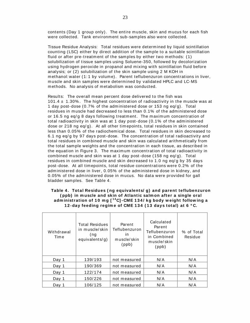
Results: The overall mean percent dose delivered to the fish was 101.4 ± 1.30%. The highest concentration of radioactivity in the muscle was at 1 day post-dose (0.7% of the administered dose or 153 ng eq/g). Total residues in muscle had decreased to less than 0.1% of the administered dose or 16.5 ng eq/g 8 days following treatment. The maximum concentration of total radioactivity in skin was at 1 day post-dose (0.1% of the administered dose or 218 ng eq/g). At all other timepoints, total residues in skin contained less than 0.05% of the radiochemical dose. Total residues in skin decreased to 6.1 ng eq/g by 97 days post-dose. The concentration of total radioactivity and total residues in combined muscle and skin was calculated arithmetically from the total sample weights and the concentration in each tissue, as described in the equation in Figure 3. The maximum concentration of total radioactivity in combined muscle and skin was at 1 day post-dose (158 ng eq/g). Total residues in combined muscle and skin decreased to 1.0 ng eq/g by 35 days post-dose. At all timepoints, total residue concentrations were 0.2% of the administered dose in liver, 0.05% of the administered dose in kidney, and 0.05% of the administered dose in mucus. No data were provided for gall bladder samples. See Table 4.
Table 4. Total Residues (ng equivalents/g) and parent teflubenzuron (ppb) in muscle and skin of Atlantic salmon after a single oral
administration of 10 mg [14C]-CME 134/kg body weight following a 12-day feeding regime of CME 134 (13 days total) at 6 ºC.
Withdrawal Time
Total Residues in muscle/skin
(ng equivalents/g)
Parent
Teflubenzuron in
muscle/skin (ppb)
Calculated
Parent Teflubenzuron in Combined muscle/skin
(ppb)
% of Total Residue
Day 1 139/193 not measured N/A N/A Day 1 190/369 not measured N/A N/A Day 1 122/174 not measured N/A N/A Day 1 150/226 not measured N/A N/A Day 1 106/125 not measured N/A N/A
24
Withdrawal
Time
Total Residues in muscle/skin
(ng equivalents/g)
Parent Teflubenzuron
in muscle/skin
(ppb)
Calculated Parent
Teflubenzuron in Combined muscle/skin
(ppb)
% of Total Residue
Day 1 Mean 153/218 not measured 158 N/A Day 8 9.0/13.5 not measured N/A N/A Day 8 21.4/35.1 not measured N/A N/A Day 8 23.6/41.1 not measured N/A N/A Day 8 11.6/21.7 not measured N/A N/A Day 8 13.0/16.9 not measured N/A N/A Day 8 20.1/42.8 not measured N/A N/A
Day 8 Mean 16.5/28.5 not measured 17.5 N/A Day 16 ND/17.0 not measured N/A N/A Day 16 10.2/19.1 not measured N/A N/A Day 16 ND/11.6 not measured N/A N/A Day 16 8.0/8.2 not measured N/A N/A Day 16 8.8/14.3 not measured N/A N/A Day 16 18.1/28.0 not measured N/A N/A
Day 16 Mean 7.5/16.4 not measured 8.29 N/A Day 24 ND/9/7 not measured N/A N/A Day 24 ND/13.3 not measured N/A N/A Day 24 ND/9.9 not measured N/A N/A Day 24 ND/9.6 not measured N/A N/A Day 24 10.4/10.1 not measured N/A N/A Day 24 ND/7.4 not measured N/A N/A
Day 24 Mean 1.7/10.0 not measured 2.43 N/A Day 35 ND/14.2 not measured N/A N/A Day 35 ND/5.6 not measured N/A N/A Day 35 ND/9.8 not measured N/A N/A Day 35 ND/9.8 not measured N/A N/A Day 35 ND/7.1 not measured N/A N/A Day 35 ND/18.8 not measured N/A N/A
Day 35 Mean ND/10.9 not measured 1.01 N/A Day 50 9.8/4.1 not measured N/A N/A Day 50 6.2/10.9 not measured N/A N/A Day 50 6.2/9.1 not measured N/A N/A Day 50 7.4/10.3 not measured N/A N/A Day 50 ND/6.8 not measured N/A N/A Day 50 5.6/10.7 not measured N/A N/A
Day 50 Mean 5.9/8.7 not measured 6.15 N/A Day 75 ND/5.9 not measured N/A N/A
25
Withdrawal
Time
Total Residues in muscle/skin
(ng equivalents/g)
Parent Teflubenzuron
in muscle/skin
(ppb)
Calculated Parent
Teflubenzuron in Combined muscle/skin
(ppb)
% of Total Residue
Day 75 ND/9.8 not measured N/A N/A Day 75 ND/8.0 not measured N/A N/A Day 75 ND/ND not measured N/A N/A Day 75 ND/6.8 not measured N/A N/A
Day 75 Mean ND/7.2 not measured 0.53 N/A Day 93 NA not measured N/A N/A Day 93 NA not measured N/A N/A Day 93 NA not measured N/A N/A Day 93 NA not measured N/A N/A Day 93 NA not measured N/A N/A Day 93 NA not measured N/A N/A
Day 93 Mean NA not measured not calculated N/A Day 97 ND/3.7 not measured N/A N/A Day 97 ND/6.2 not measured N/A N/A Day 97 ND/5.2 not measured N/A N/A Day 97 ND/5.4 not measured N/A N/A Day 97 ND/7.9 not measured N/A N/A Day 97 ND/8.0 not measured N/A N/A
Day 97 Mean ND/6.1 not measured 0.65 N/A
The total tissue residues of teflubenzuron were slightly lower than the tissue residue data quantitated in Study No. NTO/007 and Study No. NTO/009, and the entire metabolic profile was not analyzed. Marker residue concentrations, but not total residue concentrations, were measured. A marker to total residue ratio was not determined from the study data. Parent drug represented at least 97% of the total residues at Day 1 and at least 58% of the total residues at Day 8.
b. Comparative Metabolism Study
Study Title – “The Biokinetics and Metabolism of [14C]-CME 134 in the Rat” - Study No. CUB 6/85
Study Director: Not provided in the submission
Study Date: December 4, 1986
Study Location: In-life Facility – Celamerck GmbH & Co. KG, Federal Republic of Germany Analytical Facility – Not provided in the submission
26
Animals: Male and female Wistar rats of the Chbb: THOM (SBF) strain weighing approximately 200 grams were dosed with radioactive compound.
Test Substance: Two batches of nonradioactive teflubenzuron (CME 134) were used with purities of 99.4% and 99.3% as confirmed by HPLC. Teflubenzuron (CME 134) was uniformly labeled with 14C in the aniline ring (specific activity of 38.7 mCi/mg). The radiochemical purity of the CME 134 used was >97% and its radioactivity confirmed by thin layer chromatography prior to dosing each test group.
Treatment Groups and Study Design: Rats were divided into six test groups and dosed by oral gavage. As shown in Table 5, all treatments were prepared as suspensions in a mixture of 1% Tylose C 30 and 1% Tween 80 (1:1) according to the following regimens: A single oral dose of 25 mg/kg body weight (BW) [14C]-CME 134 for expiration studies (Group A), a single oral dose of 25 mg/kg BW [14C]-CME 134 for excretion and metabolism studies (Group B), a 14-day repeated oral dose of 25 mg/kg BW CME 134 followed by a single oral dose of 25 mg/kg BW [14C]-CME 134 for excretion and metabolism studies (Group C), a single oral dose of 750 mg/kg BW [14C]-CME 134 for excretion and metabolism studies (Group D), a single oral dose of 25 mg/kg BW [14C]-CME 134 for blood concentration studies, and a single oral dose of 750 mg/kg BW [14C]-CME 134 for blood concentration studies (Group E).
Table 5. Treatment Design
Number and Gender Dose Parameters Measured A. 1 male, 1 female 25 mg/kg BW [14C]-CME 134 Test of expired air B. 5 males, 5 females 25 mg/kg BW [14C]-CME 134 Excretion and metabolism
C. 5 males, 5 females
25 mg/kg BW CME 134 (single dose of nonradioactive drug
daily for 14 days followed by a single dose of [14C]-CME 134)
Excretion and metabolism
D. 5 males, 5 females 750 mg/kg BW [14C]-CME 134 Excretion and metabolism E. 5 males, 5 females 25 mg/kg BW [14C]-CME 134 Blood concentrations F. 5 males, 5 females 750 mg/kg BW [14C]-CME 134 Blood concentrations
Analysis: Expired air was collected from rats in Group A. For rats in Groups B, C and D, urine and feces were collected at 24, 48, 72, 96, 120, 144, 168, and 192 hours after dosing with radioactive drug. Cage washings also were collected for these groups. For rats in Groups E and F, serial blood samples were collected before treatment and at 20, 40, 60 minutes, and 2, 4, 6, 8, 24, 48, 72, 96, 120, and 168 hours after treatment. For rats in Groups E and F, tissues were not collected for measurement of radioactivity due to the results of a previous study showing that very little radioactivity is present.
Expired air was collected in absorption towers. Volatile radioactivity was measured as [14CO2] by liquid scintillation counting. Radioactivity was measured in urine and cage washings by liquid scintillation counting. Urine samples were freeze-dried and extracted with acetone and methanol. The evaporated solutions were applied to thin layer plates for chromatographic
27
analysis. If sufficient radioactivity was present, metabolite fractions separated by thin layer chromatography (TLC) were treated either with enzyme (b-glucuronidase/aryl-sulfatase) or with hydrochloric acid and the resulting solutions separated by TLC again. Feces were combusted in an automatic sample oxidizer and then radioactivity was measured by liquid scintillation counting. Fecal samples were freeze-dried and extracted with acetone and methanol. The evaporated solutions were measured for radioactivity by liquid scintillation counting and also were applied to thin-layer chromatography plates. Extracted fecal samples that contained > 1% non-extractable radioactive residues were put through a second extraction procedure with heated methanol and separated by TLC. Blood plasma samples were centrifuged immediately after collection and the plasma was removed. Radioactivity in plasma was measured by liquid scintillation counting. The red cells were treated with sodium hydroxide and digestin after which radioactivity was determined by liquid scintillation counting. The carcasses were solubilized by adding sodium hydroxide and applying heat for 4 days. The radioactivity in the resulting fat and liquid was determined by liquid scintillation counting.
Thin-Layer Chromatography Analysis: One-dimensional TLC was used with three different solvent systems. Non-radiolabeled CME 134 and possible degradation products, 3,5-dichloro-2,4-difluoroaniline and 3,5-dichloro-2,4-di- fluorophenyl-urea, were used as reference compounds. The solvent system for CME 134 and 3,5-dichloro-2,4-difluoroaniline (combined) was acetate/cyclohexane. The solvent system for 3,5-dichloro-2,4-difluoroaniline without radiolabelled compound was dichloromethane. The amount of parent drug was calculated by subtracting the results of the two solvent systems. The solvent system for 3,5-dichloro-2,4-di-fluorophenyl-urea was ethyl acetate/cyclohexane.
HPLC Analysis: HPLC was performed with a Hibar LiChroCart column using acetonitrile/water (65:35) as the eluent and an UV detector.
UV-spectroscopy Analysis: UV-spectra of the parent drug and metabolites were recorded using an HPLC UV-photodiode array detector.
Mass Spectroscopy Analysis: Electron impact mass spectra were performed by Boehringer Ingelheim, Federal Republic of Germany using a Varian-MAT-CH7 mass spectrometer. The conditions of analysis were described.
Results: All quantities were reported as CME 134 equivalents.
28
i. Excretion Pattern of Radioactivity
Single 25 mg/kg BW Dose (Group A): No radioactivity was detected (LOD = 0.0001% of dose) in the expired air.
Single 25 mg/kg BW Dose (Group B): Within 24 hours after dosing, greater than 85% of the administered dose was eliminated in the feces. Overall, radioactivity recovered in feces was a mean of 93.3% of the administered dose for males and 90.7% of the administered dose for females. Radioactivity recovered in urine was a mean of 0.86% of the administered dose for males and 0.55% of the administered dose for females. Only 0.05% of the administered dose could be detected in the carcass eight days after treatment.
Single 25 mg/kg BW Dose (Group E): Plasma concentrations of CME 134 equivalents plateau at one hour post-treatment (0.38-0.46 mg/mL for males, 0.22-0.25 mg/mL for females). Plasma concentrations were less than 0.01 mg/mL in both sexes at the end of the study period (7 days). The concentrations of radioactivity present in whole blood were comparable to those found in plasma.
Repeated 25 mg/kg BW Dose (Group C): The results of this test group (pulsed nonradiolabeled followed by radiolabeled) were not significantly different from those of the Single Low Dose (Group B).
Single 750 mg/kg BW Dose (Group D): Within 48 hours after dosing, greater than 90% of the administered dose was recovered in the feces. After 8 days, total radioactivity recovered in urine, feces, and carcass was 0.15%, 94.2%, and <0.01% of the administered dose, respectively.
Single 750 mg/kg BW Dose (Group F): In males, plasma concentrations of CME 134 equivalents peaked at 24 hours post-treatment (mean 3.27 mg/mL). A comparable peak concentration was not identified in females but the report speculates that the peak plasma concentration in females may have occurred between 8 and 24 hours post-dose when samples were not collected. Plasma concentrations were 0.06 mg/mL for males and 0.08 mg/mL for females at the end of the study period (7 days). The concentrations of radioactivity present in whole blood were slightly less than those found in plasma.
ii. Characterization of Radioactivity in Feces
Unextractable and non-extracted radioactivity residues in feces accounted for between 0.7% and 4.5% of the administered dose in Groups B, C, and D.
Results of TLC showed that in all groups the greatest portion of radioactivity in the feces was unchanged parent drug (range 82.2%-91.4% of the administered dose). The identity of parent drug in feces was confirmed by MS, HPLC and UV-spectroscopy. The concentration of metabolites in feces ranged from 2.8% to 6.5% of the dose.
One metabolite was identified as 3,5-dichloro-2,4-difluorophenyl-urea using a reference standard and TLC, HPLC and UV-spectroscopy. An attempt at MS was not successful due to the small amount of metabolite and high amounts of impurities present. The metabolite could not be detected in all of the rats. In
29
Group D (single 750 mg/kg BW dose), the amount of this metabolite was a maximum of 0.2% of the administered dose. In Groups B and C (single 25 mg/kg BW dose), the amount of this metabolite was 0.5-1.0% of the administered dose.
A second metabolite that may have been present, 3,5-dichloro-2,4- difluoroaniline, was not definitively identified. If this second metabolite (created by cleavage of the urea bridge) was present, concentrations would be approximately 0.4-0.7% of the administered dose after the low dose and approximately 0.1-0.2% of the administered dose after the high dose.
iii. Characterization of Radioactivity in Urine
Thin-layer chromatograms could only be obtained from the urine extract of Group D (single 750 mg/kg BW dose). Traces of unchanged parent drug (0.01-0.02% of the administered dose) were found by TLC comparison with the reference standard. The report concludes that the parent drug found in urine may be due to fecal contamination.
The metabolite 3,5-dichloro-2,4-difluoro-phenyl-urea was found by TLC. HPLC investigation of the isolate TLC fraction (<0.01% of dose) showed that the compound that had the same retention time as the metabolite was present in very low concentration. Further investigation of the metabolite by UV-spectroscopy was not possible.
A large amount of polar compounds remained at the origin of the TLC plates. These isolated fractions were treated with both HCl and b-glucuronidase/arylsulfatase. TLC after treatment indicated that some of the polar metabolite fractions were conjugates.
The report concludes that, in the rat, teflubenzuron is excreted primarily as unchanged parent drug while a small percentage of the drug is metabolized either by hydroxylation of one of the phenyl rings or cleavage of the urea bridge. Approximately 90% of the dose was found in the feces as parent drug. In feces, one metabolite was identified definitively as 3,5-dichloro-2,4- difluorophenyl-urea and comprised up to 1% of the dose. A second metabolite, 3,5-dichloro-2,4-difluoroaniline, was not identified definitively but may have been present in feces at approximately 0.2% of the dose.
Study Title – “The Biliary Excretion and Metabolism of [14C]-CME 134” - Study No. Not provided in the submission
Study Director: Not provided in the submission
Study Date: October 15, 1987
Study Location: In-life Facility – Shell Agrar Company, West Germany Analytical Facility – Not provided in the submission
Animals: Male and female Wistar rats were obtained from Charles River, Kent, United Kingdom. The rats weighed approximately 200 grams during the test period. During acclimation, the rats were housed in stainless steel cages.
30
During the experimental phase, the rats were housed in restraining cages constructed of perspex and stainless steel.
Test Substance: Teflubenzuron (CME 134) was uniformly labeled with 14C in the aniline ring (specific activity of 38.7 mCi/mg). The radiochemical purity of the CME 134 used was >98% and its radioactivity confirmed by thin layer chromatography prior to dosing each test group. Reference standard of CME 134 was used, along with 3,5-dichloro-2,4-difluoro-phenylurea, and 3,5-dichloro-2,4-difluoro-aniline, para-hydroxy-aniline derivative, ortho-hydroxy-aniline derivative, and meta-hydroxy-benzoyl derivative.
Treatment Groups and Study Design: Rats were divided into two test groups. The bile ducts of all rats were cannulated prior to the dose administration while the rats were anesthetized. One test group (3 males, 3 females) received a single dose of 25 mg/kg BW [14C]-CME 134 by oral gavage. The other test group (3 males, 3 females) was administered a single dose of 750 mg/kg BW [14C]-CME 134 by gastric intubation. Each treatment was prepared as a suspension in a mixture of 1% Tylose C30 and 1% Tween 80 (1:1).
Analysis: In each test group, bile was collected until 48 hours post-dose. Urine and feces were collected at 0-24 and 24-48 hours post-dose. At 48 hours, all animals were sacrificed and the gastro-intestinal tracts, livers, and carcasses were collected.
The radioactivity in bile and urine was measured by liquid scintillation counting. Fecal samples were freeze dried and extracted twice with acetone and methanol. The samples were sonicated and heated prior to measuring concentrations of radioactivity. Gastro-intestinal tracts were extracted with methanol and then measured for radioactivity. Carcasses were digested with heat in sodium hydroxide, methanol, water and Triton X-405. Samples of extracted feces, GI tracts, and liver were combusted in an Automatic Sample Oxidiser. Pooled samples of bile, fecal extracts, and urine were analyzed for proportion of radioactive components. Bile and urine were freeze-dried and extracted with methanol. The solvent concentrates from bile and urine and the fecal extracts were analyzed by TLC using a solvent system of cyclohexane, dichloromethane, and ethyl acetate. Extracts were co-chromatographed with reference compounds. Urine and bile extracts were subjected to enzymatic treatments, alkaline hydrolysis, and acid hydrolysis.
Mass Spectrometry Analysis: Mass spectra were obtained using a VG 7070E mass spectrometer. Electron ionization spectra were obtained with an electron energy of 70 eV and a trap current of 200 mA, while chemical ionization spectra were obtained with an electron energy of 50 eV, an emission current of 200-500 mA and isobutane as the reagent gas.
Results: After both the 25 mg/kg BW and 750 mg/kg BW doses, the majority of radioactivity was found in the feces (approximately 50% of dose). For the 25 mg/kg BW dose, the sum of urinary and biliary excreted radioactivity indicated absorption of 18.58% of the administered dose for males and 16.54% of the administered dose for females. For the 750 mg/kg BW dose, the sum of urinary and biliary excreted radioactivity indicated absorption of 2.04% of the administered dose for males and 2.41% of the administered dose for females. Only small amounts of radioactivity were found in the liver and carcass.
31
The majority of activity in the feces (approximately 40% of the low dose and 56% of the high dose) was unchanged CME 134. Radioactivity in feces was associated with 3,5-dichloro-2,4-difluoro-phenylurea, para-hydroxy-aniline derivative, ortho-hydroxy-aniline derivative, and a meta-hydroxy-benzoyl derivative that accounted for approximately 2% of the administered dose.
Radioactivity in bile that was associated with CME 134 accounted for 0.2% of the administered dose. Radioactivity in bile that was associated with para- hydroxy-aniline derivative, ortho-hydroxy-aniline derivative, meta-hydroxy- benzoyl derivative and 3,5-dichloro-2,4-difluoro-aniline comprised 0.1-0.2% of the administered dose. Radioactivity associated with 3,5-dichloro-2,4-difluoro- phenylurea accounted for 0.7 to 0.8% of the administered dose. Radioactivity associated with the polar base line material was 14.1-15.5% of the administered dose for the low dose rats and 1.5-1.8% of the administered dose for the high dose rats.
The data show that the absorption of CME 134 in rats is dose-dependent with greater absorption occurring at the 25 mg/kg BW dose. The majority of the radioactivity was found in the feces as unchanged CME 134 (approximately 50% of the administered dose). The concentrations of metabolites found were typically less than 1% of the administered dose. The metabolite, 3,5-dichloro- 2,4-difluorophenyl urea, was found in feces. A substantial portion of the biotransformation products in bile was unidentified polar material (14.1-15.5% of the administered dose for the low dose rats).
Summary of Comparative Metabolism Studies
The comparative metabolism studies provided sufficient data to qualitatively compare the reside profile in the rat and in Atlantic salmon. The major metabolite in Atlantic salmon was parent drug, which was also present in the rat.
c. Tissue Residue Depletion Study
Study Title – “The determination of the residues of CME 134 (a benzoyl urea insecticide) in the target species Atlantic Salmon (Salmo salar) at 10 ºC following administration over a seven day period and provisions of samples for the determination of the metabolism and radioactive depletion of [14C]-CME 134” - Study No. NTO/010
Study Director: C.H. McGuire, B.Sc., Huntingdon Life Sciences, Suffolk, England
Study Report Date: October 3, 1996
Study Location: In-Life Facility – Atlantic Fish Health Inc. (AFHI), Prince Edward Island, Canada Analytical Facility - Huntingdon Life Sciences, Suffolk, England
Animals: Ninety Atlantic salmon (Salmo salar) held at 10 ºC
32
Test Substance: Teflubenzuron (CME 134) with 97.4% purity was used. CME 134 was radiolabeled with carbon-14 to form two radiolabeled test materials: [14C]-Benzoyl-CME 134 with a purity of 99.9% (specific activity of 64.1 mCi/mg) and [14C]-Aniline-CME 134 with a purity of 98.7% (specific activity of 38.7 mCi/mg). The radiochemical purity of both forms of [14C]-CME 134 was checked prior to the study by HPLC. The test material was prepared with equal amounts of the two radioactive forms of CME 134 and non-radiolabeled CME 134 in tetrahydrofuran (THF). THF was then removed. The day before dosing, the test material was re-suspended in dimethyl sulphoxide (DMSO). This formulation was added to fish food and administered directly into the fish by intra-oesophageal intubation.
Treatment Groups and Study Design: Teflubenzuron was administered via medicated feed at the target rate of 10 mg teflubenzuron/kg body weight for 7 consecutive days. Groups of ten fish were sampled each day on days 0, 1, 4, 8, 12, 18, 24, 35, 50 and 120 post-dose. However, one fish per group received a single oral dose of 10 mg [14C]-CME 134/kg body weight on feeding day 7, instead of the non-radiolabeled medicated feed. The following tissues were collected from each fish: muscle and skin.
Tissue Residue Analysis: Parent teflubenzuron concentrations in muscle and skin samples from fish treated with medicated feed were determined by a validated HPLC method, similar to that utilized in Study No. NTO/007, Study No. NTO/009 and Study No. NTO/013. Fortified samples of control muscle and skin were analyzed with each sample batch.
Results: Recoveries for muscle and skin ranged from 75-391% and 70-161%, of the administered dose, respectively. The mean teflubenzuron concentrations in muscle were 894 ppb at Day 1, 329 ppb at Day 4, 103 ppb at Day 8, 52 ppb at Day 12, 26 ppb at Day 18, 28 ppb at Day 24, and 37 ppb at Day 35 post-dose. In skin, mean teflubenzuron concentrations were 1,310 ppb, 353 ppb, 221 ppb, 86 ppb, 51 ppb, 40 ppb, and 43 ppb at Day 1, 4, 8, 12, 18, 24 and 35 post-dose, respectively. Mean teflubenzuron concentrations in combined muscle and skin were determined arithmetically according to Equation 3 above and were 931 ppb, 331 ppb, 116 ppb, 56 ppb, 29 ppb, 29 ppb and 38 ppb at Day 1, 4, 8, 12, 18, 24 and 35 post-dose, respectively. The mean teflubenzuron concentrations in muscle, skin and combined muscle and skin are summarized in Table 6.
The tissue samples collected on Days 50 and 120 were not analyzed because residues at days 18, 24 and 35 had stabilized at concentrations just above the method LOQ. The report concludes that the slow depletion of tissue residues after Day 18 was likely due to background concentrations of teflubenzuron present in the tank environment, caused by the water recirculating system used in the test tanks. While the study included a subset of fish that received a single dose of radioactive CME 134 (following 6 days of administration of nonradioactive medicated feed), the entire metabolic profile was not analyzed in this study. Concentrations of the marker residue (teflubenzuron), not total residues, were measured. Neither a marker to total residue ratio nor a tolerance were determined in this study.
33
Table 6. Mean Teflubenzuron Concentrations in Muscle, Skin and Combined Muscle and Skin
Study Title – “The determination of CME 134 in Atlantic salmon tissues” - Study No. NTO/011
Study Director: C.H. McGuire, B.Sc., Huntingdon Life Sciences, Suffolk, England
Study Report Date: June 22, 1995
Study Location: In-Life Facility – Not provided in the submission Analytical Facility – Pharmaco LST, Ltd., Suffolk, England
Animals: Fifty Atlantic salmon (Salmo salar) held at 7 ºC
Test Substance: Teflubenzuron (CME 134) with 97.4% purity was used and administered to fish via medicated pellets.
Treatment Groups and Study Design: Teflubenzuron was administered via medicated feed at the target rate of 10 mg teflubenzuron/kg body weight for 7 consecutive days. Groups of five fish were sampled each day on days 1, 3, 6, 9, 10, 12, 15, 25 and 32 post-dose. The following tissues were collected: muscle and skin.
Tissue Residue Analysis: Parent teflubenzuron concentrations in muscle and skin samples were determined by a validated HPLC method similar to that utilized in Study No. NTO/007, Study No. NTO/009, Study No. NTO/010 and Study No. NTO/013. Fortified samples of control muscle and skin were analyzed with each sample batch.
34
Results: Recoveries for muscle and skin were less than 70% on days 6, 10, 12, and 15 post-dose. At 1 day withdrawal, two out of five muscle samples had quantitatable residues (91 and 365 ppb). At 3 days withdrawal, two out of five muscle samples had quantitatable residues (23 and 97 ppb). Teflubenzuron residues were either not quantitatable or the muscle samples were not analyzed at the remaining withdrawal time points. At day 1 withdrawal, mean teflubenzuron residues in skin were 332 ppb. Skin samples were not analyzed at 3, 6, 9 and 10 days post-dose. After 12 days withdrawal, teflubenzuron residues in skin had decreased to between 19 and 27 ppb. After 15 days withdrawal, teflubenzuron residues in skin were not detected except for one skin sample measured at 18 days withdrawal (21 ppb). Mean teflubenzuron concentrations in combined muscle and skin were determined by arithmetically combining 86% of the teflubenzuron residue concentration in muscle with 14% of the teflubenzuron residue concentration in skin. The mean teflubenzuron concentrations in muscle, skin and calculated combined muscle and skin are summarized in Table 7.
Table 7. Teflubenzuron Concentrations in Muscle, Skin and Combined Muscle and Skin
Withdrawal Time
(Days)
Muscle (86% of reported value)
Skin (14% of reported
value)
Muscle and Skin
Combined (86%:14%)
1 <LOQ 12 12 1 78 60 138 1 <LOQ 10 10 1 <LOQ 17 17 1 313 132 445 3 <LOQ NA <LOQ 3 23.4 NA 20 3 <LOQ NA <LOQ 3 <LOQ NA <LOQ 3 96.76 NA 83 6 <LOQ NA <LOQ 6 <LOQ NA <LOQ 6 <LOQ NA <LOQ 6 <LOQ NA <LOQ 6 <LOQ NA <LOQ 9 <LOQ NA <LOQ 9 <LOQ NA <LOQ 9 <LOQ NA <LOQ 9 <LOQ NA <LOQ 9 <LOQ NA <LOQ 12 NA 21.37 2.99 12 NA <LOQ <LOQ
35
Withdrawal Time (Days)
Muscle (86% of reported value)
Skin (14% of reported
value)
Muscle and Skin
Combined (86%:14%)
12 NA <LOQ <LOQ 12 NA 26.05 3.65 12 NA 47.54 6.66 15 <LOQ ND <LOQ 15 <LOQ ND <LOQ 15 <LOQ ND <LOQ 15 <LOQ ND <LOQ 15 <LOQ ND <LOQ 18 <LOQ <LOQ <LOQ 18 <LOQ <LOQ <LOQ 18 <LOQ <LOQ <LOQ 18 <LOQ <LOQ <LOQ 18 <LOQ 20.6 2.88 25 NA <LOQ <LOQ 25 NA <LOQ <LOQ 25 NA <LOQ <LOQ 25 NA <LOQ <LOQ 25 NA <LOQ <LOQ 32 NA <LOQ <LOQ 32 NA <LOQ <LOQ 32 NA <LOQ <LOQ 32 NA <LOQ <LOQ 32 NA <LOQ <LOQ
LOQ - 20.56 ppb NA - not analyzed ND - not detected
The residue data were limited due to the numerous samples that were not analyzed or were not quantitatable. The report concludes that the method may not have been performing optimally. An initial batch of fifty fish used to obtain an average weight for the experimental fish, resulted in a lower than average weight compared to that of the actual experimental fish (177 g vs. 246 g). The report concludes that the actual mean dose administered to experimental fish was approximately 7.2 mg/kg BW/day, not the 10 mg/kg BW/day target consumption rate intended for the study. This lower dose rate may account for the low teflubenzuron tissue residue concentrations obtained in the study.
36
Study Title – “The determination of the residues of CME 134 (a benzoyl urea insecticide) in the target species Atlantic salmon (Salmo salar) at 6 ºC following administration over a fourteen day period and provision of samples for the determination of the metabolism and radioactive depletion of [14C]-CME 134” - Study No. NTO/014
Study Director: C.H. McGuire, B.Sc., Huntingdon Life Sciences, Suffolk, England
Study Report Date: October 28, 1996
Study Location: In-Life Facility – Atlantic Fish Health Inc. (AFHI), Prince Edward Island, Canada Analytical Facility - Huntingdon Life Sciences, Suffolk, England
Animals: Ninety Atlantic salmon (Salmo salar) held at 6 ºC
Test Substance: Teflubenzuron (CME 134) with 97.4% purity was used and administered via medicated pellets. CME 134 was radiolabeled with carbon-14 to form two radiolabeled test materials: [14C]-Benzoyl-CME 134 with a purity of 99.9% (specific activity of 64.1 mCi/mg) and [14C]-Aniline-CME 134 with a purity of 98.7% (specific activity of 38.7 mCi/mg). The radiochemical purity of both forms of [14C]-CME 134 was checked prior to the study by HPLC. The test material was prepared with equal amounts of the two radioactive forms of CME 134 and non-radiolabeled CME 134 in tetrahydrofuran (THF). THF was then removed. The day before dosing, the test material was re-suspended in dimethyl sulphoxide (DMSO). This formulation was added to fish food and administered directly into the fish by intra-oesophageal intubation.
Treatment Groups and Study Design: Teflubenzuron was administered via medicated feed at the target rate of 10 mg teflubenzuron/kg body weight for 14 consecutive days. Groups of nine fish were sampled each day on days 1, 8, 16, 24, 35, 50, 75, and 97 post-dose. However, one fish per group was received a single oral dose of 10 mg [14C]-CME 134/kg body weight on feeding day 14, instead of the non-radiolabeled medicated feed. The following tissues were collected from each fish: muscle and skin.
Tissue Residue Analysis: Parent teflubenzuron concentrations in muscle and skin samples were determined by a validated HPLC method, similar to that utilized in Study No. NTO/007, Study No. NTO/009, Study No. NTO/010, Study No. NTO/011 and Study No. NTO/013. Fortified samples of control muscle and skin were analyzed with each sample batch.
Results: Recoveries for muscle and skin were above 70%. The mean teflubenzuron concentrations in muscle were 405 ppb at Day 1, 63 ppb at Day 8, 45 ppb at Day 16, 41 ppb at Day 24 and 23 ppb at Day 35 post-dose. In skin, mean teflubenzuron concentrations were 443 ppb, 106 ppb, 55 ppb, 62 ppb and 45 ppb at Day 1, 8, 16, 24 and 35 post-dose, respectively. Mean teflubenzuron concentrations in combined muscle and skin were determined arithmetically according to Equation 3 above and were 407 ppb, 67 ppb, 56 ppb, 42 ppb, and 25 ppb at Day 1, 8, 16, 24 and 35 post-dose, respectively. The mean teflubenzuron concentrations in muscle, skin and combined muscle and skin are summarized in Table 8.
37
Table 8. Mean Teflubenzuron Concentrations in Muscle, Skin and Combined Muscle and Skin
The tissue samples collected on Days 50, 75, 93 and 97 post-dose were not analyzed. The report concludes that slow depletion of tissue residues after Day 16 was likely due to background concentrations of teflubenzuron present in the tank environment, caused by the water recirculating system used in the test tanks. The presence of background concentrations of teflubenzuron did not alter the conclusions for the study. While the study included fish treated with a single dose of radioactive CME 134 (in addition to repetitive administration of nonradioactive medicated feed), the entire metabolic profile was not analyzed and marker residue concentrations (teflubenzuron), not total residue concentrations, were measured. The report does not identify a marker to total residue ratio or a tolerance. The depletion characteristics of teflubenzuron in Atlantic salmon tissue were evaluated. Teflubenzuron residues in muscle, skin and combined muscle and skin peak at the Day 1 sampling and decline thereafter. The depletion of teflubenzuron tissue residues measured in this study was similar to that measured in the total residue studies (Study No. NTO/007, NTO/009, and NTO/013) and in the depletion study (Study No. NTO/010) conducted at 10 ºC. Variations in medicated feed consumption in the depletion Study No. NTO/011 resulted in low teflubenzuron tissue residues. Teflubenzuron residues in muscle, skin and combined muscle and skin peak at Day 1 withdrawal and deplete thereafter.
2. Target Tissue and Marker Residue
The target tissue is muscle with adhering skin in natural proportions, as that is considered the only edible tissue in Atlantic salmon. The marker residue is parent teflubenzuron because this is the residue present in the highest concentration and persists for the longest period of time, as shown in Study No. NTO/007, NTO/009 and NTO/013.
38
3. Import Tolerance
Total residues of teflubenzuron were below the safe concentration of total residues of 2,000 ppb and a marker to total residue ratio of 97% was observed in the pulsed dose total residue study (Study No. NTO/007). Because total residues were below the calculated safe concentration, an import tolerance could have been calculated using the pulsed dose study marker to total residue ratio (97%). Because the total residue study did not utilize a preferred study design (pulsed dose vs. continuous dose), a conservative marker to total residue ratio of 80% was utilized in the import tolerance calculation. Utilizing the calculated safe concentration of total residues of 2,000 ppb, CVM could have assigned an import tolerance as high as 1,600 ppb for residues of teflubenzuron in Atlantic salmon muscle with adhering skin in natural proportions. However, because the import tolerance value requested was 500 ppb, which represents 25% of the safe concentration, and is lower than the calculated import tolerance, we assign this lower value, 500 ppb, as the import tolerance for residues of diflubenzuron in Atlantic salmon muscle with adhering skin in natural proportions.
4. Withdrawal Period
Withdrawal periods are not assigned for import tolerances.
G. Analytical Method for Residues:
The FDA monitors for teflubenzuron using a multi-residue pesticide monitoring procedure. The LC-MS/MS analytical procedure (LOD <10 ppb) is described in Laboratory Information Bulletin (LIB) 4464 (Development and Validation of a Multiresidue Determination for Pesticides by LC-MS/MS). In addition, an LC-MS analytical procedure (LOD <10 ppb) was developed and validated specifically for the detection of teflubenzuron residues in salmon tissue (LIB 4463: A Rapid LC/MS Method for Determination of Teflubenzuron in Salmon Tissue). Both LIBs are available from the Office of Regulatory Science/Office of Regulatory Affairs/FDA via the agency FOI request channel (http://www.fda.gov/regulatoryinformation/foi/howtomakeafoiarequest/default.htm)
Both procedures perform at concentrations significantly lower than the assigned import tolerance of 500 ppb.
III.
AGENCY CONCLUSIONS:
These data support the establishment of an import tolerance of 0.5 parts per million teflubenzuron in muscle and skin of Atlantic salmon as provided under Sec. 512(a)(6) of the Food, Drug, and Cosmetic Act.