28
FRESH-WATER DINOFLAGELLATES OF MARYLAND I ;-. K. H. THOMPSON May, 1947 Publication No. 67 I CHESAPEAKE BIOLOGICAL LABORATORY Solomows Island, Maryland

FRESH-WATER DINOFLAGELLATES OF MARYLAND
I
;-. K. H. THOMPSON
May, 1947
Publication No. 67 I
CHESAPEAKE BIOLOGICAL LABORATORY
Solomows Island, Maryland

State of Maryland
DEPARTMENT OF RESEARCH AND EDUCATIOK
Corn missz'o n ers :
B. H. Willier, Chairntan ....................................................................................... Baltimore
........................................................................................................ H. R. Bassett Crisfield
&I-oyd M. Bertholf ................ ................ ................................................................ Westminster
.......................................................................................................... E. N. Cory C l e g e Park
.......................................................................................... Franklin D. IIay..: CentreviIle
Director:
...................................................................... ......................... R. V. Truit t -. Solomons Island
State Weather Service :
....................... ........................................................... G. N. Brancato, Meteorologist in Charge .... Baltimore
Chesapeake Biological Laboratory :
Edwin M. Barry, B. S., Education Assistant 6. F. Beaven, M . A., Biologist I , Oyster Inwestigations
Coit M . Coker, M. A., Biologis t I I , Fishery Investigations
Alice TV, Cronin, B. A., Chemist ZZZ
L. Eugene Cronin, PhD., Biologist ZI, Crnb Znoestigations
George B. Gray, B. S., Administrative Assistant
Harry A. Hensel, Jr., Inwestigaior
Theodore M. Miller, B. S., Fishery Technologist Harvey Mister, Captain, Fish Culturist
Lola h?l. Parks, B . A., Librarian
Fred W . Sieling, B. S., Biologist Zl, O y s t e r I n v e s t i g a t i o n s
R. V . Truitt, PhD., Biologis t
Doris M. V?oodburn, Secretary

FRESH-WATER DINOFLAGELLATES OF MARYLAND
The coastal plane region of the middle Atlantic states has numer- ous boggy swamps and small, shallow, eutropic ponds. Those from which c~llec~tions have been made, in Maryland, are rich in decaying vegetable matter and have a muddy bottorrt of some depth. This generally has extensive areas covered with species of Utrichularia, Potomogeton, Myriophyllum, Elodea, Typha and Pontederia.
Collections from such habitats in the summer of 1942 and again, at (different times, in the years 1946 and 1947 yielded an abundance of dinoflagellate forms. Some of these were insufficient in numbers to allow for proper identification, so the present paper does not give a complete list for the state.
The list lof fresh-water Dinolflagellates reported for lthe United States is small, 21 in all. The majority of these xre thecate species in the genTra Peridinium and Glenodinium with one in the genus Hemidinizim and two in Cerntium. There are but three species of unarmored Dinoflagellates reported.
Of the 20 identified species reported on in this paper, two are new to science and ten have not been reporlted previously for tlie . United States. The greater number of the latter are in the "naked" or unarmored group. This is due not only to the fact Ithat the unarmored forms are more prevalent than 'the armoi-ed in the fresh- water ponds horn which collections were made but also $to the fact that, in most cases, there was an abundance 01 individuals of each species. Thus a study could be made of both the living cells ant1 of ones which had been immobilized.
Asquick killing, without loss of cell shape, is obtained by h,olding the inverted and uncovered water mount over iodine crystals. The cell contents turn black and there is oftell a slight lifting of the pellicle. Prolonged treatment with iodine fumes (2-3 minutes) causes the pellicle to be lifted more (or the pratoplast to shrink more) . In such a preparation the fact that the pellicle is really composed of numerous, very small, thin plates becomes evident. In profile the sutures of the plates are seen as slight, opaque swellings, giving a

beaded appearance to the otherwise clear pellicle. The value ol such quick killing is that measurements can be made of a largc number of individuals and the extremes in size can be obtained as well as an accurate location alf the girdle with reference to the middle of the cell.
The unarmored Dinoflagellates are >probably more widespread in the United Sltates than the literature would indicate. Unfortunately they lose their lshape in preservatives and have no durable shell that muld be studied in preserved collections. The writer, however, feels that the collection method used by him has aided greatly in obtaining a more concentrated number of individuals. The collect- ing jar is filled with the squeezings from mats of filamentous algae, submerged aquatics, scrapings from culms (i.e. Typha) and some portions 01 the unsqueezed aquatics, preferably the older portions bearing decayed leaves. Afler half an hour or a longer exposure to diffuse light most of the motile organisms will be concenltrated at the surface on the light side. Cysts are generally present in the gelatinous slime and debris squeezed from the planits or scraped from the culms. Quite frequently excjstrnent will be found occur- ring during the next day or two. Such a concentrated collection should be examined as soon as possible for it spoils very soon unless kept cool. The adequate numbers of individuals obtained in this manner, permitting a ,thorough study of both living and killed cells, com- pensates for the lack od a hard shell, a lack which makes these forms difficult of determination and which is probably behind the paucity of species reported for this country.
'The following is a classification of the 20 species presented in this paper.
CLASS DINOPHYCEAE ORDER Gymnodiniales
Family Gymnodiniaceae * Gymnodinium albulurn Lindem. * Gymnodiniz~m ordinatum Sku~a * Gymnodinium triceratium Skuja * Gymnodinizlrn marylandicum Sp. Nov. * Gymnodinium cestocoetes Sp. Nov. * Gymnodinium neglecturn (Schilling) Lindem. * Gymnodinium fuscurn (Ehrenberg) Stein * Gymnodinizlnz viride Penard

* Massartia Alz~sei (Danysz) Schiller * Gyrodinium j9zlsillzlm Schilling
Family Glenodiniopsidaceae Hemidinium nasutum Stein
* Hemidinium ochracez~m Levander
Family Glenodiniaceae * Glenodinium Elpatiewsky i (Ostenf .) Schiller
Glenodinium quadridens (Stein) Schiller
Family Peridiniaceae Peridinizim inconspicziurn Lemm. Peridinium umbonatum Stein Peridinium Volzii Lemm. Peridinium Willei Huitf.-Kaas. Peridiniunz cinctum ( 0 . F. M.) Ehrenb.
Family Heterodiniaceae Ceratiz~m hirundinella (0 . F. M.) Rergh
Gymnodiniaceae Gymnodinium albulum LINDEM. Plate I, Figs. 1, 2.
Cells cylindrical with rounded apices, 9-25 p long. Girdle equa- torial; sulcus evident in lower half of cell only, either narrow or broad, running completely or incompletely to (the antapex. T h e upper half of the cell as wide as or b roade~ than the lower half. Stigma absent. Chromat~pho~res 3-5, present or absent, yellow-brown to green, discoidal and parietal. Flagellum equal to or slightly longer than the cell body. Orange to red oil globules may be present.
T h e Maryland material measured 12-16 p long and 9-1 1 p wide. Numerous refractive granules were present in the peripheral cytoplasm.
Lindemann found this species in swamps containing brackish water, but Schiller (1933, p. 330) states that he found it in a fresh- water lake, Attersee, from the surface dJown to a depth of 20 meters. Schiller (1933, p. 339) described, tentatively, lthe species G. pro-
* Not previously reported for the United States.

fundunz which he also collected in Attersee from a depth of 10-30 meters. This species, in the literature, is indistinguishable from G. albulum. Also, the narrow V-shaped sulcus on the lower half of the cell, described for G. projundurn, occurred as a variable in the Maryland material of G. albulum. The dimensions of G. profundurn, 13-14 p long by 11 p wide, are included in the extremes given by Lindcmann Ifor G. albulum and within those of the Maryland material. Schiller (1933, pp. 433, 434) describes and figures an unidentified species also collected in Attersee and at depths below 25 meters. Of this material he saps the girdle was visible only as a shallow trough around the cells; chrorna~tophores were lacking; and the cells measured 9-10 p long by 7-8 p wide. Again the size range falls within that of G. albulum. The shape of the cell and the lack of a distinct girdle and of chromat~~ph~ores is also paralleled in the Maryland material. The Iatter was collected from a small swamp pond that had just cleared of a two-week ice and snow cover. Some individuals were colorless, some pale yellow and others had more brownislh chromatopllores. I t is known that G. albulum occurs in a colorless form. When first mounted on a microscope slide the cells are very active, but come to rest very soon, losing thcir flagella anld the characteristic shape of the swimming cell. T h e girdle be- comes a shallow depression, as seen in profile, and ithe sulcus dis- appears. The cells also exhibit some twisting and bulging movements before coming to a complete rest. Finally the girdle depressions become entirely obliterated and the cell is a short cylinder wi,th rounded poles.
From the foregoing account of the bverla,pping of the Maryland material with G. albulum, G. profundurn, and Schiller's G. spec., the writer feels that the Iatter two are variants and depth forms of the earlier )described G. albulum.
T h e Maryland material was collected in Fcbruary and March, 1947, near Belcamp, Maryland, from a small swamp at the head of Bush River estuary, but separated from it by highway fill.
Gymnodinium ordinatum SKUJA (1939). Plate I, Figs. 3, 4. Cells dorm-ventrally flattened, oval in front view, 12-15 p long,
11-13 p wide and 8 p thick. Girdle slightly subequatorial. Sulcus on lower half only. Trailing flagellum equal to or twice the length of the cell. Chromatophores yellow-brown, parietal, irregularly discoidal, and generally four in number. One or two rcd oil globules may be present. Stigma wanting.

The Maryland material measured 14-15 p long and 10.5-12 p wide. There were four or five discoidal, roughlly oval chromato- phores which were olive-brown to yellow-brown in color. The trailing flagellum was twice the body length. Collected in February from under ice, Belcamp, Maryland.
Gymnodinium triceratium SKUJA (1939). Plate I, Figs. 5, 6.
Cells slightly flattened dmso-ventrally; 10-14 p long, 9-1 1 p wide, and 7-9 p thick. The girdle is equatorial Upper half broadly rounded, the lower produced into two or three conical processes. Chromatophores !three to five, parietal, irregularly discoidal, yellow- brown. Stigma absent. One or two red oil g150bules may be present. Flagellum one and one-half to ltwo times cell length.
The Maryland material measured 13-15 p long, and 9-1 1 p wide. There were four to five discoidal, parietal, olive-brown to brown chromatophores present. The flagellum was approximately one and one-half times the cell length. Collected in February 1947 from under ice at Belcamp and again in April from a small swamp pond at Priest Bridge, Maryland.
Gymnodinium marylandicum SP. Nov.". Plate I, Figs. 7-9.
Cellulae a fronte visae ovatae, dorso-ventraliter complanatae, 17-22 p long., 13-19 p lat. Cingulum medium, spiram autem descendentem ad sinistrum formans, eetrelnitatibus ad sulcum 1-1y2 plo lati tudinem cinguli summo tis. Sulcus ver ticalis au t obliquus utraque extremitate acutuin sinum formans. Dimidia cellulae superius inferiusque magnitudine £ere aequa. Dimidium superivs late rotunda- tum; inferius truncato-conicum, angustum laltunve, antapice rotunda- to. Margo superior cinguli paululum se expandens sed rotundatus; margo inferior rotundatus. Flagellum posticum 1-1 1/2 plo lungius -
quam cellula. Stigma non repertum. Chro~nat~ophori 4-6, discoidei atque #parietales, flaveo-brunnei ad brunneos, se diffundentes. Unus plusve olei globulus parvus, luteo-ruber ad nitido-rubrum intterdum adest. Cystis ovata, membrana levi, 18-24 p longitudine, 14-21 p latitudine.
Cells ovoid in face view, dorso-ventrally flattened; 17-22 p long, 13-19 p wide. Girdle median 'but forming a descending spiral to the left, the ends, at the sulcus, displaced 1 - 1 s times the width of tlie girdle. Sulcus vertical or oblique, forming a sharp sinus at each
* The writer is indebted to Dr. Hannah Croasdale for the writing of the Latin diagnoses in this paper.
7

end. Upper and lower halves of the cell nearly equal. Upper half broadly rounded; lower, a truncated cone, in outline, either narrow or broad, with rounded antapex. Upper margin of girdle slightly Aaring but rounded. Lower margin rounded. Trailing flagellum 1 - 1 s times cell length. S,tigma wanting. Chromatophores 4-6, dis- coidal and parietal, golden-brown to brown, becoming diffuse. One to several orange-red to bright-red oil globules may be present. Cyst oval, smooth-walled; 18-24 p long, 14-21 p wide.
Collected in February and March from water flowing slowly from under ice, Belcamp, Maryland.
Gymnodinium cestocoetes SP. NOV. Plate I, Figs. 10-14.
Cellulae a fronte visae ovatae, d,orso-ventraliter paululum com- planatae; 23-25 p long., 19-21 p lat. Cingulum supramedium, pro- funde canaliculatum, margine superiore imminente, margine inferiore ro-tunadato. Sulcus vix maniEestus nisi u t sinus non profundus in superiore cellulae parte, in parte inferiore primum angustus deinde marginibus abrupte definitis Iatus factus. Pars superior cellulae a fronte visa hemisphaerica; pars inferior conica, antapice rotundato. Flagellum posyicum longitudine aequum corpori aut paulo longius. Stigma non repertum. Chromatophori parvi, mullti, elongati, ad peripheriain cellulae radiatiin clispositi.
Cystis ovata a,ut late elliptica, membrana firma, aculeis aciformi- I
bub, ut videtur, ubtecta. lnvolucrum homogeneum gelatinosumque circumdans aut comprehenclens aculeos, postea dissolvens, hanc mem- branam firma extrinsecus relinquens. Unus plusve olei globus ruber necnon vacuola magna excentrica plerumque shunt.
Cells ovoid in face view, solnewhat dorso-ventrally flattened; deeply grooved with the upper margin overhanging. Lower margin rounded. Sulcus barely evident as a shallow sinus in the upper portion of the cell; in the lower portion, at first narrow then broadening with sharply defined margins. Upper portion of cell hemispherical in face view. Lower portion conical with a rounded antapex. Trailing flagellum equal to cell length or slightly longer. Stigma wanting. Chromat~ophures small, numerous, elongate and radially disposed at the periphery ,of the cell.
Cyst oval or broadly elliptic with a firm wall seemingly beset with needle-like prickles. These within, or part of, a homogenous gelatin- ous envelope which becomes dissolved away leaving the firm wall exterior. One or more red oil globules usually present and generally a large excentric vacuole.

In some cysts there may be a thin or thicker layer, of a gelatinous nature, between the protoplast and the firm cell wall. Since this was observed in tllc older, larger and smooth-walled cysts the writer suspects it to be a germination manifestation.
In germination the wall expands considerably and the protoplast divides to form two new individuals. These xi-e released thvough a further dissolution of the cyst wall.
In the division 'of motile cells cytokinesis starts immeclia~ely alter the cells come to rest and is completed within two hours. The daughter cells become independently motile before final separation.
The specific name of 6. cestocoetes is compounded of two Greek words, cesto, girdle, and coetes, sunk in olr deep in, in reference to the 'deep groove of the girdle which is overhung by the superior margln.
The cyst of this species recalls that of G. pseudo$xxlustre (M7olosz.) Schiller from which it differs in being oval rather than spherical. The motile cell differs in being much smaller, in lacking a stigma and in the deeply cleft girdle that is supramedian rather than median as in G. psezidopalustre.
Collected Jan. and Feb. 1947 from a woodland swamp near Dublin, Maryland. Collected again from under ice in Feb. and March, from a small swamp at the head of Bush River estuary, near Belcamp, Maryland. The swamp is separated from the river by a highway fill.
Gymnodinium neglectum (SCHILLING) Lindem. (= Glenodinium neglectum SCHILLING) . Plate I, Figs. 15, 16.
Cells broad-oval to nearly spherical, very slightly dorso-ventrally flattened; 32-35 p s~~metimes up to 45 p long, 28-30 p wide. Girdle slightly below the middle and slightly spiraled, descending to the left, the ends being displaced about half of the width of the furrow. Sulcus not extending into the epicone and either a shallow sinus or clearly extending to the antapex of the hypocone. Hypocone broadly rounded, epicone conical to broadly rounded. Wall commposed ol numerous, small, nearly perfect hexagonal plates which are not visible on the living cell. Stigma elongate, bright to 'dark red, located in the sulcus. Flagellum 1x2 times the cell length. Chromatophores small, nu- merous, discoid-ovoid, yellow-brown to dark-brown, parietal or radial at the periphery. A red oil globule may be present.
Cyst spherical or slightly oval with a smototh wall. Within thc same size as the motile cell.

G. neglectunz was collected from swamps in the vicinity of Solo- mons, Maryland, during July and August of 1942. I t was collected again during July, Aug. and Sept. of 1946 from swamps at Lower Marlboro, Priest Bridge, and Belcamp, Marylan'd. In none of these collecttons were there individuals showing the small antapical spine that is somerimes present. The plates were observed on the discarded shells of several encysted inldividuals. They become more evident as the shell begins to dissolve and the plates to spread apart.
Gymnodinium fuscum (EHRENB.) STEIN. Plate I, Figs. 17-19.
Cells elongate-elliptic, dorso-ventrally flattened, 80-100 p long, 55-60 p wide and 25-35 p thick. Girdle median, describing a slight left descending spiral, deeply grooved with the sharp and flared upper margin overhanging. Sulcus vertical or slightly oblique enlding in a short, sharp sinus in the epicone and in a long narrow groove in the hypocone. Epicone domeshaped, apex broadly rounded. Hypocone conical in face view and often abruptly attenuated into a short blunt hyaline tail. Stigma lacking (see discussion of Maryland material below) . Trailing flagellum 1-1 1/2 times cell length. Chro- matophores numerous, lanceolate, rod-like, radially disposed at the periphery of the cell, light to Idark-brown. One or more red oil globu,les may be present. Cyst spherical or very slightly oval with a smooth wall.
Collected July, Aug., and Sept. 1942 at Solomons, Maryland; July, Aug., Sept. 1946 from Lower Marilboro, Olivet and Belcamp, Mary- land; and, Oct. through Dec. 1946 and Jan. through March 1947 &om Belcamp, Maryland. G. fuscum is the single .most widespread and most abundant of Gymnodinium species in Maryland.
The Maryland material measured 46-77 p Long and 32-45 p wide. A stigma is present. This was not 'discovered until the nature of the sulcus in the hypocone was understood by the writer. The sulcus in the hypocone is tubular; that is, shortly below the girdle the margins #of the sulcus overlap to form a tube. The right margin is overlappe6 by the left. It becomes dissipated some distance from the antapex, but the left margin may define a sharp line nearly to the antapex. In those individuals in which the sulcus is not com- plete,ly closed, a long, narrow, thin stigma can be seen lying against the right wall of the sulcus. The stigma is so thin as to be invisible when seen on edge, but is readily seen in those individuals lying at a quarter turn of the long axis. When the sulcus is closed the stigma is not visible.

Whi,le dividing cells are present in a fresh collection this activity increases after the collection has stood for some time. After 24 hours or more, most of the individuals will be found at the surface of the water in a brownish, gelatinous scum. Here there are innumerable cells in all stages of cytokinesis. Other than size and the presence of a stigma there is nothing to distinguish the Maryland material from G. fuscum as described and figured in the literature. G. fuscum has not been reported previously from the United States, but Curiha (1913) reports it from Brazil.
The 'only other species of Gymno'dinium approaching G. fuscum is G. caudatum Prescott (1944). This species has a stigma, a distinct posterior caudus and is much larger, being 164-118 p long and 65-70 p wide. Its size range falls above the published range of G. fuscum to about the same degree as that of the Maryland material falls below. Likewise, the Maryland material at times also flolrrns pointer1 or slplayed tails, particularly at the beginning of division when the initial stage of fission starts with a splayed tail. This suggests the possibility that the extremes of size in G. fuscum have not yet been defined and that through more knowledge of the species, G. caz~datz~m may be found to be the upper limits or a giant form of the species.
Gymnodinium viride PENARD. Plate I, Fig. 25.
Cells elliptic, 35 p long, 25 p wide; apex and antapex broadly rounded lor somewhat flattened. GirdJe equatorial and 'deep. Sulcus narrow, ending in a sharply narrow sinus above and nearly reaching the antapical margin below. Stigma bright red, elongate, in the upper end of the sulcus. Chromatophores numerous, radially arranged at the periphery, bright blue-green. Flagellum 1-1% times cell length. Small refractive granules are present in the cytoplasm.
The Maryland material measured 29-31.6 p long anld 22-24.7 p wide. The ~hromatoph~ores were (large and elongate-cone-shaped with the smaller end $directed inward.
Collected in July of 1942 at Solomons, Maryland and again in July of 1946 from a swamp at Priest Bridge, Maryland. This species has been reported from Brazil (da Cunha 1913) but not from the United States.
MASSARTIA CONRAD. Massartia Musei (DANYSZ.) SCHILLER (1 937) (= Gymnodinium
Musei DANYSZ.) . Plate I, Figs. 20-22.
Cells ovoid with a rounlded apex and a broadly rounded to flat

os slightly concave anEapex; dorso-ventrally flattened. The girdle lies below the middle. I t is overarched and nearly obscured by its sharply flaring upper margin. The sulcus is evident in the hypocone only where it broadens to the antapex. T h e flagellum is 1y2-2 times the cell length. A stigma is lacking. Chromatophores are dark-brown, numerous, $discoidal-elliptic, parietal or s~omewhat radiate. The only dimension given in the literature is fo ra length of 20 p.
The Maryland material measured 17.8 p wide, 19 p long, and 10.8 p thick. Collected in August 1942 from Big Fresh Creek near Solomons, Maryland.
1
T h e genus Massartia is represented in the U n i ~ e d States by one other species, M. vor-ticella (Stein) Schiller (1937), reported as Gymnoldiniurn vorticella Stein (Prescott 1927) .
Gyrodinium pusillum (SCHILLING) KOF. ET SWEZY (= Gymnodinium pusillum SCHILLING) . Plate I, Figs. 23, 24.
Cells elliptic with broadly rounded pules. Epicone smaller than hypocone. Cells dorso-ventrally flattened. Girdle describing a down- ward spiral to the left with the ends, a t the sulcus, displaced 1% times the girdle width. Sulcus a short, narrow slit into the epicone, running obliquely into the hypocone where it broadens and becomes dissipated before reaching the antapex. Flagellum 1-1y2 times the body length. Eyespot narrowly elliptic, located in the sulcus just below the girdle. Chromatophores golden-brown, few to many, discoidal and parietal. Dimensions given in the literature are 23 p long, 18.4 p wide.
T h e Maryland material measured 25-32 p lung, 18-20 p wide and 14-15 p thick. Collected in January 1947 in the headquarters of Bush Kiver estuary (fresh water) near Belcamp, Maryland.
Peridiniales Glenodiniopsidaceae
Hemidinium nasutum STEIN. Plate 11, Figs. 1-3.
Cells unsymmetrically ellipsoidal, dorso-ventrally flattened. Girdle incomp,letely encircling the cell, describing a descending left-hand spiral. Sulcus broadening and extending to the posterior margin. 1E:-espot lacking. Chromatophores small, elongate, parietal, and

yellow-brown. Flagellum 1% to 2 times body length. Dimensions in the literature are 24-28 p long.
The Maryland material measured 23-32 p long, 15-22 p wide and 8-11 p thick. Collected during July, August and September, 1942, from Big Fresh Creek near Sollomons, Maryland, and in July of the same year from Patchett's pond near Denton, Maryland. In July and August of 1946 and March of 1947 it was again c~~llected from a swamp at Priest Bridge on highway 301 at the juncture with U. S. highway 50.
This species has been reported previously from California (Allen 1920) and Iowa (Prescott 1927) .
Hemidinium ochraceum LEVANDER. Plate 11, Figs. 4-6.
Cells broadly ellipsoidal and slightly dorso-ventraJly flattened. Gir,dle incomplete with a prcminently rounded lip on the lower or posterior margin. Sulcus becoming obscure before reaching the posterior margin. The epicone is red to red-brown. The posterior end is yelIow-green. Dimensions given in the literature are 26-33 p long, 23-26 p wide.
The Maryland material measured 28.5-29.7 p long, 21 p wide and 15-18 p thick. Chromatophores were not described by Eevander. There were 5-11 large discoidal, nearly circular, parietal chromato- phores in the Maryland material. These were a yellow-brown or green color. The ante~ior red-brown and posterior green pigmenta- tion was present in aJl cells seen. Flagellum 11/22 times cell length-
Collected in December of 19416 from a small pool by an old quarry which was filled with water from melting snow. Previously known only from Finland and Sweden where Levander found it in rain- water pools in rocks.
Glenodiniaceae Glenodinium Elpatiewskyi (OSTENF.) SCHILLER. Plate 11, Figs. 7-1 1,
Cells angular but roughly [ovoid, and very slightly dorso-ventrally ilattened. Girdle very slightly spiraled downward to the left. Sulcus terminating very shortly in the epivalve, but broadening and extend- ing to the antapex in the hypovalve. The epivalve has 7 precingular and 4 apical plates. The hypovalve has 5 postcingular anmd 2 anta- pical plates. The intercalary bands of the hypovalve may be beset. with few to many minute spines. Length 30-40 p; width 22-32 p.
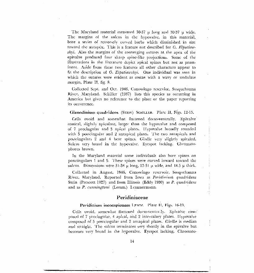
The Maryland material measured 30-37 p long and 30-37 p wide. The margins of the sulcus in tlhe hypovalve, in this material, bore a series of retrorsely curved barbs which diminisheld in size towar*d the antapex. This is a feature not described for G. EEpatiew- skyi. Also the margins of the converging sutures at the apex of the epivalve produced four sharp spine-like projections. Some of the illustrations in the literature 'depict apical spines but not as prom- inent. Aside from these two features all other characters appear to fit the description of G. Elpatiewskyi. One individual was seen in which the sutures were evident as costae with a wavy or undulate margin, Plate 11, fig. 8.
Collected Sept. and Oct. 1946, Conowingo reservior, Susquehanna River, Maryland. Schiller (1937) lists this species as occurring in America but gives no reference to 'the place or the paper reporting its occurrence.
Glenodinium quadridlens (STE,IN) SCHILLER. Plate 11, Figs. 12-15.
Cells ovoid and somewhat flattened dorso-ventral'ly. Epivalve conical, slightly apiculate, larger than the hypovalve and composed of 7 precingular an'd 5 apical plates. Mypovalve broasdly rounded with 5 p~stcingu~lar and 2 antapical plates. The two antapicals and . postcingulars 2 and 4 bear spines. Gir'dle very slightly spiraled. Sulcus very br'oad in the hypovalve. Eyespot lacking. Chromato- phores brown.
In the Maryland material some individuals also bore spines on po~tcingul~rs 1 and 5. These spines were curved inward toward the sulcus. Dimensions were 31-38 p long, 27-31 p wide, and 18.5 p thick.
Collected in August, 1946, Canowingo reservoir, Susquehanna River, Maryland. Reported from Iowa as Peridinium quadridens Stein (Prescott 1927) and fr'om Illinois (Eddy 1930) as P. quadridens and as P. czlnningtoni (Lemm.) Lemmermann.
Peridiniaceae
Peridinium inconspicuum LEMM. Plate 11, Figs. 16-19.
Cells ovoid, somewhat flattened dorso-ventrally. Epivalve com- posed of 7 precingular, 4 apical, and 2 intercalary plates. Hypovalve composed of 5 postcingular and 2 antapical plates. Girdle is median and straight. The sulcus terminates very shortly in the epivalve but becomes very broad i s the hypovalve. Eyespot lacking. Chromato-

phores numerous, parietal and brown. Cells 15-24 p long, 12-20 p wide.
The Maryland material measured 24.8 p long and 21 p wide. Collected in August, 1942, from Big Fresh Creek near Solomons, Maryland. Previously reported from Illinois by Eddy (1930) .
Peridinium umbonatum STEIN. Plate 11, Figs. 20-24.
Ce,lls ovoid and clearly flattened dorso-ventrally. Epivalve much larger than the hypovalve, composed of 7 precingular, 4 apical and 2 intercalary plates. The latter two tsouching. Hypovalve of 5 postcingular anld 2 antapical plates. Girdle very slightly spiraled. Stigma lacking. Chromatophores light to dark brown, numerous, discoidal and parietal. Cells 35-40 p long and 30 p wide.
The Maryland material measured 19.8-28.5 p long, and 15.5-26 p wide. This is the single most widespread and abundant of thecate forms in Maryland. It exhibited wide variation in shape including Lemmermann's variety inaequale in which the hypovalve is much narrower than the epivalve. Collected in July, Aug., Sept. 1942 at Solomons, Md. and July of that year from Patchetts pond, near Denton, Md; April 1946 through March 1947 from Olivet, Lower Marlboro, Priest Bridge, Belcamp, Webster, Rising Sun and Dublin, Maryland. These ~~ollections were made from permanent swamps, and from temporary po'ols in roadside ditches, from warm summer waters and from under the ice in winter. Cysts were more abundant in the winter collections. These are b~oadly ellipsoidal with a firm smraaoth wall. The chromatophores are parietal. Few to several orange or reld oil globules may be present also. The protoplast is highly vacuolate with large vacuoles interspersed with strands of cytoplasm radiating from the center to the periphery.
Previously reported from Illinois (Eddy 1930).
Peridinium Volzii LEMMERMANN. Plate 111, Figs. 1-4.
Cells somewhat spherical in face view, slightly flattened dorso- ventrally. Girdle median, spiraled. Sulcus reaching about half way into the epivalve, broadening slightly below the girdle and extending to the posterior margin of the hypovalve. Epivalve composed of 7
eprecingular, 4 apical an'd 3 intercalary plates with the second inter- calary touching the third, fourth and fifth precingulars. Hypovalve composed of 5 postcingular and 2 anltapical $plates. The plates are strongly areolate and the intercalary bands are generally wide and

heavily striated. Chromatophores brown, discoisdal and parietal. Cells about as wide as long; 38-60 p in diameter.
The Maryland material measured 49.5-53 p in diameter. Collected in March 1947, Priest Bridge swamp, Maryland.
Previously reported from Minnesota (Eddy 1930) and from Mas- sachusetts (Prescott and Croasdale 1937) .
Peridinium Willei HHITF.-KAAS. Plate 111, Figs. 5-8. Cells circular in face view and slightly to greatly flattened dorso-
ventrally. Girdle spiraled, slightly belsow the middle of the cell. Sulcus extending into the epivalve but little, gradually broadening as it extends to the #posterior margin of the hypovalve. Plate formula same as that of P. Volzii, but the intercalary and 2, 3, 4 apical plates are elongated laterally and the first apical is very much larger than that of P. Volzii. The plates are strongly areolate and the inter- calary bands are heavily striated. Chromatophores, dark-brown, numerous, and parietal. Cells 40-60 p long and 45-70 p wide.
The Maryland Material measured 52-68 p long and 62-68 p wide. An eyespot was evident in this material, though t'his structure is not mentioned in the literature. It lies to the right side on the lateral wall of the sulcus about midway between the girdle and ata- pex. At the posterior end, the margirns of the sulcus are continuous with the margin of the adjacent intercalary banlds. Here they flare out as very evident costae with finger-like thickenings. Collected in January 1947 from Belcamp and again in May from Priest Bridge, Maryland. Previously reported from Iowa (Prescott 1927) and from Ten(ness6e (Esddy 1930) .
Peridinium cinctum (MULLER) EHRENBERG. Plate IV, Figs. 1-5. Cells ovoid to spherical, slightly flattened dorso-ventrally. Girdle
median and slightly spiraled. Sulcus extends very little into the epivalve and reaches the posterior margin af the hypovalve. The second anterior intercalary #plate touches the third ansd fourth pre- cingulars, otherwise the plate formula is the same as that of the two preceding species. An eyespot is lacking. Chromatophores are dark-brown, numerous anld parietal. Cells 40-60 p long, 35-55 p wide. The plates are areolate and the intercalary bands heavily striated.
The Maryland material measured 52-73 p long and 60-75.5 p wide. Cysts of this species were also collected. These are spherical and surrounded by a gelatinous material which includes the remnanli of the theca. The protoplast is highly vacuolate and strands of cy10

plasm radiate from the center to the periphery. The chrornatophores are parietal and gathered into small rosettes. One or more large recl oil globules may be present. Collected in July, Aug. and Sept., 1942, near Solfomons, Md., and again intermittently froin June 1946 through May, 1947, from lo we^ Marlboro, Priest Bridge, Belcamp and Webster, Maryland. Previously reported from Iowa, California, Illi- nvia, Wisconsin, Tennessee and Massachusetts (Allen 1920, Prescott 1927, Eddy 1930, Prescott and Croasdale 1937) .
Heterodiniaceae Ceratium hirundinella ( 0 . F. M.) BERGH. Plate IV, Figs. 6-9. Cells insymmetrical with one apical and two or three antapical
horns. Epivalve composed of 3 precingular and 2 apical plates. Hypovalve composed of 3 postcingular and one antapical plate. Plates heavily sculptured with areolae. Cells variable in size and number 'of antapical horns; 95-400 p lo'ng. Chromatophores yellow- brown, numerous aqd parietal.
Collecteld July, Aug. and Sept., 1946, Conowingo Reservoir, Susque- hanna River, Maryland. Cosmopolitan. Previously reported from many localities in the United States.
T h e cysts of this species were relatively abundant h plankton sanlples taken during August of 1946. The chromatophores are indistinguishable i n these, the cells being of an even dark-brown coJor. A large red oil globule may be present. The cysts are tetra- hedral with stout to slender horn-like processes a t the apices.
LITERATURE CITED ALLEN, W. E. 1920.-Quantitative and statistical study of the San
Joaquin in and near Stockton, California. Univ. of Calif. Pub. Zool. 22: 1-292
CUNHA, A. da 1913.-Beitrage zur Kenntilis der Protozoenfauna Bra- siliens. Mem. Inst. Oswaldo Cruz 5, 101 T.9,10
EDDY, S. 1930.-The fresh-water armored or thecate Dinoflagellates. Trans. Amer. Miccosc. Soc. 49: 277-32.1 8 plates
PRESCOTT, G. W, 1927.-Motile algae of Iowa. Univ. Iowa Stud. Nat. Hist. XII, (6) : 5-40, F1. I-X
-- 1944.-New species and varieties of Wisconsin Algae. Farlowia 1, (3) : 347-385, i fig., 5 plates.
PRESCOTT, G. W. and CROASDAEE, H. T. 1937-New or noteworthy fresh-water algae of Massachusetts. Trans. Amer. Microsc. So@. 56: 269-282, 3 plates.
SCHIELER, J. 1933.-Dinoflagellatae (Peridineae) in E. Rabenhorst's Krytogamen-Flora, Part 1, vol. 10. Leipzig, 617 pages, 631 figs.
--- 1937.-ibid, Part 2, vol. 10, 589 pages, 612 figs. SMUJA, H. 1939.-Beitrag zur Algenflora Lettlands 11. Materiali Latvijas
algu florai 11. Acta Horti Rotanici. Universitatis Latviensis XI- XII: 41-161, 11 plates

PLATE I
All figures X808
Figs . 1. 2 . Gymnodinium albulum Lindemann .................................... P • 5
Fi.gs . 3. 4 . Gyrnnodinium ordinaturn Skuja ................................................ Pa ti
Figs . 5. 6 . Gymnodiniuna tricerntium Skuja ., ............................................. P- 7 6
Figs . 7.9 . Gymnodiniunz marylandicum Thompson ...................... P . 7
Figs . 10.14 . Gy mnodinium cestocoetes Thompson ............................... P-
Fig . 10 division in germination of a cyst . Fig . 11 cyst . Fig . 14 division of a motilc cell .
* . Figs . 15. 16 . Gymnodinium neglectum (Schilling) Lindem ....... p 9
Fig . 15 newly encysted cell still surrounded by old theca . ............................ . Figs . 17-1 9 . Gymnodinium fuscunz (Ehrenb. ) Stein p 10
. ...................................... . . Figs 20.22 AIassartia Musei (Danysz.) Schiller p I1
Fig . 22 cyst . ...... . . . . Figs 23, 24 Gyrodiniz~m pusillurn (Schilling) Kof et Swezy p 12
. ........................................................ . . Fig 25 Gymnodinium viride Penard 1) 11
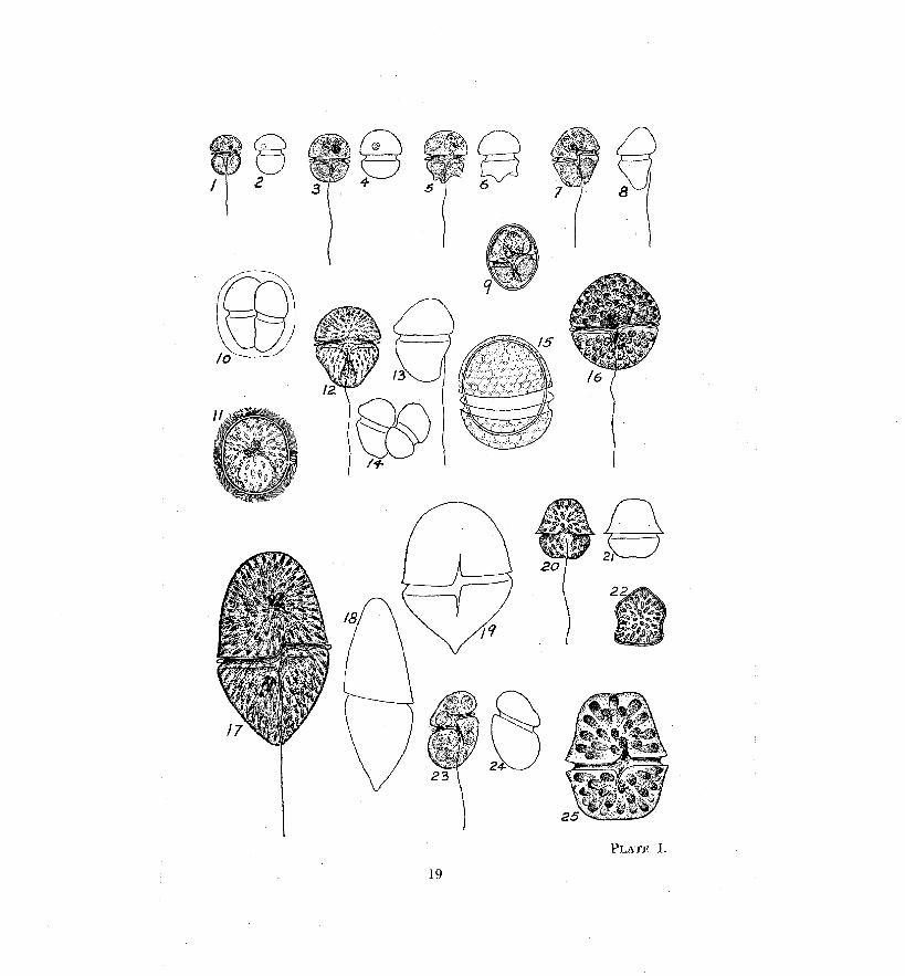

PLATE 11
All figures X808
Figs. 1-3. Hemitliniunz nasutzl~n Stein ........................................................... p. 12 Fig. 1 ventral view.
Fig. 2 side view.
Fig. 3 dorsal view.
Figs. 4-6: Hemidinium oclzrnceum Levander ..................................... p. 13
ventral, side and dorsal views.
.............. Figs. 7-1 1. Glendinium Elj!mtiewskyi (Ostenf.) Schiller p. 13
Fig. 7, 8 dorsal view.
Fig. 9 epivalve view.
Fig. 10 ventral view.
Fig. 11 hypovalve view.
Figs. 12-15. Glenudinium. quadridens (Stein) Schiller ..................... p. 14
Fig. 12 dorsal view.
Fig. 13 epivalve view.
Fig. 14 ventral view.
Fig. 15 hypovalve view.
Figs. 16-19. Peridinium inconspicuz~rn Lemmermann ........................ p. 14
Fig. 16 epivalvc view.
Fig. 17 ventral view.
Fig. 1s dorsal view.
Fig. 19 hypovalve view.
...................................................... Figs. 20-24. Peridiniz~rn umbonatum. Stein p. 15 Fig. 20 epivalve view. Fig. Z1 ventral view. Fig. 22 hypovalve view. Fig. 23 dorsal view.
Fig. 24 cyst.


PLATE I11
All figures X808
...................................................... Figs. 1-4. Peridinium Volzii Lemmermann p. 15 Fig. 1 epivalve view.
Fig. 2 ventral view.
Fig. 3 dorsal view.
Fig. 4 hypovalve view.
......................................................... Figs. 5-8. Peridinium Willei Huitf.-Kaas p. 16
Fig. 5 epivalve view.
Fig. 6 dorsal view.
Fig. 7 ventral view.
Fig. 8 hypovalve view.
PLATE IV
Figs. 1-5. (X808) Peridinium cinctz~m (Muller) Ehrenb ............. p. 16
Fig. 1 epivalve view.
Fig. 2 ventral view.
Fig. 3 dorsal view.
Fig. 4 hypovalve view. Fig. 5 cyst.
......... Figs. 6-9. ( ~ 4 0 4 ) Ceratium lzirundinellk (0. F. M.) Bergh p 17 Fig. 6 ventral view. Figs. 7, 8 cysts. Fig. 9 dorsal view.

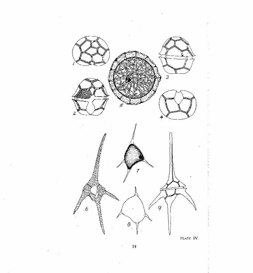
PLATE IV.
24

PUBLICATIONS "
DEPARTMENT OF RESEARCH AND EDUCATION Chesapeake Biological Laboratory
Herewith are listed the publications, both reprinted and regular, of the Department of Research and Education and its Chesapeake Biological Laboratory. Numbers 1-41 were printed as Contributions. Thereafter, athey were issued as Publications. This series is to be continued in re- porting results from the Department's research programme. Future treatises dealing with education and economic problems will be issued in a new series, Educational Series, 15 of which are listed herein.
1. Aspects of the Oyster Season in Maryland-Truitt, R. V., 1927. State Press.
2, The Fishes of Maryl'and-Truitt, R. V., B. A. Bean and H. W. Fowler, 1930fl State Press."
3. Recent Oyster Researches on Chesapeake Bay in Maryland-Truitt, R. V., 1930. State Press.
4. The (Oyster and the Oyster Industry of Maryland-Truitt, R. V., 1931. State Press."
5. Scientific Fisheries Work in Maryland-Truitt, R. V., 1932. Trans. Am. Fish. Sot.*
6. Growth of Mya arenaria L. in the Bay of Fundy Region-Newcombe, C. L., Vol. 13, 1935. Jour. of dr2es."
'7. Striped Bass Investigations in the !Chesapeake Bay-Truitt, R. V. and V. D. Vladykov,-1937. Trans. Am. Fish Soc.
8. 1936 Annual Report, Chesapeake Biological Laboratory-Truitt, R. V., 1937. State Press.*
9. The Importance of Sport Fishing in Maryland-Truitt, R. V. and V. D. Vlad-ykov, 1936. Trans. Am. Fish. Soc.*
16. Variations in Growth Indices of Mua arenaria L. on the Atlantic Coast of North America-Newcombe, c.-L. and H. Kessler, 1936. Ecology, 17: No. 3."
11. The Oyster Packing Industry of Baltimore-Nichol, A. J., 1937: Weant Press.
12. Baltimore and the Oyster Industry-Truitt, R. V., 1937. Watkins Press.
13. V'ariations of Dactylometra quinquecirrha--littleford, R. A. and R. V. Truitt, Science, 1937."
14. Utilization of Marine Products-Miller, T. M., 1947. Weant Press. Weant Press.
15. Variations in Growth Indices sf Venus mercenaria L. Fsoni Widely Separated Environments of the Atlantic Coas&Newcornbe, C . L., S. J. Thompson and H. Kessler, Vol. 16.'
16. Notes on Maryland Algae-Bold, Harold C. Bztll. Torrey Bot. Club- Can. Jour. of Res., 1 9 3 8 4 o i n t cont. with University of Maryland.
17. Populations of the Shad, Alosa sapidissima, along the Atlantic Coast Regions-Vladykov, V. D. and D. 8. Wallace, Trans. Am. Fish. Soc., 1937."
18, Migration in the Striped Bass, Roccus lineaius, of the Chesapeake Bay-Wadykov, V. D. and D. H. Wallace, Trans. Am. Soc., 1937."
19. Oxygen-Poor Waters of the Chesapeake Bay-Newcombe, C. L. and William A. Rorne, Science, Vol. 88, 1938."
28. The Relative Growth of Parts in the Blue Crab, Callinectes aapidus Rathbun-Gray, Ellen H. 'and Curtis L. Newcornbe, Growth, Vol. 2, 1938."
Those starred (9 have been exhausted and therefore, are not available f o r distribution.

21. Studies on the Physiics and Chemistry of Estuarine Waters in Chesa- peake Bay-Newcombe, Curtis L., William A. Horne and Boland B, Shepherd, Journl, Mar. Res., Vo1. 11, 1939.
22. 1937 Annual Report, Chesapeake Biological Laboratory-Truitt, R. V., 138. State Press."
23. The 1938 Program of the Chesapeake Biological Laboratory-Newcornbe, C. L., Collecting Net, 1938."
24. Studies of Moulting in Callinectes sapidus Rathbun-Gray, Ellen H. and Curtis L. Newcombe, Growtlz, Vol. 11, No. 4, 1939.*
25. Progresls of the Rock and Shad Research Work a t the Chesapeake Biplogical Laboratory-Wallace, D. H. and R. V. Truitt, Trans. Am. Fzsh. Soc., 1938."
26. Sport Fishing in Maryland-Truitt, R. V., 1938. Weant Press.* 27. Our Water Resources and Their Conservation-Truitt, R. V., 1939.
Weant Press." 28. The Distribution of Phosphates in the Chesapeake Bay-Newcornbe,
Curtis L. and Andrew G. Lang, Pro., Anz. Phil. Soc., Vol. 81, 1939." 29. Distribution of Rathkea-Littleford, Robert A,, Nature, Vol. 143, 1939." 30. 1938 Annual Report of the Chesapeake Biological Labo~atosy-Truitt,
R. V., 1939. State Press." 31. The Life Cycle of Dactylometra quinqueci~rha, L. Aggassiz in =the
Chesapeake Bay-Littleford, Robert A., Biol. Bull., Vul. LXXVII, 1939. 32. Variations in the Phosphorus Content of Estuarine Waters of the
Chesapeake Bay-Near Solomons Island, Maryland-Newcombe, G. L. and H. F. Brust, Jour. Mar. Res., Val. 111, 1940.
33. Crab Mortality on Chesapeake Bay Shedding Floats-Beaven, G. F. and R. V. Truitt, An. Rpt. Md. Cons. Dept., 1939.
34. 1939 Annual Report, Chesapeake Biological Laboratory-Truitt, R. V., 1940. Weant Press.
35. Preliminary Report on Respiratory Studies of Littorinla irroratu L.- Newcombe, Curtis L. with Charles Miller and Donald W. Chappell, Nature, Vd . 137, 1936.*
36. Validity of Concentric Rings of Mya arenaria, L. for Determining Age-Newcornbe, C. L., Nature, Vol. 137, 1936."
37. An Experimental Study of Certain Quantitative Plankton Methods- Newcombe, C. L., Robert A. Littleford and Boland B. Shepherd, Ecology, Vol. 21, 1940.
38. Observations 'on the Alkalinity of Estuarine Waters of the Chesa~eake Bay Near Solomons Island, Maryland-Brust, Harry F. and C. L. Newcombe, Jour. Mar. Res., Vol. LII, 1940.
39. Sexual Development of the Croaker (Microogon undulatus) and Dis- tri,butions of the Early Stages in the Chesapeake Bay-Wallace, David H., Trans. Am. Fish. Soc., 1940.
40. 1940 Annual Report, Chesapeake giological Laboratory-Truitt, R. V., 1941. Wean6 Press."
41. Maryland Commercial Fish Hatchery Operations, 1940-Wallace, David H. and R. V. Truitt, 1941. Weaat Press."
42. Seafood and the DieLLemon,, J. M. and R. V. Truitt, 1941. Stnfe Press. 43. Studies of the.Effects of Industrial Pollution in the Lower Patapsco
River Area-Olson, R. A., 1941. Maxur-Nicholson Press. 44. The Taxonomv and Distribution of the Blorinp Saonqes (Cliorzidae)
Alonx the Atlantic Coast of North America-Old, Marcus C. , 1941. Muzq~r-Nicholson Press.
45. A Rapid Response Thermocouple Unit of High Sensitivity for the Determination of Temperature Stratification in Natural Waters- Olson, R. A., 1941. Weant Press.
46. Ovarian Growth and Ovulation in the Mature Blue Crab, Callinectes saoidus Rathbun-Hard, W. L., 1942. Weant Press.
47. 1941 Annual Report, Chesapeake Biological Laboratory-Truitt, R. V., 1942. Weant Press."
Those starred (*) have been exhausted and therefore, are not available f o r distribution.
2 6
48. Control of Fishing Intensity in Maryland-Hammer, Ralph C. and R. V. T'ruitt, l'rans. Am. Fish. Soc., 1942."
49. The Zoeal Stage of the Blue Crab-Churchill, E. P., 1942. Maurice Lesser Press.
50. Observations on the Feeding Habits of Marlin-Wallace, David H., 1942. Weant Press.*
51. Ecological and Life History Aspects of the Red-Jointed Fiddler Crab, Uca Minax (LeConte), in the Regions of Solomons Island, Maryland- Gray, Ellen H., 1942. Mazirice Lesser Press."
52. Experimental Sponge Crab Plantings and Crab Larvae Distribution in lthe Region of Crisfield, Maryland-Graham, James G. and G. F. Beauen, 1942. Weant Press."
53. A Study of the Crab Pot a s a Fcishing Gear-Davis, Charies C., 1942. Weant Press."
54. Maryland Commercial Fish Hatchery Operations, 1941-42-Truitt, R. V., 1942. Weand Press.
55. Cephalomonas, A New Genus of the Volvocales-Noe Higinbotham, 1942. Joint cont. with Osborne Bot. Lab., Yale University. Bull., Torrey Bot. Club, 1942.
56. 1942 Annual Report, Chesapeake Biological Laboratory-Beaven, G. F., 1944. Weant Press.
57. Indications of Compensatory Growth in the Striped Bass, Riccus sazatilis Walbaum, as Revealed by a Study of the Scales-Tiller, Richard E., 1943. Weant Press.
58. The Larval Stages of the Calanoid Copepod Eurgtemora hirundoides (Nordquist)-Davis, Charles C., 1944. Weant Press.
59. E,bullition of Gases from Marsh and Lake Waters-Conger, Paul S., 1944. Weant Press.
60. Maryland Commercial Fish Hatchery Operations, 1943-Hammer, Ralph, C., 1944. Weant Press.
81. On Four ~ d e c i e s of Copepoda New to Chesapeake ~ a y , ' w i t h Description of a New Variety of Paracalanus crassirostris Dahl-Davis, Charles C., 1944. Weant Press.
62. Histochemical Observations on Glycogen in the Liver of $he Blue Crab, Callinectes sapidus Rathbun-Sister Leonide Regan, 1944. Weant Press.
63. A Survey of the Bryozoa of Chesapeake Bay-Osburn, Raymond C., 1944. Weantt Press.
64. Environmental Characteristics of a River Estuary-Nash, Carroll B., 1947. Jour. Mar, Res., vol. VI, No. 3.
65. Some Observations on Seasonal Variations in Planlrton Population, Patuxent River, Maryland, 1943-1945-Morse, Dorothy Clum, 1947. Weant Press.
66. Anatomy and Histology of the Male Reproductive Systeni of Callinectes sapidus Rathbun-Cronin, Lewis Eugene, 1947. Journal of Morphology, Vol. 81, No. 2, September.
67. Fresh-Water Dinoflagellates of Maryland-Thompson, R. N., 1947. Weant Press.
E D U C A T I O N A L SERIES. 1. The Maryland Fishery Management Plan-Tiller, R. E., 1944. Wennt
Press. 2. The Maryland Management Plan and the Rock Fishery-Tiller, R. E.,
1944. Weant Press. 3. Maryland Laws Governing Natural Resources-1944. Mou~ice Eesse7-
Press. 4. Svmposium-1944 (Maryland Conservation Forum. Weant Press. 5. The Marvland Management Plan and the Shad Fishery-Tiller, R. E.,
1944. Weant Press. --
Those starred (*) have been exhausted and therefore, are not available for distribution.

Fishery Management Plan Productive-Tiller, R. E., 1944. Weant Press. The Oyster-'fruitt, R. V., 1944. Reprinted from BIOS: Vol. XV, No. 3. O1ctolber, 1944. Maryland's Oyster Problem-Beaven, G. F., 1945. French-Bray Co. Maryland Stream Flow Records-Singewald, J. T., Jr., 1945. French- Bray Co. Maryland Board of Natural Resources Officials and Organizations Con- cerned with the Conservation of Natural Resources-Department ~f Research and Education, Solomons, Maryland, 1945. Maryland Comniercial Fish Hatchery Operations-Hammer, Ralph C., 1946. Weant Press. Trees of Maryland-Kaylor, Joseph F., 1946. Weant Press. Gr,ound Water in the Baltimore Area, Maryland-B~ennett, Robert R., 1946. Weant Press. Utilization of Marine Products-Miller, T. M., 1947. Weant Press. Maryland Commercial Fish Hatchery Operations-Coker, Coit M., 1947. Weant Press.


