Frolich, Human Anatomy, Mechanics of Movemen MECHANICS OF MOVEMENT Tissues and Structures Involved Muscle Nerve Bone Cartilage What are Tendons? Role of Joints Mechanics of Joints Making it all work
Transcript
Frolich, Human Anatomy, Mechanics of Movement
MECHANICS OF MOVEMENT
Tissues and Structures Involved Muscle Nerve Bone Cartilage
What are Tendons? Role of Joints Mechanics of Joints Making it all work
Frolich, Human Anatomy, Mechanics of Movement
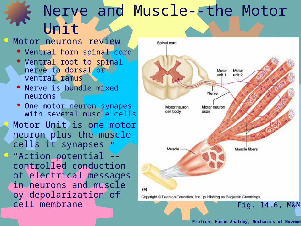
Nerve and Muscle--the Motor Unit Motor neurons review
Ventral horn spinal cord Ventral root to spinal nerve to
dorsal or ventral ramus Nerve is bundle mixed
neurons One motor neuron synapes
with several muscle cells Motor Unit is one motor
neuron plus the muscle cells it synapses
“Action potential”--controlled conduction of electrical messages in neurons and muscle by depolarization of cell membrane
Fig. 14.6, M&M
Frolich, Human Anatomy, Mechanics of Movement
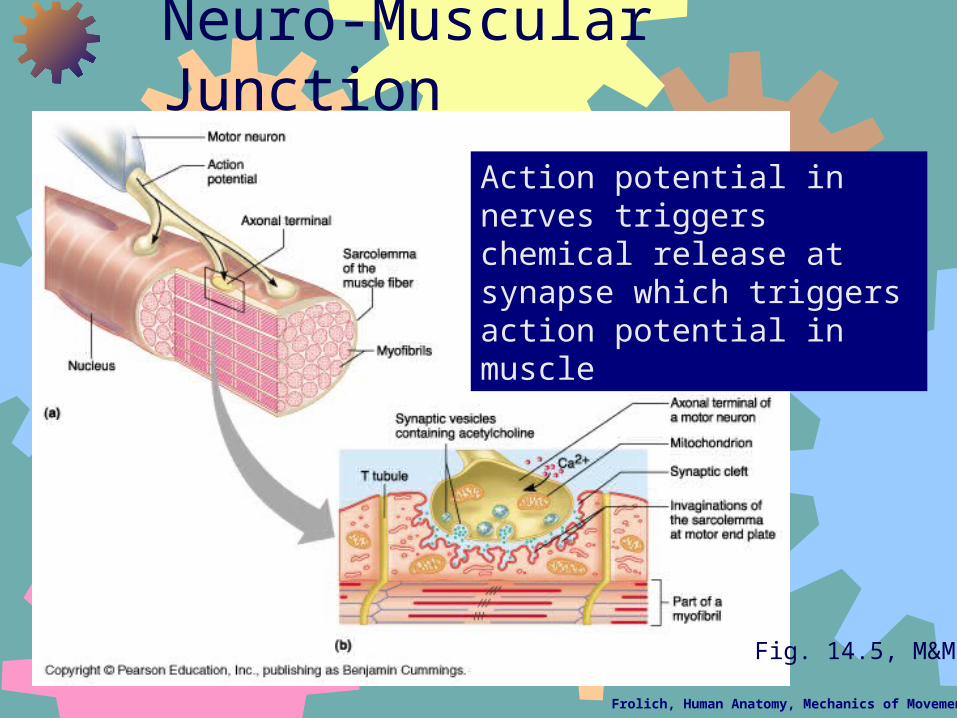
Neuro-Muscular Junction
Action potential in nerves triggers chemical release at synapse which triggers action potential in muscle
Fig. 14.5, M&M
Frolich, Human Anatomy, Mechanics of Movement
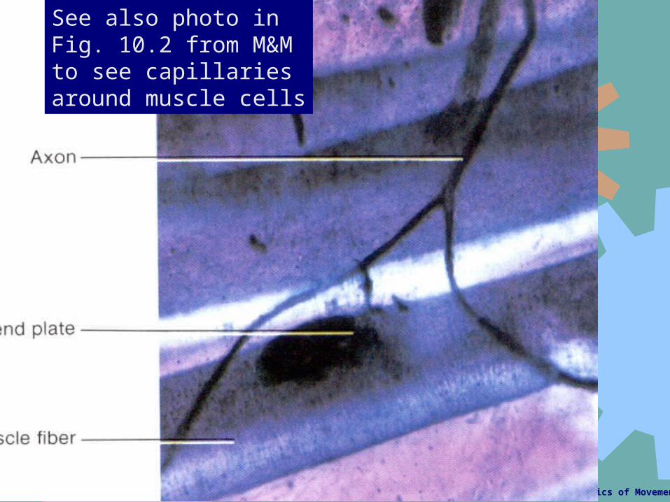
See also photo in Fig. 10.2 from M&M to see capillaries around muscle cells
Frolich, Human Anatomy, Mechanics of Movement
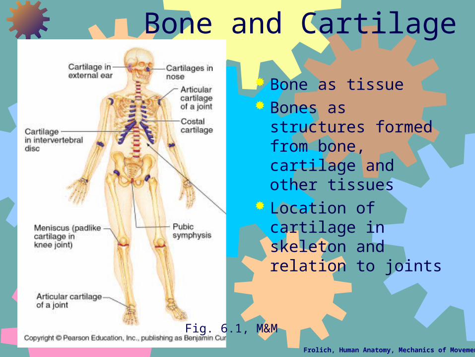
Bone and Cartilage
Bone as tissue Bones as structures
formed from bone, cartilage and other tissues
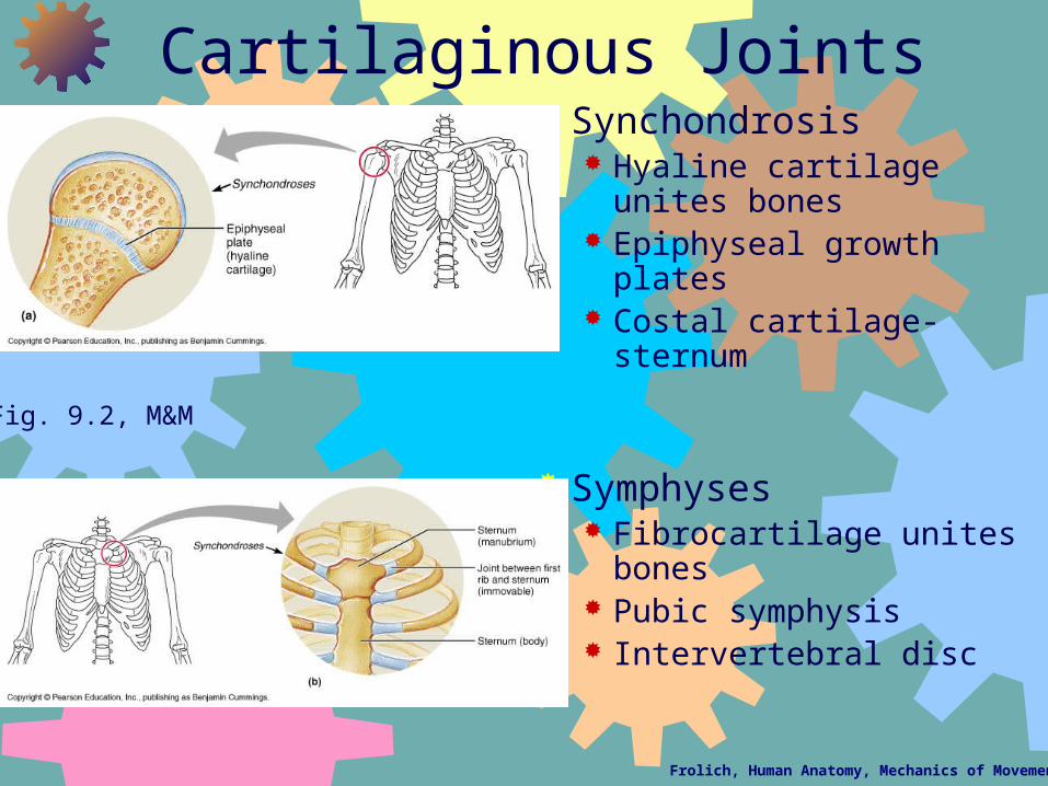
Location of cartilage in skeleton and relation to joints
Fig. 6.1, M&M
Frolich, Human Anatomy, Mechanics of Movement
HOW MOVEMENT HAPPENS: Muscles Pull on Tendons to Move Bones at Connections called Joints or Articulations
Frolich, Human Anatomy, Mechanics of Movement
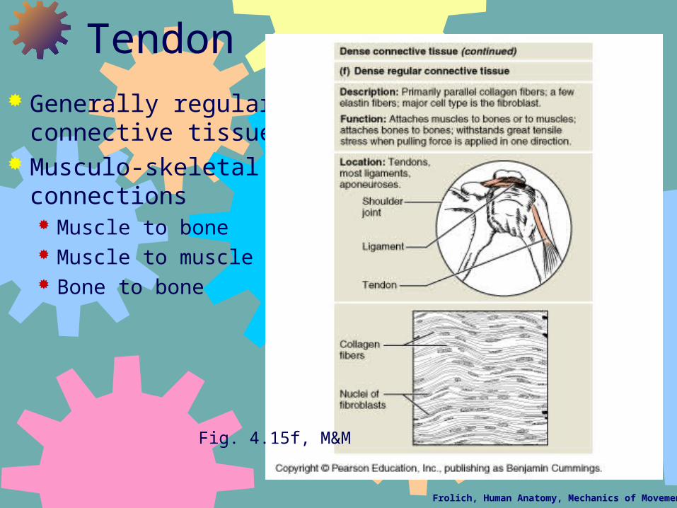
Tendon Generally regular
connective tissue Musculo-skeletal
connections Muscle to bone Muscle to muscle Bone to bone
Fig. 4.15f, M&M
Frolich, Human Anatomy, Mechanics of Movement
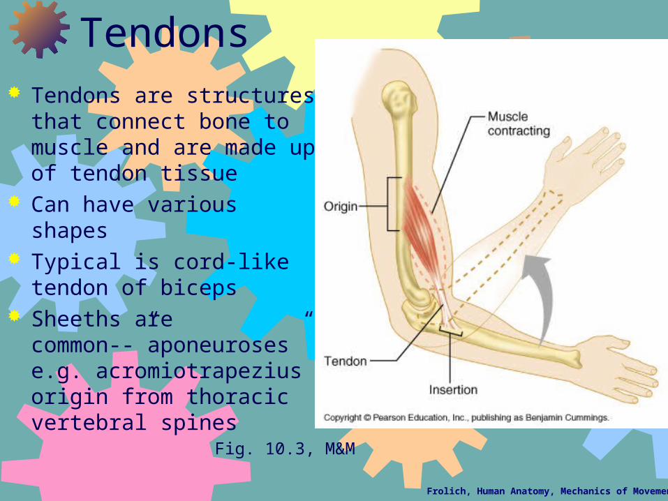
Tendons Tendons are structures that
connect bone to muscle and are made up of tendon tissue
Can have various shapes Typical is cord-like tendon of
biceps Sheeths are
common--”aponeuroses” e.g. acromiotrapezius origin from thoracic vertebral spines
Fig. 10.3, M&M
Frolich, Human Anatomy, Mechanics of Movement
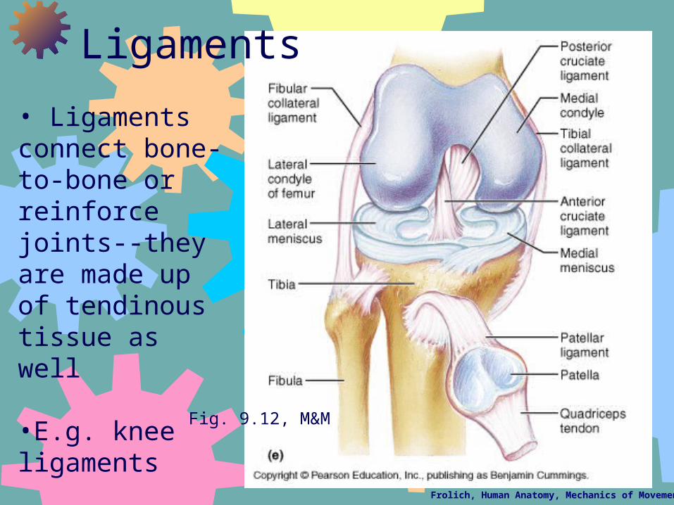
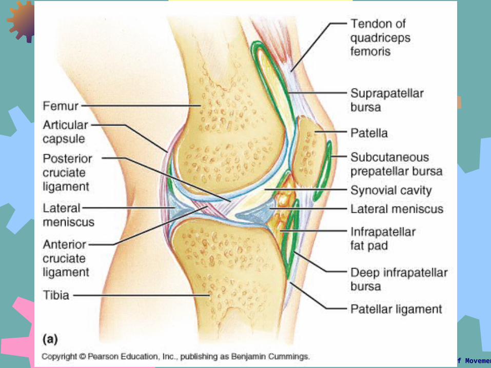
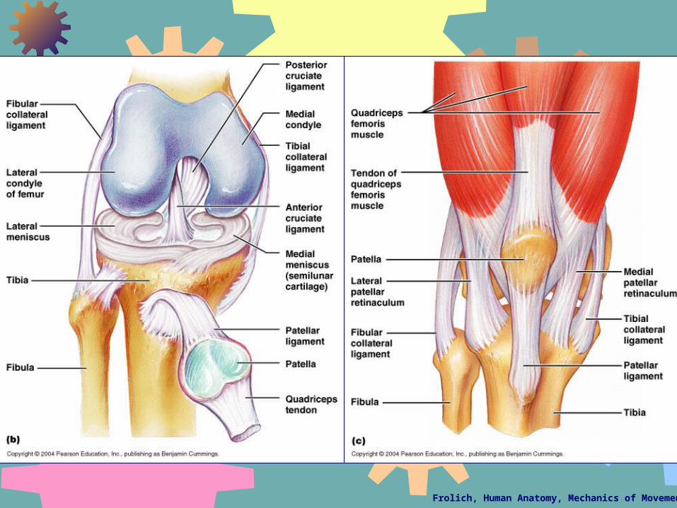
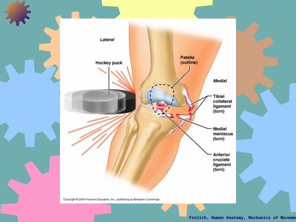
• Ligaments connect bone-to-bone or reinforce joints--they are made up of tendinous tissue as well
•E.g. knee ligaments
Ligaments
Fig. 9.12, M&M
Frolich, Human Anatomy, Mechanics of Movement
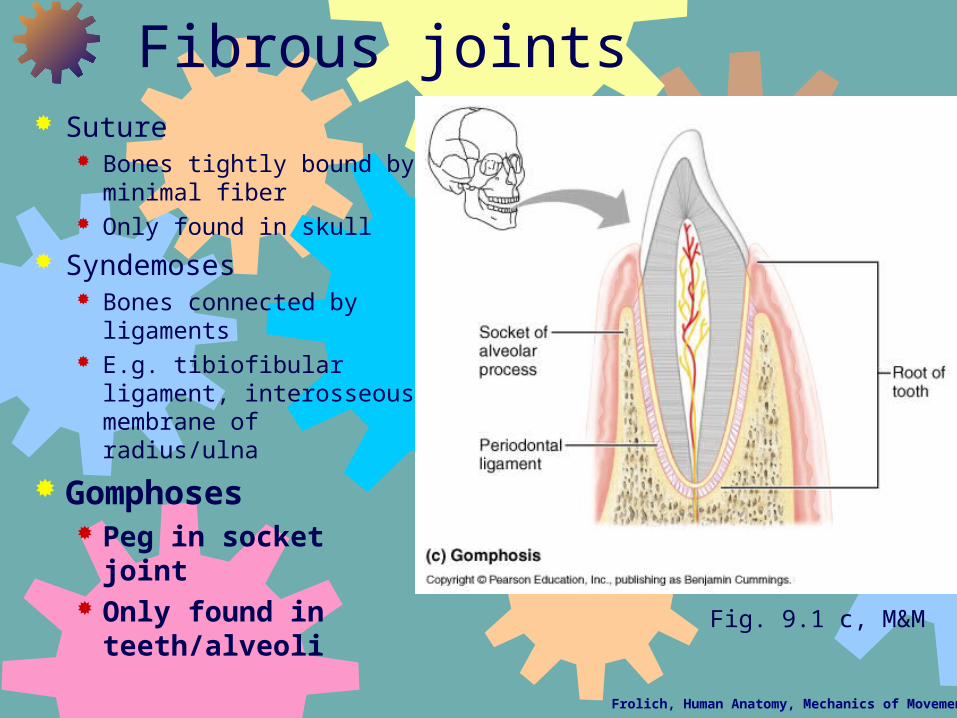
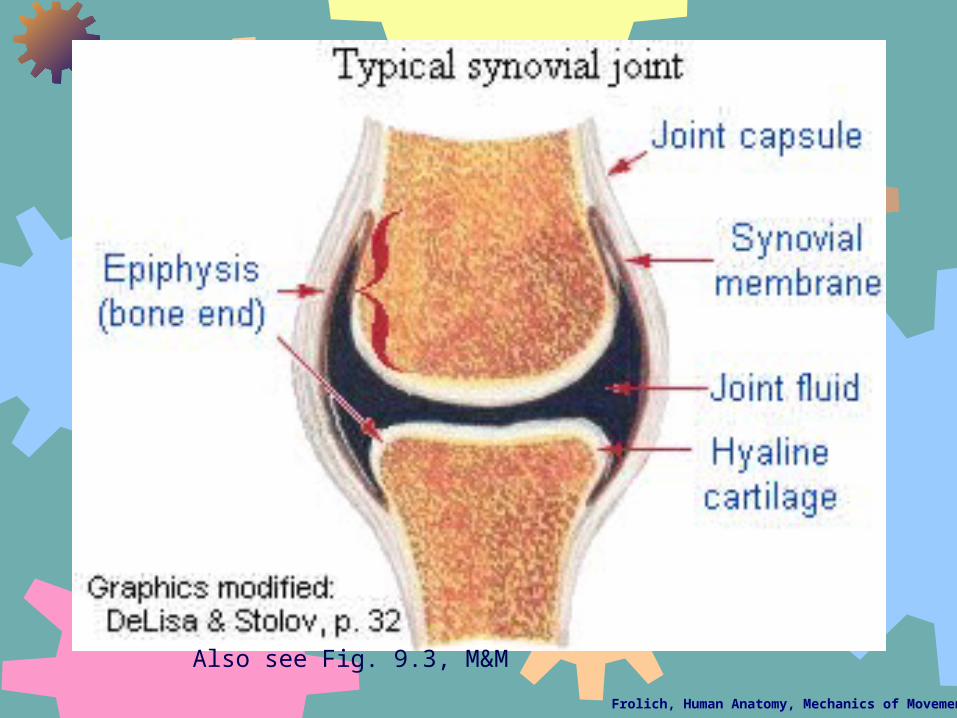
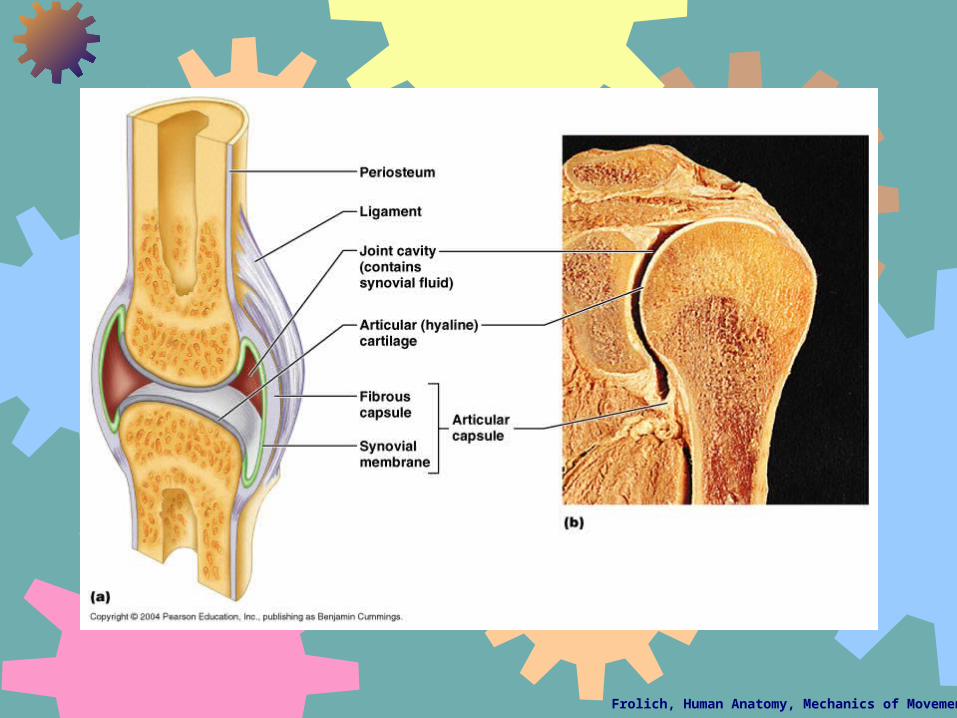
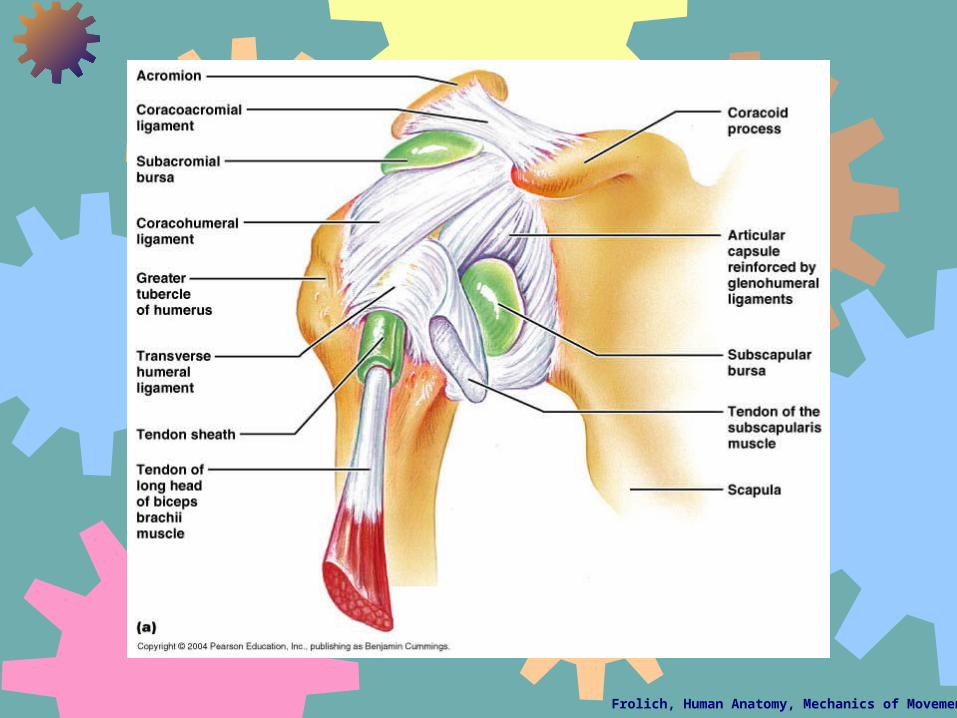
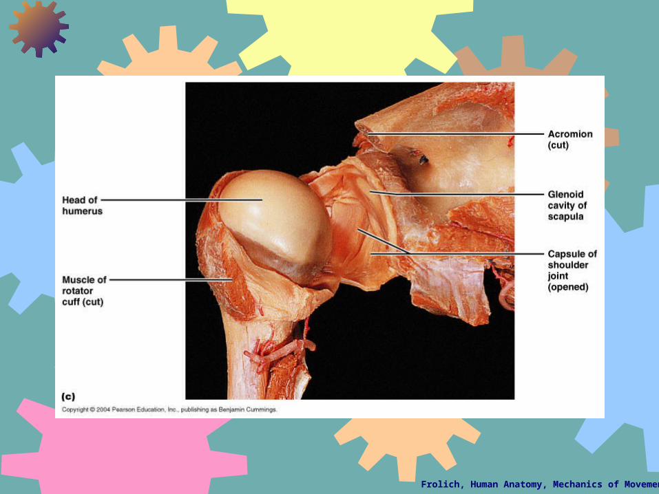
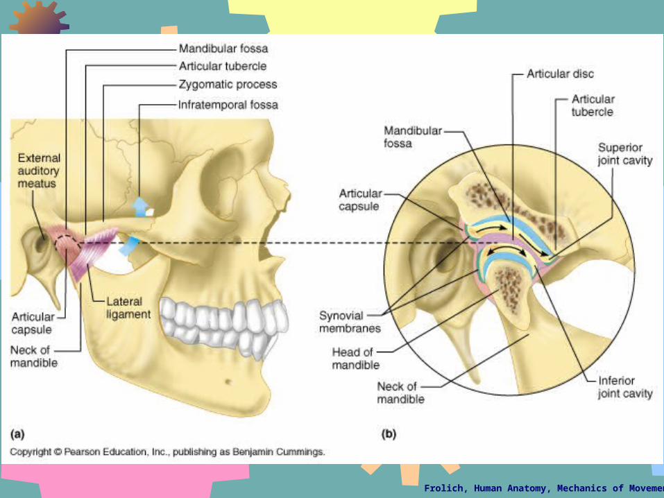
Joints or Articulations
Connections between bonesUsually, but not always allow for