62
From Gene to Protein Chapter 17 A.P. Biology Rick Knowles
| Date post: | 23-Dec-2015 |
| Category: |
Documents |
| Upload: | sarah-norman |
| View: | 216 times |
| Download: | 0 times |

From Gene to Protein
Chapter 17
A.P. Biology
Rick Knowles
• Overview: The Flow of Genetic Information • The information content of DNA
– Is in the form of specific sequences of nucleotides along the DNA strands.
• The DNA inherited by an organism– Leads to specific traits by dictating the synthesis of
proteins
• The process by which DNA directs protein synthesis, gene expression– Includes two stages, called transcription and
translation

• The ribosome– Is part of the cellular machinery for translation,
polypeptide synthesis
Figure 17.1

One Gene-One Enzyme Hypothesis
• 1941- Beadle and Tatum- created mutants in the fungus Neurospora using X-rays.
• Exposed wild-type spores to the mutagen.• Change growth from a complete to minimal
media.• Mutants no longer able to synthesize
essential organics (e.g. amino acids)

• Using genetic crosses– They determined that their mutants fell into three classes,
each mutated in a different gene
Figure 17.2
Working with the mold Neurospora crassa, George Beadle and Edward Tatum had isolated mutants requiring arginine in their growth medium and had shown genetically that these mutants fell into three classes, each defective in a different gene. From other considerations, they suspected that the metabolic pathway of arginine biosynthesis included the precursors ornithine and citrulline. Their most famous experiment, shown here, tested both their one gene–one enzyme hypothesis and their postulated arginine pathway. In this experiment, they grew their three classes of mutants under the four different conditions shown in the Results section below.
The wild-type strain required only the minimal medium for growth. The three classes of mutants had different growth requirements
EXPERIMENT
RESULTS
Class IMutants
Class IIMutants
Class IIIMutantsWild type
Minimal medium(MM)(control)
MM +Ornithine
MM +Citrulline
MM +Arginine(control)

CONCLUSIONFrom the growth patterns of the mutants, Beadle and Tatum deduced that each mutant was unable to carry out one step in the pathway for synthesizing arginine, presumably because it lacked the necessary enzyme. Because each of their mutants was mutated in a single gene, they concluded that each mutated gene must normally dictate the production of one enzyme. Their results supported the one gene–one enzyme hypothesis and also confirmed the arginine pathway. (Notice that a mutant can grow only if supplied with a compound made after the defective step.)
Class IMutants(mutationin gene A)
Class IIMutants(mutationin gene B)
Class IIIMutants(mutationin gene C)Wild type
Gene A
Gene B
Gene C
Precursor Precursor Precursor Precursor
Ornithine Ornithine Ornithine Ornithine
Citrulline Citrulline Citrulline Citrulline
Arginine Arginine Arginine Arginine
EnzymeA
EnzymeB
EnzymeC
A A A
B B B
C C C

One Gene-One Enzyme• Beadle and Tatum- found several mutants.• Found a different site for each enzyme.• Each mutant had a different mutation at a
different site (enzyme) on the chromosome.• Each mutant had a defect in a different
enzyme.• Concluded: one gene encodes one enzyme.

One Gene-One Enzyme Hypothesis
• Genetic traits are expressed as a result of the activities of enzymes.
• Many enzymes contain multiple subunits.• Each subunit encoded by a differernt
gene.• Now, referred to as one gene-one
polypeptide.

Basic Principles of Transcription and Translation
• Transcription– Is the synthesis of RNA under the direction of DNA– Produces messenger RNA (mRNA)
• Translation– Is the actual synthesis of a polypeptide, which occurs
under the direction of mRNA– Occurs on ribosomes

• In prokaryotes– Transcription and translation occur together
Figure 17.3a
Prokaryotic cell. In a cell lacking a nucleus, mRNAproduced by transcription is immediately translatedwithout additional processing.
(a)
TRANSLATION
TRANSCRIPTIONDNA
mRNA
Ribosome
Polypeptide

• In eukaryotes– RNA transcripts are modified before becoming true
mRNA
Figure 17.3b
Eukaryotic cell. The nucleus provides a separatecompartment for transcription. The original RNAtranscript, called pre-mRNA, is processed in various ways before leaving the nucleus as mRNA.
(b)
TRANSCRIPTION
RNA PROCESSING
TRANSLATION
mRNA
DNA
Pre-mRNA
Polypeptide
Ribosome
Nuclearenvelope

Central Dogma
• Cells are governed by a cellular chain of command
DNA RNA Protein

• During transcription– The gene determines the sequence of bases along the
length of an mRNA molecule
Figure 17.4
DNAmolecule
Gene 1
Gene 2
Gene 3
DNA strand(template)
TRANSCRIPTION
mRNA
Protein
TRANSLATION
Amino acid
A C C A A A C C G A G T
U G G U U U G G C U C A
Trp Phe Gly Ser
Codon
3 5
35

Copyright © 2005 Pearson Education, Inc. publishing as Benjamin Cummings
Cracking the Code
• A codon in messenger RNA
– Is either translated into an amino acid or serves as a translational stop signal
Figure 17.5
Second mRNA baseU C A G
U
C
A
G
UUUUUCUUAUUG
CUUCUCCUACUG
AUUAUCAUAAUG
GUUGUCGUAGUG
Met orstart
Phe
Leu
Leu
lle
Val
UCUUCCUCAUCG
CCUCCCCCACCG
ACUACCACAACG
GCUGCCGCAGCG
Ser
Pro
Thr
Ala
UAUUAC
UGUUGC
Tyr Cys
CAUCACCAACAG
CGUCGCCGACGG
AAUAACAAAAAG
AGUAGCAGAAGG
GAUGACGAAGAG
GGUGGCGGAGGG
UGGUAAUAG Stop
Stop UGA StopTrp
His
Gln
Asn
Lys
Asp
Arg
Ser
Arg
Gly
U
CA
GUCAG
UCAG
UCAG
Fir
st m
RN
A b
ase
(5
en
d)
Th
ird
mR
NA
bas
e (3
en
d)
Glu

Copyright © 2005 Pearson Education, Inc. publishing as Benjamin Cummings
Almost a Universal Code
• In laboratory experiments
– Genes can be transcribed and translated after being transplanted from one species to another
Figure 17.6

Molecular Components of Transcription
• RNA synthesis–Is catalyzed by RNA polymerase, which
pries the DNA strands apart and hooks together the RNA nucleotides.
–Follows the same base-pairing rules as DNA, except that in RNA, uracil substitutes for thymine.

Copyright © 2005 Pearson Education, Inc. publishing as Benjamin Cummings
Synthesis of an RNA Transcript
• The stages of transcription are
– Initiation
– Elongation
– Termination
Figure 17.7
PromoterTranscription unit
RNA polymerase
Start point
53
35
35
53
53
35
53
35
5
5
Rewound
RNA
RNA
transcript
3
3Completed RNA transcript
Unwound
DNA
RNA
transcript
Template strand of DNA
DNA
1 Initiation. After RNA polymerase binds to the promoter, the DNA strands unwind, and the polymerase initiates RNA synthesis at the start point on the template strand.
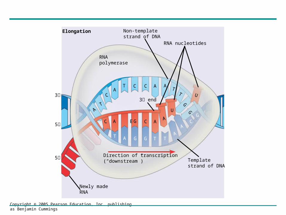
2 Elongation. The polymerase moves downstream, unwinding theDNA and elongating the RNA transcript 5 3 . In the wake of transcription, the DNA strands re-form a double helix.
3 Termination. Eventually, the RNAtranscript is released, and the polymerase detaches from the DNA.
Copyright © 2005 Pearson Education, Inc. publishing as Benjamin Cummings
Elongation
RNApolymerase
Non-templatestrand of DNA
RNA nucleotides
3 end
C A E G C AA
U
T A G G T TA
AC
G
U
AT
CA
T C C A AT
T
GG
3
5
5
Newly madeRNA
Direction of transcription(“downstream”) Template
strand of DNA

Copyright © 2005 Pearson Education, Inc. publishing as Benjamin Cummings
RNA Polymerase Binding and Initiation of Transcription
• Promoters -DNA sequences that signal the initiation of RNA synthesis.
• Transcription factors- other proteins that help eukaryotic RNA polymerase recognize promoter sequences
Figure 17.8Figure 17.8
TRANSCRIPTION
RNA PROCESSING
TRANSLATION
DNA
Pre-mRNA
mRNA
Ribosome
Polypeptide
T A T AAA AAT AT T T T
TATA box Start point TemplateDNA strand
53
35
Transcriptionfactors
53
35
Promoter
53
355
RNA polymerase IITranscription factors
RNA transcript
Transcription initiation complex
Eukaryotic promoters1
Several transcriptionfactors
2
Additional transcriptionfactors
3


Elongation of the RNA Strand
• As RNA polymerase moves along the DNA– It continues to untwist the double helix, exposing
about 10 to 20 DNA bases at a time for pairing with RNA nucleotides.

Termination of Transcription
• The mechanisms of termination- are different in prokaryotes and eukaryotes

• Concept 17.3: Eukaryotic cells modify RNA after transcription
• Enzymes in the eukaryotic nucleus– Modify pre-mRNA in specific ways before the
genetic messages are dispatched to the cytoplasm

Copyright © 2005 Pearson Education, Inc. publishing as Benjamin Cummings
Alteration of mRNA Ends
• Each end of a pre-mRNA molecule is modified in a particular way
– The 5 end receives a modified nucleotide cap
– The 3 end gets a poly-A tail
Figure 17.9
A modified guanine nucleotideadded to the 5 end
50 to 250 adenine nucleotidesadded to the 3 end
Protein-coding segment Polyadenylation signal
Poly-A tail3 UTRStop codonStart codon
5 Cap 5 UTR
AAUAAA AAA…AAA
TRANSCRIPTION
RNA PROCESSING
DNA
Pre-mRNA
mRNA
TRANSLATIONRibosome
Polypeptide
G P P P
5 3

Copyright © 2005 Pearson Education, Inc. publishing as Benjamin Cummings
Split Genes and RNA Splicing
• RNA splicing
– Removes introns and joins exons
Figure 17.10
TRANSCRIPTION
RNA PROCESSING
DNA
Pre-mRNA
mRNA
TRANSLATION
Ribosome
Polypeptide
5’ CapExon Intron
1
5’
30 31
Exon Intron
104 105 146
Exon 3’Poly-A tail
Poly-A tail
Introns cut out andexons spliced together
Codingsegment
5’ Cap1 146
3’ UTR3’ UTR
Pre-mRNA
mRNA

Copyright © 2005 Pearson Education, Inc. publishing as Benjamin Cummings
• Is carried out by spliceosomes in some cases
Figure 17.11
RNA transcript (pre-mRNA)
Exon 1 Intron Exon 2
Other proteinsProtein
snRNA
snRNPs
Spliceosome
Spliceosomecomponents
Cut-outintron
mRNA
Exon 1 Exon 2
5’
5’
5’
1
2
3
Ribozymes• Ribozymes-
– Are catalytic RNA molecules that function as enzymes and can splice RNA
• The presence of introns-– Allows for alternative RNA splicing.

Copyright © 2005 Pearson Education, Inc. publishing as Benjamin Cummings
• Proteins often have a modular architecture
– Consisting of discrete structural and functional regions called domains
• In many cases
– Different exons code for the different domains in a protein
Figure 17.12
GeneDNA
Exon 1 Intron Exon 2 Intron Exon 3
Transcription
RNA processing
Translation
Domain 3
Domain 1
Domain 2
Polypeptide

Some Differences between Prokaryotic and
Eukaryotic Protein Synthesis

Prokaryotes Eukaryotes
• Lack introns- no mRNA processing.
• Begin translation before transcription is finished.
• Have introns- mRNA is spliced.
• mRNA completely formed before translation.
• mRNA has a 5’ methyl G cap added


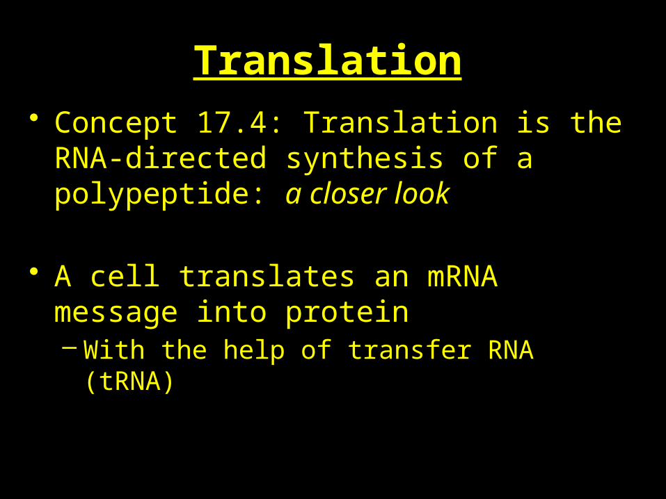
Translation• Concept 17.4: Translation is the RNA-directed
synthesis of a polypeptide: a closer look
• A cell translates an mRNA message into protein– With the help of transfer RNA (tRNA)
Copyright © 2005 Pearson Education, Inc. publishing as Benjamin Cummings
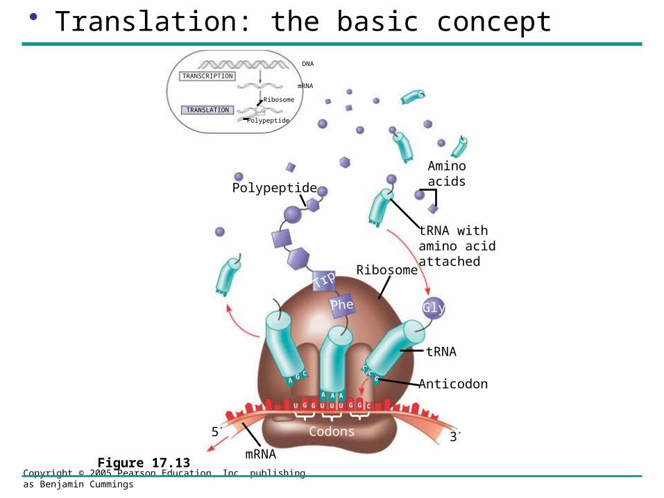
• Translation: the basic concept
Figure 17.13
TRANSCRIPTION
TRANSLATION
DNA
mRNA
Ribosome
Polypeptide
Polypeptide
Aminoacids
tRNA withamino acidattached
Ribosome
tRNA
Anticodon
mRNA
Trp
Phe Gly
AG C
A A A
CC
G
U G G U U U G G C
Codons5 3

• Molecules of tRNA are not all identical– Each carries a specific amino acid on one end– Each has an anticodon on the other end
Copyright © 2005 Pearson Education, Inc. publishing as Benjamin Cummings
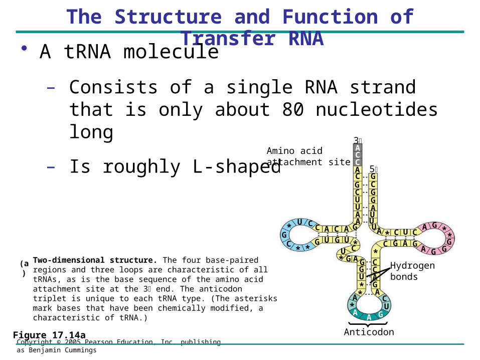
The Structure and Function of Transfer RNA
ACC
• A tRNA molecule
– Consists of a single RNA strand that is only about 80 nucleotides long
– Is roughly L-shaped
Figure 17.14a
Two-dimensional structure. The four base-paired regions and three loops are characteristic of all tRNAs, as is the base sequence of the amino acid attachment site at the 3 end. The anticodon triplet is unique to each tRNA type. (The asterisks mark bases that have been chemically modified, a characteristic of tRNA.)
(a)
3
CCACGCUUAA
GACACCU*
GC
* *G U G U *CU
* G AGGU**A
*A
A GUC
AGACC*
C G A GA G G
G*
*GA
CUC*AUUUAGGCG5
Amino acidattachment site
Hydrogenbonds
Anticodon
A

Copyright © 2005 Pearson Education, Inc. publishing as Benjamin Cummings
A tRNA
Figure 17.14b
(b) Three-dimensional structure
Symbol used in this book
Amino acidattachment site
Hydrogen bonds
AnticodonAnticodon
A A G
5’
3’
3’ 5’
(c)

Copyright © 2005 Pearson Education, Inc. publishing as Benjamin Cummings
• A specific enzyme called an aminoacyl-tRNA synthetase
– Joins each amino acid to the correct tRNA
Figure 17.15
Amino acid
ATP
Adenosine
Pyrophosphate
Adenosine
Adenosine
Phosphates
tRNA
P P P
P
P Pi
Pi
Pi
P
AMP
Aminoacyl tRNA(an “activatedamino acid”)
Aminoacyl-tRNAsynthetase (enzyme)
Active site binds theamino acid and ATP.
1
ATP loses two P groupsand joins amino acid as AMP.
2
3 AppropriatetRNA covalentlyBonds to aminoAcid, displacingAMP.
Activated amino acidis released by the enzyme.
4

Ribosomes
• Ribosomes:– Facilitate the specific coupling of tRNA anticodons
with mRNA codons during protein synthesis

Copyright © 2005 Pearson Education, Inc. publishing as Benjamin Cummings
The Ribosomal Subunits
– Are constructed of proteins and RNA molecules named ribosomal RNA or rRNA
Figure 17.16a
TRANSCRIPTION
TRANSLATION
DNA
mRNA
Ribosome
Polypeptide Exit tunnelGrowingpolypeptide
tRNAmolecules
EP
A
Largesubunit
Smallsubunit
mRNA
Computer model of functioning ribosome. This is a model of a bacterial ribosome, showing its overall shape. The eukaryotic ribosome is roughly similar. A ribosomal subunit is an aggregate of ribosomal RNA molecules and proteins.
(a)
53

Copyright © 2005 Pearson Education, Inc. publishing as Benjamin Cummings
• The ribosome has three binding sites for tRNA
– The P site
– The A site
– The E site
Figure 17.16b
E P A
P site (Peptidyl-tRNAbinding site)
E site (Exit site)
mRNAbinding site
A site (Aminoacyl-tRNA binding site)
Largesubunit
Smallsubunit
Schematic model showing binding sites. A ribosome has an mRNA binding site and three tRNA binding sites, known as the A, P, and E sites. This schematic ribosome will appear in later diagrams.
(b)
Copyright © 2005 Pearson Education, Inc. publishing as Benjamin Cummings
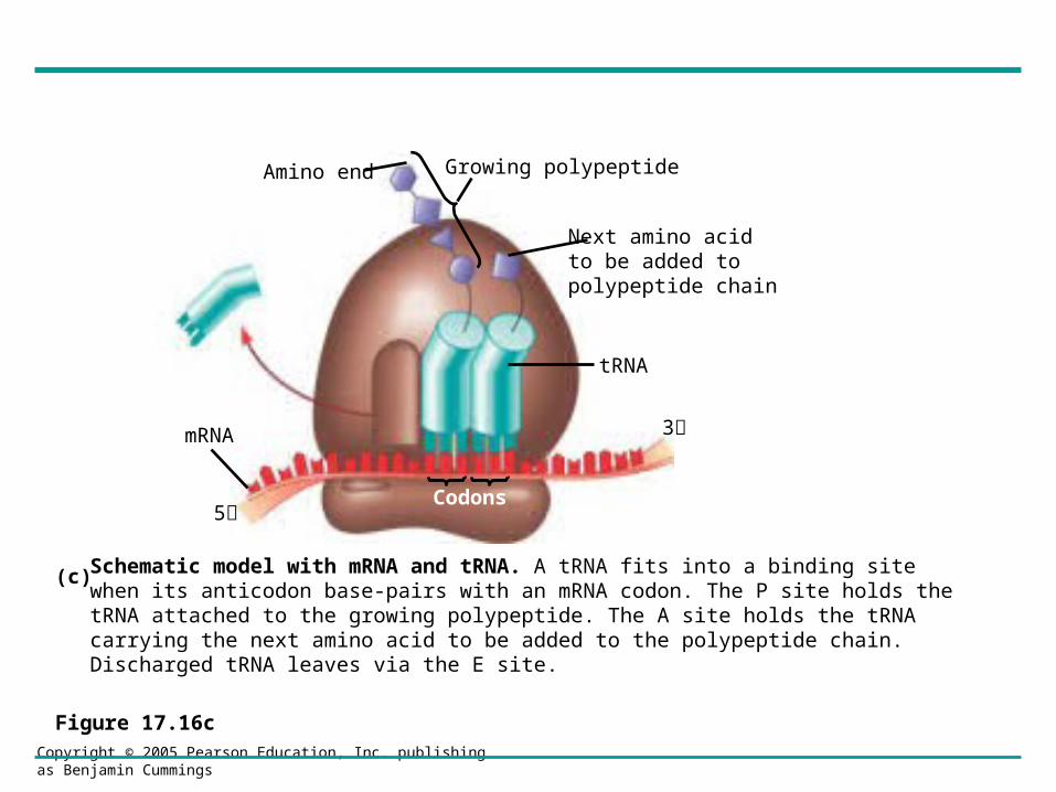
Figure 17.16c
Amino end Growing polypeptide
Next amino acidto be added topolypeptide chain
tRNA
mRNA
Codons
3
5
Schematic model with mRNA and tRNA. A tRNA fits into a binding site when its anticodon base-pairs with an mRNA codon. The P site holds the tRNA attached to the growing polypeptide. The A site holds the tRNA carrying the next amino acid to be added to the polypeptide chain. Discharged tRNA leaves via the E site.
(c)

Copyright © 2005 Pearson Education, Inc. publishing as Benjamin Cummings
Building a Polypeptide
• We can divide translation into three stages
– Initiation
– Elongation
– Termination

Copyright © 2005 Pearson Education, Inc. publishing as Benjamin Cummings
Ribosome Association and Initiation of Translation
• The initiation stage of translation
– Brings together mRNA, tRNA bearing the first amino acid of the polypeptide, and two subunits of a ribosome
Largeribosomalsubunit
The arrival of a large ribosomal subunit completes the initiation complex. Proteins called initiationfactors (not shown) are required to bring all the translation components together. GTP provides the energy for the assembly. The initiator tRNA is in the P site; the A site is available to the tRNA bearing the next amino acid.
2
Initiator tRNA
mRNA
mRNA binding site Smallribosomalsubunit
Translation initiation complex
P site
GDPGTP
Start codon
A small ribosomal subunit binds to a molecule of mRNA. In a prokaryotic cell, the mRNA binding site on this subunit recognizes a specific nucleotide sequence on the mRNA just upstream of the start codon. An initiator tRNA, with the anticodon UAC, base-pairs with the start codon, AUG. This tRNA carries the amino acid methionine (Met).
1
MetMet
U A CA U G
E A
3
5
5
3
35 35
Figure 17.17

Copyright © 2005 Pearson Education, Inc. publishing as Benjamin Cummings
Elongation of the Polypeptide Chain
• In the elongation stage of translation
– Amino acids are added one by one to the preceding amino acid
Figure 17.18
Amino endof polypeptide
mRNA
Ribosome ready fornext aminoacyl tRNA
E
P A
E
P A
E
P A
E
P A
GDPGTP
GTP
GDP
2
2
site site5
3
TRANSCRIPTION
TRANSLATION
DNA
mRNARibosome
Polypeptide
Codon recognition. The anticodon of an incoming aminoacyl tRNA base-pairs with the complementary mRNA codon in the A site. Hydrolysisof GTP increases the accuracy andefficiency of this step.
1
Peptide bond formation. An rRNA molecule of the large subunit catalyzes the formation of a peptide bond between the new amino acid in the A site and the carboxyl end of the growing polypeptide in the P site. This step attaches the polypeptide to the tRNA in the A site.
2
Translocation. The ribosome translocates the tRNA in the A site to the P site. The empty tRNA in the P site is moved to the E site, where it is released. The mRNA moves along with its bound tRNAs,bringing the next codon to be translated into the A site.
3

Copyright © 2005 Pearson Education, Inc. publishing as Benjamin Cummings
Termination of Translation
• The final stage of translation is termination
– When the ribosome reaches a stop codon in the mRNA
Figure 17.19
Release factor
Freepolypeptide
Stop codon(UAG, UAA, or UGA)
5
3 3
5
35
When a ribosome reaches a stop codon on mRNA, the A site of the ribosome accepts a protein called a release factor instead of tRNA.
1 The release factor hydrolyzes the bond between the tRNA in the P site and the last amino acid of the polypeptide chain. The polypeptide is thus freed from the ribosome.
2 3 The two ribosomal subunits and the other components of the assembly dissociate.

Copyright © 2005 Pearson Education, Inc. publishing as Benjamin Cummings
Polyribosomes
• A number of ribosomes can translate a single mRNA molecule simultaneously
– Forming a polyribosome
Figure 17.20a, b
Growingpolypeptides
Completedpolypeptide
Incomingribosomalsubunits
Start of mRNA(5 end)
End of mRNA(3 end)
Polyribosome
An mRNA molecule is generally translated simultaneously by several ribosomes in clusters called polyribosomes.
(a)
Ribosomes
mRNA
This micrograph shows a large polyribosome in a prokaryotic cell (TEM).
0.1 µm(b)

Completing and Targeting the Functional Protein
• Polypeptide chains-undergo modifications after the translation process.
• After translation– proteins may be modified in ways that affect their
three-dimensional shape.
Copyright © 2005 Pearson Education, Inc. publishing as Benjamin Cummings
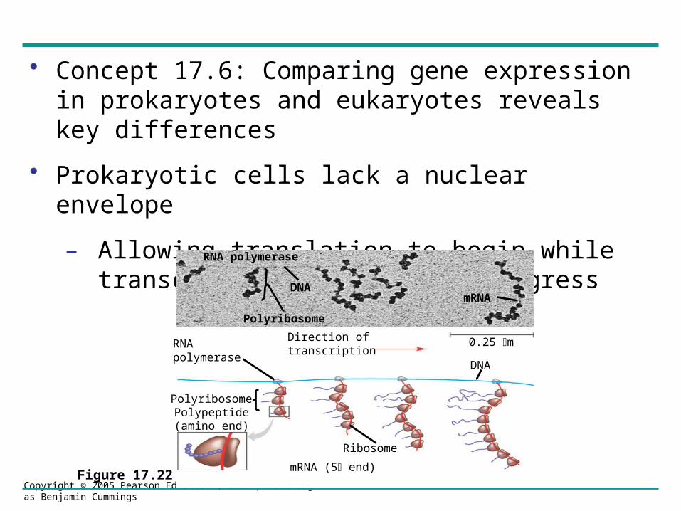
• Concept 17.6: Comparing gene expression in prokaryotes and eukaryotes reveals key differences
• Prokaryotic cells lack a nuclear envelope
– Allowing translation to begin while transcription is still in progress
Figure 17.22
DNA
Polyribosome
mRNA
Direction oftranscription
0.25 mRNApolymerase
Polyribosome
Ribosome
DNA
mRNA (5 end)
RNA polymerase
Polypeptide(amino end)


• Concept 17.7: Point mutations can affect protein structure and function
• Mutations:– Are changes in the genetic material of a cell
• Point mutations:– Are changes in just one base pair of a gene
Copyright © 2005 Pearson Education, Inc. publishing as Benjamin Cummings
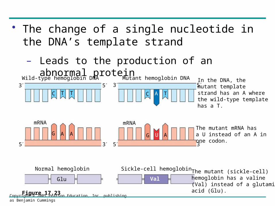
• The change of a single nucleotide in the DNA’s template strand
– Leads to the production of an abnormal protein
Figure 17.23
In the DNA, themutant templatestrand has an A where the wild-type template has a T.
The mutant mRNA has a U instead of an A in one codon.
The mutant (sickle-cell) hemoglobin has a valine (Val) instead of a glutamic acid (Glu).
Mutant hemoglobin DNAWild-type hemoglobin DNA
mRNA mRNA
Normal hemoglobin Sickle-cell hemoglobin
Glu Val
C T T C A T
G A A G U A
3 5 3 5
5 35 3

Types of Point Mutations
• Point mutations within a gene can be divided into two general categories– Base-pair substitutions– Base-pair insertions or deletions

Copyright © 2005 Pearson Education, Inc. publishing as Benjamin Cummings
Base-pair Substitution
– Is the replacement of one nucleotide and its partner with another pair of nucleotides
– Can cause missense or nonsense
Figure 17.24
Wild type
A U G A A G U U U G G C U A AmRNA
5Protein Met Lys Phe Gly
Stop
Carboxyl endAmino end
3
A U G A A G U U U G G U U A A
Met Lys Phe Gly
Base-pair substitution
No effect on amino acid sequenceU instead of C
Stop
A U G A A G U U U A G U U A A
Met Lys Phe Ser Stop
A U G U A G U U U G G C U A A
Met Stop
Missense A instead of G
NonsenseU instead of A

Copyright © 2005 Pearson Education, Inc. publishing as Benjamin Cummings
Insertions and Deletions
– Are additions or losses of nucleotide pairs in a gene
– May produce frameshift mutations
Figure 17.25
mRNA
Protein
Wild type
A U G A A G U U U G G C U A A5’
Met Lys Phe Gly
Amino end Carboxyl end
Stop
Base-pair insertion or deletion
Frameshift causing immediate nonsense
A U G U A A G U U U G G C U A
A U G A A G U U G G C U A A
A U G U U U G G C U A A
Met Stop
U
Met Lys Leu Ala
Met Phe GlyStop
MissingA A G
Missing
Extra U
Frameshift causing extensive missense
Insertion or deletion of 3 nucleotides:no frameshift but extra or missing amino acid
3’
Copyright © 2005 Pearson Education, Inc. publishing as Benjamin Cummings
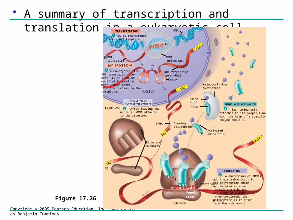
• A summary of transcription and translation in a eukaryotic cell
Figure 17.26
TRANSCRIPTION
RNA is transcribedfrom a DNA template.
DNA
RNApolymerase
RNAtranscript
RNA PROCESSING
In eukaryotes, theRNA transcript (pre-mRNA) is spliced andmodified to producemRNA, which movesfrom the nucleus to thecytoplasm.
Exon
Poly-A
RNA transcript(pre-mRNA)
Intron
NUCLEUSCap
FORMATION OFINITIATION COMPLEX
After leaving thenucleus, mRNA attachesto the ribosome.
CYTOPLASM
mRNA
Poly-A
Growingpolypeptide
Ribosomalsubunits
Cap
Aminoacyl-tRNAsynthetase
Aminoacid
tRNAAMINO ACID ACTIVATION
Each amino acidattaches to its proper tRNAwith the help of a specificenzyme and ATP.
Activatedamino acid
TRANSLATION
A succession of tRNAsadd their amino acids tothe polypeptide chainas the mRNA is movedthrough the ribosomeone codon at a time.(When completed, thepolypeptide is releasedfrom the ribosome.)
Anticodon
A CC
A A AU G G U U U A U G
U ACE A
Ribosome
1
Poly-A
5
5
3
Codon
2
3 4
5


Prokaryotes Eukaryotes• mRNA begins at
an AUG start codon, no cap.
• Multiple genes on one mRNA.
• 70S ribosome used.
• mRNA has a poly A tail added at the 3’.
• A single gene on one mRNA.
• 80S ribosome used.


Other Differences in Gene Organization• Tandem Clusters- several hundred
genes organized together; Ex. rRNA genes.
• Multigene Families- genes very different from each other, but encode similar proteins; Ex. globin genes.
• Pseudogenes- silent copies of genes inactivated by mutations.


