From sneeze to wheeze: Non-invasive studies on asthma and rhinitis Aronsson, David 2009 Link to publication Citation for published version (APA): Aronsson, D. (2009). From sneeze to wheeze: Non-invasive studies on asthma and rhinitis. Lund University: Faculty of Medicine. Total number of authors: 1 General rights Unless other specific re-use rights are stated the following general rights apply: Copyright and moral rights for the publications made accessible in the public portal are retained by the authors and/or other copyright owners and it is a condition of accessing publications that users recognise and abide by the legal requirements associated with these rights. • Users may download and print one copy of any publication from the public portal for the purpose of private study or research. • You may not further distribute the material or use it for any profit-making activity or commercial gain • You may freely distribute the URL identifying the publication in the public portal Read more about Creative commons licenses: https://creativecommons.org/licenses/ Take down policy If you believe that this document breaches copyright please contact us providing details, and we will remove access to the work immediately and investigate your claim.
Transcript
LUND UNIVERSITY
PO Box 117221 00 Lund+46 46-222 00 00
From sneeze to wheeze: Non-invasive studies on asthma and rhinitis
Aronsson, David
2009
Link to publication
Citation for published version (APA):Aronsson, D. (2009). From sneeze to wheeze: Non-invasive studies on asthma and rhinitis. Lund University:Faculty of Medicine.
Total number of authors:1
General rightsUnless other specific re-use rights are stated the following general rights apply:Copyright and moral rights for the publications made accessible in the public portal are retained by the authorsand/or other copyright owners and it is a condition of accessing publications that users recognise and abide by thelegal requirements associated with these rights. • Users may download and print one copy of any publication from the public portal for the purpose of private studyor research. • You may not further distribute the material or use it for any profit-making activity or commercial gain • You may freely distribute the URL identifying the publication in the public portal
Read more about Creative commons licenses: https://creativecommons.org/licenses/Take down policyIf you believe that this document breaches copyright please contact us providing details, and we will removeaccess to the work immediately and investigate your claim.
If you know the enemy and know yourself, your victory will not stand in doubt;
if you know Heaven and know Earth, you may make your victory complete.
Sun Tzu
6
7
TABLE OF CONTENTS LIST OF PAPERS ......................................................................................................... 9 ABBREVIATIONS...................................................................................................... 11 INTRODUCTION ....................................................................................................... 13
ALLERGIC RHINITIS.................................................................................................... 13 ASTHMA .................................................................................................................... 15 THE AIRWAYS ........................................................................................................... 17 THE ALLERGIC REACTION .......................................................................................... 18
Sensitisation ......................................................................................................... 18 Early and late response........................................................................................ 20
AIRWAY INFLAMMATION AND THE UNITED AIRWAYS CONCEPT................................. 20 United airways concept........................................................................................ 20 The role of the small airways ............................................................................... 24
RESULTS AND COMMENTS................................................................................... 47 PAPER I - ALLERGIC RHINITIS WITH OR WITHOUT CONCOMITANT ASTHMA: DIFFERENCE IN PERCEPTION OF DYSPNOEA AND LEVELS OF FRACTIONAL EXHALED NITRIC OXIDE 47 PAPER II - PERIPHERAL NITRIC OXIDE IS INCREASED IN RHINITIC PATIENTS WITH ASTHMA COMPARED TO BRONCHIAL HYPERRESPONSIVENESS.................................... 49 PAPER III - CYSTEINYL-LEUKOTRIENE LEVELS IN SPUTUM DIFFERENTIATE ASTHMA FROM RHINITIS PATIENTS WITH OR WITHOUT BRONCHIAL HYPERRESPONSIVENESS.... 50 PAPER IV - ALLERGIC RHINITIS WITH HYPERRESPONSIVENESS DIFFER FROM ASTHMA IN DEGREE OF PERIPHERAL OBSTRUCTION DURING METHACHOLINE CHALLENGE TEST.................................................................................................................................. 52 PAPER V - CHARACTERIZATION OF AIRWAY REACTIVITY TO METHACHOLINE, MANNITOL AND EUCAPNIC HYPERVENTILATION IN MILD ASTHMATICS ...................... 54
GENERAL DISCUSSION AND FUTURE PERSPECTIVES ................................ 57 POPULÄRVETENSKAPLIG SAMMANFATTNING PÅ SVENSKA .................. 63 ACKNOWLEDGEMENTS ........................................................................................ 65 REFERENCES ............................................................................................................ 69 APPENDIX................................................................................................................... 87
9
LIST OF PAPERS1
This thesis is based on the following papers, which will be referred to in the text by their roman numerals (I-V): I. Aronsson D, Tufvesson E, Bjermer L Allergic rhinitis with or without concomitant asthma: difference in perception of dyspnoea and levels of fractional exhaled nitric oxide. Clin Exp Allergy. 2005 Nov;35(11):1457-61. II. Tufvesson E, Aronsson D, Ankerst J, George SC, Bjermer L Peripheral nitric oxide is increased in rhinitic patients with asthma compared to bronchial hyperresponsiveness. Respir Med. 2007 Nov;101(11):2321-6. III. Tufvesson E, Aronsson D, Bjermer L Cysteinyl-leukotriene levels in sputum differentiate asthma from rhinitis patients with or without bronchial hyperresponsiveness. Clin Exp Allergy. 2007 Jul;37(7):1067-73 IV. Aronsson D, Tufvesson E, Ankerst J, Bjermer L Allergic rhinitis with hyper-responsiveness differ from asthma in degree of peripheral obstruction during metacholine challenge test. Clin Physiol Funct Imaging. 2008 Mar;28(2):81-5. V. Aronsson D, Tufvesson E, Bjermer L Characterization of airway reactivity to methacholine, mannitol and eucapnic hyperventilation in mild asthmatic. Submitted to Clin Resp Journal.
AHR2 Airway hyperresponsiveness APC Antigen presenting cells BHR2 Bronchial hyperresponsiveness CANO Alveolar concentration of nitric oxide CD Cluster of differentiation Cys-LTs Cysteinyl leukotrienes DTT Dithiothreitol ECP Eosinophil cationic protein EIB Exercise induced bronchoconstriction EVH Eucapnic voluntary hyperventilation FENO Fractional exhaled nitric oxide FEV1 Forced expiratory volume in one second Fres Resonant frequency FVC Forced vital capacity ICS Inhaled corticosteroid Ig Immunoglobulin IL Interleukin IOS Impulse oscillometry J'awNO Proximal nitric oxide flux LT Leukotriene MCh Methacholine NO Nitric oxide PBS Phosphatebuffered saline PEF Peak expiratory flow R Resistance RAST Radioallergosorbent test SPT Skin prick test X Reactance
2 In paper I-IV the term bronchial hyperresponsiveness is used and in paper V the term airway hyperresponsiveness is used. For the purpose of this thesis the terms are interchangeable. For the sake of simplicity the term airway hyperresponsiveness is used when not referring to a specific paper.
12
13
INTRODUCTION
Allergic rhinitis Allergic rhinitis is a global problem that causes major disability and
illness in all ethnic groups and ages. The economic impact on the society
is often hard to estimate due to low direct costs, but the indirect cost is
substantial, since allergic rhinitis affects work performance, sleep, school
and social life [1]. For example, the total expenditures 2005 to treat
(health care and prescription treatment) allergic rhinitis were estimated to
$11.2 billion for USA alone [2]. Prevalence of allergic rhinitis can be as
high as 25-40 % in some countries and seems to be rising, especially in
parts of the world with previously low prevalence numbers [3-8]. In
Sweden, studies on military recruits show an increase in prevalence of
nasal symptoms of allergic rhinitis from 4 % during the fifties to over 15
% in the mid-seventies [9]. The most common aeroallergens in Sweden
are pollen (birch, timothy, mugworth), animal dander (cat, dog, horse),
house dust mites and moulds.
Rhinitis is defined as an inflammation of the lining of the nose and is
characterized by nasal symptoms including anterior or posterior
rhinorrhoea, sneezing, nasal blockage and/or itching of the nose. These
symptoms occur during two or more consecutive days for more than 1
hour on most days [10]. The most common cause of rhinitis is most
likely infection (i.e. common cold). Allergic rhinitis is clinically defined
as a symptomatic disorder of the nose, caused by an IgE-mediated
inflammation of the nasal membranes, and is often associated with ocular
symptoms [1].
14
Genetic as well as environmental factors influence development of
allergic rhinitis. The patterns of inheritance are complex and the recent
increase in the prevalence of allergic rhinitis cannot be explained by
genetic factors alone [11]. Exposures to inhaled allergens cause allergic
rhinitis, while food allergens rarely are the cause of isolated nasal
symptoms. Other suggested risk factors include exposure to air
pollutants, birth weight, prematurity, ethnicity and various lifestyle and
environmental factors in the western industrial areas [12-16]. In 1989,
Strachan proposed that infection and unhygienic conditions may protect
against development of allergy [17]. This so-called “hygiene-hypothesis”
has since then been developed and explored but no unified concept has
yet emerged [18]. Like for the risk factors mentioned above, further
research is needed.
Traditionally, allergic rhinitis has been subdivided into seasonal,
perennial and occupational, based on the time of exposure and following
symptoms, where seasonal allergic rhinitis is most commonly caused by
outdoor allergens such as molds and pollen. Perennial allergic rhinitis on
the other hand is associated with indoor allergens (eg house dust mites)
[19]. However, this classification is to a large degree based on the
causing allergens, and is not entirely satisfactory as a majority of the
patients are sensitized to many different allergens, and symptoms may
vary. Therefore, this classification has been gradually abandoned in
favour of the terms intermittent and persistent allergic rhinitis, which is
solely based on the duration of symptoms [1].
The diagnosis of allergic rhinitis is based mainly on patient symptom
history. Diagnosis can be aided by objective tests based on the
15
demonstration of allergen-specific IgE in the skin (e.g. skin prick test) or
in the blood (e.g. RAST) [20].
Asthma Asthma is a serious global health problem. It has been defined, based on
its functional consequences:
Asthma is a chronic inflammatory disorder of the airways in which many
cells and cellular elements play a role. The chronic inflammation is
associated with airway hyperresponsiveness that leads to recurrent
episodes of wheezing, breathlessness, chest tightness, and coughing,
particularly at night or early in the morning. These episodes are usually
associated with widespread, but variable, airflow obstruction within the
lung that is often reversible either spontaneously or with treatment [21].
Asthma is a problem worldwide, with an estimated 300 million affected
individuals. Prevalence ranges from 1-18 %, depending on location.
Annual worldwide deaths from asthma have been estimated at 250000
and mortality does not appear to correlate well with prevalence. [21, 22].
Recently, a decrease in prevalence has been recorded in North America
and Western Europe. However, increasing asthma symptom prevalence
in Africa, Latin America and parts of Asia indicate that the global burden
of asthma is continuing to rise, but the global prevalence differences are
lessening [23]. The rate of asthma seems to increase as communities
adopt western lifestyles and become urbanised.
The international patterns of asthma prevalence are not explained by the
current knowledge of the causes of asthma. Research into the causes of
16
asthma and the efficacy of primary and secondary intervention strategies
represent key priority areas in the field of asthma research [22].
As with allergic rhinitis, both genetic and environmental factors play a
role in the development of the disease. Asthma has a heritable
component, but the mechanisms seem complex [24, 25]. A specific gene
connected to asthma is yet to be found [26]. Rather, several genes
associated to asthma have been identified [27]. Interestingly, genes that
influence the response to asthma treatment, such as glucocorticosteroids,
have been identified [28].
A number of environmental factors have been suggested as influencing
the risk of developing asthma, eg indoor and outdoor allergens,
infections, tobacco smoke, diet, air pollution and various occupational
sensitizers [29-34]. Protective factors include being raised in a rural
setting, having older siblings and being exposed to certain infections [35-
37]. This is in line with the “hygiene hypothesis” mentioned above.
Diagnosis of asthma is to a large degree based on medical history where
symptoms such as episodic breathlessness, wheezing, chest tightness and
cough are key indicators of the disease. Seasonal variability of
symptoms, family history of asthma, childhood eczema and exercise
related symptoms are other factors that may indicate asthma [38]. Lung
function testing such as spirometry and peak expiratory flow provides
possibilities to further strengthen the diagnosis. Typical for asthma is the
reversibility of lung function abnormalities [39, 40]. Another hallmark of
asthma is the propensity of the airways to react with narrowing to non-
allergic stimuli such as cold air, smoke or heavy perfumes. This is
referred to as airway hyperresponsiveness and can be demonstrated in the
clinic with various provocative agents, such as methacholine (MCh) or
17
histamine [41, 42]. Further investigations include exploration of possible
allergies and, recently, testing to establish presence of allergic
inflammation in the airways.
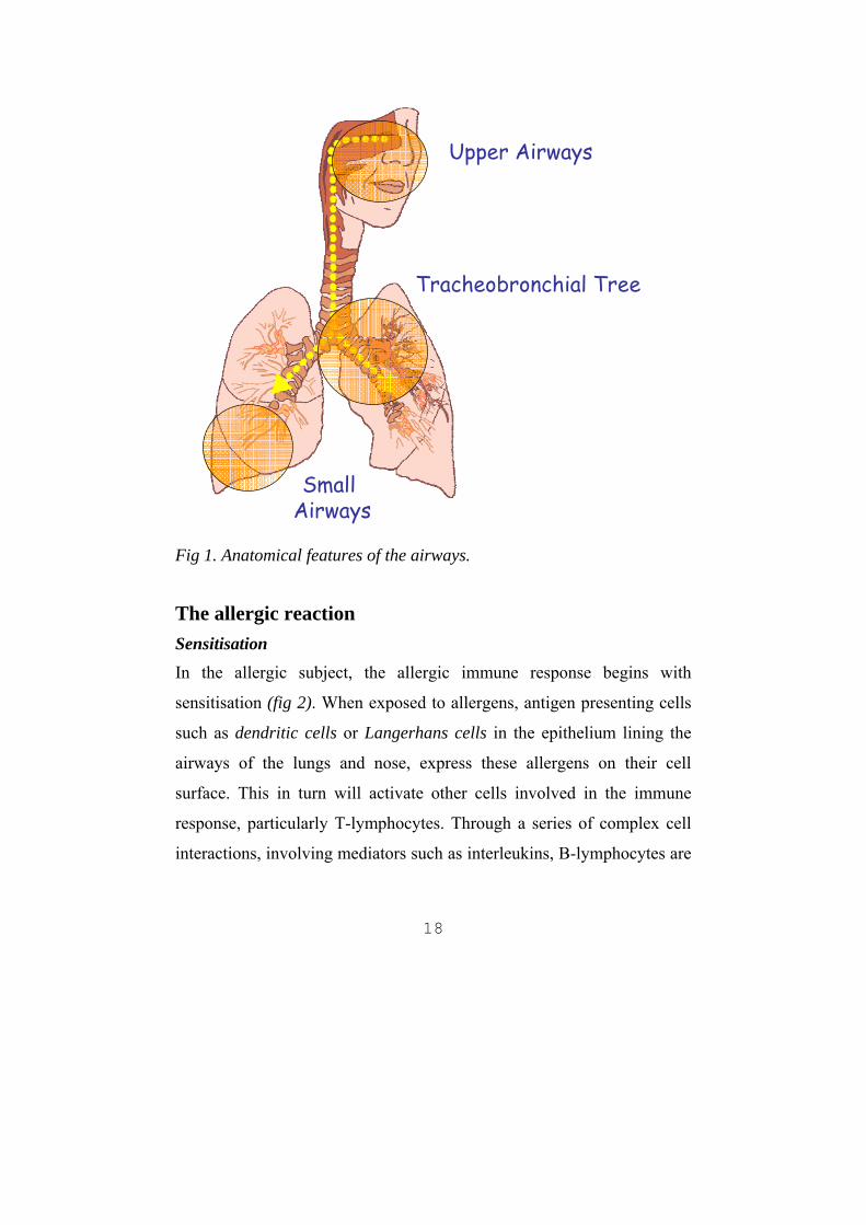
The Airways When inhaling, the air passes through the mouth or the nose down the
pharynx and the larynx. Together with the paranasal sinuses, these
anatomical features constitute the upper airways (fig 1). The air then
enters the tracheobronchial tree (lower airways), starting with the
trachea. The first 16 branchings, or generations, of the airways are called
the conducting zone, since no gas exchange takes place here. The
transitional zone runs through generation 17-19 and consists of the
respiratory bronchioles, where the functional unit of the gas exchange in
the lung, the alveoli, first appears. The respiratory zone (generation 20-
23) contains alveolar ducts and alveolar sacs, and this is where most of
the gas exchange takes place. The bronchioles beyond generation 7-8,
where the diameter is less than 2 mm, are sometimes referred to as the
small airways. The small airways provide only 10 % of total airway
resistance, even though it accounts for approximately 80 % of the total
lung surface area [43].
Upper Airways
Tracheobronchial Tree
Small Airways
Fig 1. Anatomical features of the airways.
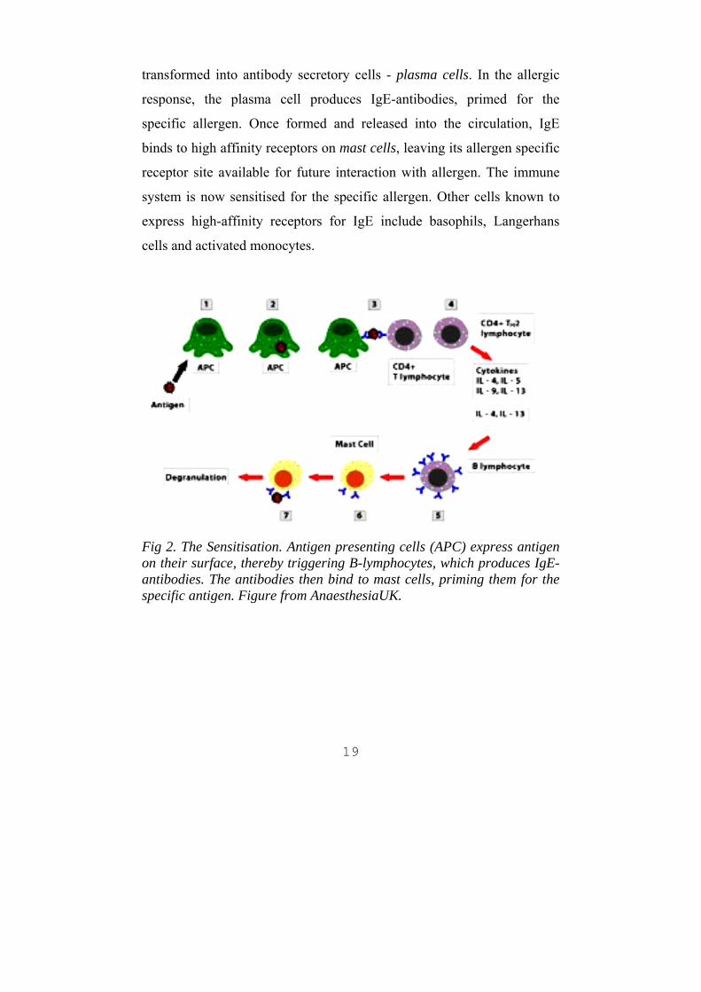
The allergic reaction Sensitisation In the allergic subject, the allergic immune response begins with
sensitisation (fig 2). When exposed to allergens, antigen presenting cells
such as dendritic cells or Langerhans cells in the epithelium lining the
airways of the lungs and nose, express these allergens on their cell
surface. This in turn will activate other cells involved in the immune
response, particularly T-lymphocytes. Through a series of complex cell
interactions, involving mediators such as interleukins, B-lymphocytes are
18
transformed into antibody secretory cells - plasma cells. In the allergic
response, the plasma cell produces IgE-antibodies, primed for the
specific allergen. Once formed and released into the circulation, IgE
binds to high affinity receptors on mast cells, leaving its allergen specific
receptor site available for future interaction with allergen. The immune
system is now sensitised for the specific allergen. Other cells known to
express high-affinity receptors for IgE include basophils, Langerhans
cells and activated monocytes.
19
Fig 2. The Sensitisation. Antigen presenting cells (APC) express antigen on their surface, thereby triggering B-lymphocytes, which produces IgE-antibodies. The antibodies then bind to mast cells, priming them for the specific antigen. Figure from AnaesthesiaUK.
20
Early and late response When the now sensitised subject is re-exposed to the allergen, binding of
the allergen to IgE triggers the immune system to initiate a more
aggressive and rapid memory response. The early-phase allergic response
is that which occurs within 30 minutes of allergen exposure. Cross-
linking of a sufficient number of mast cell/basophil-bound IgE antibodies
by allergen initiates a process of intra-cellular signalling which leads to
degranulation of cells and release of primary inflammatory mediators,
such as histamine and cysteinyl leukotrienes. The symptoms induced are
dependent on the affected organ, and include bronchoconstriction in the
lower airways, wheal-and-flare reaction in the skin and rhinorrhea in the
nose.
A late-phase response commonly occurs 3-8 hours after allergen
exposure. The phase is dominated by recruitment, tissue infiltration and
activation of eosinophils, macrophages and lymphocytes [44].
Mechanisms involved in the initiation of the late-phase cellular response
are not entirely clear, but most likely involve multiple cells and
mediators. T helper 2 cells have been suggested to have a central role in
directing the allergic inflammation [45, 46].
Airway inflammation and the united airways concept United airways concept
The increase in the prevalence of asthma has been associated with an
increase in atopic sensitisation, and is paralleled by similar increases in
other allergic disorders such as eczema and rhinitis [47]. Most patients
with asthma have rhinitis [48]. Of 7219 patients with asthma in the UK,
76 % reported symptoms of rhinitis. Of this 76 %, half said that their
21
rhinitis made their asthma worse [49]. Many patients with allergic rhinitis
have an increased bronchial reactivity to methacholine or histamine [50,
51]. It is also known that patients with rhinitis have an increased risk of
developing clinical asthma over time [52]. The presence of airway
hyperresponsiveness together with atopic manifestations in childhood
increases this risk [53]. This close connection has led to the concept of
“one airway one disease” or united airways [54, 55]. One model that has
been proposed is that the two conditions are manifestations of one
syndrome and that the more severe the rhinitis, the more severe the
asthma [56]. However, it is not clear whether allergic rhinitis represents
an earlier clinical manifestation of allergic disease in atopic subjects who
will later go on to developing asthma or whether the nasal disease itself is
causative for asthma [1].
Thus, allergic rhinitis and asthma are commonly associated, and the nasal
and bronchial mucosa is similar in many ways. There are also
differences. The nose and bronchi have different embryologic origin, and
smooth muscle is present only in the bronchi [57]. Still, segmental
bronchial provocation can induce nasal inflammation in patients with
allergic rhinitis and, conversely, nasal allergen challenge can induce
bronchial inflammation [58, 59]. Different theories have been suggested
on how this distant interaction can be explained. For example, locally
produced inflammatory mediators could affect distant leukocytes through
systemic circulation, or circulating leukocytes could become activated
when passing through the affected tissue [60].
In both allergic rhinitis and asthma, inflammation of the airways is
strongly associated with airway hyperresponsiveness and symptoms. The
acute inflammatory response includes well known reactions such as
22
bronchoconstriction, plasma exudation and mucus hypersecretion in the
lungs, and itching, sneezing, rhinorrhea and blockage in the nose [61,
62]. The inflammation involves infiltration of inflammatory cells such as
activated mast cells, eosinophils and T cells in the airway wall and at the
airway surface [46, 63]. In asthma, over 100 different mediators are
recognized to be involved and mediate the inflammatory response in the
airways [64]. Even structural cells of the airways such as epithelial cells,
smooth muscle cells and fibroblasts have been shown to synthesize and
release inflammatory mediators [65-67]. The eosinophil cationic protein
(ECP) is a secretory ribonuclease, which is found in the eosinophilic
leukocyte [68]. Levels of ECP can be measured in various body fluids
(eg sputum, serum, saliva) and have been shown to correlate well with
airway inflammation but not airway hyperresponsiveness. Thus, it can be
useful in assessing asthma severity, compliance with anti-inflammatory
asthma therapy and as a guide to tailing down inhaled corticosteroid
therapy [69].
Overproduction of IgE plays a critical role in the inflammatory process in
both allergic rhinitis and asthma, and is the result from complex
interaction between B-cells, T-cells, mast cells and basophils through
various inflammatory mediators [70, 71]. Key mediators are the cysteinyl
leukotrienes (CysLTs), a family of inflammatory lipid mediators
synthesized from arachidonic acid by several cells, including mast cells,
eosinophils and macrophages. Receptors for CysLTs can be found in
both bronchial and nasal mucosa, and production of CysLTs is increased
in patients with allergic rhinitis and asthma. They appear to play a role in
both the early and late phase of the allergic reaction, and are involved in
recruitment and maturation of inflammatory cells [72, 73].
23
While the acute inflammation phase has previously been in focus, it is
being increasingly recognized that chronic inflammation is an important
aspect of asthma [74]. This chronic inflammation may result in structural
changes in the airway, referred to as airway remodeling. These structural
changes include fibrosis resulting from deposition of extra cellular matrix
components such as collagen, smooth muscle cell hyperplasia and
hypertrophy, hyperplasia of mucus-secreting cells, and new vessel
formation (angiogenesis) [75]. This remodeling may explain the
irreversible lung function abnormalities experienced in some asthmatics,
even in remission [76]. Glycosaminoglycans are essential extracellular
matrix molecules which regulate tissue flexibility. Hyaluronan is a
glucosaminoglycan, and as such an important part of early connective
tissue repair. Hyaluronan deposition around and internal to the smooth
muscle would be expected to oppose the effect of smooth muscle
contraction [77]. Elevated levels of hyaluronan are commonly seen in
bronchoalveolar lavage in patients with fibrosing inflammatory
conditions, and thus can be regarded as a potential marker of tissue
remodelling [78, 79].
In allergic rhinitis, remodeling is still poorly understood and the
pathological extent of nasal remodeling as well as its clinical
consequences is unclear [80, 81].
24
The role of the small airways As mentioned above, the small airways provide only 10 % of the total
airway resistance [43]. This has led to the small airways being termed
“the silent zone” since airflow obstruction within them causes little
change in conventional tests of lung function [82]. However, it is known
that asthmatic inflammation is present in the small airways [83].
Although inflammation in the large central airways has been the subject
of numerous asthma studies, inflammation in the small distal airways
remains largely unexamined because of the relative inaccessibility of
these structures. However, growing evidence suggest that small airway
inflammation is not clinically silent in asthma. By the use of a fiberoptic
bronchoscope wedged into a subsegmental bronchus, Wagner et al found
a sevenfold increase in peripheral airway pressure in mild asthmatics
compared to healthy subjects, even though the lung function appeared
normal [84]. Increased numbers of lymphocytes and eosinophils have
been shown to be uniformly distributed throughout the large and small
airways of mild and severe asthmatic persons as compared with control
cases [85]. Small airway remodeling may be the explanation for the
development of irreversible airflow obstruction [86]. Nocturnal asthma is
associated with an increase in night-time distal lung inflammation, as
evidenced by the accumulation of alveolar tissue eosinophils,
macrophages and CD4+ lymphocytes. Interestingly, only alveolar (and
not central airway) eosinophilia correlated with overnight reduction in
lung function [87, 88]. The presence of an enhanced inflammatory
process in the small airways is consistent with an increase in the
peripheral airway resistance [89]. The involvement of the small airways
25
seems to be particularly prominent in fatal asthma [90]. Distal lung
disease appears to increase the risk of recurrent asthma exacerbation [91].
The introduction of high-resolution computed tomography allows
assessment of the contribution of small airways to deficits in lung
function. Results of such imaging suggest that the small airways may
play a significant role in airway hyperresponsiveness in asthmatics [92,
93].
In conclusion, all these findings suggest that the small airways are of
utmost importance in the development and progress of asthma, and
subsequently also plays an important role in the treatment of the disease.
The clinical implications of small airways disease on the united airway
concept are still not clear.
Monitoring airway inflammation Invasive vs. non-invasive techniques
The nature and extent of pulmonary diseases can be assessed by direct
invasive bronchoscopy with bronchial washings, biopsy, and/or
bronchoalveolar lavage. While bronchoscopy can provide valuable
information, it requires well trained personnel, and can be demanding on
the patient. The last few decades, new promising non-invasive techniques
to monitor lung function and airway inflammation have been developed:
Nitric oxide
Nitric oxide (NO) was initially described as an endothelium-derived
relaxing factor [94]. It can be measured in exhaled air, and is produced in
the nose and paranasal sinuses, as well as in the bronchial tree [95, 96].
26
Levels of exhaled NO increase during active asthma and allergic rhinitis
[97, 98]. Thus, high levels of NO may reflect ongoing inflammation in
the airways of the patients, and can therefore be regarded as a non-
invasive potential clinical tool to monitor asthma [99]. Indeed, exhaled
NO has been shown to correlate with other inflammation indicators, such
as induced sputum eosinophilia and bronchial reactivity, in steroid-naïve
asthmatics [100]. Exhaled NO arises from the airway and alveolar
compartments, and recently, new analytical methods have been
developed to characterize these sources [101]. Through models of the NO
exchange dynamics, the exhalation flow rate dependence of the exhaled
NO concentrations have been explained. This allows for discrimination
of the NO contribution in the different compartments of the lung. Put
simply, by measuring NO at different exhalation flows it is possible to
approximate the NO concentration in the peripheral region as well as the
conducting airways. Thus, exhaled NO may provide further
pathophysological understanding of the pattern of inflammation in
various airway diseases.
Nasal NO concentrations are very high relative to the lower respiratory
tract in humans, and has been proposed as a surrogate marker for
inflammation in allergic rhinitis, but results have not been as consistent
as in asthma [97, 102, 103].
Induced Sputum
The aim of sputum induction is to collect a sample of secretions from the
lower airways in subjects who do not produce sputum spontaneously,
which allows access to cell subsets and inflammatory biomarkers which
27
may help in the diagnosis and monitoring of the airway disease.
Nebulised isotonic or hypertonic solutions are used to induce production
of expectorate. The expectorate can then be processed and analysed for
biomarkers of disease. The method has been well validated and reference
values for healthy adults have been published [104, 105]. It is well
known that the level of ECP and eosinophilic cell count in sputum are
higher in asthmatics than in healthy subjects, and eosinophil cell count
has been used as a successful tool to guide asthma treatment adjustment
[45, 69, 106, 107]. Induced sputum is also a potential tool for
phenotyping asthma: high percentages of lymphocytes have been found
in ski asthma, whereas eosinophils and neutrophils were increased in
asymptomatic swimmers and runners respectively [108-110]. Future
research may find novel biomarkers [111].
Evaluation of lung function Spirometry While patient history and clinical examination are important in the
diagnosis of asthma, they do not provide any reliable information on the
extent of the airway obstruction. Spirometry is the traditional method for
measuring lung function and has been used for decades, and
recommendations on standardisation have been published [39, 112]. It is
a physiological test that measures how an individual inhales or exhales
volumes of air as a function of time. This is most commonly expressed as
the forced vital capacity (FVC), which is the volume delivered during an
expiration made as forcefully and completely as possible starting after
full inspiration, and the forced expiratory volume in one second (FEV1),
which is the volume delivered during the first second of the FVC
28
manoeuvre. These, and other parameters, can be used to evaluate the
subject’s lung function compared to reference values, and also to some
degree characterize the type of impairment (eg obstructive or restrictive
lung disease). It can also be used to assess the reversibility of the
obstruction, by comparing results before and after treatment with
bronchodilators or inhaled steroids [39]. This greatly enhances diagnostic
confidence, as patients with asthma frequently have poor perception of
symptom severity, especially if their asthma is longstanding [113]. Thus,
spirometry can provide complementary information about different
aspects of asthma control.
Impulse Oscillometry The forced oscillation technique is a non-invasive method with which to
measure respiratory mechanics, and was first used in the fifties [114,
115]. Impulse oscillometry (IOS) is a variant of this technique, and it
measures airway impedance by sending a pulse-shaped sound wave
produced by a loudspeaker into the lungs of a spontaneously breathing
subject and looking for changes in flow in response to the dilatory effect
of the applied energy. The oscillations provide a measure of total airway
impedance, which reflects both resistive elements of the airways
(resistance, R) and viscoelastic and inertive forces in the lungs and the
chestwall (reactance, X). By applying sound waves of different
frequencies during different phases of the respiratory cycle, the
instrument can measure resistance, defined as the opposition of the
respiratory system to the flow of air, at different levels in the respiratory
tree. Reactance is the sum of inertance, which is the inertive force of the
29
air column in the conducting airways, and capacitance, which reflects the
elastic properties of the peripheral lung. The inertive force of the air
column is a physical property of air, and is normally not of any interest in
human studies. The inertive part of the total reactance increases with
higher frequencies of the sound pulse (the air column must be moved
more frequently). Resonant frequency (Fres) is the frequency where the
inertance and capacitance are equal in magnitude and opposite in sign
(phase), and is measured in Hz. Thus, inertive forces dominate at
frequencies above Fres whereas elastic forces are increasingly related to
frequencies below Fres. Low frequent reactance is usually reported as X5
(reactance at 5Hz). X5 reflects changes to the lung periphery and is non-
specific. Increased negative values can be seen both in restrictive and
obstructive disease.
While spirometry is a valuable method for measuring lung function, it is
effort dependent and careful instructions to the patient on how to perform
the expiratory manoeuvre is required. The forced oscillation technique,
on the other hand, requires minimal cooperation from the patients, since
the forced oscillations are superimposed on the normal breathing, thus
avoiding the need for any special breathing manoeuvre or any noticeable
interference with respiration. The minimal influence on respiratory
properties is particularly important when assessing airway
hyperresponsiveness. The forced oscillation technique also provides the
possibility to assess airflow obstruction in the peripheral airways,
something that conventional lung function tests can not do. However, one
should be aware of the fact that the IOS model is based on theoretical
assumptions. Very few physiological correlations between forced
oscillation technique parameters and direct evidence of airway function
30
have been published. Therefore, one has to be careful of drawing too
extensive conclusions from the results.
Airway hyperresponsiveness It is not unusual for patients with asthma to have a normal spirometry,
yet experience airway narrowing in response to a stimulus that would be
innocuous in a healthy person [41, 116]. This hyperreactivity of the
airways is termed bronchial or airway hyperresponsiveness.
The mechanisms behind airway hyperresponsiveness are not completely
understood. Excessive contraction of airway smooth muscle due to
increased contractility of smooth muscle cells could be one important
factor [117]. The thickness of the airway wall from necropsy specimens
is greater in subjects with fatal asthma than in those with milder disease
and in non-asthmatics, and oedema and structural changes in the airway
wall could amplify airway narrowing due to contraction of smooth
muscle for geometric reasons [90, 118, 119]. Airway remodeling changes
could decrease the radial constraint provided by connective tissue
elements, allowing excessive airway smooth muscle shortening, even
though some data actually suggest that airway remodeling may provide
protection against airway narrowing [119, 120]. Finally, epithelial
damage from ongoing inflammation may allow greater amounts of
bronchoconstrictor mediators to reach smooth muscle cells, sensory
nerves or other cells involved in airway narrowing [121].
While it is not entirely clear what drives the chronic airway
hyperresponsiveness, fluctuations in the extent of eosinophilic
inflammation may underlie changes in the degree of hyperresponsiveness
seen during the course of the disease. Eliminating eosinophilic
31
inflammation by glucocorticoid treatment improves airway
hyperresponsiveness, although it does not eliminate it completely [122].
Likewise, avoiding allergens that may trigger inflammation only
improves, but does not eliminate, airway hyperresponsiveness [123].
Almost all asthmatics exhibit increased responsiveness, especially during
symptomatic episodes. Airway hyperresponsiveness has also been
described in patients with allergic rhinitis, as well as in other pulmonary
diseases such as chronic obstructive pulmonary disease [50, 124, 125].
Hence, the presence of airway hyperresponsiveness does not necessarily
mean that the patient has asthma. However, lack of airway
hyperresponsiveness to a large degree excludes asthma. Thus,
measurement of the degree of hyperresponsiveness may help establish a
diagnosis of asthma [126, 127].
Challenge testing Airway hyperresponsiveness can be demonstrated by several different
provocation tests. They are usually divided into two groups: direct and
indirect provocation. In direct challenge the provoking substance is
assumed to act directly on the receptors of effector cells such as smooth
muscle cells, endothelial cells and/or mucus producing cells, and hereby
inducing bronchial obstruction. The effect is believed to be only partly
dependent on present inflammation, and it may be present even in
patients with chronic changes, i.e. remodelling [128]. Indirect challenge
on the other hand, is presumed to be acting on inflammatory cells,
causing them to release mediators, which in turn triggers smooth muscle
cell constriction [42, 129]. Thus, in theory, a positive result to indirect
challenge requires inflammation present in the airways.
Methacholine chloride (acetyl-β-methylcholine) is a
parasympathomimetic synthetic analog of acetylcholine. It stimulates
[130]. Methacholine challenge is a commonly used direct test and has
been shown to identify airway hyperresponsiveness with high sensitivity
(fig 3.) [41, 131, 132]. A negative test can to a high degree exclude
asthma as the cause of a patient’s symptom, while a positive test has less
diagnostic specificity.
Fig 3. Example of reaction patterns to direct challenge testing (e.g. methacholine or histamine). The concentration of the inhaled provocative substance that triggers a 20 % fall in FEV1 (PC20) determines the degree of airway hyperresponsiveness.
Indirect tests on the other hand are generally less sensitive. The
triggering mechanism in exercise induced bronchoconstriction (EIB) is
believed to be the loss of water via evaporation from the airway surface.
32
33
This water loss is believed to cause airway narrowing through thermal
and osmotic effects of the dehydration [133, 134]. Cold, dry air is more
provocative than warm, humid air [135].
Eucapnic voluntary hyperventilation (EVH) simulates the
hyperventilation achieved during exercise and it is assumed that
individuals sensitive to the provocation are reacting to the increased
ventilation per se, possible due to drying of the airway surface liquid and
increased osmolarity. EVH have shown high sensitivity in identifying
patients with EIB [136].
Mannitol challenge is a fairly new method of applying an osmotic
stimulus that mimics the effects of the dehydration caused by
hyperventilation during exercise. Mannitol is a polyol (sugar alcohol),
and is a potent osmotic stimulus [137]. Mannitol hyperresponsivness
have proven to predict the response to corticosteroid therapy in
asthmatics [138]. It can be used to identify patients with asthma with EIB
[139]. However, it has been shown to be less sensitive than MCh in
identifying airway hyperresponsiveness [140].
34
35
AIMS
The overall aim of this thesis was to study the distribution of
inflammation and obstruction in asthmatics, by using non-invasive
methods, and to compare the results to results from patients with allergic
rhinitis. Five studies are included in this thesis with the following
specific aims:
I. To investigate whether patients with allergic rhinitis and asthma
differed from rhinitis with or without bronchial
hyperresponsiveness in degree of perception of dyspnoea and
airway inflammation, measured as fractional exhaled nitric oxide.
II. To assess peripheral and proximal NO concentration in rhinitic
subjects, and to correlate the peripheral NO concentration to the
peripheral obstruction in response to methacholine.
III. To measure induced sputum Cys-LT, as well as markers of
remodelling and eosinophilic inflammation in sputum from patients
with rhinitis with or without BHR, comparing the results with
patients with rhinitis and clinical asthma.
IV. To compare the degree of involvement of the peripheral airways
during methacholine challenge test in asthmatics and patients with
allergic rhinitis with or without BHR by using the impulse
oscillometry technique.
36
V. To investigate whether different direct and indirect stimuli induces
different patterns of obstruction, recorded as central and peripheral
resistance. Also to see whether baseline resistance could predict a
positive response to direct or indirect provocation.
37
METHODS
Study populations For paper I, patients with seasonal allergic rhinitis were recruited and
investigated with methacholine challenge testing with impulse
oscillometry, fractional nitric oxide and induced sputum, both during and
outside pollen season.
For paper II-IV, the size of the study population in paper I was
increased by further recruitment. All subjects underwent the testing
detailed above. Only those patients that were able to produce sputum
were used in paper III.
For paper V, mild asthmatics were recruited and investigated with MCh,
EVH and mannitol challenge testing, as well as impulse oscillometry and
fractional nitric oxide.
All patients attended the outpatient clinic of the department of lung
medicine in Lund. All subjects gave written informed consent, and the
ethical committee in Lund approved the studies.
Study populations are described in table 1.
allergic rhinitis, total (female)
asthma controls
Paper I 29 (17) 11 14 Paper II 51 (30) 26 12 Paper III 41 (26) 16 13 Paper IV 53 (30) 26 13 Paper V 34 14 Table 1. Study populations
38
Subject characterization All subjects were non-smokers without upper respiratory tract infection
within three weeks prior to the investigation.
Healthy controls (paper I-V)
Healthy controls had no history of allergic symptoms. A skin prick test,
SPT, (Alk Abello, Copenhagen, Denmark) was used to screen for
sensitization to a standard panel of 10 common airborne allergens (birch,
timothy, mugwort, cat, dog, horse, d. pteronyssinus, d. farinae,
aspergillus and cladosporium). Controls with positive skin prick test
were excluded. None of the controls included were hyperresponsive to
methacholine (negative challenge on a cumulative dose of 2000 microg).
Their age ranged from 19 to 56 (paper I-IV) and 24 to 61 (paper V).
Patients with seasonal allergic rhinitis (paper I-IV) Subjects with symptoms of allergic rhinitis were recruited and tested with
SPT. Only those with pure seasonal allergy were investigated, ie those
who had a positive SPT to birch, timothy and/or mugwort. Those with
confirmed sensitization to perennial allergens (animal dander, dust mites
or moulds) were excluded. Sensitization to animal dander was allowed if
the patient were not exposed to animals.
Patients with allergic rhinitis was subdivided into patients with allergic
rhinitis and no bronchial hyperresponsiveness, patients with allergic
rhinitis and bronchial hyperresponsiveness but no symptoms of asthma
and patients with allergic rhinitis and doctor’s diagnosed asthma.
39
Patients with asthma (paper I-V) In paper I-IV, some of the patients with allergic rhinitis had concomitant
asthma. They had symptoms of airway obstruction and were clinically
diagnosed mild asthmatics according to global initiative for asthma
(GINA) standards. Three of the asthmatics, used in paper II, inhaled
In paper V, the disease group consisted of patients with clinically
diagnosed mild asthma. Inhaled corticosteroid (ICS) treatment was
allowed with a maximum daily dose equivalent of 800 microg
budesonide.
Spirometry Flow-volume spirometry was used to assess pulmonary function in all
papers. A MasterScope spirometer, software version 4.5 (Erich Jaeger
GmbH, Wurzburg, Germany) was used for the flow-volume spirometry,
which was done according to the guidelines of the European Respiratory
Society [112]. The reference values were obtained from Crapo et al.
[141]. The better of two measurements of forced expiratory volume in 1 s
(FEV1) with less than 4% variation was recorded as baseline.
Borg Symptom Score Borg symptom score results are presented in paper I.
Before every flow-volume measurement the subjects were asked to grade
their perception of dyspnoea on a 10-grade scale, with 0 being no
dyspnoea at all and 10 being maximal dyspnoea (fig 4) [142]. All
subjects had a baseline dyspnoea of 0. The subjects were blinded to their
40
lung function response. Borg scores were plotted against percentage of
fall in FEV1 from baseline and linear regression analysis was used to
calculate a FEV1/Borg slope (Slope-BorgMCh) for every individual,
which was used as an index of dyspnoea.
0 nothing at all 0,5 extremely weak 1 very weak 2 weak 3 moderate 4 somewhat strong 5 strong (heavy) 6 7 very strong 8 9 10 extremely strong (maximal) Fig 4. The Borg Symptom Scale.
Methacholine challenge testing Results from methacholine challenge testing are presented in all papers.
Presence of airway hyperresponsiveness was measured with a
methacholine challenge test. First, baseline FEV1 was assessed with flow-
volume spirometry, described above. If the baseline value was below 70
% of the predicted value, the challenge was aborted. The test was carried
out with tidal volume triggered equipment (Aerosol Provocation System,
APS, Erich Jaeger GmbH, Wurzburg, Germany). The APS delivered a
cumulative dose of 2000 microg MCh in five increments (50, 150, 300,
600 and 900 microg) following an initial dose of 0.9 % NaCl. The
41
challenge was discontinued if the FEV1 declined more than 20 % during
the protocol. A positive test was defined as the cumulative dose that
caused a decline in FEV1 by 20 % or more (PD20FEV1) from baseline.
The PD20FEV1 was determined by interpolation by the last two points on
the log dose-response plot. The amounts of MCh given during every
increment of the challenge test were plotted against the corresponding
percentage fall in FEV1 from baseline. Linear regression analysis was
used to calculate a MCh/FEV1 slope (Slope-FEV1MCh) which was used as
an index of airway hyperresponsiveness. When FEV1 fell below 80 % of
the baseline value or when the total amount of 2000 microg MCh was
delivered, 400 microg of salbutamol were given to all subject
immediately after finishing the provocation. After 10-15 minutes a new
flow-volume spirometry was carried out, to ensure that the subjects were
recuperating properly.
Exhaled Nitric Oxide Data obtained from Exhaled Nitric Oxide measurements are presented in
paper I, II and V.
NO measurements were performed in accordance with International
American Thoracic Society recommendations, using a NIOX, nitric
oxide gas analyser (Aerocrine, AB, Stockholm, Sweden) [143]. Patients
were comfortably seated, inhaled NO depleted ambient air, and exhaled
at different flow rates (paper I-IV: 10, 50, 100 and 400 ml/s; paper V: 50,
100, 200 and 400 ml/s) 2–4 times depending on divergence.
Peripheral NO concentration (or alveolar concentration, CANO) and
proximal maximal NO flux (J'awNO) was approximated by plotting NO-
42
output (product of concentration and flow) against exhalation flow (at
flow 100-400 ml/s) [101]. The slope and intercept of this line
approximate CANO and J'awNO, respectively [144, 145]. Calculations
using the flow 50 ml/s were also performed, but were not used as an
increase in slope and a decrease in the intercept were observed
confirming previous reports that linearity between NO-output and flow is
valid only for approximately above 99 ml/s [146]. All NO measurements
were done prior to bronchial challenge test.
Impulse Oscillometry Results from impulse oscillometry are presented in paper II, IV and V.
During each challenge test, impulse oscillometry was used to provide
further information on the magnitude and site of obstruction in the
airways.
A Jaeger MasterScreen Impulse Oscillometry System (Erich Jaeger
GmbH, Wuerzburg, Germany) was used. Oscillometry was performed
before the challenge and after each step of the challenge, prior to the
spirometry, to avoid the influence of deep inspiration and subsequent
maximal forced expiratory maneuvers on IOS parameters. The subjects
used nose clips and were told to press the palms of their hands against the
cheeks to decrease the upper airways shunt. For about 30 seconds,
oscillometric pressure impulses were superimposed on the tidal breathing
of the subject, having a pulse sequence of 5 per second and a frequency
spectrum between 5-35 Hz. Airway resistance at 5 Hz and 20Hz (R5,
R20), reactance at 5 Hz (X5), resonant frequency (Fres) and area of
reactance integrated from 5 Hz to Fres (AX) were determined. During
43
MCh and Mannitol challenge test, IOS were performed 45 s. after each
challenge step, while FEV1 was performed 75 s. after each challenge
step. During the EVH challenge, IOS was performed 1, 3, 5, 7.5, 10, 15
and 20 min post challenge, with FEV1 performed immediately after IOS
at each step.
Induced sputum Data obtained from induced sputum are presented in paper III.
Sputum induction Sputum was induced by inhalation of nebulized isotonic saline solution
(0.9% NaCl) for 0.5, 1, 2 and 4 min, followed by a hypertonic solution
(4.5% NaCl) for 0.5, 1, 2 and 4 min. Lung function (PEF) was measured
1 min after each induction time-point, and induction was interrupted if
lung function was decreased ≥20%. Subjects were asked to rinse their
mouth and blow their nose, and try to cough between each dose of
nebulized saline. Sputum induction was continued until adequate sample
volume was obtained (mean time: 7.8min, SD: 4.4), and there was no
difference in sputum induction time among the patient groups.
Sputum processing Sputum plugs were sorted out, and treated with four volumes of 0.65 mM
dithiothreitol (DTT) in phosphatebuffered saline (PBS) for 1 h in 4 ºC.
Additional four volumes of PBS were added, followed by filtration
through a 60 mm filter and a final centrifugation (1000 g for 5 min),
44
which separated the supernatant from the cells. The supernatant was
frozen until later analysis.
Sputum analysis Sputum was analyzed for cysteinyl-leukotrienes and LTB4 using EIA
(detection limit 13 and 6 pg/mL, respectively) from Cayman Chemical
(Ann Arbor, MI, USA). Before analysis of subsequent assays, sputum
was dialysed to PBS to eliminate the amount of DTT. ECP was measured
using the UniCap ECP kit (detection limit 0.5 ng/mL, Pharmacia
Diagnostics, Uppsala Sweden), IL-8 and IL-13 using Quatikine
(detection limit 3.5 and 32 pg/mL, respectively, R&D Systems,
Abingdon, UK), hyaluronan and laminin using ELISAs (detection limit
10 ng/mL for both assays) from Echelon Biociences incorporated (Salt
Lake City, UT, USA) and Chemicon International (Temecula, CA,
USA), respectively. The total protein concentration was measured using a
Bio-Rad Protein Assay (Bio-Rad Laboratories.
Inc., Hercules, CA, USA). All values were adjusted to the total amount of
protein in sputum (and presented as amount per microgram total protein)
to abolish differences due to sputum heterogeneity. Samples were run in
duplicate with a maximum in between variation of 5%. All tests were
commercially standardized and further standardization for the use of
sputum analysis was performed.
Mannitol challenge testing Results from mannitol challenge testing are presented in paper V.
45
Pre-challenge spirometry was performed as described for MCh challenge
above. A mannitol powder kit (AridolTM; Pharmaxis, Frenchs Forest,
Australia) was used in conjunction with a dry powder inhaler device to
administer a cumulative dose of 635 mg in 8 increments according to the
manufacturer’s instructions. The challenge was discontinued if the FEV1
declined more than 15 % from baseline or if a between-dose fall of >10
% occurred, which was considered a positive test [147]. After the
challenge, the subject received an inhalation of 400 microg salbutamol
and a new spirometry was performed 10-15 minutes later to ensure that
the subject was recuperating properly. The PD15FEV1 was determined by
interpolation by the last two points on the log dose-response plot. The
Mannitol/FEV1 slope was calculated in the same way as the slope for
MCh (see above).
Eucapnic Voluntary Hyperventilation Results from Eucapnic Voluntary Hyperventilation are presented in paper
V.
Pre-challenge spirometry was performed as described for MCh challenge
above. The patients were instructed to hyperventilate for 4 minutes, at 85
% of maximum voluntary ventilation (30 x Baseline FEV1), guided by a
reservoir balloon. In order to maintain eucapnia, the dry air ventilation
device (Ailos Medical AB, Karlstad, Sweden) administered hypercapnic
air (5 % CO2). The air inspired was dry and at room temperature. The
spirometry was repeated together with IOS post challenge at 1, 3, 5, 7.5,
10, 15 and 20 minutes. Thereafter the subject received an inhalation of
400 microg salbutamol and a new spirometry was performed 10-15
46
minutes later to ensure that the subject was recuperating properly. A drop
of FEV1 >10 % compared to baseline was regarded as a positive test
(EVH 10) [127].
Statistical Analysis Generally, as the data could not be assumed to have a normal
distribution, non-parametric tests were used. The Mann-Whitney U-test
was used for comparison between two groups (paper I-V). Statistical
comparison between more than two groups was done with Kruskal-
Wallis test for independent samples (paper I-V). Spearman correlation
coefficient was used to determine correlation between groups (paper I-
III, V). In the case of paired samples, Wilcoxon’s test was used (paper I-
III). A p-value of less than 0.05 was considered significant.
47
RESULTS AND COMMENTS
Paper I - Allergic rhinitis with or without concomitant asthma: difference in perception of dyspnoea and levels of fractional exhaled nitric oxide It is well established that allergic rhinitis and asthma are closely linked
entities and more than 75 % of the patients with asthma reports
concomitant rhinitis [148]. Asthma is also closely associated to BHR,
and a large part of patients with rhinitis alone show a reactive pattern in
bronchial provocation tests, even though they have no symptoms of
clinical asthma [50]. The fact that patients can react with airway
obstruction to bronchial challenge, which is a hallmark of asthma,
without experiencing symptoms is curious and could possible be
explained by difference in degree and/or geographical distribution of
inflammation. In this study, we aimed to investigate the degree of
perception of dyspnoea and airway inflammation in patients with allergic
rhinitis with or without concomitant asthma, both during and outside
pollen season.
We found that 12 out of 18 patients with allergic rhinitis without asthma
had bronchial hyperresponsiveness to methacholine, which is in line with
previous observations. We also found increased inflammatory activity,
measured as FENO, during pollen season in asthma patients, but not in
those with rhinitis alone. There was a correlation between the degree of
inflammation and the degree of BHR in the asthma patients, but not in
patients with allergic rhinitis and BHR. This may indicate that the
pathogenesis of BHR is dependant on several factors, and that ongoing
inflammation is more linked to BHR in asthmatics.
Interestingly, patients with asthma had a greater perception of the
obstruction induced during the methacholine challenge test, compared to
patients with rhinitis and BHR (fig 5). No correlation of symptoms and
FENO levels could be found, indicating that the presence or absence of
symptoms could not be explained by degree of inflammation alone. Thus,
the reason for this difference in perception is still unknown.
Psychological factors could play a role, and possibly could awareness of
obstruction increase over time. Another explanation could be that
symptoms may be dependent on geographical distribution of
inflammation, specifically the degree of peripheral airway involvement.
Fig 5. Slopes of Borg/FEV1 for controls, patients with rhinitis alone with and without bronchial hyper-responsiveness (BHR) and patients with rhinitis and asthma during season and off season.
48
49
Paper II - Peripheral nitric oxide is increased in rhinitic patients with asthma compared to bronchial hyperresponsiveness Based on the conclusions in paper I, in this paper we hypothesised that
involvement of the peripheral airways differs between patients with
rhinitis and concomitant asthma and patients with (or without) BHR. In
recent years it has been possible to measure NO at different exhalation
flow, and approximate the NO concentration in the peripheral region as
well as the conducting airways [144]. Thus, in theory, it is possible to
study the geographical distribution of inflammation in the airways.
Involvement of peripheral airways can also be estimated by impulse
oscillometry, where different responsive patterns to different frequencies
reflect peripheral properties of the respiratory tract [149].
We found increased peripheral NO concentrations in patients with
rhinitis and concomitant asthma compared to patients with rhinitis only,
while patients with rhinitis and BHR represented an intermediate step
between those with rhinitis only and those with asthma (fig 6). Increased
proximal NO concentrations was also seen in asthmatics, but not in
patients with rhinitis and BHR. Furthermore, we found a correlation
between peripheral NO concentration and degree of peripheral
obstruction during methacholine challenge test, while no correlation were
seen between proximal NO concentrations and peripheral obstruction
parameters. Thus, those with highly reactive peripheral airways also
seemed to have a higher degree of ongoing small airway inflammation.
Overall, results from this study seem to strengthen our hypothesis that
asthmatics have more widespread inflammation, which includes the small
airways. Interestingly, the three subjects in the asthmatic group that had
anti-inflammatory treatment (ICS), still showed signs of a high level of
peripheral inflammation as well as peripheral obstruction.
Fig 6. Peripheral NO concentration. Concentration of peripheral NO (CANO) assessed by measuring exhaled NO at several exhalation flow rates in patients with rhinitis (R), rhinitis with bronchial hyperresponsiveness (R+BHR), rhinitis and concomitant asthma (R+A) and healthy controls (Ctrl).
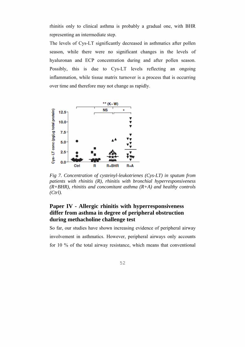
Paper III - Cysteinyl-leukotriene levels in sputum differentiate asthma from rhinitis patients with or without bronchial hyperresponsiveness The transition from allergic rhinitis to clinical asthma is probably a
gradual one, with bronchial hyperresponsiveness possibly representing an
intermediate step. Our findings in paper II seem to strengthen this theory.
Previous studies have shown that levels of eosinophils and eosinophilic
cationic protein (ECP) in induced sputum are increased in patients with
rhinitis and BHR, but not as high as in patients with rhinitis and asthma
50
51
[150, 151]. It is well known that both the level of ECP and eosinophilic
cell count in sputum are higher in asthmatics than in healthy subjects,
with a correlation to disease severity [45] [69, 152].
Cysteinyl-leukotrienes (Cys-LTs) are actively involved in the
inflammation in asthma and rhinitis, Cys-LTs are known to be elevated
in sputum from asthmatics and have been shown to be correlated to
eosinophil cell count [153, 154].
Hyaluronan is a glucosaminoglycan, and as such an important part of
early connective tissue repair, and can be regarded as a potential marker
of tissue remodelling [78, 79].
In this paper we wanted to measure induced sputum Cys-LTs, as well as
markers of remodelling and eosinophilic inflammation in sputum from
patients with rhinitis with or without BHR, comparing the results with
patients with rhinitis and clinical asthma.
We found increased levels of Cys-LT and hyaluronan in sputum in
asthmatics compared to patients with rhinitis with or without BHR (fig
7). Asthmatics had a slightly higher concentration of ECP compared to
patients with rhinitis and BHR, but the difference was not significant.
This indicates that there is more inflammatory turnover of the connective
tissue in rhinitis patients with asthma compared with BHR only, and that
Cys-LT driven inflammation is present in the asthmatic group. While
patients with rhinitis and BHR had significantly lower levels of CYS-LT
compared to asthmatics, they still had slightly higher levels of ECP and
Cys-LT compared to patients with rhinitis only. This might indicate an
initiated inflammatory process in the airways that may later lead to the
development of asthma, strengthening our hypothesis that transition from
rhinitis only to clinical asthma is probably a gradual one, with BHR
representing an intermediate step.
The levels of Cys-LT significantly decreased in asthmatics after pollen
season, while there were no significant changes in the levels of
hyaluronan and ECP concentration during and after pollen season.
Possibly, this is due to Cys-LT levels reflecting an ongoing
inflammation, while tissue matrix turnover is a process that is occurring
over time and therefore may not change as rapidly.
Fig 7. Concentration of cysteinyl-leukotrienes (Cys-LT) in sputum from patients with rhinitis (R), rhinitis with bronchial hyperresponsiveness (R+BHR), rhinitis and concomitant asthma (R+A) and healthy controls (Ctrl).
Paper IV - Allergic rhinitis with hyperresponsiveness differ from asthma in degree of peripheral obstruction during methacholine challenge test So far, our studies have shown increasing evidence of peripheral airway
involvement in asthmatics. However, peripheral airways only accounts
for 10 % of the total airway resistance, which means that conventional
52
53
tests of the lung function (e.g. FEV1) fail to accurately reflect changes in
peripheral resistance [43, 155, 156]. Hence, the term “silent zone” is
sometimes used for the small airways [157]. Impulse oscillometry is a
forced oscillation technique that allows for discrimination between
central and peripheral obstruction [158].
In this paper we wanted to compare the degree of involvement of the
peripheral airways in asthmatics and patients with allergic rhinitis with or
without BHR, specifically the degree of peripheral airway obstruction
during methacholine challenge test, by using impulse oscillometry.
We found that while patients with rhinitis and asthma and patients with
rhinitis and BHR showed similar reactivity to methacholine, asthmatics
had significantly more increase in parameters indicating peripheral
obstruction (i.e. dR5-R20, AX, X5) during the methacholine challenge
test (fig 8). The proximal resistance (i.e. R20) followed a similar pattern
in all groups. Thus, both patients with asthma and patients with rhinitis
and BHR reacted to methacholine with decrease in FEV1, and while the
degree of obstruction in the bronchi seemed to be similar, asthmatics had
signs of a higher degree of peripheral involvement. Possibly, this could
explain our previous findings that asthmatics have a greater perception of
bronchial obstruction.
Fig 8. Slope-AXMCh for controls, patients with AR with or without BHR and patients with AR and concomitant asthma.
Paper V - Characterization of airway reactivity to methacholine, mannitol and eucapnic hyperventilation in mild asthmatics In the previous papers we have found evidence of peripheral airway
obstruction in asthmatics, triggered by methacholine challenge tests.
Provocation testing for identifying airway hyperresponsiveness in
asthmatics has become increasingly important in the diagnosis of asthma
and for monitoring the effect of treatment. Methacholine challenge is a
well established provocation test; it is a direct test, ie it acts directly on
the receptors of effector cells such as smooth muscle cells, endothelial
cells and/or mucus producing cells, and hereby inducing bronchial
obstruction. Hence, it is believed to be only partly dependent on present
inflammation. Indirect challenge on the other hand, is presumed to be
acting on inflammatory cells, causing them to release mediators, which in
turn triggers smooth muscle cell constriction. Indirect challenges could
54
55
thus possibly provide more information about underlying inflammation
in the airways.
While methacholine challenge testing is a very sensitive tool for
detecting airway hyperresponsiveness, not all asthmatics react to indirect
testing. Specifically, exercise induced obstruction is a common feature in
asthma, but not all asthmatics suffer from it. Exercise challenge is
believed to cause obstruction through hyperventilation, which could
cause drying of the airway surface liquid and increased osmolarity. Two
other examples of indirect challenges are eucapnic voluntary
hyperventilation and mannitol provocation test, which both apply
osmotic stimuli to the airways. Since EIB seems to occur more in
peripheral airways than in central airways [159], it is plausible to assume
that different pattern of reaction to various challenge tests may identify
different asthma phenotypes.
In this study we compare the reactive pattern during direct and indirect
challenge testing in patients with mild asthma, by using impulse
oscillometry. We also investigated whether baseline resistance could
predict the outcome of either challenge test.
We found that 5 patients were negative to all tests. Of the remaining 29
patients, 27 were positive to direct testing (methacholine) and 23 were
positive to indirect testing (either EVH or mannitol). Thus, even in mild
asthmatics, a majority of the patients are positive to indirect testing.
Interestingly, even though EVH and Mannitol challenges are thought to
trigger the same mechanisms, not all patients positive to EVH were
positive to mannitol. This indicates that the tests are not fully
interchangeable. However, the limits for what constitutes a positive result
differ between the tests (10 % fall in FEV1 for EVH and 15 % fall for
56
mannitol), and when changed to 10 % fall in FEV1 for both tests, the
result became more similar.
No difference in broncho-constrictive pattern could be identified during
the different provocation tests; the obstruction induced seemed to follow
the same geographical pattern regardless of the triggering stimuli.
However, those with a positive mannitol provocation had a lower FEV %
pred and signs of more peripheral airway involvement at baseline. This
supports the idea that peripheral airway involvement is an important
predictor of asthma airway reactivity.
57
GENERAL DISCUSSION AND FUTURE PERSPECTIVES
Asthma is a serious global health problem that has increased rapidly in
prevalence in the western world during the last decades, and is now
increasing rapidly in the developing world as well. The main challenge
for asthma researchers today is to find a way to prevent this is increase in
prevalence. While factors influencing the development and expression of
asthma are known, and the pathological features of asthma are
increasingly well described, the exact link between exposure to risk
factors and the development of chronic airway inflammation are not yet
fully understood. Until we know more, the possibilities to stop the
development of asthma with pharmacological intervention will be
limited, and focus will be on identifying and reducing exposure to risk
factors. Hopefully, future research results will shed more light on the
pathophysiological connection between allergic rhinitis and asthma. The
findings in this thesis suggest that advanced allergic airway disease
includes involvement of more peripheral parts of the lung. If indeed the
progress from allergic rhinitis to asthma is dependent on geographical
spreading of the airway inflammation to the peripheral airways, it might
be theoretically possible to stop this progress with pharmacological
therapy, thus hindering the development of asthma. This, of course, is
dependant on gaining knowledge of the specific mechanisms driving the
inflammation, which already are under extensive scrutiny from asthma
researchers around the world. However, it should be of particular interest
to elucidate the process behind the involvement of the peripheral airways.
58
Is this the step that completes the progress from allergic rhinitis with
airway hyperresponsiveness to full blown asthma?
Interestingly, airway hyperresponsiveness is fairly common in patients
with allergic rhinitis without concomitant clinical asthma. In paper I, we
show that these rhinitis patients do not experience symptoms from
bronchial obstruction to the same degree as asthmatics do. Thus, the
obstruction of the large airways alone cannot explain the dyspnoea
experienced by asthmatics. Indeed, the relationship between
inflammation in the airways of a patient and either the symptoms of
asthma or airway hyperresponsiveness is not simple. Dyspnoea is
multifactorial and the exact mechanism that causes dyspnoea in
asthmatics is not fully understood. The sense of respiratory effort,
chemoreceptor stimulation, mechanical stimuli arising in lung and chest
wall receptors, and neuroventilatory dissociation may all contribute to the
sensation of dyspnoea [160]. In asthma, it is speculated that
hyperinflation of the lung is a great contributor to dyspnoea [161]. In our
studies, it is unclear if asthmatics had more hyperinflated lungs compared
to patients with rhinitis and airway hyperresponsiveness. We did find a
higher degree of peripheral involvement during the methacholine
challenge in the asthmatics. It is tempting to try to explain the difference
of dyspnoea in the two groups by referring to difference in peripheral
involvement of the peripheral airway, especially since it is in the
peripheral airways that the actual primary function of the lungs, the gas-
exchange, takes place. However, we found no correlation between degree
of peripheral airway involvement and degree of dyspnoea. The reason for
the higher degree of symptoms in the asthmatic group remains unclear.
59
Current guidelines recommend “that patients with persistent allergic
rhinitis should be evaluated for asthma by history, chest examination and,
if possible and when necessary, assessment of airflow of obstruction
before and after bronchodilator” [1], to catch the development of asthma
in patient with allergic rhinitis. With emerging insight in the importance
of the small airways, small airway involvement should be considered in
patients with asthma. Monitoring the small airways is not an easy feat,
though. We have seen in this thesis that IOS can provide information on
resistance and other properties of the small airways. Fractional exhaled
NO can be used to evaluate presence of ongoing peripheral inflammation.
These tools are as of yet not easily implemented in the clinical practice.
Development of new techniques, or improvement of current technology,
could in the future facilitate a more comprehensive assessment of the
airways.
Asthma is a heterogenous disease. Different variants include exercise-
induced bronchoconstriction and cough-variant asthma. Correct
characterization of the disease could have implications for the treatment
and exploring the degree of peripheral involvement could be an
important part of phenotyping the airway inflammation. In paper V, we
found that different provocation tests were not fully interchangeable, and
that positive results may reflect different phenotypes. For example,
patients with a positive mannitol challenge generally had more evidence
of peripheral airway involvement at baseline. Further research on this
area is needed. In our study, we tested a broad sample of mild asthmatics.
It would be of special interest to investigate specific variants of the
asthma disease to elucidate if the pattern of inflammation and airway
resistance differs between different asthma groups.
60
Ideally, we should strive for the ability to cure asthma, if we cannot fully
prevent it. While no curative treatment exist today, it is possible to
reverse the bronchospasm with bronchodilators. We have access to a
variety of anti-inflammatory drugs that block parts of the inflammatory
response, in particular the inhaled corticosteroids which have been the
mainstay in asthma treatment for over 30 years. Corticosteroids
accomplish its effect by inducing the recruitment of the nuclear enzyme
histone deacetylase 2 (HDAC2) to multiple activated inflammatory
genes, which leads to deacetylation of the hyperacetylated genes, thereby
suppressing inflammation [162]. However, not all patients with asthma
respond to treatment with corticosteroids, even in high doses. Neither do
corticosteroids seem to prevent reduction of lung function over time,
which indicates that the remodelling process in the airways is not
affected by corticosteroid treatment [163]. It is of particular interest that
most current ICS are delivered in a suspension with a particle size of
>2mm. Thus, it is possible to have an untreated, persistent inflammation
in the small airways despite high-dose ICS treatment [164]. Also,
treatment with intra-nasal corticosteroids for concurrent rhinitis in
asthmatics has been found to have a limited benefit in reducing asthma
morbidity in some studies [165, 166]. Current guidelines recommend
treatment of not only the lower but also the upper airways [1]. In light of
this, the increased involvement of peripheral airways in asthmatics found
in this thesis would further stress the need for treating the entire airway
system. Since long-term treatment with oral steroids has severe side-
effects, and presence of steroid resistant inflammation makes it less
effective in some instances, new alternatives are desirable. There already
exists other oral treatments in the form of antileukotrienes (e.g.
61
montelukast), but although antileukotrienes have had some clinical effect
in asthma, they are generally less effective and more expensive than
Kristofer. Discussing science (and other things…) in a more casual
atmosphere have been invaluable.
My Friends, for not asking me too many questions about my research! It
has been nice to think about something else now and then.
67
My parents, Bo and Britten for trying your best to answer all those
questions I had as a child. My sister, Elisabet, for giving me room to
chatter back then. Now, the roles are reversed!
Cilla, for your endless patience, understanding and love, and Hillevi, for
changing my life completely. I love you!
These studies were funded by Swedish Heart and Lung Foundation,
Swedish Reasearch Council, Swedish Asthma and Allergy Association’s
Research Foundations, and National Institutes of Health Grant
HL070645.
68
69
REFERENCES
[1]. Bousquet J, Van Cauwenberge P, Khaltaev N. Allergic rhinitis and its impact on asthma. J Allergy Clin Immunol 2001;108(5 SUPPL):S147-334. [2]. Soni A. Allergic Rhinitis: Trends in use and expenditures, 2000 and 2005. MEPS Statistical Brief 2008;204. [3]. Sly RM. Changing prevalence of allergic rhinitis and asthma. Ann Allergy Asthma Immunol 1999;82(3):233-48 QUIZ 248-52. [4]. Sears MR, Burrows B, Herbison GP, Holdaway MD, Flannery EM. Atopy in childhood. II. Relationship to airway responsiveness, hay fever and asthma. Clin Exp Allergy 1993;23(11):949-56. [5]. Nathan RA, Meltzer EO, Derebery J et al. The prevalence of nasal symptoms attributed to allergies in the United States: findings from the burden of rhinitis in an America survey. Allergy Asthma Proc 2008;29(6):600-8. [6]. Variations in the prevalence of respiratory symptoms, self-reported asthma attacks, and use of asthma medication in the European Community Respiratory Health Survey (ECRHS). Eur Respir J 1996;9(4):687-95. [7]. Strachan D, Sibbald B, Weiland S et al. Worldwide variations in prevalence of symptoms of allergic rhinoconjunctivitis in children: the International Study of Asthma and Allergies in Childhood (ISAAC). Pediatr Allergy Immunol 1997;8(4):161-76. [8]. Asher MI, Montefort S, Björkstén B et al. Worldwide time trends in the prevalence of symptoms of asthma, allergic rhinoconjunctivitis, and eczema in childhood: ISAAC Phases One and Three repeat multicountry cross-sectional surveys. Lancet 2006;368(9537):733-43.
70
[9]. Bråbäck L, Hjern A, Rasmussen F. Body mass index, asthma and allergic rhinoconjunctivitis in Swedish conscripts-a national cohort study over three decades. Respir Med 2005;99(8):1010-4. [10]. International Consensus Report on the diagnosis and management of rhinitis. International Rhinitis Management Working Group. Allergy 1994;49(19 SUPPL):1-34. [11]. Barnes KC, Marsh DG. The genetics and complexity of allergy and asthma. Immunol Today 1998;19(7):325-32. [12]. Krämer U, Koch T, Ranft U, Ring J, Behrendt H. Traffic-related air pollution is associated with atopy in children living in urban areas. Epidemiology 2000;11(1):64-70. [13]. Katz KA, Pocock SJ, Strachan DP. Neonatal head circumference, neonatal weight, and risk of hayfever, asthma and eczema in a large cohort of adolescents from Sheffield, England. Clin Exp Allergy 2003;33(6):737-45. [14]. Bråbäck L, Hedberg A. Perinatal risk factors for atopic disease in conscripts. Clin Exp Allergy 1998;28(8):936-42. [15]. Pattemore PK, Ellison-Loschmann L, Asher MI et al. Asthma prevalence in European, Maori, and Pacific children in New Zealand: ISAAC study. Pediatr Pulmonol 2004;37(5):433-42. [16]. Tedeschi A, Barcella M, Bo GA, Miadonna A. Onset of allergy and asthma symptoms in extra-European immigrants to Milan, Italy: possible role of environmental factors. Clin Exp Allergy 2003;33(4):449-54. [17]. Strachan DP. Hay fever, hygiene, and household size. BMJ 1989;299(6710):1259-60. [18]. Schaub B, Lauener R, von Mutius E. The many faces of the hygiene hypothesis. J Allergy Clin Immunol 2006;117(5):969-77 QUIZ 978.
71
[19]. Dykewicz MS, Fineman S. Executive Summary of Joint Task Force Practice Parameters on Diagnosis and Management of Rhinitis. Ann Allergy Asthma Immunol 1998;81(5 PT 2):463-8. [20]. Tschopp JM, Sistek D, Schindler C et al. Current allergic asthma and rhinitis: diagnostic efficiency of three commonly used atopic markers (IgE, skin prick tests, and Phadiatop). Results from 8329 randomized adults from the SAPALDIA Study. Swiss Study on Air Pollution and Lung Diseases in Adults. Allergy 1998;53(6):608-13. [21]. Global Initiative for Asthma. GINA Report, Global Strategy for Asthma Management and Prevention. wwwginasthmacom 2006;. [22]. Global Initiative for Asthma. Global Burden of Asthma. wwwginasthmacom 2004;. [23]. Pearce N, Aït-Khaled N, Beasley R et al. Worldwide trends in the prevalence of asthma symptoms: phase III of the International Study of Asthma and Allergies in Childhood (ISAAC). Thorax 2007;62(9):758-66. [24]. Holloway JW, Beghé B, Holgate ST. The genetic basis of atopic asthma. Clin Exp Allergy 1999;29(8):1023-32. [25]. Ober C. Perspectives on the past decade of asthma genetics. J Allergy Clin Immunol 2005;116(2):274-8. [26]. Wiesch DG, Meyers DA, Bleecker ER. Genetics of asthma. J Allergy Clin Immunol 1999;104(5):895-901. [27]. Ober C, Hoffjan S. Asthma genetics 2006: the long and winding road to gene discovery. Genes Immun 2006;7(2):95-100. [28]. Ito K, Chung KF, Adcock IM. Update on glucocorticoid action and resistance. J Allergy Clin Immunol 2006;117(3):522-43. [29]. Wahn U, Lau S, Bergmann R et al. Indoor allergen exposure is a risk factor for sensitization during the first three years of life. J Allergy Clin Immunol 1997;99(6 PT 1):763-9.
72
[30]. Sigurs N, Bjarnason R, Sigurbergsson F, Kjellman B. Respiratory syncytial virus bronchiolitis in infancy is an important risk factor for asthma and allergy at age 7. Am J Respir Crit Care Med 2000;161(5):1501-7. [31]. Chan-Yeung M, Malo JL. Aetiological agents in occupational asthma. Eur Respir J 1994;7(2):346-71. [32]. Strachan DP, Cook DG. Health effects of passive smoking. 6. Parental smoking and childhood asthma: longitudinal and case-control studies. Thorax 1998;53(3):204-12. [33]. Devereux G, Seaton A. Diet as a risk factor for atopy and asthma. J Allergy Clin Immunol 2005;115(6):1109-17 QUIZ 1118. [34]. Künzli N, Bridevaux PO, Liu S et al. Traffic-Related Air Pollution Correlates with Adult-Onset Asthma among Never-Smokers. Thorax 2009;. [35]. Braun-Fahrländer C. Environmental exposure to endotoxin and other microbial products and the decreased risk of childhood atopy: evaluating developments since April 2002. Curr Opin Allergy Clin Immunol 2003;3(5):325-9. [36]. Leonardi-Bee J, Pritchard D, Britton J. Asthma and current intestinal parasite infection: systematic review and meta-analysis. Am J Respir Crit Care Med 2006;174(5):514-23. [37]. Ball TM, Castro-Rodriguez JA, Griffith KA, Holberg CJ, Martinez FD, Wright AL. Siblings, day-care attendance, and the risk of asthma and wheezing during childhood. N Engl J Med 2000;343(8):538-43. [38]. Levy ML, Fletcher M, Price DB, Hausen T, Halbert RJ, Yawn BP. International Primary Care Respiratory Group (IPCRG) Guidelines: diagnosis of respiratory diseases in primary care. Prim Care Respir J 2006;15(1):20-34.
73
[39]. Pellegrino R, Viegi G, Brusasco V et al. Interpretative strategies for lung function tests. Eur Respir J 2005;26(5):948-68. [40]. Brand PL, Quanjer PH, Postma DS et al. Interpretation of bronchodilator response in patients with obstructive airways disease. The Dutch Chronic Non-Specific Lung Disease (CNSLD) Study Group. Thorax 1992;47(6):429-36. [41]. Sterk PJ, Fabbri LM, Quanjer PH et al. Airway responsiveness. Standardized challenge testing with pharmacological, physical and sensitizing stimuli in adults. Report Working Party Standardization of Lung Function Tests, European Community for Steel and Coal. Official Statement of the European Respiratory Society. Eur Respir J Suppl 1993;16:53-83. [42]. Joos GF, O'Connor B, Anderson SD et al. Indirect airway challenges. Eur Respir J 2003;21(6):1050-68. [43]. Macklem PT, Mead J. Resistance of central and peripheral airways measured by a retrograde catheter. J Appl Physiol 1967;22(3):395-401. [44]. Durham SR. Mechanisms of mucosal inflammation in the nose and lungs. Clin Exp Allergy 1998;28 Suppl 2:11-6. [45]. Venge P. Monitoring the allergic inflammation. Allergy 2004;59(1):26-32. [46]. Barnes PJ. Immunology of asthma and chronic obstructive pulmonary disease. Nat Rev Immunol 2008;8(3):183-92. [47]. Masoli M, Fabian D, Holt S, Beasley R. The global burden of asthma: executive summary of the GINA Dissemination Committee report. Allergy 2004;59(5):469-78. [48]. Bousquet J, Vignola AM, Demoly P. Links between rhinitis and asthma. Allergy 2003;58(8):691-706. [49]. Walker S, Sheikh A. Self reported rhinitis is a significant problem for patients with asthma. Prim Care Respir J 2005;14(2):83-7.
74
[50]. Alvarez MJ, Olaguibel JM, García BE, Rodríquez A, Tabar AI, Urbiola E. Airway inflammation in asthma and perennial allergic rhinitis. Relationship with nonspecific bronchial responsiveness and maximal airway narrowing. Allergy 2000;55(4):355-62. [51]. Leynaert B, Bousquet J, Henry C, Liard R, Neukirch F. Is bronchial hyperresponsiveness more frequent in women than in men? A population-based study. Am J Respir Crit Care Med 1997;156(5):1413-20. [52]. Greisner WA, Settipane RJ, Settipane GA. Co-existence of asthma and allergic rhinitis: a 23-year follow-up study of college students. Allergy Asthma Proc 1998;19(4):185-8. [53]. Porsbjerg C, von Linstow ML, Ulrik CS, Nepper-Christensen S, Backer V. Risk factors for onset of asthma: a 12-year prospective follow-up study. Chest 2006;129(2):309-16. [54]. Passalacqua G, Ciprandi G, Canonica GW. The nose-lung interaction in allergic rhinitis and asthma: united airways disease. Curr Opin Allergy Clin Immunol 2001;1(1):7-13. [55]. Krouse JH. The unified airway--conceptual framework. Otolaryngol Clin North Am 2008;41(2):257-66 V. [56]. Togias A. Rhinitis and asthma: evidence for respiratory system integration. J Allergy Clin Immunol 2003;111(6):1171-83 QUIZ 1184. [57]. Simons FE. Allergic rhinobronchitis: the asthma-allergic rhinitis link. J Allergy Clin Immunol 1999;104(3 PT 1):534-40. [58]. Braunstahl GJ, Kleinjan A, Overbeek SE, Prins JB, Hoogsteden HC, Fokkens WJ. Segmental bronchial provocation induces nasal inflammation in allergic rhinitis patients. Am J Respir Crit Care Med 2000;161(6):2051-7.
75
[59]. Bonay M, Neukirch C, Grandsaigne M et al. Changes in airway inflammation following nasal allergic challenge in patients with seasonal rhinitis. Allergy 2006;61(1):111-8. [60]. Togias A. Systemic effects of local allergic disease. J Allergy Clin Immunol 2004;113(1 SUPPL):S8-14. [61]. Kay AB. Allergy and allergic diseases. First of two parts. N Engl J Med 2001;344(1):30-7. [62]. Kay AB. Allergy and allergic diseases. Second of two parts. N Engl J Med 2001;344(2):109-13. [63]. Broide DH. The pathophysiology of allergic rhinoconjunctivitis. Allergy Asthma Proc 2007;28(4):398-403. [64]. Barnes PJ, Chung KF, Page CP. Inflammatory mediators of asthma: an update. Pharmacol Rev 1998;50(4):515-96. [65]. John M, Hirst SJ, Jose PJ et al. Human airway smooth muscle cells express and release RANTES in response to T helper 1 cytokines: regulation by T helper 2 cytokines and corticosteroids. J Immunol 1997;158(4):1841-7. [66]. Levine SJ. Bronchial epithelial cell-cytokine interactions in airway inflammation. J Investig Med 1995;43(3):241-9. [67]. Saunders MA, Mitchell JA, Seldon PM et al. Release of granulocyte-macrophage colony stimulating factor by human cultured airway smooth muscle cells: suppression by dexamethasone. Br J Pharmacol 1997;120(4):545-6. [68]. Boix E, Torrent M, Sánchez D, Nogués MV. The antipathogen activities of eosinophil cationic protein. Curr Pharm Biotechnol 2008;9(3):141-52. [69]. Koh GC, Shek LP, Goh DY, Van Bever H, Koh DS. Eosinophil cationic protein: is it useful in asthma? A systematic review. Respir Med 2007;101(4):696-705.
76
[70]. Poole JA, Rosenwasser LJ. The role of immunoglobulin E and immune inflammation: implications in allergic rhinitis. Curr Allergy Asthma Rep 2005;5(3):252-8. [71]. Smurthwaite L, Durham SR. Local IgE synthesis in allergic rhinitis and asthma. Curr Allergy Asthma Rep 2002;2(3):231-8. [72]. Peters-Golden M, Gleason MM, Togias A. Cysteinyl leukotrienes: multi-functional mediators in allergic rhinitis. Clin Exp Allergy 2006;36(6):689-703. [73]. Wenzel SE. The role of leukotrienes in asthma. Prostaglandins Leukot Essent Fatty Acids 2003;69(2-3):145-55. [74]. Redington AE, Howarth PH. Airway wall remodelling in asthma. Thorax 1997;52(4):310-2. [75]. Holgate ST. Airway inflammation and remodeling in asthma: current concepts. Mol Biotechnol 2002;22(2):179-89. [76]. Brown PJ, Greville HW, Finucane KE. Asthma and irreversible airflow obstruction. Thorax 1984;39(2):131-6. [77]. Paré PD, Roberts CR, Bai TR, Wiggs BJ. The functional consequences of airway remodeling in asthma. Monaldi Arch Chest Dis 1997;52(6):589-96. [78]. Bjermer L, Lundgren R, Hällgren R. Hyaluronan and type III procollagen peptide concentrations in bronchoalveolar lavage fluid in idiopathic pulmonary fibrosis. Thorax 1989;44(2):126-31. [79]. Nilsson K, Henriksson R, Hellström S, Tengblad A, Bjermer L. Hyaluronan reflects the pre-fibrotic inflammation in irradiated rat lung: concomitant analysis of parenchymal tissues and bronchoalveolar lavage. Int J Radiat Biol 1990;58(3):519-30. [80]. Salib RJ, Howarth PH. Remodelling of the upper airways in allergic rhinitis: is it a feature of the disease? Clin Exp Allergy 2003;33(12):1629-33.
77
[81]. Bousquet J, Jacot W, Vignola AM, Bachert C, Van Cauwenberge P, Jacquot W. Allergic rhinitis: a disease remodeling the upper airways? J Allergy Clin Immunol 2004;113(1):43-9. [82]. Bjermer L. History and future perspectives of treating asthma as a systemic and small airways disease. Respir Med 2001;95(9):703-19. [83]. Hamid Q, Song Y, Kotsimbos TC et al. Inflammation of small airways in asthma. J Allergy Clin Immunol 1997;100(1):44-51. [84]. Wagner EM, Liu MC, Weinmann GG, Permutt S, Bleecker ER. Peripheral lung resistance in normal and asthmatic subjects. Am Rev Respir Dis 1990;141(3):584-8. [85]. Carroll N, Cooke C, James A. The distribution of eosinophils and lymphocytes in the large and small airways of asthmatics. Eur Respir J 1997;10(2):292-300. [86]. Merkus PJ, van Pelt W, van Houwelingen JC et al. Inhaled corticosteroids and growth of airway function in asthmatic children. Eur Respir J 2004;23(6):861-8. [87]. Kraft M, Martin RJ, Wilson S, Djukanovic R, Holgate ST. Lymphocyte and eosinophil influx into alveolar tissue in nocturnal asthma. Am J Respir Crit Care Med 1999;159(1):228-34. [88]. Kraft M, Djukanovic R, Wilson S, Holgate ST, Martin RJ. Alveolar tissue inflammation in asthma. Am J Respir Crit Care Med 1996;154(5):1505-10. [89]. Yanai M, Sekizawa K, Ohrui T, Sasaki H, Takishima T. Site of airway obstruction in pulmonary disease: direct measurement of intrabronchial pressure. J Appl Physiol 1992;72(3):1016-23. [90]. Carroll N, Elliot J, Morton A, James A. The structure of large and small airways in nonfatal and fatal asthma. Am Rev Respir Dis 1993;147(2):405-10.
78
[91]. in 't Veen JC, Beekman AJ, Bel EH, Sterk PJ. Recurrent exacerbations in severe asthma are associated with enhanced airway closure during stable episodes. Am J Respir Crit Care Med 2000;161(6):1902-6. [92]. Goldin JG, McNitt-Gray MF, Sorenson SM et al. Airway hyperreactivity: assessment with helical thin-section CT. Radiology 1998;208(2):321-9. [93]. Zeidler MR, Goldin JG, Kleerup EC et al. Small airways response to naturalistic cat allergen exposure in subjects with asthma. J Allergy Clin Immunol 2006;118(5):1075-81. [94]. Palmer RM, Ferrige AG, Moncada S. Nitric oxide release accounts for the biological activity of endothelium-derived relaxing factor. Nature 1987;327(6122):524-6. [95]. Lundberg JO, Farkas-Szallasi T, Weitzberg E et al. High nitric oxide production in human paranasal sinuses. Nat Med 1995;1(4):370-3. [96]. Lundberg JO, Weitzberg E, Lundberg JM, Alving K. Nitric oxide in exhaled air. Eur Respir J 1996;9(12):2671-80. [97]. Alving K, Weitzberg E, Lundberg JM. Increased amount of nitric oxide in exhaled air of asthmatics. Eur Respir J 1993;6(9):1368-70. [98]. Kharitonov SA, Rajakulasingam K, O'Connor B, Durham SR, Barnes PJ. Nasal nitric oxide is increased in patients with asthma and allergic rhinitis and may be modulated by nasal glucocorticoids. J Allergy Clin Immunol 1997;99(1 PT 1):58-64. [99]. Taylor DR. Nitric oxide as a clinical guide for asthma management. J Allergy Clin Immunol 2006;117(2):259-62. [100]. Jatakanon A, Lim S, Kharitonov SA, Chung KF, Barnes PJ. Correlation between exhaled nitric oxide, sputum eosinophils, and methacholine responsiveness in patients with mild asthma. Thorax 1998;53(2):91-5.
79
[101]. George SC, Hogman M, Permutt S, Silkoff PE. Modeling pulmonary nitric oxide exchange. J Appl Physiol 2004;96(3):831-9. [102]. Arnal JF, Didier A, Rami J et al. Nasal nitric oxide is increased in allergic rhinitis. Clin Exp Allergy 1997;27(4):358-62. [103]. Wilson AM, Dempsey OJ, Sims EJ, Lipworth BJ. Subjective and objective markers of treatment response in patients with seasonal allergic rhinitis. Ann Allergy Asthma Immunol 2000;85(2):111-4. [104]. Pizzichini E, Pizzichini MM, Efthimiadis A et al. Indices of airway inflammation in induced sputum: reproducibility and validity of cell and fluid-phase measurements. Am J Respir Crit Care Med 1996;154(2 PT 1):308-17. [105]. Belda J, Leigh R, Parameswaran K, O'Byrne PM, Sears MR, Hargreave FE. Induced sputum cell counts in healthy adults. Am J Respir Crit Care Med 2000;161(2 PT 1):475-8. [106]. Jayaram L, Pizzichini MM, Cook RJ et al. Determining asthma treatment by monitoring sputum cell counts: effect on exacerbations. Eur Respir J 2006;27(3):483-94. [107]. Green RH, Brightling CE, McKenna S et al. Asthma exacerbations and sputum eosinophil counts: a randomised controlled trial. Lancet 2002;360(9347):1715-21. [108]. Karjalainen EM, Laitinen A, Sue-Chu M, Altraja A, Bjermer L, Laitinen LA. Evidence of airway inflammation and remodeling in ski athletes with and without bronchial hyperresponsiveness to methacholine. Am J Respir Crit Care Med 2000;161(6):2086-91. [109]. Helenius IJ, Rytilä P, Metso T, Haahtela T, Venge P, Tikkanen HO. Respiratory symptoms, bronchial responsiveness, and cellular characteristics of induced sputum in elite swimmers. Allergy 1998;53(4):346-52.
80
[110]. Bonsignore MR, Morici G, Riccobono L et al. Airway inflammation in nonasthmatic amateur runners. Am J Physiol Lung Cell Mol Physiol 2001;281(3):L668-76. [111]. Vignola AM, Rennar SI, Hargreave FE et al. Standardised methodology of sputum induction and processing. Future directions. Eur Respir J Suppl 2002;37:51s-5S. [112]. Miller MR, Hankinson J, Brusasco V et al. Standardisation of spirometry. Eur Respir J 2005;26(2):319-38. [113]. Killian KJ, Watson R, Otis J, St Amand TA, O'Byrne PM. Symptom perception during acute bronchoconstriction. Am J Respir Crit Care Med 2000;162(2 PT 1):490-6. [114]. Dubois AB, Brody AW, Lewis DH, Burgess BF. Oscillation mechanics of lungs and chest in man. J Appl Physiol 1956;8(6):587-94. [115]. Oostveen E, MacLeod D, Lorino H et al. The forced oscillation technique in clinical practice: methodology, recommendations and future developments. Eur Respir J 2003;22(6):1026-41. [116]. Lötvall J, Inman M, O'Byrne P. Measurement of airway hyperresponsiveness: new considerations. Thorax 1998;53(5):419-24. [117]. Black JL. Asthma--more muscle cells or more muscular cells? Am J Respir Crit Care Med 2004;169(9):980-1. [118]. Hogg JC, Pare PD, Moreno R. The effect of submucosal edema on airways resistance. Am Rev Respir Dis 1987;135(6 PT 2):S54-6. [119]. Wang L, McParland BE, Paré PD. The functional consequences of structural changes in the airways: implications for airway hyperresponsiveness in asthma. Chest 2003;123(3 SUPPL):356S-62S. [120]. McParland BE, Macklem PT, Pare PD. Airway wall remodeling: friend or foe? J Appl Physiol 2003;95(1):426-34.
81
[121]. Jeffery PK, Wardlaw AJ, Nelson FC, Collins JV, Kay AB. Bronchial biopsies in asthma. An ultrastructural, quantitative study and correlation with hyperreactivity. Am Rev Respir Dis 1989;140(6):1745-53. [122]. Lundgren R, Söderberg M, Hörstedt P, Stenling R. Morphological studies of bronchial mucosal biopsies from asthmatics before and after ten years of treatment with inhaled steroids. Eur Respir J 1988;1(10):883-9. [123]. Peroni DG, Boner AL, Vallone G, Antolini I, Warner JO. Effective allergen avoidance at high altitude reduces allergen-induced bronchial hyperresponsiveness. Am J Respir Crit Care Med 1994;149(6):1442-6. [124]. Ramsdale EH, Morris MM, Roberts RS, Hargreave FE. Asymptomatic bronchial hyperresponsiveness in rhinitis. J Allergy Clin Immunol 1985;75(5):573-7. [125]. Ramsdale EH, Morris MM, Roberts RS, Hargreave FE. Bronchial responsiveness to methacholine in chronic bronchitis: relationship to airflow obstruction and cold air responsiveness. Thorax 1984;39(12):912-8. [126]. Cockcroft DW. Bronchoprovocation methods: direct challenges. Clin Rev Allergy Immunol 2003;24(1):19-26. [127]. Anderson SD, Brannan JD. Methods for "indirect" challenge tests including exercise, eucapnic voluntary hyperpnea, and hypertonic aerosols. Clin Rev Allergy Immunol 2003;24(1):27-54. [128]. Ward C, Pais M, Bish R et al. Airway inflammation, basement membrane thickening and bronchial hyperresponsiveness in asthma. Thorax 2002;57(4):309-16. [129]. Van Schoor J, Joos GF, Pauwels RA. Indirect bronchial hyperresponsiveness in asthma: mechanisms, pharmacology and implications for clinical research. Eur Respir J 2000;16(3):514-33.
82
[130]. Birnbaum S, Barreiro TJ. Methacholine challenge testing: identifying its diagnostic role, testing, coding, and reimbursement. Chest 2007;131(6):1932-5. [131]. Cockcroft DW. How best to measure airway responsiveness. Am J Respir Crit Care Med 2001;163(7):1514-5. [132]. Hargreave FE, Ramsdale EH, Sterk PJ, Juniper EF. Advances in the use of inhalation provocation tests in clinical evaluation. Chest 1985;87(1 SUPPL):32S-5S. [133]. Anderson SD, Daviskas E. The mechanism of exercise-induced asthma is. J Allergy Clin Immunol 2000;106(3):453-9. [134]. McFadden ER, Gilbert IA. Exercise-induced asthma. N Engl J Med 1994;330(19):1362-7. [135]. Carlsen KH, Anderson SD, Bjermer L et al. Exercise-induced asthma, respiratory and allergic disorders in elite athletes: epidemiology, mechanisms and diagnosis: part I of the report from the Joint Task Force of the European Respiratory Society (ERS) and the European Academy of Allergy and Clinical Immunology (EAACI) in cooperation with GA2LEN. Allergy 2008;63(4):387-403. [136]. Rundell KW, Anderson SD, Spiering BA, Judelson DA. Field exercise vs laboratory eucapnic voluntary hyperventilation to identify airway hyperresponsiveness in elite cold weather athletes. Chest 2004;125(3):909-15. [137]. Eggleston PA, Kagey-Sobotka A, Lichtenstein LM. A comparison of the osmotic activation of basophils and human lung mast cells. Am Rev Respir Dis 1987;135(5):1043-8. [138]. Leuppi JD, Salome CM, Jenkins CR et al. Predictive markers of asthma exacerbation during stepwise dose reduction of inhaled corticosteroids. Am J Respir Crit Care Med 2001;163(2):406-12. [139]. Brannan JD, Koskela H, Anderson SD, Chew N. Responsiveness to mannitol in asthmatic subjects with exercise- and
83
hyperventilation-induced asthma. Am J Respir Crit Care Med 1998;158(4):1120-6. [140]. Porsbjerg C, Rasmussen L, Thomsen SF, Brannan JD, Anderson SD, Backer V. Response to mannitol in asymptomatic subjects with airway hyper-responsiveness to methacholine. Clin Exp Allergy 2007;37(1):22-8. [141]. Crapo RO, Morris AH, Gardner RM. Reference spirometric values using techniques and equipment that meet ATS recommendations. Am Rev Respir Dis 1981;123(6):659-64. [142]. Chetta A, Castagnaro A, Foresi A et al. Assessment of breathlessness perception by Borg scale in asthmatic patients: reproducibility and applicability to different stimuli. J Asthma 2003;40(3):323-9. [143]. ATS/ERS recommendations for standardized procedures for the online and offline measurement of exhaled lower respiratory nitric oxide and nasal nitric oxide, 2005. Am J Respir Crit Care Med 2005;171(8):912-30. [144]. Tsoukias NM, George SC. A two-compartment model of pulmonary nitric oxide exchange dynamics. J Appl Physiol 1998;85(2):653-66. [145]. Tsoukias NM, Tannous Z, Wilson AF, George SC. Single-exhalation profiles of NO and CO2 in humans: effect of dynamically changing flow rate. J Appl Physiol 1998;85(2):642-52. [146]. Pedroletti C, Zetterquist W, Nordvall L, Alving K. Evaluation of exhaled nitric oxide in schoolchildren at different exhalation flow rates. Pediatr Res 2002;52(3):393-8. [147]. Anderson SD, Brannan J, Spring J et al. A new method for bronchial-provocation testing in asthmatic subjects using a dry powder of mannitol. Am J Respir Crit Care Med 1997;156(3 PT 1):758-65.
84
[148]. Leynaert B, Neukirch C, Kony S et al. Association between asthma and rhinitis according to atopic sensitization in a population-based study. J Allergy Clin Immunol 2004;113(1):86-93. [149]. Smith HJ, Reinhold P, Goldman MD. Forced oscillation technique and impulse oscillometry. Eur Respir Mon. ; 2005:31, pp 72-105. [150]. Alvarez MJ, Olaguibel JM, Garcia BE, Tabar AI, Urbiola E. Comparison of allergen-induced changes in bronchial hyperresponsiveness and airway inflammation between mildly allergic asthma patients and allergic rhinitis patients. Allergy 2000;55(6):531-9. [151]. Foresi A, Leone C, Pelucchi A et al. Eosinophils, mast cells, and basophils in induced sputum from patients with seasonal allergic rhinitis and perennial asthma: relationship to methacholine responsiveness. J Allergy Clin Immunol 1997;100(1):58-64. [152]. Louis R, Lau LC, Bron AO, Roldaan AC, Radermecker M, Djukanovic R. The relationship between airways inflammation and asthma severity. Am J Respir Crit Care Med 2000;161(1):9-16. [153]. Brightling CE, Ward R, Woltmann G et al. Induced sputum inflammatory mediator concentrations in eosinophilic bronchitis and asthma. Am J Respir Crit Care Med 2000;162(3 PT 1):878-82. [154]. Higashi N, Taniguchi M, Mita H, Osame M, Akiyama K. A comparative study of eicosanoid concentrations in sputum and urine in patients with aspirin-intolerant asthma. Clin Exp Allergy 2002;32(10):1484-90. [155]. Hogg JC, Macklem PT, Thurlbeck WM. Site and nature of airway obstruction in chronic obstructive lung disease. N Engl J Med 1968;278(25):1355-60. [156]. Green M. Treating large and small airways. Int J Clin Pract Suppl 1998;96:2-4.
85
[157]. Woolcock AJ, Vincent NJ, Macklem PT. Frequency dependence of compliance as a test for obstruction in the small airways. J Clin Invest 1969;48(6):1097-106. [158]. Goldman MD, Saadeh C, Ross D. Clinical applications of forced oscillation to assess peripheral airway function. Respir Physiol Neurobiol 2005;148(1-2):179-94. [159]. Kaminsky DA, Bates JH, Irvin CG. Effects of cool, dry air stimulation on peripheral lung mechanics in asthma. Am J Respir Crit Care Med 2000;162(1):179-86. [160]. Manning HL, Schwartzstein RM. Pathophysiology of dyspnea. N Engl J Med 1995;333(23):1547-53. [161]. Lougheed MD, Lam M, Forkert L, Webb KA, O'Donnell DE. Breathlessness during acute bronchoconstriction in asthma. Pathophysiologic mechanisms. Am Rev Respir Dis 1993;148(6 PT 1):1452-9. [162]. Barnes PJ. How corticosteroids control inflammation: Quintiles Prize Lecture 2005. Br J Pharmacol 2006;148(3):245-54. [163]. Long-term effects of budesonide or nedocromil in children with asthma. The Childhood Asthma Management Program Research Group. N Engl J Med 2000;343(15):1054-63. [164]. Thongngarm T, Silkoff PE, Kossack WS, Nelson HS. Hydrofluoroalkane-134A beclomethasone or chlorofluorocarbon fluticasone: effect on small airways in poorly controlled asthma. J Asthma 2005;42(4):257-63. [165]. Adams RJ, Fuhlbrigge AL, Finkelstein JA, Weiss ST. Intranasal steroids and the risk of emergency department visits for asthma. J Allergy Clin Immunol 2002;109(4):636-42. [166]. Taramarcaz P, Gibson PG. Intranasal corticosteroids for asthma control in people with coexisting asthma and rhinitis. Cochrane Database Syst Rev 2003;4:CD003570.
86
[167]. Barnes PJ. Anti-leukotrienes: here to stay? Curr Opin Pharmacol 2003;3(3):257-63. [168]. Humbert M, Beasley R, Ayres J et al. Benefits of omalizumab as add-on therapy in patients with severe persistent asthma who are inadequately controlled despite best available therapy (GINA 2002 step 4 treatment): INNOVATE. Allergy 2005;60(3):309-16. [169]. Bjermer L, Diamant Z. Current and emerging nonsteroidal anti-inflammatory therapies targeting specific mechanisms in asthma and allergy. Treat Respir Med 2004;3(4):235-46. [170]. Newman KB, Lynch DA, Newman LS, Ellegood D, Newell JD. Quantitative computed tomography detects air trapping due to asthma. Chest 1994;106(1):105-9. [171]. Robinson PD, Goldman MD, Gustafsson PM. Inert Gas Washout: Theoretical Background and Clinical Utility in Respiratory Disease. Respiration 2009;.