48
From Swiss Inst of Bioinformatics https://serendip.brynmawr.edu/oneworld/virus EBOLA VIRUS
| Date post: | 25-Dec-2015 |
| Category: |
Documents |
| Upload: | william-lesley-ross |
| View: | 214 times |
| Download: | 0 times |

From Swiss Inst of Bioinformatics
https://serendip.brynmawr.edu/oneworld/virus
EBOLA VIRUS
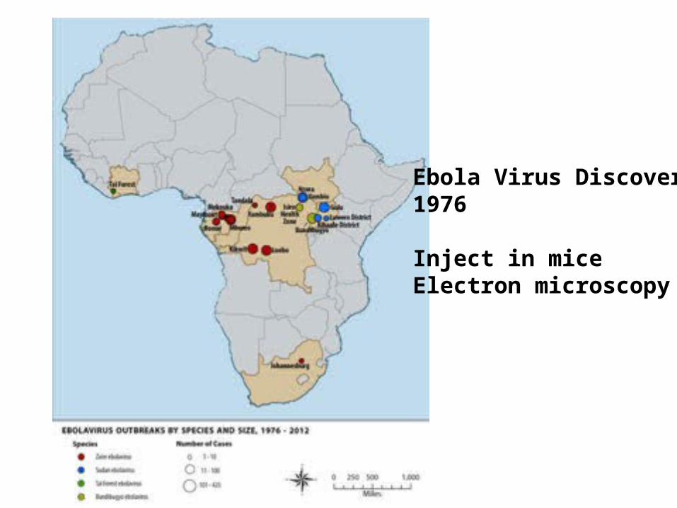
Ebola Virus Discovery1976
Inject in miceElectron microscopy


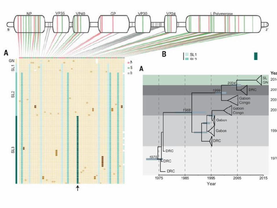
Transmission of EBOV (and search for reservoir species)
Microbes and Infection, 2005,The Natural History of Ebola Virus in Africa
Some infections occurFollowing bat sightings.Related viruses have bat reservoirs
Experiments have failed toreproducibly infect a wide varietyof putative reservoir species with EBOV
Susceptible to EBOVGorillaChimpanzeeDuiker(laboratory mice)

Ghana 2008-2011 Survey
• Methodology—surveyed fever patients Central/North Ghana—18 hospitals/285 patients– PCR to amplify EBOV L gene– ELISA (hepatitis) – Sequencing
• No Ebola• Major illness Hepatitis B & C (adults), A (kids)
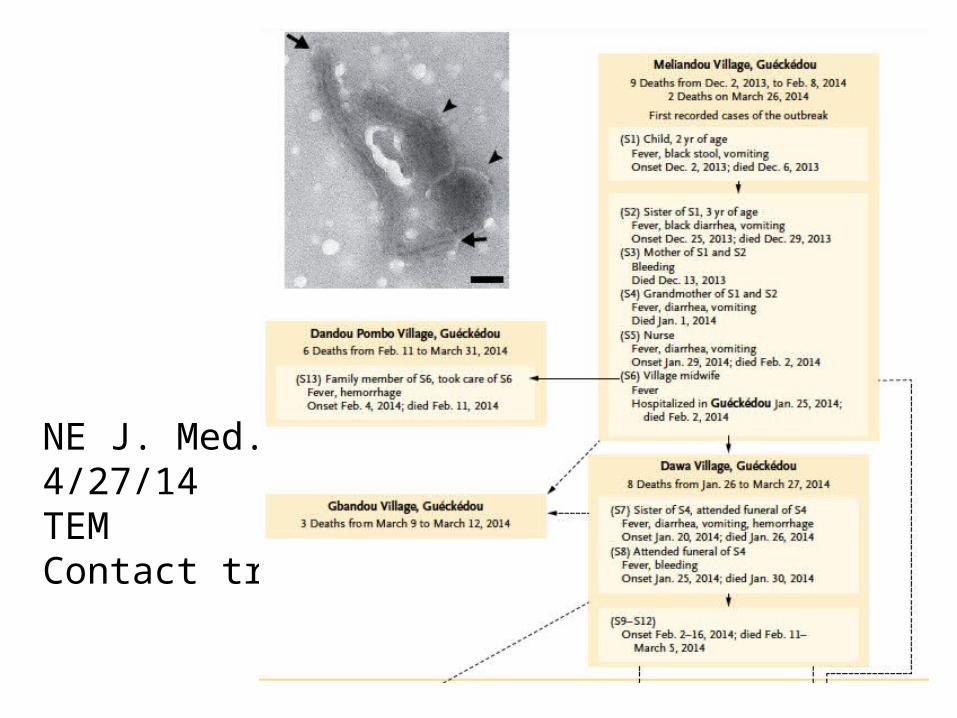
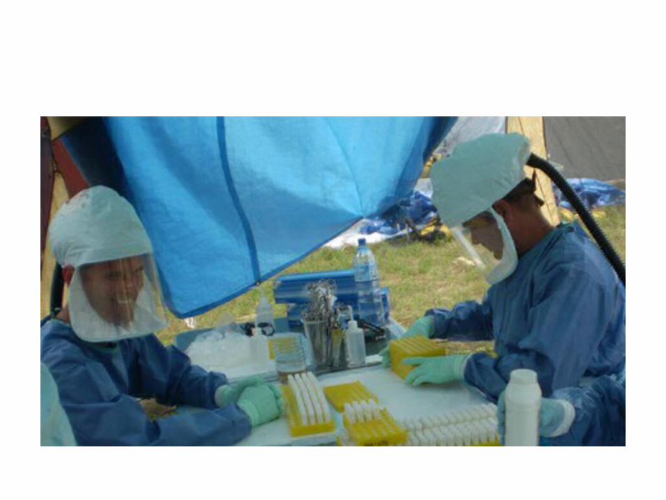
NE J. Med.4/27/14TEMContact tracing

Diagnosis –PCRRT-PCR to find high viral loadsGenome sequencingViral Growth cell culture/FL-anti Ebola AbEMphylogeny


Eurosurveillance, Volume 19, Issue 36, 11 September 2014 Rapid communications
Early transmission dynamics of Ebola virus disease (EVD), West Africa, March to August 2014 H Nishiura , G Chowell
Math modelling
Transmission RatesRt need to be<1 to control epidemic
Rt= number of peopleInfected by each patient
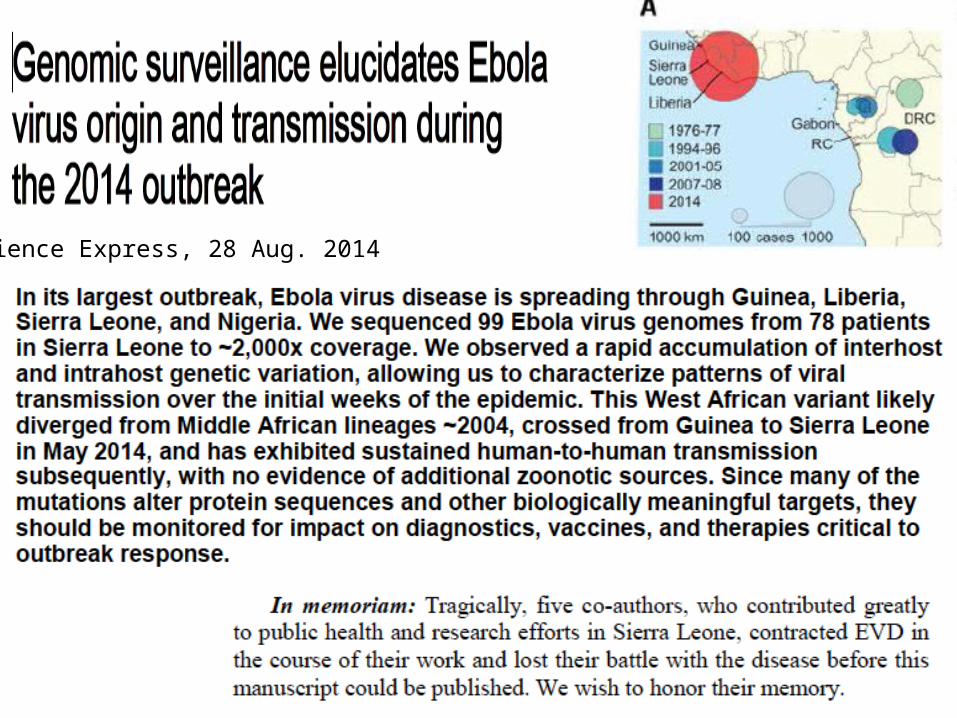
Science Express, 28 Aug. 2014
Pathogenesis of Ebola-Hartman et al, CDC
• FilovirusesHemorraghic fever/vascular dyfunction– Ebola (Zaire & Sudan)– Marburg– Reston & Ivory Coast (animals)
• 4-10 day incubation period; 50-90% fatal• Non-specific initial symptoms (fever, pains,
nausea)• Virus and antibodies detectable with symptoms

Ebola pathogenesis
• Coagulation connection—TF (tissue factor) levels increased Ebola macrophages engulfed in fibrin but Protein C level decreased (decreases coagulation)
• Animal fatality reduced by anti-coagulation factor
• Endothelial leakageshock, hemorrhage Glycoprotein GP1, GP2 connection not clear
Ebola pathogeneis
• Fatal patients—bleeding, coagulation defects, high viral levels many organs, low antibody levels
• Transmission body fluids, not aerosol• Entry—mucous tissue, cuts, common receptor,
replicate many cell types• Immunosuppression—enters dendritic (immature
immune) cellslymph system. • Dendritic cells coordinate innate/adaptive immune
response—cytokine signalling (VP35), T-cells, interferon• Lymphocyte apoptosis
Ebola Goals
• History and Geography• Pathogenesis• Biochemistry & Molecular Biology (and
limitations & unknowns)– Nucleic Acids– 7 Proteins– Implications for understanding biology– Therapeutic implications

Annual Rev Genetics, 1998, Conzelman
Replication/Transcription

“universal” procedure for RT-PCRExtract RNA from 140 uL serum 20 min reverse transcriptase 50°C95°C 5 min10 “precycles” 95°C 5 s, 60-55°C 5s, 72°C 25 sec40 cycles 95°C (denature), 56°C 10 s (anneal), 72°C 10 s (elongate)
$$$ equipment/hoursUnder development—10 min test for “field”
Advertising e-mail arrived Aug. 8, 2014

Comprehensive Functional Analysis of N-Linked Glycans on Ebola Virus GP1—Lenneman et al MBIO Aug. 1, 2014
Endosome proteolytic processing
Why study GP1/GP2?• Viral entry—RBD=receptor binding site• Vaccine development• Understand role of conserved glycosylation—• Mutate conserved N, Y so GP1 will be expressed –sugar
– Pseudovirus in tissue culture• Results
– Sugar removal allows protein production, increases viral entry (including macrophages), cell binding, protease susceptibility, cathespin B independence, but decreases binding NPC1 receptor
– Ca 2+ binds receptor lectins that bind glycans has some reduced binding when glycosylation is removed
– Near complete sugar removal allows greater anti-sera recognition for GP1 core only• Authors suggest GLYCAN SHIELD more important than entry reduction as
reason evolutionary conservation.

Ebola Vaccine Development• Want anti-GP antibodies (acute infection)• Want longer term protection (CD8/T
cell/cytokine)• Adenovirus can deliver GP, but many are
immune to human vectorsChimp adenovirus
• Use non-replicating form of virus• Booster with MVA protects 100% Macaques

Nature Medicine, 7 Sept. 2014

10 month challenge with lethal Ebola Dose
Ebola Polymerase L associates withTopoisomerase I

Topoisomerase I• Known to be important in
viral replication/transcription
• Strand breaking is important
• Inhibition of Top I results in less Ebola replication
• Ebola changes localization of TopI
• Therapeutic implications??
J. Virology Aug. 2013, Takahashi et al

L gene—RdRpRNA dependent RNA polymerase
• 2000 amino acids• Two domains
– Polymerase– Transcription factor
• Template is RNP
• Humans don’t have this enzyme
• Good therapeutic target• Like HIV RT???• Why so little research?
Co-factor
Potential PPIs for VP35(based on VP35 biochemistry/structure)
NMR—solution experimentVerifies that drugs bind.Perturbs chemical shift.Binding pocket mutants haveno chemical shift change

Do drugs disrupt NP/VP35-IID interaction? Stop polymerase?
Pull down assay withamylose beads.If VP35/IID binds NP,an NP-His band is visible.
2 drugs inhibit binding
Ebola polymerase complexIncludesEBOV L, VP30, VP35, NP
Several drugs show doseDependent inhibition
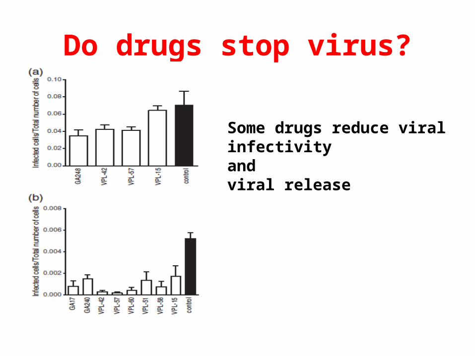
Do drugs stop virus?
Some drugs reduce viralinfectivityandviral release

Ebola Virus Modulates Transforming Growth Factor Signaling andCellular Markers of Mesenchyme-Like Transition in Hepatocytes
• Proteomics/Kinomics Approach• How does EBOV affect global signalling?– How are phosphorylation patterns changed?– Therapeutic targets?
Kindrachuk et al J. Virology September, 2014
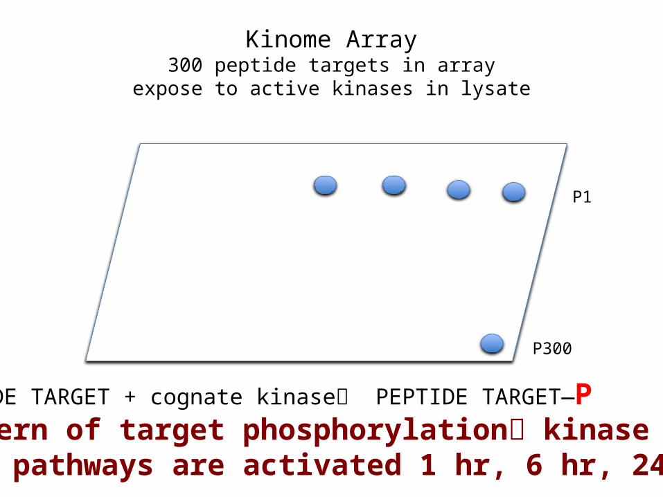
Kinome Array300 peptide targets in array
expose to active kinases in lysate
P1
P300
PEPTIDE TARGET + cognate kinase PEPTIDE TARGET—PPattern of target phosphorylation kinase IDWhat pathways are activated 1 hr, 6 hr, 24 hr?

Results
• TGF-b pathway up-regulated (secretion TGF-b and VEGF confirmed by ELISA)
• Use inhibitors of pathway TGF-b, P13K/AKT, MAPK/EFK, raf, JNK, PKC
• Early use of some inhibitorsReduced mouse fatalities
EMT (epithelial to mesenchyme transition)
• TGF-b normal roles—wound healing, cell growth/differentiation, migration, immune response
• What is happening to EBOV-infected cells?• Epithelial tissues first
infected—adherens/tight junctions disassembled. Gene expression pattern changes to reduce epithelial and increase mesenchyme cytoskeletal expression

Western blots/phosphorylationEMT protein TGF- b pathway P antibodies
Article error—figure legend switched 8&9
EMT—local plus systemic effects

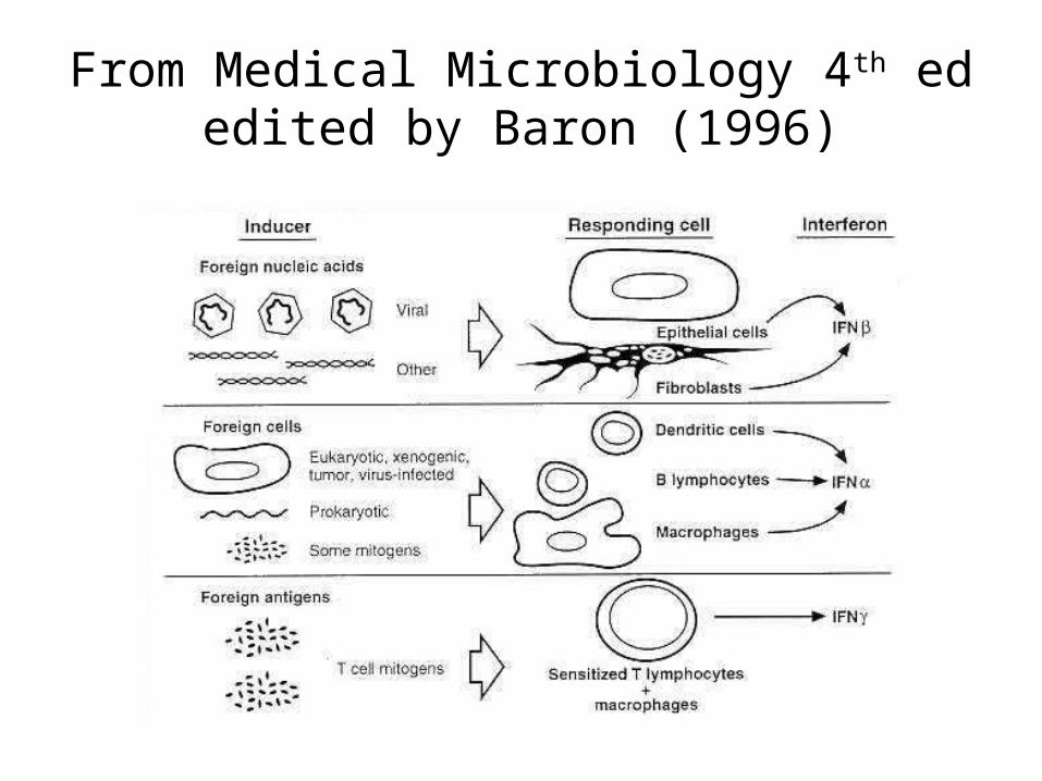
From Medical Microbiology 4th ededited by Baron (1996)

Interferon protects uninfected cellsby changing gene expression
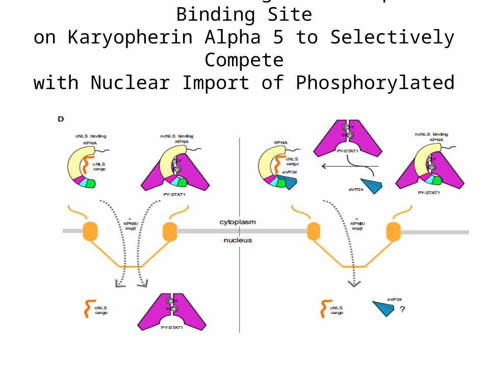
Ebola Virus VP24 Targets a Unique NLS Binding Siteon Karyopherin Alpha 5 to Selectively Compete
with Nuclear Import of Phosphorylated STAT1 Xu et al Cell Host & Microbe, 2014

Proposed Model
• STAT1 must enter nucleus for interferon response• STAT1 binds to KPNA for nuclear transport• eVP24 binds strongly to KPNA• eVP24 competes with STAT1 for same KPNA binding
site• eVP24 and STAT1 binding sites overlap and are distinct
from binding sites for normal transported molecules• eVP24 prevents normal interferon response by
blocking STAT1 transport to nucleus, but does allow entry other molecules

Evidence for modelCrystallography structure—shows hydrophobic and H-bondingat interface very strong bindingeVP24/KPNA
KPNA can be severely truncatedand still bind eVP24
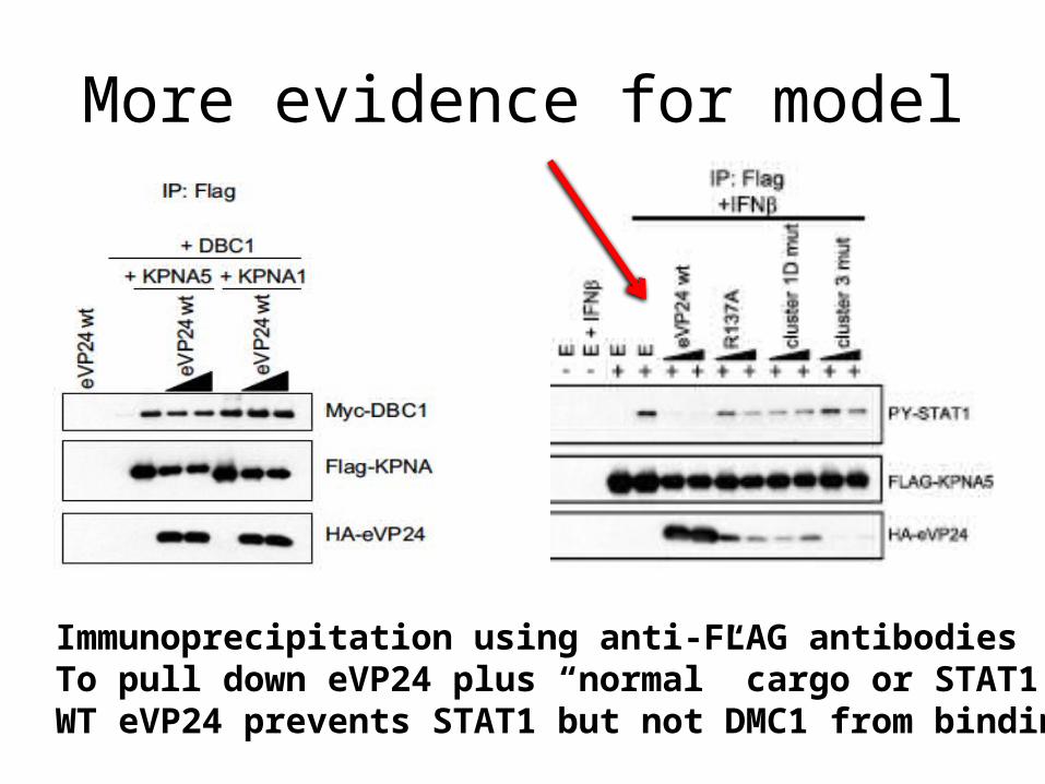
More evidence for model
Immunoprecipitation using anti-FLAG antibodiesTo pull down eVP24 plus “normal” cargo or STAT1WT eVP24 prevents STAT1 but not DMC1 from binding

Last evidence for model
Assay tests whether interferoncan induce promoter
WT eVP24 reducesExpression by >90%
Marburg Virus mVP24has different sequencein key positions anddoes not block STAT1nuclear entry or interferonresponse.


![OCB Ebola Review Summary Report Finalcdn.evaluation.msf.org/.../ocb_ebola_review_summary_report_final_3… · OCB EBOLA REVIEWOCB EBOLA REVIEW SUMMARY REPORT [[[[AprilAprilApril 2012200112016666]]]]](https://static.documents.pub/doc/80x56/5b05e1847f8b9ad1768c04f0/ocb-ebola-review-summary-report-ebola-reviewocb-ebola-review-summary-report-aprilaprilapril.jpg)


