[Frontiers in Bioscience 12, 2631-2645, January 1, 2007] 2631 What triggers cell-mediated mineralization? Leonie F.A. Huitema 1,2 , Arie B. Vaandrager 1 1 Department of Biochemistry and Cell Biology, Faculty of Veterinary Medicine, and Graduate School of Animal Health, Utrecht University, Utrecht, The Netherlands, 2 Department of Equine Sciences, Faculty of Veterinary Medicine, and Graduate School of Animal Health, Utrecht University, Utrecht, The Netherlands TABLE OF CONTENTS 1. Abstract 2. Introduction 3. Different types of mineralization 3.1. Intramembranous ossification 3.2. Endochondral ossification 3.3. Pathological calcification 4. Mechanism of cell-mediated mineral deposition 4.1. The role of matrix vesicles 4.2. The role of cell death 4.3. The role of nucleating proteins 4.4. The role of mineralization inhibitors 5. Perspectives 6. Acknowledgement 7. References 1. ABSTRACT Mineralization is an essential requirement for normal skeletal development, but under certain pathological conditions organs like articular cartilage and cardiovascular tissue are prone to unwanted mineralization. Recent findings suggest that the mechanisms regulating skeletal mineralization may be similar to those regulating pathological mineralization. In general, three forms of cell- mediated mineralization are recognized in an organism: intramembranous ossification, endochondral ossification and pathological mineralization. This review summarizes recent work that tried to elucidate how cell-mediated mineralization is initiated and regulated. To explain mineralization, several theories have been proposed. One theory proposes that mineralization is initiated within matrix vesicles (MVs). A second, not mutually exclusive, theory proposes that phosphate induces apoptosis, and that apoptotic bodies nucleate crystals composed of calcium and phosphate. A third theory suggests that mineralization is mediated by certain non-collagenous proteins, which associate with the extracellular matrix. Regardless of the way mineralization is initiated, the organism also actively inhibits mineralization by specific proteins and removal of an inhibitor may also induce mineralization. Although many studies greatly contributed to a better understanding of the mechanisms regulating cell-mediated mineralization, many questions remain about the mechanisms that trigger cell-mediated mineralization and how this process is regulated. Further investigation is necessary to develop in the future novel therapeutic strategies to prevent pathological mineralization. 2. INTRODUCTION Mineralization is an essential requirement for normal skeletal development, which is generally accomplished through the function of two cell types, osteoblasts and chondrocytes (1). In contrast, soft tissues do not mineralize under normal conditions. However, under certain pathological conditions some tissues like articular cartilage and cardiovascular tissues are prone to mineralization (Table 1) (2;3). Mineralization of articular cartilage contributes to significant morbidity because of its association with joint inflammation and worsening of the progression of osteoarthritis (4). Articular cartilage calcification occurs in association with aging, degenerative joint disease (e.g. osteoarthritis), some genetic disorders and various metabolic disorders (4-7). Similarly, arterial calcification occurs with advanced age, atherosclerosis, metabolic disorders, including end stage renal disease and diabetes mellitus, and some genetic disorders (8). Arterial calcification contributes to hypertension and increased risks of cardiovascular events, leading to morbidity and mortality (9). While pathological mineralization has long been considered to result from a mere physiochemical precipitation of calcium and phosphate, recent studies have provided evidence that soft tissue mineralization is a regulated process, which has many similarities with bone formation (10;11). For now it is unclear why soft tissues have the tendency to mineralize. Therefore, the purpose of this review is to discuss the components involved in cell-mediated mineralization.
Transcript
[Frontiers in Bioscience 12, 2631-2645, January 1, 2007]
2631
What triggers cell-mediated mineralization? Leonie F.A. Huitema1,2, Arie B. Vaandrager1
1 Department of Biochemistry and Cell Biology, Faculty of Veterinary Medicine, and Graduate School of Animal Health, Utrecht University, Utrecht, The Netherlands, 2 Department of Equine Sciences, Faculty of Veterinary Medicine, and Graduate School of Animal Health, Utrecht University, Utrecht, The Netherlands TABLE OF CONTENTS 1. Abstract 2. Introduction 3. Different types of mineralization 3.1. Intramembranous ossification 3.2. Endochondral ossification 3.3. Pathological calcification 4. Mechanism of cell-mediated mineral deposition 4.1. The role of matrix vesicles 4.2. The role of cell death 4.3. The role of nucleating proteins 4.4. The role of mineralization inhibitors 5. Perspectives 6. Acknowledgement 7. References 1. ABSTRACT
Mineralization is an essential requirement for normal skeletal development, but under certain pathological conditions organs like articular cartilage and cardiovascular tissue are prone to unwanted mineralization. Recent findings suggest that the mechanisms regulating skeletal mineralization may be similar to those regulating pathological mineralization. In general, three forms of cell-mediated mineralization are recognized in an organism: intramembranous ossification, endochondral ossification and pathological mineralization. This review summarizes recent work that tried to elucidate how cell-mediated mineralization is initiated and regulated. To explain mineralization, several theories have been proposed. One theory proposes that mineralization is initiated within matrix vesicles (MVs). A second, not mutually exclusive, theory proposes that phosphate induces apoptosis, and that apoptotic bodies nucleate crystals composed of calcium and phosphate. A third theory suggests that mineralization is mediated by certain non-collagenous proteins, which associate with the extracellular matrix. Regardless of the way mineralization is initiated, the organism also actively inhibits mineralization by specific proteins and removal of an inhibitor may also induce mineralization. Although many studies greatly contributed to a better understanding of the mechanisms regulating cell-mediated mineralization, many questions remain about the mechanisms that trigger cell-mediated mineralization and how this process is regulated. Further investigation is necessary to develop in the future novel therapeutic strategies to prevent pathological mineralization.
2. INTRODUCTION
Mineralization is an essential requirement for normal skeletal development, which is generally accomplished through the function of two cell types, osteoblasts and chondrocytes (1). In contrast, soft tissues do not mineralize under normal conditions. However, under certain pathological conditions some tissues like articular cartilage and cardiovascular tissues are prone to mineralization (Table 1) (2;3). Mineralization of articular cartilage contributes to significant morbidity because of its association with joint inflammation and worsening of the progression of osteoarthritis (4). Articular cartilage calcification occurs in association with aging, degenerative joint disease (e.g. osteoarthritis), some genetic disorders and various metabolic disorders (4-7). Similarly, arterial calcification occurs with advanced age, atherosclerosis, metabolic disorders, including end stage renal disease and diabetes mellitus, and some genetic disorders (8). Arterial calcification contributes to hypertension and increased risks of cardiovascular events, leading to morbidity and mortality (9). While pathological mineralization has long been considered to result from a mere physiochemical precipitation of calcium and phosphate, recent studies have provided evidence that soft tissue mineralization is a regulated process, which has many similarities with bone formation (10;11). For now it is unclear why soft tissues have the tendency to mineralize. Therefore, the purpose of this review is to discuss the components involved in cell-mediated mineralization.
Cell-mediated mineralization
2632
3. DIFFERENT TYPES OF MINERALIZATION 3.1. Intramembranous ossification
Clavicles and bones in the regions of the craniofacial skeleton develop by intramembranous ossification, the principle of which is shown in Figure 1, panel 1. During intramembranous ossification, osteoblasts originating from mesenchymal cell condensations are responsible for bone and matrix deposition (12). During this mesenchymal cell condensation, high densities of mesenchymal cells aggregate and start to produce an extracellular matrix (13). Subsequently, mesenchymal cells acquire the typical columnar shape of osteoblasts, increase the synthesis of alkaline phosphatase (APase), and begin to secrete bone matrix, which consists mainly of type I collagen (Figure 1, panel 1A). Osteoblasts than generate phosphate (Pi) by an increase of APase, which is necessary for bone formation (14;15). Numerous ossification centres then develop and eventually fuse (12). Finally, osteoblasts undergo apoptosis (~ 70 %) or terminally differentiate to form osteocytes (~ 30%), which become entrapped in the mineralized bone matrix (14;16;17). Osteocytes are in fact osteoblasts buried within the mineralized bone matrix (Figure 1, panel 1B). Once embedded, they cease their ossification activity and communicate with each other and with cells at the bone surface via a meshwork of cell processes that run through canaliculi in the bone matrix (16). A key regulator of osteoblast differentiation and function is the transcription factor core binding factor alpha 1 (Cbfa1) (12;18-21). Cbfa1 is targeted to the promotors of several bone proteins, such as osteocalcin, bone sialoprotein, APase and type I collagen (18). No bone tissue is formed in Cbfa1 null mice, although a complete cartilaginous skeleton is formed, indicating that Cbfa1 is not essential for chondrogenesis (19).
To maintain bone structure and skeletal growth,
remodeling is necessary. The remodeling of bone that has been formed by either intramembranous ossification or endochondral ossification (see below) consists of a strict coupling of bone resorption by osteoclasts and bone formation by osteoblasts that continue throughout life, with a positive balance during growth and with a negative balance during ageing. Osteoclasts are multinucleated giant cells formed from hemopoietic precursors of the monocyte and macrophage series (22). They attach to the bone surface by sealing a resorbing compartment that they acidify by secreting H+ ions. This will than dissolve the bone mineral, and subsequently expose the organic matrix to proteolytic enzymes that degrade it (22). 3.2. Endochondral ossification
Vertebrate long bones form through a process called endochondral ossification, in which a cartilage template produced by chondrocytes, is formed and replaced by bone (Figure 1, panel 2) (12). During the process of endochondral ossification, mesenchymal cell condensation results in differentiation into chondrocytes (13). Chondrocytes then produce a framework of skeletal cartilage template that will subsequently be replaced by bone (12). It has been suggested that this cartilage template in long bones is formed because the mineralized
extracellular matrix of bone limits interstitial growth. To achieve rapid and directional growth in an organism, an intermediate structure (cartilage) that can function under high load, and at the same time generate space for new bone formation, would then be very helpful (23). Generally four zones are recognized during endochondral ossification (Figure 1, panel 2A-D). The upper zone is the resting zone (Figure 1, panel 2A), chondrocytes are small and dormant, and mainly type II collagen is produced. Subsequently, in the proliferative zone chondrocytes start to proliferate in vertical columns (Figure 1, panel 2B). The next zone is the hypertrophic zone (Figure 1, panel 2C), where the chondrocytes start to enlarge, produce type X collagen, and increase the synthesis of APase. Finally, hypertrophic chondrocytes induce mineralization (Figure 1, panel 2D), which is correlated with increased levels of Pi in the mineralization zone (24-26). Furthermore, terminally differentiated chondrocytes in the growth plate are deleted from the cartilage by programmed cell death (23). In this final zone, osteoprogenitor cells and hemoatopoietic stem cells arrive via the newly formed blood vessels, which penetrate through the transverse septa of mineralizing hypertrophic chondrocytes. Osteoprogenitor cells beneath the site of vascular invasion differentiate into osteoblasts that aggregate on the surface of the calcified cartilage and deposit bone matrix (osteoid; Figure 1, panel 2E). Bone resorbing osteoclasts start to remodel the newly formed bone (27).
The transcription factor SOX9 is mainly
expressed in resting and proliferating chondrocytes, while it is switched off in hypertrophic chondrocytes (28). SOX9 is required for expression of cartilage specific extracellular matrix components, such as collagen types II, IX and XI (12;28). In humans, mutations in SOX9 result in a skeletal malformation syndrome called campomelic dysplasia (29;30). In addition, no chondrocyte specific markers are expressed in SOX9 null cells in mouse chimeras (31). Interestingly, low Cbfa1 expression is detected in hypertrophic chondrocytes, which suggests that hypertrophic chondrocytes may undergo a phenotypic transition towards the osteoblastic phenotype (12). In addition, in Cbfa1 null mice hypertrophic chondrocytes in the femur and humerus are absent (21). The hypothesis that chondrocytes transdifferentiate into osteoblasts is supported by microscopic examinations of cells present at the chondro-osseous junction in the growth plate, which have suggested that indeed chondrocytes differentiate into osteoblasts (32-35). Based on this observation, it is speculated that there are two subpopulations of chondrocytes, one which becomes apoptotic, while the other population trans-differentiates into osteoblasts (32). 3.3. Pathological calcification.
Soft tissues in organisms, like soft tissue structures in joints, skin, heart, blood vessels, kidneys, muscles, lungs, etc. do not mineralize under normal conditions (3). However, as shown in Table 1, under certain pathological conditions some organs mineralize. This is also called pathological mineralization or dystrophic calcification (36). In particular vascular smooth muscle cells in blood vessels are prone to mineralization (Figure 1,
Cell-mediated mineralization
2633
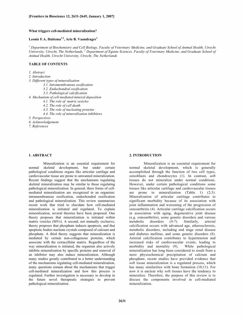
Figure 1. Simplified schematic representation of different forms of cell mediated mineralization. Hatched area represent mineral. Intramembranous ossification. A. Mesenchymal cells aggregate and form nodules (ossification centre), where the cells will differentiate into osteoblasts. B. Bone is formed and remodeled by osteoblasts and osteoclasts. Osteocytes become entrapped in the mineralized matrix. In endochondral ossification 4 zones are recognized in the epiphyseal growth plate. A) resting zone: chondrocytes are small and dormant, mainly type II collagen is produced, B) proliferative zone: chondrocytes start to proliferate in vertical columns, type II collagen is produced, C. Hypertrophic zone: chondrocytes enlarge and start to produce type X collagen, D) Mineralizing zone: hypertrophic chondrocytes start to mineralize, produce matrix vesicles (MVs) and finally die, Type I collagen is now produced E) Osteoblasts grow on the mineralization sheath and produce bone and type I collagen. Some osteoblasts become entrapped in the mineralized matrix and become osteocytes. Osteoclasts remodel newly formed bone. Pathological mineralization (e.g. atherosclerosis). A. Vascular smooth muscle cells in a blood vessel, B. As a result of an atherosclerotic lesion, vascular smooth muscle cells differentiate into osteoblast-like cells and start to induce mineralization. panel 3A-B). Recent studies have provided evidence that pathological vascular calcification is a regulated process, which has many similarities with bone formation (3;11;37;38). Furthermore, expression of a variety of bone-associated proteins has been found in atherosclerotic plaques (39;40). In addition, it has been reported that 10 to 30 % of the cells in a smooth muscle cell culture system undergo a dramatic phenotypic transition in the presence of relatively high phosphate (> 2 mM). This is characterized by the loss of smooth muscle cell lineage marker expression and upregulation of genes related to the osteogenic phenotype (10;41-43). This suggests that vascular smooth muscle cells undergo phenotypic transition to osteochondroprogenitor-like cells. Moreover, formation of complete bone tissue and bone marrow has been demonstrated in calcified
arteries, a phenomenon also called heterotopic (extraosseous) calcification (44;45). In addition, regulated changes in chondrocyte differentiation and viability characteristically seen in growth plate chondrocytes can also occur in mineralizing articular cartilage chondrocytes as a result of osteoarthritis (4). 4. MECHANISM OF CELL-MEDIATED MINERAL DEPOSITION
To induce cell-mediated mineralization, the organism has to create an environment with a local increase of calcium and/or phosphate (Pi), and subsequently organize the nucleation of these ions in an ordered fashion. It has been reported that Pi levels increase considerably from the proliferative to the
Cell-mediated mineralization
2634
Table 1. Forms of pathological mineralization Organ Disease Clinical pattern Etiology References Joints Tendonitis Calcification of the tendon Inflammation, injury, overload 137, 138 Arthritis Calcification of articular cartilage Inflammation, injury, overload, aging 38 Bursitis Calcification of bursal walls Inflammation, overload, aging 139-141 Bone spurs Bony projections that grow along the edges of the
cells Inflammation, injury, aging, high blood pressure
44, 145
Muscles Myositis ossificans Bone formation in the muscles Injury 146 Lung Metastatic pulmonary calcification Calcification in alveolar septa End stage renal disease 147, 148 Pulmonary ossification Bone formation in alveolar compartments Inflammation 148 Skin Cutaneous ossification Bone formation in the skin Injury, inflammation 149 Eyes Cataracts Calcification of the lens Aging 150 Kidney Kidney stones Calcium oxalate or calcium phosphate stones Inherited, mineral imbalance 151 Brain Bilateral striopallidodentate calcinosis Symmetric calcification of the basal ganglia Aging, inherited, infection
152, 153
Spinal cord Neurogenic heterotopic ossification Bone formation in spinal cord Injury 154 Tumor Osteosarcoma Bone cancer that may metastasize elsewhere Unknown 155
hypertrophic zone in the growth plate (25;26). Furthermore, patients with end-stage renal disease (ESRD) develop vascular calcification, which is correlated with an increased serum Pi concentration that typically exceeds 2.0 mM (normal level: 1.4 mM) (10;46). In addition, APase, which cleaves phospho-compounds to inorganic phosphate, is highly increased in mineralization competent cells like osteoblasts, hypertrophic chondrocytes and mineralizing vascular smooth muscle cells. The role of APase in mineralization is essential, since APase deficient mice show impaired skeletal mineralization (47). However, the identity of the physiological organic Pi substrate is not defined.
There is much debate regarding the biochemical mechanisms that initiate mineralization subsequent to the increase in calcium and/or Pi. Figure 2 summarizes the principal factors that are believed to play a role in the process of tissue mineralization. One theory proposes that mineralization is initiated within matrix vesicles (MVs) (48). A second, not mutually exclusive, theory proposes that Pi induces apoptosis, and that apoptotic bodies nucleate crystals composed of calcium and Pi (23;49). A third theory suggests that mineralization is mediated by certain non-collagenous proteins (NCPs), which associate with the extracellular matrix (50-52). Organisms also actively inhibit mineralization by secreting specific proteins and removal of an inhibitor may induce mineralization as well (Figure 2) (53). 4.1. The role of matrix vesicles
It has been hypothesized that cell-mediated mineralization is induced within matrix vesicles (MVs). MVs are cell-derived extracellular membrane enclosed particles, about 0.1-1 micrometer in size (48). In a mineralization inducing environment, they are proposed to bud off the cell plasma membrane in a polarized fashion to the longitudinal septal matrix in the growth plate and to the newly formed osteoid under the mineral facing surfaces of osteoblasts in bone (48;54). MVs have been isolated from mineralizing odontoblasts, osteoblasts, chondrocytes and vascular smooth muscle cells (55-57). This is generally performed after a crude
collagenase digestion (typically collagenase 500 U/ml at 37 °C for 3 hours). After gentle vortexing, MVs are then harvested by differential centrifugation. To this end, the collagenase digest is centrifuged at 13,000 x g for 20 minutes, and the resulting cells and cell debris are discarded. Subsequently, the supernatant is spun at 100,000 x g for 1 hour, which results in a pellet that contains MVs (58-62).
Many studies have been directed at the elucidation of the mechanism of MVs mineralization. In those studies, MVs isolated from tissues or cell culture systems were induced to calcify. This is generally performed by incubating isolated MVs in a so called synthetic cartilage lymph medium which is a physiological buffer containing approximately 2 mM calcium and 1.5 mM Pi (inorganic phosphate) (47;56;60;63-66). From these studies it was concluded that MVs have to be mineralization competent to nucleate calcium phosphate, because isolated MVs from non-mineralizing tissues do not calcify in synthetic cartilage lymph (59;67). Unlike the mineralization competent MVs, these latter MVs did not express Annexin V and had a lower APase activity (47;59). These observations suggest that not all MVs are equivalent and that only mineralizing tissue can produce mineralization competent MVs. These studies are complicated by the presence of vesicles derived from apoptotic cells (apoptotic bodies) in the matrix, which have properties similar but not identical to MVs (see next section) (61).
Several reports suggest that MVs do not
contain crystals at the time of their release from the cell, but that the first crystalline mineral appears after the MV has been immobilized in selected areas of the collagen matrix (48). The mineral crystal is then formed by concentrating calcium and phosphate at the inner leaflet of the vesicle membrane, which has been reported to be enriched in phosphatidylserine (PS) (68). Once the mineral has reached a certain size it ruptures the vesicle membrane and contributes to the extracellular matrix (48). The mechanisms by which the crystals break down or penetrate through the membrane are not fully understood.
Cell-mediated mineralization
2635
Figure 2. Schematic diagram of factors proposed to be involved in cell mediated mineralization. Bold arrows indicate that an increase of Pi rather than calcium induces cell-mediated mineralization. (1) Tissue fluid contains calcium and phosphate. An increase of Pi may induce mineralization. (2) Cells can also generate a local increase of Pi, when they increase APase-levels. The increased Pi induces: (3) the production of MVs, which contain APase and also generate Pi, (4) cell death, which results in the formation of apoptotic bodies, (5) production of nucleating proteins, which are secreted in the extracellular matrix. (6) To control mineralization, the cells constitutively produce mineralization inhibitors. (7) Tissue fluid also contains mineralization inhibitors, principally fetuin that originates from serum.
Because MVs are enclosed by a membrane, transport proteins are required to mediate the influx of mineral ions into these particles. Uptake of phosphate (Pi) is critical for the formation of minerals within MVs. It has been shown that the sodium dependent type III Pi
transporter Glvr-1 is mainly expressed in the growth plate by early hypertrophic chondrocytes, which are the MVs producing cells (69). Furthermore, MVs isolated from chicken epiphyseal cartilage have been shown to contain a sodium dependent Pi transport system (70). However, other
Cell-mediated mineralization
2636
studies report that MV mineralization is not strictly sodium dependent, suggesting the presence of other Pi transporters as well (63). MVs also have the potential to cleave phospho-compounds to inorganic phosphate, resulting in a local increase of Pi concentration, as APase has been shown to be enriched in the membrane of MVs (48;71).
Annexin II, V and VI have been reported to mediate the influx of calcium into the vesicles, by forming hexamers in the PS enriched MV membrane (72;73). Furthermore, Annexin V binds type II and X collagen and this interaction has been shown to stimulate its calcium channel activity (60). The role of Annexin V was further established through suppression by siRNA, which resulted in inhibited mineralization, while overexpression stimulated mineralization (74;75). Annexin expression has also been found to be increased in hypertrophic chondrocytes from the growth plate (76). However, Annexin V knockout mice did not show an impaired skeletal phenotype and neither were the in vitro calcification properties of the isolated annexin (-/-) chondrocytes significantly impaired (77). Possibly other members of the annexin family compensate the deficiency in annexin V. 4.2. The role of cell death
There is a strong correlation between mineralization and cell death. Especially pathological mineralization has often been associated with apoptotic or necrotic processes (11). Furthermore, terminally differentiated chondrocytes in the growth plate are deleted from the cartilage by programmed cell death (23;36). Inorganic phosphate (Pi), whose concentration is reported to increase from the proliferative to the hypertrophic zone in the growth plate, has been shown to be a potent apoptogen (78;79). A Pi-induced intracellular effect was evidenced, since Na-Pi transporter inhibitors were shown to inhibit apoptosis in parallel with mineralization. However, it should be noted that Pi-induced cell death was strongly synergized by the extracellular calcium concentration (69;80;81). A critical role of Pi in apoptosis was also established in mice affected with hypophosphatemia. These animals contain an expanded layer of late non hypertrophic chondrocytes in the growth plate, which is associated with a decrease in the number of apoptotic hypertrophic chondrocytes (82). In contrast, patients affected with hyperphosphatemia show pathological mineralization, which correlated with an increase in cell death (83;84).
Pi is not the only agent that induces cell death during mineralization, since pathological mineralization also occurs in the absence of hyperphosphatemia, indicating the role of additional factors. In agreement with this, it has been reported that no calcification occurred in vessels with calcium and Pi concentrations of 1.8 mM and 3.8 mM respectively, but mechanical injury resulted in extensive calcification under these conditions (85).
Until now it is not clear how apoptosis contributes to mineralization. Possibly, dying or injured cells may become highly permeable to calcium and phosphate, and may concentrate these ions beyond their
solubility product, facilitating heterogeneous nucleation and crystal growth. It has been proposed that an early step in apoptosis is externalization of phosphatidylserine (PS) (86). PS has been shown to have a high affinity for calcium and may act as a nucleator for calcium phosphate crystal formation (36;87). However, it has been reported that mineral-PS interactions can retard crystal growth (88). This suggests that although PS has the capacity to nucleate calcium phosphate in cells undergoing apoptosis, other factors, probably produced by living cells, are necessary to induce crystal growth. It has also been suggested that apoptotic bodies derived from dying cells may act as nucleating mineralization centres in a similar way as described for MVs. Apoptotic bodies isolated from cell culture systems have been shown to precipitate calcium phosphate when incubated in a synthetic cartilage lymph (56;89-91). However, the precipitation capacity was less in apoptotic bodies when compared to MVs (56). This indicates that MVs have a stronger capacity to induce crystal growth, which was supported by Kirsch et al. (61) who reported that apoptotic bodies do not contain APase and the calcium channel forming annexins II, V and VI.
Inhibiting apoptosis with a general caspase inhibitor has been shown to inhibit mineralization in cell culture systems by approximately 40%, indicating a role of apoptosis in mineralization, but also a role of factors other than apoptosis in the process of mineralization (78;92).
Recently, it has been proposed that mineralizing hypertrophic growth plate chondrocytes are not dying by a classical form of apoptosis, because, in contrast to in vitro cell culture systems, they do not produce apoptotic bodies in vivo (23;93). Instead, it was speculated that they eliminate themselves by a process of autophagocytosis. This hypothesis is supported by ultramicroscopic examination of hypertrophic growth plate chondrocytes, showing that dying chondrocytes contain autophagic vacuoles (autophagosomes) and cell remnants that are blebbed off, indicative of autophagocytosis (23;93;94). This specific form of cell death is also called chondroptosis (93). Possibly, the blebs generated by these cells have mineralizing capacities. 4.3. The role of nucleating proteins
Mineralization has also been proposed to be regulated by non-collagenous proteins (NCPs) found in the organic matrix of bone (52;95-99). The NCPs are reported to constitute 5-10% of the total extracellular matrix and can be classified into four groups (Table 2). These groups include proteoglycans, glycoproteins, the γ carboxy glutamic acid (gla)-containing proteins and the serum associated proteins (95;99;100). Of these proteins mainly glycoproteins have been demonstrated to play a critical role in the initiation and growth of the calcium phosphate mineral phase (Table 3). Glycoproteins are proteins that are modified posttranslationally by glycosylation, phosphorylation and sulfatation. Some glycoproteins contain an RGD (Arg-Gly-Asp) sequence that interacts with the integrin receptor family. Glycoproteins that contain an RGD sequence are bone sialoprotein, BAG-75,
Cell-mediated mineralization
2637
Table 2. The non-collagenous proteins regulating mineralization can be classified into four groups Proteoglycans Glycoproteins γ carboxy glutamic acid (gla)-containing proteins Serum proteins versican decorin biglycan hyaluronate
Table 3. Proteins that have been associated with mineralization Protein Mouse mutant Phenotype In vitro effect on mineralization References Annexin V Anxa5 -/- No obvious altered phenotype Mediates influx of calcium into vesicles, siRNA
inhibits mineralization 72-74, 77
Alkaline phosphatase TNAP -/- Hypophosphatemia and impaired growth Release of inorganic Pi from organic phospho-compounds
71, 156, 157
Bone sialioprotein (BSP)
ND1 ND1 Nucleates calcium phosphate 51, 52, 158
BAG-75 ND1 ND1 Sequesters millimolar quantities of Pi 51, 52
Dentin matrix protein-1
dmp1 -/- Decreased mineral to matrix ratio in bones Facilitates hydroxyapatite crystal growth (nonphosphorylated)
95, 101, 111, 112
Fibronectin Fn -/- Non viable Facilitates hydroxyapatite crystal growth 113, 114 Fetuin Fetuin -/- Vascular and soft tissue calcification Inhibits precipitation of calcium and phosphate 119, 121, 122 Matrix GLA protein MGP -/- Vascular, valve and cartilage calcification ND1 123 Osteopontin OPN -/- Enhanced valve implants calcification inhibits mineralization when phosphorylated 128, 129 Osteonectin ON -/- Increased volume of adipose tissue May retard crystal growth 96, 134, 135, 159 Osteocalcin OC -/- Increased bone mass May retard crystal growth 96, 134-136
1 ND: Not determined dentin matrix protein-1, fibronectin, osteopontin and thromobospondin (99;101).
In vitro studies have shown that bone sialoprotein (BSP) can nucleate apatite crystals. BSP is an anionic phosphoprotein that is expressed almost exclusively in mineralized tissues (102;103). Furthermore, it has been demonstrated that after treatment with organophosphate for 4-8 hours, BSP localizes to the extracellular matrix in osteoblastic cultures, well before the first appearance of apatite crystals (104-106). This suggests that Pi (which is probably produced by APase) triggers BSP secretion into the extracellular matrix where it can subsequently nucleate calcium phosphate in metastable solutions. In addition, it has been reported that another noncollagenous bone matrix protein, bone acidic glycoprotein-75 (BAG-75) predicts the location of mineral nucleation, and possibly recruits BSP (51;52). Purified BAG-75 can self-associate into supramolecular spherical complexes and sequesters millimolar quantities of Pi, which indicates that BAG-75 generates a localized Pi source for crystal nucleation reactions (107). Interestingly, it has been proposed that BSP is associated with a population of vesicle-like structures (defined as crystal ghosts), which are 500-800 nm in size. However, BSP did not associate with the smaller 50-300 nm vesicle population (52). An important role of BSP in mineralization has been further established by the observation that transfection of BSP cDAN into non-mineralizing MC3T3-E1 subclones can restore the ability to form mineral deposits (51;108). On the basis of this information, BSP is likely to be involved with early mineral deposition. So far, the phenotype of BSP null mice is not known.
Another NCP, dentin matrix protein-1 (DMP1),
has been reported to facilitate hydroxyapatite growth
(95;100). A role of DMP1 in mineralization was suggested when it was shown to be mainly expressed during dentin mineralization, and later also in osteoblasts (109-111). Furthermore, DMP1 null mice have a decreased mineral to matrix ratio in bones (112). In an in vitro biomineralization model DMP1 has been shown to undergo a conformational change upon calcium binding and to subsequently assemble calcium phosphate nuclei into ordered protein-mineral complexes. This results in an inhibiting effect on spontaneous calcium phosphate precipitation. Thus, DMP1 could sequester and stabilize newly formed calcium phosphate clusters (95). In addition, another in vitro study reported that DMP-1-induced crystal growth and proliferation is dependent on its degree of phosphorylation, because nonphosphorylated DMP1 acts a nucleators while the phosphorylated form inhibits nucleation (101).
Another NCP, fibronectin also has been shown to facilitate hydroxyapatite growth in the presence of a hydroxyapatite seed, and a close association between fibronectin and hydroxyapatite has been found in vivo (113;114). Fibronectin, like DMP-1, has an inhibiting effect on spontaneous calcium phosphate precipitation (95;114). Therefore, it has been postulated that DMP-1 and fibronectin play a structural role in crystal growth, rather than a nucleating role. 4.4. The role of mineralization inhibitors
In mammals, mineralization is generally controlled by two serum NCPs, fetuin and matrix Gla protein (Table 3) (53;115). It has been proposed that the biological function of these proteins is to maintain high metastable blood calcium phosphate levels and to inhibit unwanted (soft tissue) mineralization. Fetuin is synthesized in the liver and found in high concentrations in mammalian serum. Because of its high affinity to hydroxyapatite it is
Cell-mediated mineralization
2638
also found in bone and teeth (116-118). Fetuin knockout mice spontaneously develop widespread soft tissue calcifications, including significant myocardial calcification (119). In humans fetuin deficiency is associated with inflammation and vascular calcification (115;120). In vitro studies demonstrate that fetuin inhibits precipitation of supersaturated solution of calcium and Pi by formation of a high molecular mass fetuin-mineral complex (121). This complex prevents growth, aggregation and precipitation of calcium phosphate (122).
Matrix Gla protein is an extracellular matrix protein that is generally expressed by chondrocytes and vascular smooth muscle cells (123). Mice deficient in matrix Gla protein are normal at birth but develop severe calcification of all arteries (and cartilage) within weeks. These mice die at around 8 weeks of age, mostly due to a rupture of the aorta (123). Interestingly, it has recently been reported that MVs isolated from vascular smooth muscle cells that are induced to mineralize contain fetuin and matrix Gla protein (56;124). It has been speculated that this may be a defence mechanism of the cell to limit excessive mineralization (56;124).
The NCP osteopontin (OPN) has also been shown to control crystal growth (125). In addition, Pi has been proposed to be a specific signal for upregulation of OPN gene expression, which supports its regulatory role during mineralization (15;126). In agreement with this, gluteraldehyde fixed porcine aortic valves implanted into OPN null mice mineralized to a much greater extent than those implanted in wild type mice (127). However, the inhibiting effect of OPN on mineralization is dependent on the extent of phosphorylation of OPN, because OPN does not inhibit mineralization after dephosphorylation by APase (128-130). In vivo, OPN is a protein that is normally found in mineralized tissue, but also in epithelial lining cells of numerous organs, and body fluids, including urine, saliva, milk and bile (129;131). Next to inhibiting mineralization, OPN also regulates bone cell adhesion and osteoclast function in the skeleton (129). When OPN attaches to osteoclasts, it stimulates the acidification of the local environment, which will allow for the dissolution of the mineral (129).
Another NCP, osteonectin (ON), also known as SPARC (secreted protein, acidic, rich in cysteine), is a calcium binding matrix protein found in many tissues undergoing remodeling (132). Several in vitro studies have demonstrated that ON can inhibit crystal nucleation and retard crystal growth (133-135), although Hunter et al. (1996) found no effect (96). An explanation for the different results may be that different model systems were used, or the difference in ON concentrations tested. In vivo, ON deficient mice show an increased volume of adipose tissue and a decreased osteoblast and osteoclast number, resulting in osteopenia. This suggests that ON rather plays an important role in cell differentiation as well (132).
In vitro, similar results as with ON were obtained for the NCP osteocalcin (OC), which is also known as bone Gla protein (BGP) (96;99;133-135). In addition, in vivo
transgenic OC deficient mice demonstrate an increase in bone mass, which suggests that OC indeed limits mineralization (136). 5. PERSPECTIVES
Pathological mineralization can have severe clinical consequences (Table 1). For example, articular cartilage calcification is one of the major degenerative diseases of the skeleton and leads to cartilage destruction, severe pain and joint stiffness (4). In addition, vascular calcification may lead to mortality (8;9). Although many investigations greatly contributed to a better understanding of the mechanisms regulating cell-mediated mineralization, many questions remain about the mechanisms that trigger cell-mediated mineralization and how this process is regulated. For instance, it is still not clear whether one type of vesicles induces mineralization, or whether more types of vesicles are involved. This might possibly be different between different tissues. In addition, it is unclear where the mineral exactly nucleates. For example, does the first mineral develop extracellularly in vesicles? Or does it start intracellularly, which will then result in the formation of vesicles? Furthermore, various proteins have been shown to be involved in mineralization, but it is unclear how these proteins are related mechanistically. Therefore, a detailed understanding of the roles of the various hypotheses and the coordinated interaction between them will provide novel therapeutic strategies to prevent pathological mineralization. 6. ACKNOWLEDGEMENT
Authors acknowledge Dr. P.R. van Weeren, Prof. A. Barneveld and Prof. J.B. Helms for critical reading of the manuscript.
7. REFERENCES 1. Kronenberg H. M.: Developmental regulation of the growth plate. Nature 423, 332-336 (2003) 2. Giachelli C. M.: Ectopic calcification: gathering hard facts about soft tissue mineralization. A J Pathol 154, 671-675 (1999) 3. Giachelli C. M.: Inducers and inhibitors of biomineralization: lessons from pathological calcification. Ortho Craniofac Res 8, 229-231 (2005) 4. Terkeltaub R. A.: What does cartilage calcification tell us about osteoarthritis? J Rheumatol 29, 411-415 (2002) 5. Karpouzas G. A. and R. A. Terkeltaub: New developments in the pathogenesis of articular cartilage calcification. Curr Rheumatol Rep. 1, 121-127 (1999) 6. Maldonado I., A. M. Reginato and A. J. Reginato: Familial calcium crystal diseases: what have we learned? Curr Opin Rheumatol. 13, 225-233 (2001) 7. Zhang Y. and M. A. Brown: Genetic studies of chondrocalcinosis. Curr Opin Rheumatol 17, 330-335 (2005)
Cell-mediated mineralization
2639
8. Wilson P. W., L. I. Kauppila, C. J. O'Donnell, D. P. Kiel, M. Hannan, J. M. Polak and L. A. Cupples: Abdominal aortic calcific deposits are an important predictor of vascular morbidity and mortality. Circulation 103, 1529-1534 (2001) 9. Rutsch F. and R. Terkeltaub: Deficiencies of physiologic calcification inhibitors and low-grade inflammation in arterial calcification: lessons for cartilage calcification. Joint Bone Spine 72, 110-118 (2005) 10. Giachelli C. M.: Vascular calcification: in vitro evidence for the role of inorganic phosphate. J Am Soc Nephrol 14, S300-S304 (2003) 11. Magne D., M. Julien, C. Vinatier, F. Merhi-Soussi, P. Weiss and J. Guicheux: Cartilage formation in growth plate and arteries: from physiology to pathology. Bioessays 27, 708-716 (2005) 12. Olsen B. R., A. M. Reginato and W. Wang: Bone development. Annu Rev Cell Dev Biol 16, 191-220 (2000) 13. Behonick D. J. and Z. Werb: A bit of give and take: the relationship between the extracellular matrix and the developing chondrocyte. Mech Dev 120, 1327-1336 (2003) 14. Mackie E. J.: Osteoblasts: novel roles in orchestration of skeletal architecture. Int J Biochem Cell Biol 35, 1301-1305 (2003) 15. Beck, Jr. G. R.: Inorganic phosphate as a signaling molecule in osteoblast differentiation. J Cell Biochem 90, 234-243 (2003) 16. Franz-Odendaal T. A., B. K. Hall and P. E. Witten: Buried alive: how osteoblasts become osteocytes. Dev Dyn 235, 176-190 (2006) 17. Knothe Tate M. L., J. R. Adamson, A. E. Tami and T. W. Bauer: The osteocyte. Int J Biochem Cell Biol 36, 1-8 (2004) 18. Ducy P., R. Zhang, V. Geoffroy, A. L. Ridall and G. Karsenty: Osf2/Cbfa1: a transcriptional activator of osteoblast differentiation. Cell 89, 747-754 (1997) 19. Komori T., H. Yagi, S. Nomura, A. Yamaguchi, K. Sasaki, K. Deguchi, Y. Shimizu, R. T. Bronson, Y. H. Gao, M. Inada, M. Sato, R. Okamoto, Y. Kitamura, S. Yoshiki and T. Kishimoto: Targeted disruption of Cbfa1 results in a complete lack of bone formation owing to maturational arrest of osteoblasts. Cell 89, 755-764 (1997) 20. Liu W., S. Toyosawa, T. Furuichi, N. Kanatani, C. Yoshida, Y. Liu, M. Himeno, S. Narai, A. Yamaguchi and T. Komori: Overexpression of Cbfa1 in osteoblasts inhibits osteoblast maturation and causes osteopenia with multiple fractures. J Cell Biol 155, 157-166 (2001)
21. Karsenty G.: Minireview: transcriptional control of osteoblast differentiation. Endocrinology 142, 2731-2733 (2001) 22. Martin T. J. and N. A. Sims: Osteoclast-derived activity in the coupling of bone formation to resorption. Trends Mol Med 11, 76-81 (2005) 23. Shapiro I. M., C. S. Adams, T. Freeman and V. Srinivas: Fate of the hypertrophic chondrocyte: microenvironmental perspectives on apoptosis and survival in the epiphyseal growth plate. Birth Defects Res C Embryo Today 75, 330-339 (2005) 24. Erlebacher A., E. H. Filvaroff, S. E. Gitelman and R. Derynck: Toward a molecular understanding of skeletal development. Cell 80, 371-378 (1995) 25. Shapiro I. M. and A. Boyde: Microdissection--elemental analysis of the mineralizing growth cartilage of the normal and rachitic chick. Metab Bone Dis Relat Res 5, 317-326 (1984) 26. Wuthier R. E.: Involvement of cellular metabolism of calcium and phosphate in calcification of avian growth plate cartilage. J Nutr 123, 301-309 (1993) 27. White A. and G. Wallis: Endochondral ossification: a delicate balance between growth and mineralisation. Curr Biol 11, R589-R591 (2001) 28. Goldring M. B., K. Tsuchimochi and K. Ijiri: The control of chondrogenesis. J Cell Biochem 97, 33-44 (2006) 29. Foster J. W., M. A. Dominguez-Steglich, S. Guioli, G. Kowk, P. A. Weller, M. Stevanovic, J. Weissenbach, S. Mansour, I. D. Young, P. N. Goodfellow and .: Campomelic dysplasia and autosomal sex reversal caused by mutations in an SRY-related gene. Nature 372, 525-530 (1994) 30. Wagner T., J. Wirth, J. Meyer, B. Zabel, M. Held, J. Zimmer, J. Pasantes, F. D. Bricarelli, J. Keutel, E. Hustert, U. Wolf, Tommerup, W. Schempp and G. Scherer: Autosomal sex reversal and campomelic dysplasia are caused by mutations in and around the SRY-related gene SOX9. Cell 79, 1111-1120 (1994) 31. Bi W., J. M. Deng, Z. Zhang, R. R. Behringer and B. de Crombrugghe: Sox9 is required for cartilage formation. Nat Genet 22, 85-89 (1999) 32. Bianco P., F. D. Cancedda, M. Riminucci and R. Cancedda: Bone formation via cartilage models: the "borderline" chondrocyte. Matrix Biol 17, 185-192 (1998) 33. Galotto M., G. Campanile, G. Robino, F. D. Cancedda, P. Bianco and R. Cancedda: Hypertrophic chondrocytes undergo further differentiation to osteoblast-like cells and participate in the initial bone formation in developing chick embryo. J Bone Miner Res 9, 1239-1249 (1994)
Cell-mediated mineralization
2640
34. Riminucci M., J. N. Bradbeer, A. Corsi, C. Gentili, F. Descalzi, R. Cancedda and P. Bianco: Vis-a-vis cells and the priming of bone formation. J Bone Miner Res 13, 1852-1861 (1998) 35. Thesingh C. W., C. G. Groot and A. M. Wassenaar: Transdifferentiation of hypertrophic chondrocytes into osteoblasts in murine fetal metatarsal bones, induced by co-cultured cerebrum. Bone Miner 12, 25-40 (1991) 36. Speer M. Y. and C. M. Giachelli: Regulation of cardiovascular calcification. Cardiovasc Pathol 13, 63-70 (2004) 37. Demer L. L. and Y. Tintut: Mineral exploration: search for the mechanism of vascular calcification and beyond: the 2003 Jeffrey M. Hoeg Award lecture. Arterioscler Thromb Vasc Biol 23, 1739-1743 (2003) 38. Kirsch T.: Determinants of pathological mineralization. Curr Opin Rheumatol 18, 174-180 (2006) 39. Bobryshev Y. V.: Transdifferentiation of smooth muscle cells into chondrocytes in atherosclerotic arteries in situ: implications for diffuse intimal calcification. J Pathol 205, 641-650 (2005) 40. Tyson K. L., J. L. Reynolds, R. McNair, Q. Zhang, P. L. Weissberg and C. M. Shanahan: Osteo/chondrocytic transcription factors and their target genes exhibit distinct patterns of expression in human arterial calcification. Arterioscler Thromb Vasc Biol 23, 489-494 (2003) 41. Steitz S. A., M. Y. Speer, G. Curinga, H. Y. Yang, P. Haynes, R. Aebersold, T. Schinke, G. Karsenty and C. M. Giachelli: Smooth muscle cell phenotypic transition associated with calcification: upregulation of Cbfa1 and downregulation of smooth muscle lineage markers. Circ Res 89, 1147-1154 (2001) 42. Shioi A., Y. Nishizawa, S. Jono, H. Koyama, M. Hosoi and H. Morii: Beta-glycerophosphate accelerates calcification in cultured bovine vascular smooth muscle cells. Arterioscler Thromb Vasc Biol 15, 2003-2009 (1995) 43. Bostrom K., K. E. Watson, S. Horn, C. Wortham, I. M. Herman and L. L. Demer: Bone morphogenetic protein expression in human atherosclerotic lesions. J Clin Invest 91, 1800-1809 (1993) 44. Deneke T., K. Langner, P. H. Grewe, E. Harrer and K. M. Muller: Ossification in atherosclerotic carotid arteries. Z Kardiol 90 Suppl 3, 106-115 (2001) 45. Seifert G.: Heterotopic (extraosseous) calcification (calcinosis). Etiology, pathogenesis and clinical importance. Pathologe 18, 430-438 (1997) 46. Block G. A., T. E. Hulbert-Shearon, N. W. Levin and F. K. Port: Association of serum phosphorus and calcium x phosphate product with mortality risk in chronic
hemodialysis patients: a national study. Am J Kidney Dis 31, 607-617 (1998) 47. Anderson H. C., J. B. Sipe, L. Hessle, R. Dhanyamraju, E. Atti, N. P. Camacho and J. L. Millan: Impaired calcification around matrix vesicles of growth plate and bone in alkaline phosphatase-deficient mice. Am J Pathol 164, 841-847 (2004) 48. Anderson H. C.: Molecular biology of matrix vesicles. Clin Orthop Relat Res 266-280 (1995) 49. Gibson G.: Active role of chondrocyte apoptosis in endochondral ossification. Microsc Res Tech 43, 191-204 (1998) 50. Glimcher M. J.: Mechanism of calcification: role of collagen fibrils and collagen-phosphoprotein complexes in vitro and in vivo. Anat Rec 224, 139-153 (1989) 51. Gorski J. P., A. Wang, D. Lovitch, D. Law, K. Powell and R. J. Midura: Extracellular bone acidic glycoprotein-75 defines condensed mesenchyme regions to be mineralized and localizes with bone sialoprotein during intramembranous bone formation. J Biol Chem 279, 25455-25463 (2004) 52. R. J. Midura J. P., A. Wang, D. Lovitch, D. Law, K. Powell and J. P. Gorski: Bone acidic glycoprotein-75 delineates the extracellular sites of future bone sialoprotein accumulation and apatite nucleation in osteoblastic cultures. J Biol Chem 279, 25464-25473 (2004) 53. Schinke T. and G. Karsenty: Vascular calcification--a passive process in need of inhibitors. Nephrol Dial Transplant 15, 1272-1274 (2000) 54. Borg T. K., R. Runyan and R. E. Wuthier: A freeze-fracture study of avian epiphyseal cartilage differentiation. Anat Rec 99, 449-457 (1981) 55. Hoshi K. and H. Ozawa: Matrix vesicle calcification in bones of adult rats. Calcif Tissue Int 66, 430-434 (2000) 56. Reynolds J. L., A. J. Joannides, J. N. Skepper, R. McNair, L. J. Schurgers, D. Proudfoot, W. Jahnen-Dechent, P. L. Weissberg and C. M. Shanahan: Human vascular smooth muscle cells undergo vesicle-mediated calcification in response to changes in extracellular calcium and phosphate concentrations: a potential mechanism for accelerated vascular calcification in ESRD. J Am Soc Nephrol 15, 2857-2867 (2004) 57. Wuthier R. E., J. E. Chin, J. E. Hale, T. C. Register, L. V. Hale and Y. Ishikawa: Isolation and characterization of calcium-accumulating matrix vesicles from chondrocytes of chicken epiphyseal growth plate cartilage in primary culture. J Biol Chem 260, 15972-15979 (1985) 58. Kirsch T. and R. E. Wuthier: Stimulation of calcification of growth plate cartilage matrix vesicles by
Cell-mediated mineralization
2641
binding to type II and X collagens. J Biol Chem 269, 11462-11469 (1994) 59. Kirsch T., H. D. Nah, I. M. Shapiro and M. Pacifici: Regulated production of mineralization-competent matrix vesicles in hypertrophic chondrocytes. J Cell Biol 137, 1149-1160 (1997) 60. Kirsch T., G. Harrison, E. E. Golub and H. D. Nah: The roles of annexins and types II and X collagen in matrix vesicle-mediated mineralization of growth plate cartilage. J Biol Chem 275, 35577-35583 (2000) 61. Kirsch T., W. Wang and D. Pfander: Functional differences between growth plate apoptotic bodies and matrix vesicles. J Bone Miner Res 18, 1872-1881 (2003) 62. Hsu H. H. and H. C. Anderson: Evidence of the presence of a specific ATPase responsible for ATP-initiated calcification by matrix vesicles isolated from cartilage and bone. J Biol Chem 271, 26383-26388 (1996) 63. Wu L. N., G. R. Sauer, B. R. Genge, W. B. Valhmu and R. E. Wuthier: Effects of analogues of inorganic phosphate and sodium ion on mineralization of matrix vesicles isolated from growth plate cartilage of normal rapidly growing chickens. J Inorg Biochem 94, 221-235 (2003) 64. Hsu H. H. and N. P. Camacho: Isolation of calcifiable vesicles from human atherosclerotic aortas. Atherosclerosis 143, 353-362 (1999) 65. Hsu H. H., O. Tawfik and F. Sun: Mechanisms of calcification by vesicles isolated from atherosclerotic rabbit aortas. Biochim Biophys Acta 1563, 18-22 (2002) 66. Hsu H. H., N. C. Camacho, O. Tawfik and F. Sun: Induction of calcification in rabbit aortas by high cholesterol diets: roles of calcifiable vesicles in dystrophic calcification. Atherosclerosis 161, 85-94 (2002) 67. Hsu H. H., N. P. Camacho, F. Sun, O. Tawfik and H. Aono: Isolation of calcifiable vesicles from aortas of rabbits fed with high cholesterol diets. Atherosclerosis 153, 337-348 (2000) 68. Majeska R. J., D. L. Holwerda and R. E. Wuthier: Localization of phosphatidylserine in isolated chick epiphyseal cartilage matrix vesicles with trinitrobenzenesulfonate. Calcif Tissue Int 27, 41-46 (1979) 69. Palmer G., J. Zhao, J. Bonjour, W. Hofstetter and J. Caverzasio: In vivo expression of transcripts encoding the Glvr-1 phosphate transporter/retrovirus receptor during bone development. Bone 24, 1-7 (1999) 70. Montessuit C., J. P. Bonjour and J. Caverzasio: Expression and regulation of Na-dependent P(i) transport in matrix vesicles produced by osteoblast-like cells. J Bone Miner Res 10, 625-631 (1995)
71. Whyte M. P.: Hypophosphatasia and the role of alkaline phosphatase in skeletal mineralization. Endocr Rev 15, 439-461 (1994) 72. Kirsch T., H. D. Nah, D. R. Demuth, G. Harrison, E. E. Golub, S. L. Adams and M. Pacifici: Annexin V-mediated calcium flux across membranes is dependent on the lipid composition: implications for cartilage mineralization. Biochemistry 36, 3359-3367 (1997) 73. Matsuda R., N. Kaneko and Y. Horikawa: Presence and comparison of Ca2+ transport activity of annexins I, II, V, and VI in large unilamellar vesicles. Biochem Biophys Res Commun 237, 499-503 (1997) 74. Wang W., J. Xu and T. Kirsch: Annexin V and terminal differentiation of growth plate chondrocytes. Exp Cell Res 305, 156-165 (2005) 75. Balcerzak M., E. Hamade, L. Zhang, S. Pikula, G. Azzar, J. Radisson, J. Bandorowicz-Pikula and R. Buchet: The roles of annexins and alkaline phosphatase in mineralization process. Acta Biochim Pol 50, 1019-1038 (2003) 76. Kirsch T., B. Swoboda and H. Nah: Activation of annexin II and V expression, terminal differentiation, mineralization and apoptosis in human osteoarthritic cartilage. Osteoarthritis Cartilage 8, 294-302 (2000) 77. Brachvogel B., J. Dikschas, H. Moch, H. Welzel, M. K. von der, C. Hofmann and E. Poschl: Annexin A5 is not essential for skeletal development. Mol Cell Biol 23, 2907-2913 (2003) 78. Magne D., G. Bluteau, C. Faucheux, G. Palmer, C. Vignes-Colombeix, P. Pilet, T. Rouillon, J. Caverzasio, P. Weiss, G. Daculsi and J. Guicheux: Phosphate is a specific signal for ATDC5 chondrocyte maturation and apoptosis-associated mineralization: possible implication of apoptosis in the regulation of endochondral ossification. J Bone Miner Res 18, 1430-1442 (2003) 79. Mansfield K. R. Rajpurohit and I. M. Shapiro: Extracellular phosphate ions cause apoptosis of terminally differentiated epiphyseal chondrocytes. J Cell Physiol 179, 276-286 (1999) 80. Mansfield K., C. C. Teixeira, C. S. Adams and I. M. Shapiro: Phosphate ions mediate chondrocyte apoptosis through a plasma membrane transporter mechanism. Bone 28, 1-8 (2001) 81. Mansfield K., B. Pucci, C. S. Adams and I. M. Shapiro: Induction of apoptosis in skeletal tissues: phosphate-mediated chick chondrocyte apoptosis is calcium dependent. Calcif Tissue Int 73, 161-172 (2003) 82. Sabbagh Y., T. O. Carpenter and M. B. Demay: Hypophosphatemia leads to rickets by impairing caspase-mediated apoptosis of hypertrophic chondrocytes. Proc Natl Acad Sci USA 102, 9637-9642 (2005)
Cell-mediated mineralization
2642
83. Ketteler M., V. Brandenburg, W. Jahnen-Dechent, R. Westenfeld and J. Floege: Do not be misguided by guidelines: the calcium x phosphate product can be a Trojan horse. Nephrol Dial Transplant 20, 673-677 (2005) 84. Takeda E. H. Yamamoto, K. Nashiki, T. Sato, H. Arai and Y. Taketani: Inorganic phosphate homeostasis and the role of dietary phosphorus. J Cell Mol Med 8, 191-200 (2004) 85. Lomashvili K. A., S. Cobbs, R. A. Hennigar, K. I. Hardcastle and W. C. O'Neill: Phosphate-induced vascular calcification: role of pyrophosphate and osteopontin. J Am Soc Nephrol 15, 1392-1401 (2004) 86. Bratton D. L., V. A. Fadok, D. A. Richter, J. M. Kailey, L. A. Guthrie and P. M. Henson: Appearance of phosphatidylserine on apoptotic cells requires calcium-mediated nonspecific flip-flop and is enhanced by loss of the aminophospholipid translocase. J Biol Chem 272, 26159-26165 (1997) 87. Schoen F. J., J. W. Tsao and R. J. Levy: Calcification of bovine pericardium used in cardiac valve bioprostheses. Implications for the mechanisms of bioprosthetic tissue mineralization. Am J Pathol 123, 134-145 (1986) 88. Boskey A. L. and B. L. Dick: The effect of phosphatidylserine on in vitro hydroxyapatite growth and proliferation. Calcif Tissue Int 49, 193-196 (1991) 89. Cheung H. S. and L. M. Ryan: Phosphocitrate blocks nitric oxide-induced calcification of cartilage and chondrocyte-derived apoptotic bodies. Osteoarthritis Cartilage 7, 409-412 (1999) 90. Hashimoto S., R. L. Ochs, F. Rosen, J. Quach, G. McCabe, J. Solan, J. E. Seegmiller, R. Terkeltaub and M. Lotz: Chondrocyte-derived apoptotic bodies and calcification of articular cartilage. Proc Natl Acad Sci USA 95, 3094-3099 (1998) 91. Lotz M., S. Hashimoto and K. Kuhn: Mechanisms of chondrocyte apoptosis. Osteoarthritis Cartilage 7, 389-391 (1999) 92. Proudfoot D., J. N. Skepper, L. Hegyi, M. R. Bennett, C. M. Shanahan and P. L. Weissberg: Apoptosis regulates human vascular calcification in vitro: evidence for initiation of vascular calcification by apoptotic bodies. Circ Res 87, 1055-1062 (2000) 93. Roach H. I., T. Aigner and J. B. Kouri: Chondroptosis: a variant of apoptotic cell death in chondrocytes? Apoptosis 9, 265-277 (2004) 94. Roach H. I. and N. M. Clarke: Physiological cell death of chondrocytes in vivo is not confined to apoptosis. New observations on the mammalian growth plate. J Bone Joint Surg Br 82, 601-613 (2000)
95. He G., S. Gajjeraman, D. Schultz, D. Cookson, C. Qin, W. T. Butler, J. Hao and A. George: Spatially and temporally controlled biomineralization is facilitated by interaction between self-assembled dentin matrix protein 1 and calcium phosphate nuclei in solution. Biochemistry 44, 16140-16148 (2005) 96. Hunter G. K., P. V. Hauschka, A. R. Poole, L. C. Rosenberg and H. A. Goldberg: Nucleation and inhibition of hydroxyapatite formation by mineralized tissue proteins. Biochem J 317, 59-64 (1996) 97. Hunter G. K., M. S. Poitras, T. M. Underhill, M. D. Grynpas and H. A. Goldberg: Induction of collagen mineralization by a bone sialoprotein--decorin chimeric protein. J Biomed Mater Res 55, 496-502 (2001) 98. Butler W. T., J. E. Finch, Jr. and C. V. Desteno: Chemical character of proteins in rat incisors. Biochim Biophys Acta 257, 167-171 (1972) 99. Donley G. E. and L. A. Fitzpatrick: Noncollagenous matrix proteins controlling mineralization; possible role in pathologic calcification of vascular tissue. Trends Cardiovasc Med 8, 199-206 (1998) 100. He G., T. Dahl, A. Veis and A. George: Nucleation of apatite crystals in vitro by self-assembled dentin matrix protein 1. Nat Mater 2, 552-558 (2003) 101. Tartaix P. H., M. Doulaverakis, A. George, L. W. Fisher, W. T. Butler, C. Qin, E. Salih, M. Tan, Y. Fujimoto, L. Spevak and A. L. Boskey: In vitro effects of dentin matrix protein-1 on hydroxyapatite formation provide insights into in vivo functions. J Biol Chem 279, 18115-18120 (2004) 102. Bianco P., L. W. Fisher, M. F. Young, J. D. Termine and P. G. Robey: Expression of bone sialoprotein (BSP) in developing human tissues. Calcif Tissue Int 49, 421-426 (1991) 103. Chen J. K., H. S. Shapiro, J. L. Wrana, S. Reimers, J. N. Heersche and J. Sodek: Localization of bone sialoprotein (BSP) expression to sites of mineralized tissue formation in fetal rat tissues by in situ hybridization. Matrix 11, 133-143 (1991) 104. McQuillan D. J., M. D. Richardson and J. F. Bateman: Matrix deposition by a calcifying human osteogenic sarcoma cell line (SAOS-2). Bone 16, 415-426 (1995) 105. Stanford C. M., P. A. Jacobson, E. D. Eanes, L. A. Lembke and R. J. Midura: Rapidly forming apatitic mineral in an osteoblastic cell line (UMR 106-01 BSP). J Biol Chem 270, 9420-9428 (1995) 106. Wang A., J. A. Martin, L. A. Lembke and R. J. Midura: Reversible suppression of in vitro biomineralization by activation of protein kinase A. J Biol Chem 275, 11082-11091 (2000)
Cell-mediated mineralization
2643
107. Gorski J. P., E. A. Kremer, Y. Chen, S. Ryan, C. Fullenkamp, J. Delviscio, K. Jensen and M. D. McKee: Bone acidic glycoprotein-75 self-associates to form macromolecular complexes in vitro and in vivo with the potential to sequester phosphate ions. J Cell Biochem 64, 547-564 (1997) 108. Wang D., K. Christensen, K. Chawla, G. Xiao, P. H. Krebsbach and R. T. Franceschi: Isolation and characterization of MC3T3-E1 preosteoblast subclones with distinct in vitro and in vivo differentiation/mineralization potential. J Bone Miner Res 14, 893-903 (1999) 109. D'Souza R. N., A. Cavender, G. Sunavala, J. Alvarez, T. Ohshima, A. B. Kulkarni and M. MacDougall: Gene expression patterns of murine dentin matrix protein 1 (Dmp1) and dentin sialophosphoprotein (DSPP) suggest distinct developmental functions in vivo. J Bone Miner Res 12, 2040-2049 (1997) 110. George A., R. Silberstein and A. Veis: In situ hybridization shows Dmp1 (AG1) to be a developmentally regulated dentin-specific protein produced by mature odontoblasts. Connect Tissue Res 33, 67-72 (1995) 111. MacDougall M., T. T. Gu, X. Luan, D. Simmons and J. Chen: Identification of a novel isoform of mouse dentin matrix protein 1: spatial expression in mineralized tissues. J Bone Miner Res 13, 422-431 (1998) 112. Ling Y., H. F. Rios, E. R. Myers, Y. Lu, J. Q. Feng and A. L. Boskey: DMP1 depletion decreases bone mineralization in vivo: an FTIR imaging analysis. J Bone Miner Res 20, 2169-2177 (2005) 113. Daculsi G., P. Pilet, M. Cottrel and G. Guicheux: Role of fibronectin during biological apatite crystal nucleation: ultrastructural characterization. J Biomed Mater Res 47, 228-233 (1999) 114. Couchourel D., C. Escoffier, R. Rohanizadeh, S. Bohic, G. Daculsi, Y. Fortun and M. Padrines: Effects of fibronectin on hydroxyapatite formation. J Inorg Biochem 73, 129-136 (1999) 115. Ketteler M., C. Vermeer, C. Wanner, R. Westenfeld, W. Jahnen-Dechent and J. Floege: Novel insights into uremic vascular calcification: role of matrix Gla protein and alpha-2-Heremans Schmid glycoprotein/fetuin. Blood Purif 20, 473-476 (2002) 116. Brown W. M., N. R. Saunders, K. Mollgard and K. M. Dziegielewska: Fetuin--an old friend revisited. Bioessays 14, 749-755 (1992) 117. Wendel M., D. Heinegard and A. Franzen: A major non-collagenous 62 kDa protein from rat bone mineralized matrix is identical to pp63 a phosphorylated glycoprotein from liver. Matrix 13, 331-339 (1993)
118. Takagi Y., H. Shimokawa, M. Suzuki, H. Nagai and S. Sasaki: Immunohistochemical localization of alpha 2HS glycoprotein in dentin. Calcif Tissue Int 47, 40-45 (1990) 119. Merx M. W., C. Schafer, R. Westenfeld, V. Brandenburg, S. Hidajat, C. Weber, M. Ketteler and W. Jahnen-Dechent: Myocardial stiffness, cardiac remodeling, and diastolic dysfunction in calcification-prone fetuin-A-deficient mice. J Am Soc Nephrol 16, 3357-3364 (2005) 120. Ketteler M.: Fetuin-A and extraosseous calcification in uremia. Curr Opin Nephrol Hypertens 14, 337-342 (2005) 121. Schinke T., C. Amendt, A. Trindl, O. Poschke, W. Muller-Esterl and W. Jahnen-Dechent: The serum protein alpha2-HS glycoprotein/fetuin inhibits apatite formation in vitro and in mineralizing calvaria cells. A possible role in mineralization and calcium homeostasis. J Biol Chem 271, 20789-20796 (1996) 122. Price P. A. and J. E. Lim: The inhibition of calcium phosphate precipitation by fetuin is accompanied by the formation of a fetuin-mineral complex. J Biol Chem 278, 22144-22152 (2003) 123. Luo G., P. Ducy, M. D. McKee, G. J. Pinero, E. Loyer, R. R. Behringer and G. Karsenty: Spontaneous calcification of arteries and cartilage in mice lacking matrix GLA protein. Nature 386, 78-81 (1997) 124. Reynolds J. L., J. N. Skepper, R. McNair, T. Kasama, K. Gupta, P. L. Weissberg, W. Jahnen-Dechent and C. M. Shanahan: Multifunctional roles for serum protein fetuin-a in inhibition of human vascular smooth muscle cell calcification. J Am Soc Nephrol 16, 2920-2930 (2005) 125. Boskey A. L., M. Maresca, W. Ullrich, S. B. Doty, W. T. Butler and C. W. Prince: Osteopontin-hydroxyapatite interactions in vitro: inhibition of hydroxyapatite formation and growth in a gelatin-gel. Bone Miner 22, 147-159 (1993) 126. Beck G. R. Jr., E. Moran and N. Knecht: Inorganic phosphate regulates multiple genes during osteoblast differentiation, including Nrf2. Exp Cell Res 288, 288-300 (2003) 127. Steitz S. A., M. Y. Speer, M. D. McKee, L. Liaw, M. Almeida, H. Yang and C. M. Giachelli: Osteopontin inhibits mineral deposition and promotes regression of ectopic calcification. Am J Pathol 161, 2035-2046 (2002) 128. Gericke A., C. Qin, L. Spevak, Y. Fujimoto, W. T. Butler, E. S. Sorensen and A. L. Boskey: Importance of phosphorylation for osteopontin regulation of biomineralization. Calcif Tissue Int 77, 45-54 (2005) 129. Giachelli C. M. and S. Steitz: Osteopontin: a versatile regulator of inflammation and biomineralization. Matrix Biol 19, 615-622 (2000)
Cell-mediated mineralization
2644
130. Jono S., C. Peinado and C. M. Giachelli: Phosphorylation of osteopontin is required for inhibition of vascular smooth muscle cell calcification. J Biol Chem. 275, 20197-20203 (2000) 131. Sodek J., B. Ganss and M. D. McKee: Osteopontin. Crit Rev Oral Biol Med 11, 279-303 (2000) 132. Alford A. I. and K. D. Hankenson: Matricellular proteins: Extracellular modulators of bone development, remodeling, and regeneration. Bone (2006) 133. Doi Y., T. Horiguchi, S. H. Kim, Y. Moriwaki, N. Wakamatsu, M. Adachi, K. Ibaraki, K. Moriyama, S. Sasaki and H. Shimokawa: Effects of non-collagenous proteins on the formation of apatite in calcium beta-glycerophosphate solutions. Arch Oral Biol 37, 15-21 (1992) 134. Menanteau J., W. F. Neuman and M. W. Neuman: A study of bone proteins which can prevent hydroxyapatite formation. Metab Bone Dis Relat Res 4, 157-162 (1982) 135. Romberg R. W., P. G. Werness, B. L. Riggs and K. G. Mann: Inhibition of hydroxyapatite crystal growth by bone-specific and other calcium-binding proteins. Biochemistry 25, 1176-1180 (1986) 136. Ducy P., C. Desbois, B. Boyce, G. Pinero, B. Story, C. Dunstan, E. Smith, J. Bonadio, S. Goldstein, C. Gundberg, A. Bradley and G. Karsenty: Increased bone formation in osteocalcin-deficient mice. Nature 382, 448-452 (1996) 137. Archer R. S., J. I. Bayley, C. W. Archer and S. Y. Ali: Cell and matrix changes associated with pathological calcification of the human rotator cuff tendons. J Anat 182 ( Pt 1), 1-11 (1993) 138. Uhthoff H. K.: Calcifying tendinitis. Ann Chir Gynaecol 85, 111-115 (1996) 139. Crevenna R., M. Keilani, G. Wiesinger, P. Nicolakis, M. Quittan and V. Fialka-Moser: Calcific trochanteric bursitis: resolution of calcifications and clinical remission with non-invasive treatment. A case report. Wien Klin Wochenschr 114, 345-348 (2002) 140. Rufai A., J. R. Ralphs and M. Benjamin: Structure and histopathology of the insertional region of the human Achilles tendon. J Orthop Res 13, 585-593 (1995) 141. Stahnke M., D. C. Mangham and A. M. Davies: Calcific haemorrhagic bursitis anterior to the knee mimicking a soft tissue sarcoma: report of two cases. Skeletal Radiol 33, 363-366 (2004) 142. Peng B., S. Hou, Q. Shi and L. Jia: Experimental study on mechanism of vertebral osteophyte formation. Chin J Traumatol 3, 202-205 (2000)
143. Thurston A. J.: Bone spurs: mechanism of production of different shapes based on observations in Dupuytren's diathesis. ANZ J Surg 72, 290-293 (2002) 144. Mohler E. R., III: Mechanisms of aortic valve calcification. Am J Cardiol 94, 1396-402 (2004) 145. Vattikuti R. and, D. A. Towler: Osteogenic regulation of vascular calcification: an early perspective. Am J Physiol Endocrinol Metab 286, E686-E696 (2004) 146. Vanden Bossche L. and G. Vanderstraeten: Heterotopic ossification: a review. J Rehabil Med 37, 129-136 (2005) 147. Chan E. D., D. V. Morales, C. H. Welsh, M. T. McDermott and M. I. Schwarz: Calcium deposition with or without bone formation in the lung. Am J Respir Crit Care Med 165, 1654-1669 (2002) 148. Chung M. J., K. S. Lee, T. Franquet, N. L. Muller, J. Han and O. J. Kwon: Metabolic lung disease: imaging and histopathologic findings. Eur J Radiol 54, 233-245 (2005) 149. Conlin P. A., L. P. Jimenez-Quintero and R. P. Rapini: Osteomas of the skin revisited: a clinicopathologic review of 74 cases. Am J Dermatopathol 24, 479-483 (2002) 150. Chen K. H., W. T. Cheng, M. J. Li, D. M. Yang and S. Y. Lin: Calcification of senile cataractous lens determined by Fourier transform infrared (FTIR) and Raman microspectroscopies. J Microsc 219, 36-41 (2005) 151. Moe O. W.: Kidney stones: pathophysiology and medical management. Lancet 367, 333-344 (2006) 152. Baba Y., D. F. Broderick, R. J. Uitri, M. L. Hutton and Z. K. Wszolek: Heredofamilial brain calcinosis syndrome. Mayo Clin Proc 80, 641-651 (2005) 153. Manyam B. V.: What is and what is not 'Fahr's disease'. Parkinsonism Relat Disord 11, 73-80 (2005) 154. Van Kuijk A. A., A. C. Geurts and H. J. van Kuppevelt: Neurogenic heterotopic ossification in spinal cord injury. Spinal Cord 40, 313-326 (2002) 155. Wang L. L.: Biology of osteogenic sarcoma. Cancer J 11, 294-305 (2005) 156. Narisawa S., N. Frohlander and J. L. Millan: Inactivation of two mouse alkaline phosphatase genes and establishment of a model of infantile hypophosphatasia. Dev Dyn 208, 432-446 (1997) 157. Tesch W., T. Vandenbos, P. Roschgr, N. Fratzl-Zelman, K. Klaushofer, W. Beertsen and P. Fratzl: Orientation of mineral crystallites and mineral density during skeletal development in mice deficient in tissue nonspecific alkaline phosphatase. J Bone Miner Res 18, 117-125 (2003)
Cell-mediated mineralization
2645
158. Hunter G. K. and H. A. Goldberg: Nucleation of hydroxyapatite by bone sialoprotein. Proc Natl Acad Sci USA 90, 8562-8565 (1993) 159. Framson P. E. and E. H. Sage: SPARC and tumor growth: where the seed meets the soil? J Cell Biochem 92, 679-690 (2004) Key Words: Mineralization, Calcification, Matrix Vesicles, Apoptosis, Non-Collagenous Proteins, Fetuin, Review Send correspondence to: Dr Arie B. Vaandrager, Department of Biochemistry and Cell Biology, Faculty of Veterinary Medicine, Utrecht University, P.O. Box 80.176, 3508 TD Utrecht, the Netherlands, Tel: 31-30-2535378, Fax : 31-30-2535492, E-mail: [email protected] http://www.bioscience.org/current/vol12.htm