OPERATIVE NUANCES FRONTOBASAL INTERHEMISPHERIC TRANS-LAMINA TERMINALIS APPROACH FOR SUPRASELLAR LESIONS Amir R. Dehdashti, M.D. Department of Neurosurgery, Geneva University Hospitals, Geneva, Switzerland Nicolas de Tribolet, M.D. Department of Neurosurgery, Geneva University Hospitals, Geneva, Switzerland Reprint requests: Amir R. Dehdashti, M.D., Department of Neurosurgery, Centre Hospitalier Universitaire Vaudois, 46 Rue de Bugnon, Lausanne 1011, Switzerland. Email: [email protected]Received, April 15, 2004. Accepted, October 25, 2004. THE FRONTOBASAL INTERHEMISPHERIC APPROACH for suprasellar tumors cur- rently incorporates technological advancements and refinements in patient selection, operative technique, and postoperative care. This technique is a valid choice for the removal of suprasellar lesions with extension into the third ventricle without major sequelae related to the surgical approach. The method described here reflects the combination of the frontal interhemispheric and trans-lamina terminalis approaches. KEY WORDS: Craniopharyngioma, Interhemispheric, Lamina terminalis, Suprasellar tumors, Third ventricle Neurosurgery 56[ONS Suppl 2]:ONS-418–ONS-424, 2005 DOI: 10.1227/01.NEU.0000157027.80293.C7 F rontal craniotomy, isolating an osseous flap only from the anterior wall of the frontal sinus, was initially used only to approach the frontal sinus (5). It was used by Dandy (1) as a craniotomy to expose and cure cerebrospinal fluid fistulae through the poste- rior wall of the frontal sinus. It was then de- veloped and adapted to the different patho- logical conditions of the base. The microsurgical technique aimed at preserving olfaction during a subfrontal approach of the anterior cranial fossa was described for the first time by Suzuki (14) in the treatment of aneurysms of the anterior communicating ar- tery (AComA). The approach to the anterior cranial fossa for intracranial lesions can be performed by a frontal transsinusal approach (if the frontal sinuses are sufficiently large), a suprasinus transfrontal approach, or a frontobasal or pte- rional approach. Suprasellar lesions are poten- tial challenges for surgical treatment. Intrasel- lar or intracisternal tumors located in the subdiaphragmatic portion can be treated via a transsphenoidal approach. Tumors extending to or located in the third ventricle can be treated via the transcallosal approach. For sellar and suprasellar tumors with extension into the third ventricle, a subfrontal or pteri- onal approach can be used (2, 8). Tumors protruding from the sellar- suprasellar region to the third or lateral ven- tricle or septum pellucidum present a partic- ular difficulty, with a risk of producing damage to the optic pathways and the hypo- thalamus. Fahlbusch et al. (2), Oi et al. (8), and Suzuki et al. (15) have previously described an interhemispheric approach for the treatment of such lesions. The advantage of this ap- proach lies in its limited brain retraction. In addition, the arteries and veins coursing along the exposed dorsal and medial surfaces of the frontal lobe and over the corpus callosum can always be saved. Nevertheless, this surgical approach is reported to be complex, and post- operative psychological problems and olfac- tory tract damage are other inherent disad- vantages. This article describes a modified version of the traditional frontal interhemi- spheric approach and basal interhemispheric approach combined with the trans-lamina ter- minalis approach, which is a frontobasal inter- hemispheric approach for lesions protruding from the sellar-suprasellar region to the third ventricle or septum pellucidum. This ap- proach provides a good view of the structures of the infundibulohypophyseal axis and tends not to require strong retraction of the frontal lobes, thus preserving olfactory tracts (3, 6, 7, 11, 13). Among the lesions that grow primarily within the third ventricle are optic gliomas, pituitary adenomas, craniopharyngiomas, me- ningiomas, cavernous angiomas, and rarely, arteriovenous malformations (AVMs). In cases of suprasellar lesions that displace the third ventricle inferoposteriorly, however, this approach provides a wide operative field and consequently good operative results without significant damage to brain tissue itself. Once again, if the frontal sinuses are sufficiently large, a transsinusal craniotomy can provide the same access. ONS-418 | VOLUME 56 | OPERATIVE NEUROSURGERY 2 | APRIL 2005 www.neurosurgery-online.com
Transcript
OPERATIVE NUANCES
FRONTOBASAL INTERHEMISPHERIC TRANS-LAMINA
TERMINALIS APPROACH FOR SUPRASELLAR LESIONS
Amir R. Dehdashti, M.D.Department of Neurosurgery,Geneva University Hospitals,Geneva, Switzerland
Nicolas de Tribolet, M.D.Department of Neurosurgery,Geneva University Hospitals,Geneva, Switzerland
Reprint requests:Amir R. Dehdashti, M.D.,Department of Neurosurgery,Centre Hospitalier UniversitaireVaudois, 46 Rue de Bugnon,Lausanne 1011, Switzerland.Email: [email protected]
Received, April 15, 2004.
Accepted, October 25, 2004.
THE FRONTOBASAL INTERHEMISPHERIC APPROACH for suprasellar tumors cur-rently incorporates technological advancements and refinements in patient selection,operative technique, and postoperative care. This technique is a valid choice for theremoval of suprasellar lesions with extension into the third ventricle without majorsequelae related to the surgical approach. The method described here reflects thecombination of the frontal interhemispheric and trans-lamina terminalis approaches.
KEY WORDS: Craniopharyngioma, Interhemispheric, Lamina terminalis, Suprasellar tumors, Third ventricle
Frontal craniotomy, isolating an osseousflap only from the anterior wall of thefrontal sinus, was initially used only to
approach the frontal sinus (5). It was used byDandy (1) as a craniotomy to expose and curecerebrospinal fluid fistulae through the poste-rior wall of the frontal sinus. It was then de-veloped and adapted to the different patho-logical conditions of the base. Themicrosurgical technique aimed at preservingolfaction during a subfrontal approach of theanterior cranial fossa was described for thefirst time by Suzuki (14) in the treatment ofaneurysms of the anterior communicating ar-tery (AComA).
The approach to the anterior cranial fossafor intracranial lesions can be performed by afrontal transsinusal approach (if the frontalsinuses are sufficiently large), a suprasinustransfrontal approach, or a frontobasal or pte-rional approach. Suprasellar lesions are poten-tial challenges for surgical treatment. Intrasel-lar or intracisternal tumors located in thesubdiaphragmatic portion can be treated via atranssphenoidal approach. Tumors extendingto or located in the third ventricle can betreated via the transcallosal approach. Forsellar and suprasellar tumors with extensioninto the third ventricle, a subfrontal or pteri-onal approach can be used (2, 8).
Tumors protruding from the sellar-suprasellar region to the third or lateral ven-tricle or septum pellucidum present a partic-ular difficulty, with a risk of producingdamage to the optic pathways and the hypo-thalamus. Fahlbusch et al. (2), Oi et al. (8), and
Suzuki et al. (15) have previously described aninterhemispheric approach for the treatmentof such lesions. The advantage of this ap-proach lies in its limited brain retraction. Inaddition, the arteries and veins coursing alongthe exposed dorsal and medial surfaces of thefrontal lobe and over the corpus callosum canalways be saved. Nevertheless, this surgicalapproach is reported to be complex, and post-operative psychological problems and olfac-tory tract damage are other inherent disad-vantages. This article describes a modifiedversion of the traditional frontal interhemi-spheric approach and basal interhemisphericapproach combined with the trans-lamina ter-minalis approach, which is a frontobasal inter-hemispheric approach for lesions protrudingfrom the sellar-suprasellar region to the thirdventricle or septum pellucidum. This ap-proach provides a good view of the structuresof the infundibulohypophyseal axis and tendsnot to require strong retraction of the frontallobes, thus preserving olfactory tracts (3, 6, 7,11, 13). Among the lesions that grow primarilywithin the third ventricle are optic gliomas,pituitary adenomas, craniopharyngiomas, me-ningiomas, cavernous angiomas, and rarely,arteriovenous malformations (AVMs). Incases of suprasellar lesions that displace thethird ventricle inferoposteriorly, however, thisapproach provides a wide operative field andconsequently good operative results withoutsignificant damage to brain tissue itself. Onceagain, if the frontal sinuses are sufficientlylarge, a transsinusal craniotomy can providethe same access.
The frontobasal interhemispheric trans-lamina terminalisapproach is suitable for lesions located in the anterior part ofthe third ventricle, especially for those that develop anteriorlyfrom the line joining the anterior ridge of the foramen ofMonro and the cerebral aqueduct. For lesions of the pinealregion, complete excision is not possible by opening only thelamina terminalis. Moreover, this approach is facilitated whenoptic nerves are short.
CLINICAL PRESENTATIONS
The common clinical manifestations of suprasellar lesionsare increased intracranial pressure, endocrine system dysfunc-tion, visual field defects, and hemorrhage in cases of AVM.
PREOPERATIVE WORKUP
Endocrine evaluation and neuropsychological examinationare routinely performed before admission. Replacement ofhormones can be done before operation if needed. Computedtomography and magnetic resonance imaging (MRI) are bothessential for identifying the tumor margin and the surgicalplan.
OPERATIVE TECHNIQUE (FORSUPRASELLAR TUMORS)(see video at web site)
Positioning
The patient is supine after induction of general anesthesiaand intubation. The patient’s head is fixed in a four-pointheadrest centrally. The vertex is rotated approximately 15degrees toward the floor, placing the head in slight extension.Lumbar drainage of cerebrospinal fluid and mannitol (0.5g/kg) allows the brain to relax and minimizes the need forretraction.
Incision
The scalp is incised through the galea, beginning 1 cmanterior to the tragus and staying within the hairline, follow-ing a bicoronal incision. The galea is elevated from the peri-cranium by use of sharp dissection. At the superior temporalline on each side, the connective tissue layer over the temporalfascia contiguous medially with the pericranium is elevatedwith the galeal layer. Elevation of the scalp flap is continuedforward, preserving the bilateral supraorbital nerves adherentto the galea. The galeal layer is separated from the pericra-nium until it reaches the supraorbital rim. The pericranium isincised separately and is elevated along the vertical line up tothe supraorbital nerve bilaterally. The periosteum is elevatedalong the midline to the nasofrontal suture, and finally, a largepericranial flap is elevated and held anteriorly.
Craniotomy
A bifrontal craniotomy isperformed under the incisedarea of the pericranium.First, a burr hole is placed oneach side at the junction ofthe orbital ridge, the zygo-matic process of the frontalbone, and the linea tempora-lis (the pterional keyhole).The third burr hole is placedon the midline 4 cm awayfrom the nasofrontal suture.Using the craniotome, a cra-niotomy is performed as low as possible on the orbital roofs.The base of the craniotomy is drilled with a high-speed drill oran oscillating saw (Fig. 1). After release of all dural attach-ments, the bifrontal bone flap is cut in one piece. The frontalsinus has been opened, the mucous membrane is removed,and the internal bone lamina of the sinus is rongeured away todecrease the dead space. The residual bone is removed with ahigh-speed drill. The frontonasal canal is plugged with tem-poral muscle.
Dural Incision
With the aid of the microscope, the dura is opened trans-versely along the anterior orbital bone edge as far forward aspossible to minimize damage to the frontal bridging veins.Then the dura is elevated in a U-shape posteriorly, and theanterior sagittal sinus is sectioned and ligated in its mostanterior portion. The falx cerebri is also cut.
Intradural Dissection
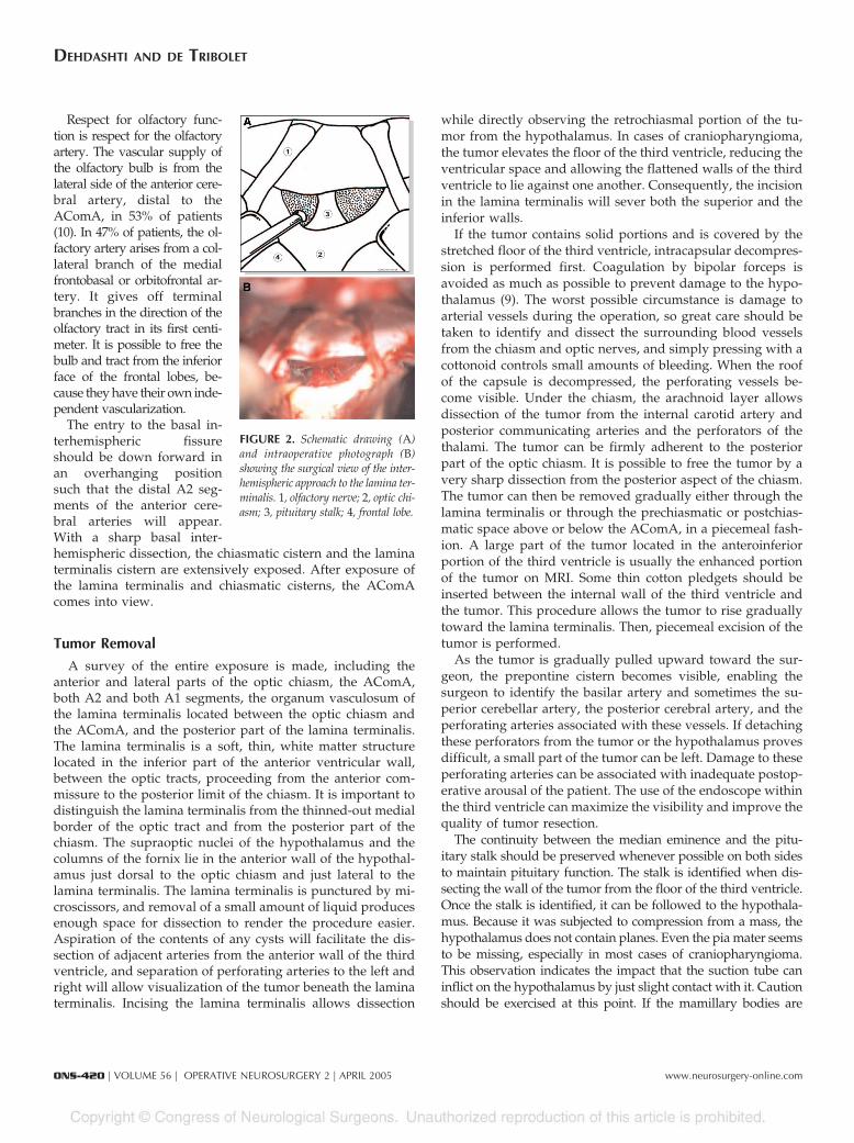
The constant existence of an arachnoidal cistern surround-ing all the olfactory structures on the inferior face of the frontallobes gives a microsurgical plane of cleavage. The arachnoid issharply divided, and dissection of the olfactory tracts, alter-nating between the left and the right, is performed. If a uni-lateral olfactory tract is completely dissected before the start ofthe dissection of the other side, there is the danger of avulsionof the contralateral olfactory tract. It is important to sharplydissect the olfactory tracts in parallel, proceeding by alternat-ing between the left and the right sides. In the dissection ofthese tracts, pressure should not be applied inferiorly butrather in a superior direction. Inadvertent traction during theoperation can lead to the complete avulsion of the olfactorytract or bulb and loss of olfaction. The olfactory bulb can bereinforced with fibrin glue, fixing it to the cribriform plate. Theolfactory tract should not be allowed to become dry during theoperation. The olfactory bulb, together with the olfactory tract,is separated from the orbital surface of the frontal lobe bilat-erally as far as the olfactory trigonal region. At the completionof dissection of the olfactory tracts, a small portion of bothoptic nerves should be visible beneath the arachnoid (Fig. 2).
FIGURE 1. Illustration showing thesite of the craniotomy. As noted in thetext, a craniotomy can also be per-formed in smaller format and even lim-ited to the extension of the frontalsinus.
Respect for olfactory func-tion is respect for the olfactoryartery. The vascular supply ofthe olfactory bulb is from thelateral side of the anterior cere-bral artery, distal to theAComA, in 53% of patients(10). In 47% of patients, the ol-factory artery arises from a col-lateral branch of the medialfrontobasal or orbitofrontal ar-tery. It gives off terminalbranches in the direction of theolfactory tract in its first centi-meter. It is possible to free thebulb and tract from the inferiorface of the frontal lobes, be-cause they have their own inde-pendent vascularization.
The entry to the basal in-terhemispheric fissureshould be down forward inan overhanging positionsuch that the distal A2 seg-ments of the anterior cere-bral arteries will appear.With a sharp basal inter-hemispheric dissection, the chiasmatic cistern and the laminaterminalis cistern are extensively exposed. After exposure ofthe lamina terminalis and chiasmatic cisterns, the AComAcomes into view.
Tumor Removal
A survey of the entire exposure is made, including theanterior and lateral parts of the optic chiasm, the AComA,both A2 and both A1 segments, the organum vasculosum ofthe lamina terminalis located between the optic chiasm andthe AComA, and the posterior part of the lamina terminalis.The lamina terminalis is a soft, thin, white matter structurelocated in the inferior part of the anterior ventricular wall,between the optic tracts, proceeding from the anterior com-missure to the posterior limit of the chiasm. It is important todistinguish the lamina terminalis from the thinned-out medialborder of the optic tract and from the posterior part of thechiasm. The supraoptic nuclei of the hypothalamus and thecolumns of the fornix lie in the anterior wall of the hypothal-amus just dorsal to the optic chiasm and just lateral to thelamina terminalis. The lamina terminalis is punctured by mi-croscissors, and removal of a small amount of liquid producesenough space for dissection to render the procedure easier.Aspiration of the contents of any cysts will facilitate the dis-section of adjacent arteries from the anterior wall of the thirdventricle, and separation of perforating arteries to the left andright will allow visualization of the tumor beneath the laminaterminalis. Incising the lamina terminalis allows dissection
while directly observing the retrochiasmal portion of the tu-mor from the hypothalamus. In cases of craniopharyngioma,the tumor elevates the floor of the third ventricle, reducing theventricular space and allowing the flattened walls of the thirdventricle to lie against one another. Consequently, the incisionin the lamina terminalis will sever both the superior and theinferior walls.
If the tumor contains solid portions and is covered by thestretched floor of the third ventricle, intracapsular decompres-sion is performed first. Coagulation by bipolar forceps isavoided as much as possible to prevent damage to the hypo-thalamus (9). The worst possible circumstance is damage toarterial vessels during the operation, so great care should betaken to identify and dissect the surrounding blood vesselsfrom the chiasm and optic nerves, and simply pressing with acottonoid controls small amounts of bleeding. When the roofof the capsule is decompressed, the perforating vessels be-come visible. Under the chiasm, the arachnoid layer allowsdissection of the tumor from the internal carotid artery andposterior communicating arteries and the perforators of thethalami. The tumor can be firmly adherent to the posteriorpart of the optic chiasm. It is possible to free the tumor by avery sharp dissection from the posterior aspect of the chiasm.The tumor can then be removed gradually either through thelamina terminalis or through the prechiasmatic or postchias-matic space above or below the AComA, in a piecemeal fash-ion. A large part of the tumor located in the anteroinferiorportion of the third ventricle is usually the enhanced portionof the tumor on MRI. Some thin cotton pledgets should beinserted between the internal wall of the third ventricle andthe tumor. This procedure allows the tumor to rise graduallytoward the lamina terminalis. Then, piecemeal excision of thetumor is performed.
As the tumor is gradually pulled upward toward the sur-geon, the prepontine cistern becomes visible, enabling thesurgeon to identify the basilar artery and sometimes the su-perior cerebellar artery, the posterior cerebral artery, and theperforating arteries associated with these vessels. If detachingthese perforators from the tumor or the hypothalamus provesdifficult, a small part of the tumor can be left. Damage to theseperforating arteries can be associated with inadequate postop-erative arousal of the patient. The use of the endoscope withinthe third ventricle can maximize the visibility and improve thequality of tumor resection.
The continuity between the median eminence and the pitu-itary stalk should be preserved whenever possible on both sidesto maintain pituitary function. The stalk is identified when dis-secting the wall of the tumor from the floor of the third ventricle.Once the stalk is identified, it can be followed to the hypothala-mus. Because it was subjected to compression from a mass, thehypothalamus does not contain planes. Even the pia mater seemsto be missing, especially in most cases of craniopharyngioma.This observation indicates the impact that the suction tube caninflict on the hypothalamus by just slight contact with it. Cautionshould be exercised at this point. If the mamillary bodies are
FIGURE 2. Schematic drawing (A)and intraoperative photograph (B)showing the surgical view of the inter-hemispheric approach to the lamina ter-minalis. 1, olfactory nerve; 2, optic chi-asm; 3, pituitary stalk; 4, frontal lobe.
injured, the patient can go intoprolonged coma and will ex-perience short-term memorydisturbances.
One should avoid a vigor-ous retraction of the tumorcapsule in cases with signifi-cant adherence to the floor ofthe third ventricle, becausethis maneuver could result ina bitemporal hemianopsia orhypothalamic dysfunction.The capsule can be removed ifit is adequately debulked,with gentle retraction. Onlyafter the tumor capsule hasbeen dissected and carefullydelimited and it has been de-termined that damage tobrain tissue will not be in-curred should excision of thetumor and the capsule itselfbe performed (Fig. 3). The tu-mor capsule is usually sepa-rated by a surrounding bar-rier of glial cells from thenormal brain, facilitating dis-section; however, in cases of craniopharyngioma, it is hard toperform complete excision without damaging neural tissue, be-cause fingers of tumor invade the surroundings (4, 12). Denselycalcified tumor may be adherent to the medial aspect of thecarotid artery. If difficulty is encountered to separate a calcifiedtumor from the wall of the carotid, a subtotal resection is pre-ferred, combined with adjunctive radiotherapy.
In achieving complete hemostasis, one should rinse theoperative field repeatedly with normal saline. The formationof hematoma around the vessels is likely to cause vasospasm,and therefore, the importance of complete hemostasis must bekept in mind (Fig. 4).
Closure
The dura is closed in a watertight manner. The frontonasalducts are covered by muscle, bone, fibrin glue, and a pedicledperiosteal layer. Fibrin glue is used to seal all of the gapsaround it. The bone flap is fixed with round maxillofacialmicroplates to cover the frontal burr holes, and the skin isclosed in two layers. A subgaleal drain is left for 24 hours.
POSTOPERATIVE COURSE
An incision in the flattened anterior wall of the third ventriclehas been made, and considerable trauma to the third ventriclehas been incurred. Therefore, corticosteroids should be contin-ued and tapered over several days, and water and electrolytebalance should be checked at hourly intervals during the first 72
hours. The body weightshould be checked twice dailyduring the hospital stay. Acontrol MRI scan is per-formed routinely within 48hours after surgery to confirmthe complete removal of thetumor or to show the residualpart. A postoperative endo-crine workup is essential toevaluate the need for hor-mone replacement.
We have performed this ap-proach in 14 operations dur-ing the past 5 years. Eight ofthese cases were for suprasel-lar craniopharyngiomas, threefor meningiomas, two for sub-callosal AVMs, and one for acavernoma. Postoperatively, atransient diabetes insipiduswas observed in three pa-tients, hormonal disturbanceswere noted in four (which re-mained permanent in two),and obesity was present intwo patients. There was noadditional visual deficit andno mortality at 6-monthfollow-up.
Illustrative Cases
The following are brief de-scriptions of two patients inwhom this approach was used.
Patient 1
The patient was a 44-year-old woman with a third ventricularcraniopharyngioma who presented with amenorrhea, polydipsia, andbitemporal superior hemianopsia. The endocrine workup showedpanhypopituitarism. A computed tomographic scan showed calcifica-tions in the floor of the third ventricle (Fig. 5A). MRI showed the solid,contrast-enhancing part of the tumor involving the floor of the thirdventricle and a cystic part occupying the anterior third ventricle (Fig.5, B and C). With the frontobasal trans-lamina terminalis approach,the tumor was totally resected and the pituitary stalk, which wasinvolved by the tumor, also had to be excised. Postoperative MRIconfirmed complete resection, and the patient showed no new neu-rological deficits but continued experiencing panhypopituitarism (Fig.5D). The visual field defect improved.
Patient 2
The patient was a 26-year-old man who bled from a subcallosalAVM extending along the anterior wall of the third ventricle andalong the medial wall of the frontal horns of the lateral ventricles (Fig.6, A and B). With the same approach, the feeding vessels from theAComA and both A2 segments could be controlled (Fig. 6C). The
FIGURE 3. Schematic drawing (A)and intraoperative photograph (B)showing tumor removal. 1, olfactorynerve; 2, optic chiasm; 3, tumor; 4,AComA; 5, left A2; 6, right A2; 7,precocious right callosomarginalartery; 8, third ventricle.
FIGURE 4. Schematic drawing (A)and intraoperative photograph (B)showing final inspection before closure.Note the three surgical windows to thethird ventricle established during sur-gery: the prechiasmatic window (8), thepostchiasmatic window under theAComA (9), and the postchiasmaticwindow above the AComA (10). 1, ol-factory nerve; 2, optic chiasm; 3, fron-tal lobe; 4, AComA; 5, left A2; 6, rightA2; 7, precocious right callosomarginalartery.
venous drainage was into both basilar veins of Rosenthal. Completeresection of the AVM was confirmed on a postoperative angiogram(Fig. 6D). The patient had recovered fully from the hemorrhagic insultand the surgery at 6-month follow-up.
ADVANTAGES
1. The operative field is wide, allowing visualization ofboth optic nerves as well as the chiasm and, behind orabove it, the AComA, the lamina terminalis, and bothA2 segments, and below the chiasm, both internal ca-rotid arteries, the posterior communicating arteries,their perforating branches, and the pituitary stalk.
2. No damage is done to the brain tissue except to thelamina terminalis itself, which, depending on the natureof the lesion, is often widened and thinned.
3. This approach is also suitable for lesions located on themidline of the anterior fossa.
DISADVANTAGES
1. The frontal sinus must be opened, so treatment of thefrontal sinus must be performed with extreme cautionto prevent infection.
2. Additional care is needed when the superior sagittalsinus is divided. The ligation of the severed end of thesinus may become loosened during operation or afterclosure of the dura, and hemorrhage from the venoussinus may occur.
COMPLICATIONS
1. Diabetes insipidus should be considered a virtual inev-itability. It is consequently essential to measure urineoutput at 1-hour intervals during and after operationand to check the water and electrolyte balance, serumand urine osmolality, and natrium.
2. Hormone disturbance may occur if the pituitary stalk issectioned.
3. Hypothalamic dysfunction may occur if the vascularsupply of the hypothalamus is damaged. Lesioning ofthe ventromedial part of the hypothalamus causes hy-perphagia, lesioning of the lateral part causes aphagiaand loss of weight, and lesioning of the anterior hypo-thalamus causes hyperthermia, obesity, somnolence, fitsof rage, and precocious puberty.
FIGURE 5. Patient 1. A, com-puted tomographic scan showing apartially calcified lesion in thefloor of the third ventricle. B and C, axial (B) and sagittal (C) MRI scansshowing a partially cystic suprasellar lesion; the enhancing part of thetumor is located in the anteroinferior part of the third ventricle. D, postop-erative MRI scan confirming complete resection of the tumor, diagnosed asa craniopharyngioma.
FIGURE 6. Patient 2. A and B, angio-grams before operation showing anAVM in the subcallosal area (A, lateralview; B, anteroposterior view). C, in-traoperative photograph of the fronto-basal interhemispheric trans-laminaterminalis approach to the AVM. D,postoperative angiogram showing dis-appearance of the AVM.
The frontobasal interhemispheric approach offers safe ac-cess to suprasellar tumors, including craniopharyngiomas.Anatomic preservation of the pituitary stalk, hypothalamicstructure, perforating vessels, anterior communicating com-plex, the visual pathway, and the olfactory nerves is oftenpossible. However, accurate neuroendocrine, electrolyte, andneuropsychological control is critical even years after surgery.
2. Fahlbusch R, Honegger J, Paulus W, Huk W, Buchfelder M: Surgical treat-ment of craniopharyngiomas: Experience with 168 patients. J Neurosurg90:237–250, 1999.
3. Fuzitsu K: Anterior interhemispheric approach to the disease involving thethird ventricle and/or the suprasellar region [in Japanese]. No ShinkeiGeka 26:667–677, 1998.
4. Ghatak MR, Hirano A, Zimmerman HM: Ultrastructure of craniopharyngi-oma. Cancer 27:1465–1475, 1971.
5. Hoffmann R: Osteoplastic operations on frontal sinuses for chronic suppu-ration. Ann Otol 13:598–608, 1904.
6. Maira G, Anile C, Colosimo C, Cabezas D: Craniopharyngiomas of the thirdventricle: Trans-lamina terminalis approach. Neurosurgery 47:857–865, 2000.
7. Ohata K, Hakuba A, Nagai K, Morino M, Iwa Y: A biorbitofrontobasalinterhemispheric approach for suprasellar lesions. Mt Sinai J Med 64:217–221, 1997.
8. Oi S, Samii A, Samii M: Operative techniques for tumors in the thirdventricle. Op Tech Neurosurg 6:205–214, 2003.
9. Page RB: Diencephalic structures at risk in third ventricular surgery, inApuzzo MLJ (ed): Surgery of the Third Ventricle. Baltimore, Williams &Wilkins, 1987, pp 553–556.
10. Passagia JG, Chirossel JP, Favre JJ, Gay E, Reyt E, Righini C, Chaffanjon P:Surgical approaches to the anterior fossa and preservation of olfaction. AdvTech Stand Neurosurg 25:195–241, 1999.
11. Shibuya M, Takayasu M, Suzuki Y, Saito K, Sugita K: Bifrontal basal inter-hemispheric approach to craniopharyngioma resection with or withoutdivision of the anterior communicating artery. J Neurosurg 84:951–956,1996.
12. Shilito J: Craniopharyngiomas: The subfrontal approach, or none at all. ClinNeurosurg 27:188–205, 1980.
13. Shirane R, Su CC, Kusakka Y, Jokura H, Yoshimoto T: Surgical outcomes in31 patients with craniopharyngiomas extending outside the suprasellarcistern: An evaluation of the frontobasal interhemispheric approach.J Neurosurg 96:704–712, 2002.
14. Suzuki J: Preservation of the olfactory tract in bifrontal craniotomy foranterior communicating aneurysms, and functional prognosis. J Neurosurg54:342–345, 1981.
15. Suzuki J, Katakura R, Mori T: Interhemispheric approach through the lam-ina terminalis to tumors of the anterior part of the third ventricle. SurgNeurol 22:157–163, 1984.
COMMENTS
At this time, when minimally invasive surgery has becometrendy, as if it were a status symbol for some modern
neurosurgeons, the proposal of a large approach introducedmore than two decades ago and not used very frequentlyseems to be out of order. Therefore, if only for this reason, weshould be grateful for this nice, elegant, and clear presentationof the frontobasal interhemispheric trans-lamina terminalis
approach. This very welcome contribution demonstrates howapproaches considered to be complex and risky are in realitysimple and safe even for the ordinary surgeon, as he or shegains experience. I truly hope that even the most reluctantsurgeons become convinced that this approach can offer theexposure they need to deal with midline large suprasellarlesions. Since 1992 (1), we systematically started to use thisapproach for larger craniopharyngiomas to obtain radical re-moval of the tumors associated, when possible, with the pres-ervation of the pituitary stalk. Since then, we have treated 162patients, with more than satisfactory results. In brief, weachieved total removal in 81% of our patients, with completepreservation of the pituitary stalk in 67%. Because craniophar-yngiomas are extra-axial midline lesions and they grow dis-placing structures from the midline in any direction, we be-lieve it is opportune to approach the tumor at the midline, andthe approach presented here allows preservation of the olfac-tory nerves and exposure and removal of the tumor betterthan any other approach, if I may say so. One should beextremely grateful to the authors for knowing how to clearlydemonstrate to the neurosurgical community the great poten-tiality of this approach, which I have worked with many timeswith great satisfaction.
Albino BricoloVerona, Italy
1. Bricolo A, Turazzi S, Talacchi A: Bilateral subfrontal (trans-lamina-terminalis)approach for radical resection of craniopharyngiomas with preservation ofpituitary stalk. Presented at the 6th Asian-Oceanic International Congress onSkull Base Surgery, Makuhari, Japan, November 12–15, 2001 (abstr).
Retrochiasmatic tumors and other lesions that insinuatethemselves into the floor of the third ventricle or present
in the anterior third ventricle itself are probably among themost technically challenging intracranial lesions. The authorsdescribe an interhemispheric trans-lamina terminalis ap-proach for removal of these lesions. The description of theirtechnique is clear, concise, and easy to follow, especially bythose who have at least some experience with anterior thirdventricle surgery. They describe the advantages and disadvan-tages of their approach. Among the main advantages is alesser need for frontal lobe retraction compared with otherapproaches, a greater ability to preserve olfaction, and per-haps a better visualization of the lamina terminalis and thesurrounding neurovascular structures. There is a point ofcaution, however, that is worth remembering. Just as watch-ing Ernie Elis swing a golf club may give the impression thata golf swing is essentially easy and simple to execute, which itprobably is for professionals but not for amateurs, so does thedescription of the authors’ technique appear simple andstraightforward and, in their hands, also safe. However, weshould not forget that these authors are true masters of thirdventricle surgery, which should not be construed as a simple,straightforward, and safe operation when performed by thoseless experienced with or even uninitiated in this procedure.
The relative simplicity and straightforwardness of exposingthe lamina terminalis and the surrounding neurovascularstructures by use of the authors’ approach may sway those ofus who are at present still prone to expose the retrochiasmaticlesions using the pterional orbital-clinoidal cranial base dis-section technique toward using the outer hemispheric ap-proach. Clearly, neurosurgeons endeavoring to remove retro-chiasmatic lesions would be well served to master the authors’technique. However, the real danger of the procedure, whichrequires considerable experience, starts with the exposure andremoval of the lesion situated behind the lamina terminalis.The immediate proximity of the hypothalamus and the poten-tial adherence of the tumor to the lateral hypothalamic wallscan prove treacherous to the outcome regardless of how thelamina terminalis is approached.
In short, I applaud the authors for offering us their contri-bution in exposing the lamina terminalis. However, neurosur-geons should be aware of the danger zone behind the laminaterminalis and of the potentially serious complications, suchas a hypothalamic injury, when deciding to proceed with thisoperation. There are not enough of these tumors facing neu-rosurgeons (fortunately or unfortunately) to make the averageneurosurgeon an expert with this operation. Consequently,patients harboring such tumors are probably safer in thehands of a surgeon experienced with third ventricle tumorsand in an institution in which such operations are performedon a programmatic basis.
Ivan S. CiricEvanston, Illinois
In this well-written report, Dehdashti and de Tribolet presentthe technical aspects of the frontobasal interhemispheric
trans-lamina terminalis approach for suprasellar lesions.We believe that there are a number of microsurgical tech-
nical limitations to the described approach. By definition,the trajectory entails bilateral mesial frontal lobe retraction.The working corridor is narrow and deep. Tumors thatextend laterally into the carotid cistern would not be easilyaccessible. Using the frontal sinus as the main port of entrydoes theoretically raise the risk of the introduction ofinfection.
On the other hand, the pterional transsylvian approach forlesions involving the suprasellar cistern provides a number ofadvantages. Early release of cerebrospinal fluid from the basalcisterns allows for brain relaxation and minimizes brain re-traction; early identification of the internal carotid artery andits branches protects these vessels; early exposure of the opticnerves and chiasm provides protection of the visual system;and the ability to dissect the anterior cerebral artery and itsbranches from a lateral approach allows maximal protection ofthese crucial vessels (1). For craniopharyngiomas that alsoextend superiorly into the foramen of Monro, the senior com-mentator (MGY) has advocated the addition, in the samesetting using a separate bone flap, of the interhemispherictranscallosal trajectory to the pterional transsylvian approachto achieve maximal resection (2).
M. Gazi YasargilLittle Rock, ArkansasSaleem I. AbdulraufSt. Louis, Missouri
2. Yasargil MG, Curcic M, Kis M, Siegenthaler G, Teddy PJ, Roth P: Totalremoval of craniopharyngiomas: Approaches and long-term results in 144patients. J Neurosurg 73:3–11, 1990.