1 Frontoparietal connectivity correlates with working memory performance in multiple sclerosis Authors: Alejandra Figueroa-Vargas *1 , Claudia Cárcamo 2 , Rodrigo Henríquez-Ch 3 , Francisco Zamorano 1 , Ethel Ciampi 2,4 , Reinaldo Uribe 2,4 , Macarena Vásquez 2 , Francisco Aboitiz 3 , Pablo Billeke* 1 1 Laboratorio de Neurociencia Social y Neuromodulación, Centro de Investigación en Complejidad Social (neuroCICS), Facultad de Gobierno, Universidad del Desarrollo, Santiago de Chile. 2 Departamento de Neurología, Hospital Clínico de la Pontificia Universidad Católica de Chile. 3 Departamento de Psiquiatría, Escuela de Medicina, and Centro Interdisciplinario de Neurociencias, Pontificia Universidad Católica de Chile. 4 Servicio de Neurología, Hospital Dr. Sótero del Río, Santiago de Chile. * Corresponding authors: Alejandra Figueroa-Vargas, [email protected], and Pablo Billeke, [email protected], Avenida Las Condes #12461, Torre 3, oficina #307, Las Condes, Santiago de Chile, CP: 7550000 . CC-BY-NC-ND 4.0 International license not certified by peer review) is the author/funder. It is made available under a The copyright holder for this preprint (which was this version posted May 19, 2020. . https://doi.org/10.1101/639930 doi: bioRxiv preprint
Transcript
1
Frontoparietal connectivity correlates with working memory performance in multiple sclerosis
Authors:
Alejandra Figueroa-Vargas*1, Claudia Cárcamo2, Rodrigo Henríquez-Ch3 , Francisco
Zamorano1, Ethel Ciampi2,4, Reinaldo Uribe2,4, Macarena Vásquez 2, Francisco Aboitiz3, Pablo
Billeke*1
1 Laboratorio de Neurociencia Social y Neuromodulación, Centro de Investigación en
Complejidad Social (neuroCICS), Facultad de Gobierno, Universidad del Desarrollo, Santiago
de Chile.
2 Departamento de Neurología, Hospital Clínico de la Pontificia Universidad Católica de Chile.
3 Departamento de Psiquiatría, Escuela de Medicina, and Centro Interdisciplinario de
Neurociencias, Pontificia Universidad Católica de Chile.
4 Servicio de Neurología, Hospital Dr. Sótero del Río, Santiago de Chile.
* Corresponding authors: Alejandra Figueroa-Vargas, [email protected], and Pablo
Billeke, [email protected], Avenida Las Condes #12461, Torre 3, oficina #307, Las Condes,
Santiago de Chile, CP: 7550000
.CC-BY-NC-ND 4.0 International licensenot certified by peer review) is the author/funder. It is made available under aThe copyright holder for this preprint (which wasthis version posted May 19, 2020. . https://doi.org/10.1101/639930doi: bioRxiv preprint
.CC-BY-NC-ND 4.0 International licensenot certified by peer review) is the author/funder. It is made available under aThe copyright holder for this preprint (which wasthis version posted May 19, 2020. . https://doi.org/10.1101/639930doi: bioRxiv preprint
Introduction Multiple Sclerosis is a chronic demyelinating disease of the Central Nervous System1. The
physiopathology is based on deregulation of the immune system generating motor, cognitive,
and neuropsychiatric symptoms2,3. Cognitive impairments are present in 40 to 70% of patients,
affecting their professional development, personal relationships, mood and quality of life4,5.
These symptoms can be detected from early phases of the disease and may include alterations
in information processing speed, attention, executive functions, and working memory (WM)6,7.
Since human cognition critically depends on WM, an ability that enables us to adaptively
maintain and manipulate information according to the demands of the environment, alterations
in this process seem to be a key step in cognitive alterations in patients with multiple sclerosis.
Researchers confirmed WM alterations are a habitual impairment, affecting patients early in
the course of the disease8,9.
WM is a hierarchical process that links sensory representations to specific responses, through
intermediate representations relevant to the task and action plans10. A distributed network of
brain areas participates in this process, exhibiting sustained activity during the period of WM
maintenance in the absence of sensory stimuli11. This sustained activity is generated by
reverberant discharges in an interconnected network that involves the prefrontal cortex (PFC),
the posterior parietal and temporal lobes11. Electrophysiological studies have revealed the
crucial participation of oscillatory activity in theta (4-8 Hz), alpha (8-12 Hz), and gamma (30-
100 Hz) bands in WM12,13. During the maintenance stage, memory load increases theta activity
and theta-gamma coupling in frontal and temporo-parietal regions14–16. EEG and MEG studies
have shown a greater synchrony between the frontal and parietal regions associated with the
amount of information successfully maintained in WM15,16.
The neurobiological mechanism underlying WM alterations in multiple sclerosis are not well
known. Successful WM needs the coordination of distributed brain networks that are
especially sensitive to the diffuse damage of white and grey matter found in multiple
sclerosis17,18. Several reports indicate very early alterations in cortico-cortical connectivity in
multiple sclerosis19,20. Both functional and structural brain imaging techniques have
revealed alterations in brain network connectivity patterns in patients with minimal or low
cognitive disabilities19. An EEG study found a decrease in alpha and theta band coherence
between the anterior and posterior electrodes, as well as between inter-hemispheric regions
.CC-BY-NC-ND 4.0 International licensenot certified by peer review) is the author/funder. It is made available under aThe copyright holder for this preprint (which wasthis version posted May 19, 2020. . https://doi.org/10.1101/639930doi: bioRxiv preprint
during rest. These alterations seem to be related to both cognitive deficits and subcortical lesion
burden21. Thus, a functional marker of the cognitive alterations for early stages of the disease
with minimal clinical manifestations would be relevant to address early cognitive
rehabilitation. Additionally, the identification of the precise functional alterations in the
oscillatory patterns opens the opportunity to plan specific interventions using, for example,
non-invasive brain stimulation. Therefore, the aim of our study was to assess the
neurophysiological underpinnings of alterations in the cortical circuits that support WM in
multiple sclerosis. We hypothesized that WM impairments in patients with multiple sclerosis
are due to an impairment in the maintenance of activity in the fronto-parietal network that is
reflected in a reorganization of cortical oscillatory dynamics. Specifically, we predicted that
i) multiple sclerosis alters the progressive increases of power of both theta and alpha oscillatory
activity in relation to the increases of memory load; and that ii) fronto-parietal theta
connectivity underlying successful memory information maintenance is impaired in patients
with multiple sclerosis.
To address this issue, we recorded EEG activity of forty individuals. Twenty patients had
relapsing-remitting multiple sclerosis with minimal clinical cognitive alterations and twenty
healthy control subjects (see Table 1 for demographic data) solved working memory tasks (see
Figure 1). Subjects had to memorize two, four or six consonants, generating three levels of
working memory load (see more detail in methods section). Importantly, the identification of
the precise neurophysiological dynamics underlying WM alteration in patients with multiple
sclerosis could contribute to both early detection and development of specific cognitive
rehabilitation interventions 11,22 .
Results
Behavioral
Both groups had over chance performance in all memory load conditions (Wilcoxon test,
ps<0.001, Bonferroni corrected), without differences between groups (p>0.2,
uncorrected). Patients with multiple sclerosis presented a tendency to have less decrease in
their performance in relation to the load increases (difference between load 2 and 6, control
mean 0.14, patient mean 0.09, p=0.06, Figure 2). In the following analysis, we focused on the
high load memory condition (six items) because there were more errors and there were no
differences between groups. We studied the reaction time (RT) as an index of cognitive effort,
.CC-BY-NC-ND 4.0 International licensenot certified by peer review) is the author/funder. It is made available under aThe copyright holder for this preprint (which wasthis version posted May 19, 2020. . https://doi.org/10.1101/639930doi: bioRxiv preprint
especially when an error occurred. Both groups presented longer RT for incorrect responses
than for correct responses (Wilcoxon test, ps<0.003, Bonferroni corrected) without difference
between groups. Additionally, we found that patients presented longer RT for incorrect
responses when the probe was part of the memory set (incorrect match responses, EM, Figure
2), which led to significant differences between groups (Wilcoxon tests, p=0.047). Finally, to
rule out difference of RT due to motor impairment, we calculated the difference in RT for load
memory 2, and the difference in RT using different hands to answer also during load 2. These
two measurements were no significantly different between groups (Wilcoxon test, p=0.5, p=0.9
respectively)
Time-Frequency EEG Analysis
We studied the effect of both memory load and successful memory performance on the power
of brain oscillatory activity. Regarding the effect of memory load, in the control group we
found two effects as expected, a positive modulation in theta activity and a negative
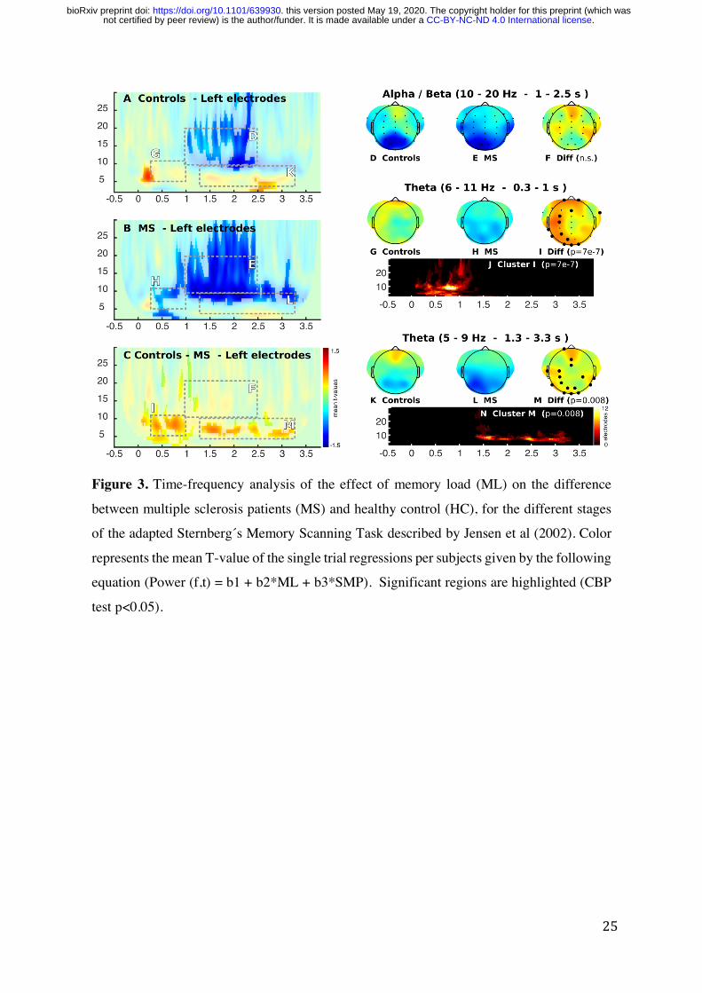
modulation in alpha/beta activity 13,23. In contrast, we did not find modulations of theta band (6-
11 Hz) in patients with multiple sclerosis, leading to a significant difference (p=7 e-7; Cluster
Based Permutation (CBP) test) between groups in the initial stage of encoding in this frequency
band (between 0.3 and 1 s, Figure 3 D-F). This difference in theta had a topographic
distribution located in electrodes of the left hemisphere (Figure 3 I). When analyzing group
differences during the maintenance stage, we observed differences in theta activity (5-9 Hz) in
the period of 1.3 to 3.3 seconds (p=0.008), which corresponded to the final part of the encoding
stage and the entire maintenance phase. The topographic distribution of this modulation was
placed over frontoparietal regions with left predominance (Figure 3 M).
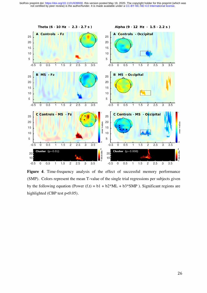
Next, we analyzed the oscillatory activity related to successful memory performance, that is,
the specific activity in the trials in which subjects correctly respond to the target stimuli in
relation to those trials where subjects make mistakes (Figure 4). We found that during the
maintenance period (2.3 to 2.7 s), patients with multiple sclerosis showed a negative
modulation in theta activity (5-11 Hz), while healthy subjects presented a positive modulation
(Figure 4A). This led to a significant difference between groups (p=0.01) showing a medial
frontal topographic distribution (Fig. 4C). Regarding the analysis of alpha activity (10 -15 Hz),
.CC-BY-NC-ND 4.0 International licensenot certified by peer review) is the author/funder. It is made available under aThe copyright holder for this preprint (which wasthis version posted May 19, 2020. . https://doi.org/10.1101/639930doi: bioRxiv preprint
Considering the results of the oscillatory activity, we carried out a connectivity analysis. We
selected a frontal electrode (Fz) and a left parietal electrode (Cp3), since modulation in theta
for both memory load and successful memory maintenance were found in these electrodes. The
following source reconstruction enabled us to infer that they represent frontal and temporo-
parietal activity respectively. We first used GC that measures statistic dependency between
signals (e.i., if one signal is useful in forescasting another signal). During the maintenance
stage, we found that healthy subjects presented an increase in the parietal-to-frontal
connectivity in the time domain, as an indicator of successful memory performance (-0.72,
p=0.01). This led to a significant difference in patients with multiple sclerosis, who did not
present this modulation (diff, p=0.009, Figure 6 and Table 2). Additionally, this
.CC-BY-NC-ND 4.0 International licensenot certified by peer review) is the author/funder. It is made available under aThe copyright holder for this preprint (which wasthis version posted May 19, 2020. . https://doi.org/10.1101/639930doi: bioRxiv preprint
.CC-BY-NC-ND 4.0 International licensenot certified by peer review) is the author/funder. It is made available under aThe copyright holder for this preprint (which wasthis version posted May 19, 2020. . https://doi.org/10.1101/639930doi: bioRxiv preprint
In this study we assessed the cortical circuits that support verbal working memory in patients
with relapsing-remitting multiple sclerosis with minimal or no burden of neurological disability
(EDSS <3 and PASAT >-1.5 SD). In the early stage of the disease, patients commonly do not
present objective cognitive alterations in the neuropsychological evaluations, however, they
manifest a subjective sensation of difficulties in their cognitive performance in daily activities
(e.g., occupational or academic tasks) 30. These patients report a common clinical pattern of
difficulties in the performance of daily cognitive tasks involving working memory, but without
clear evidence in the clinical tests applied in their evaluation routines 30–32. Accordingly, we
observed no significant differences in WM performance, but clear neurophysiological
differences between patient and control groups. In the early stage of the disease, the clinical
evaluations applied to multiple sclerosis patients without impairment often do not detect a
deterioration of this cognitive function 33–35 . The RT difference that we found could reflect an
increase in the cognitive effort necessary to correctly solve the task and could be an early
behavioral marker of WM impairment. In fact, reaction time analyses are a good marker of
attention and cognitive control dynamics 36–40 .
In spite of no clear evidence for behavioral impairments in the accuracy in the WM task,
patients with multiple sclerosis demonstrated a distributed oscillatory activity reorganization.
The ability to maintain a sustained activity in the fronto-parietal network in the absence of
sensory stimuli depends on the synchronous structured activity of different frequency ranges 41. Our working memory task elicited a recognized oscillatory activity in a frontal and parietal
network 11,42–44 . The left lateralization of the power of the theta oscillatory activity could be related
to the areas involved in language, i.e., the phonological loop, required for the specific
information of our stimuli (consonants) 13,45 . Low frequency synchronization between the
medial frontal region and temporal cortex has been demonstrated in non-verbal WM tasks,
probably reflecting cognitive control 15,23 . Indeed, our connectivity analyses showed specific
dynamics, in which frontal to parietal/temporal influences increased in function of memory
load when a subsequent successful memory performance occurs. These findings are in line
with several recent reports indicating that frontal interactions with other cortical areas are key
aspects of successful memory performance 14,46,47.
.CC-BY-NC-ND 4.0 International licensenot certified by peer review) is the author/funder. It is made available under aThe copyright holder for this preprint (which wasthis version posted May 19, 2020. . https://doi.org/10.1101/639930doi: bioRxiv preprint
In contrast, patients with multiple sclerosis demonstrated a loss of the WM oscillatory
dynamics . These patterns could represent an inefficient cognitive effort to keep the stimuli
information active in WM, which would be in accordance with the longer reaction times for
erroneous responses in high memory load. In addition to this, the patient group did not present
an increase in frontal-to-parietal connectivity 48,49. WM requires the synchronization of neural
network connections distributed in the prefrontal and parietal regions in order to integrate
complex information for generating appropriate responses. These distributed brain networks
are especially sensitive to the diffuse damage of white and gray substance found in multiple
sclerosis 17,18,50.
Thus, our results are in accordance with recent findings in patients with mild or minimal
cognitive deficit that show reorganization in electrophysiological activity 49,51 . In spite of the
fact that patients could have fewer resources to maintain the stimuli in WM they can achieve a
similar performance to healthy subjects, as long as the tasks are not too, by means of
redistribution of the remaining and available resources. It has been proposed that the central
executive may be the main component of WM that is disrupted in cognitively impaired multiple
sclerosis patients52. This proposition is supported indirectly by evidence that suggests that the
dorsolateral prefrontal cortex, which is thought to underlie executive control, is commonly
recruited when there is heavy demand placed on WM in individuals with brain injury 53. Our
findings give further evidence to support this interpretation. Indeed, the frontal low frequency
influences the gamma power in parietal and temporal areas, and this influence increases in
relation to memory load increase. Patients fail to display this influence, and this failure could
reflect the loss of the mechanisms by which the control process carried out by frontal
areas produces a successful memory performance.
Studies using fMRI have found several changes in brain activity during WM tasks including
decrease and increase in both connectivity and activity 54,55. Indeed, follow-up studies show an
inverted-U form in the evolution of the disease in resting-state functional connectivity56. Early
changes in the connectivity/functional patterns have been interpreted as compensatory changes
in prefrontal cortical regions underlying modulations of executive aspects of WM 57–59. For
instance, studies in very early states of multiple sclerosis have found medial PFC activity
increases, although later meta-analyses studies have revealed a decrease in medial prefrontal
activity. In this context, our study gives several insights into the participation of medial
prefrontal activity in working memory and its relationship with the cognitive dysfunction in
.CC-BY-NC-ND 4.0 International licensenot certified by peer review) is the author/funder. It is made available under aThe copyright holder for this preprint (which wasthis version posted May 19, 2020. . https://doi.org/10.1101/639930doi: bioRxiv preprint
demonstrate a specific electrophysiological mechanism underlying the WM deficit in Patients
with Multiple Sclerosis. Several investigations have revealed that cerebral oscillatory activity
supports different cognitive processes and have indexed their alteration in clinical populations 68–70. Indeed, recent evidence indicates that increasing theta activity by means of transcranial
Alternating Current Stimulation can improve WM performance in healthy and aging subjects 14,47. It has been seen that cognitive training can increase memory capacity and that this increase
.CC-BY-NC-ND 4.0 International licensenot certified by peer review) is the author/funder. It is made available under aThe copyright holder for this preprint (which wasthis version posted May 19, 2020. . https://doi.org/10.1101/639930doi: bioRxiv preprint
correlates with changes in connectivity of distant brain areas, specifically between frontal and
parietal regions. Therefore, the specific oscillatory features related to WM deficits identified
in our study could serve to implement non-pharmacological treatments using non-invasive
brain stimulation and cognitive training, in order to contribute to the improvement of the
quality of life of these patients.
Methods
Design and Participants
Our study is a case-control design that include 40 participants. A sample consisted of 20
patients with relapsing-remitting multiple sclerosis in an early stage, with minimal to no
clinical evidence of cognitive alterations (Table 1). According to the 2010 McDonald
Criteria, the medical diagnosis was made by a Neurologist 71 . Stable patients without episodes
of relapses in the last month, with scores of three or more on the Expanded Disability Status
Scale (EDSS), with less than -1.5 z-score of Paced Auditory Serial Addition Test (PASAT),
with non-correctable visual alterations, with a history of traumatic brain injury, neurological
and/or psychiatric pathologies, and abuse or regular consumption of drugs or alcohol were
excluded. The patients participated in the study while on their usual disease modifying
therapies (i.e., immunomodulation therapy only, without other treatment, such as
antidepressants or Benzodiazepines). The control group was composed of 20 healthy
volunteers, comparable in age, sex, manual preference, and educational level (Table 1). As well
as in the patient group, healthy subjects with non-correctable visual alterations, with a history
of neurological and/or psychiatric pathologies, traumatic brain injury, and abuse or regular
consumption of drugs or alcohol were excluded. All participants were Spanish native speakers
and provided signed informed consent prior to participation in the study. Patients underwent
neuropsychological assessment during the month previous to the EEG session. This was their
first neuropsychological evaluation. These assessments included PASAT that measures
cognitive processing speed and working memory, Symbol Digit Modalities Test (SDMT) that
measures cognitive processing speed, the Brief Visuospatial Memory Test-Revised (BVMT-
R) that measures visuospatial memory, and the World Health Organization-University of
California-Los Angeles Auditory Verbal Learning Test (WHO UCLA AVLT) that measures
verbal memory. Details are summarized in Table 1.
.CC-BY-NC-ND 4.0 International licensenot certified by peer review) is the author/funder. It is made available under aThe copyright holder for this preprint (which wasthis version posted May 19, 2020. . https://doi.org/10.1101/639930doi: bioRxiv preprint
The experimental protocol and all methods were performance in accordance to institutional
guidelines and were approved by the Ethical Committee of the Pontificia Universidad Católica
de Chile.
Sample size
For the estimation of the minimum required sample size the following parameters were
considered: a) Effect size for the mixed ANOVA statistical test (2 x 2, with interaction effects),
b) Statistical power (1- β)=.95 and c) Significance level 𝜶=.05. Considering an effect size 𝜂2
=0.09 (effect size F = 0.3 13,72), the sample size amounts to a total of 40 participants (n1=20;
n2=20).
Experimental Task
In this study we implement a modified version of Sternberg´s Memory Scanning task [Jensen,
2002]. This task consisted of a list of consonants simultaneously presented and displayed in a
circular arrangement with a fixation cross in the center of a computer monitor located 57 cm
from the subject. The letter “Y” was not included in the memory set to avoid the generation of
words that could be used as clues by the subjects. Each memory set arrangement consisted of
groups of two, four, or six consonants generating three levels of WM load. The latter refers to
the progressive number of stimuli to be stored and manipulated in WM. All the stimuli were
placed foveally, minimizing the effect generated by saccadic movements (see the experimental
task outlined in Figure 1). Each memory set was presented for 1800 ms (encoding period),
followed by a black screen with the fixation cross (maintenance period) of 2000 ms and then a
recovery period in which the fixation point was replaced by a target stimulus for 1000 ms. The
subjects were instructed to memorize the memory set and then report whether the target
stimulus was present or absent in the memory set, using the right or left hand alternately.
Subjects had 2200 ms to answer. Each subject had to respond 270 trials (90 trials for each
memory load set). The trials were presented in two main blocks divided by a pause regulated
by the subject. In addition, each main block was constituted by 15 sub-blocks, formed by 9
trials of the same memory load each. The order of the memory load was randomized. Both the
presentation of the stimuli and the recording of the test responses were done with the Software
Presentation® (Version 13.0, www.neurobs.com).
Electrophysiological Recordings
.CC-BY-NC-ND 4.0 International licensenot certified by peer review) is the author/funder. It is made available under aThe copyright holder for this preprint (which wasthis version posted May 19, 2020. . https://doi.org/10.1101/639930doi: bioRxiv preprint
Continuous EEG recordings were obtained with a 40-electrode EEG System (NuAmps ,
Neuroscan). All impedances were kept under 5kΩ. Electrode impedance was retested during
pauses to ensure stable values throughout the experiment. All electrodes were referenced to
averaged mastoids during acquisition and the signal was digitized at 1 kHz. Electro-oculogram
was obtained with four electrodes. All recordings were acquired using Scan 4.3 and stored for
off-line analysis. At the end of each session, electrode position and head points were digitalized
using a 3D tracking system (Polhemus Isotrak).
Electrophysiological data analysis
EEG signals were preprocessed using a 0.1–100 Hz band-pass filter. Eye blinks were identified
by a threshold criterion of ±100 μV, and their contribution was removed from each dataset
using Independent Component Analysis (ICA). Other remaining artifacts (e.g., muscular
artifacts) were detected by visual inspection of both the raw signal and the spectrogram. We
thus obtained 243 ± 28 artifact-free trials per subject. All artifact-free trials were transformed
into current source density (CSD) that was estimated using the spherical spline surface
Laplacian algorithm suggested by Perrin et al.,73 and implemented by Kayser and Tenke 74,75.
Induced power distribution was computed using Wavelet transform, with a 5-cycle Morlet
wavelet, in a −0.5 to 3.8 s window around the onset of the memory set stimuli. This time-
window includes 0.5 seconds of inter stimulus interval, 1.8 seconds of the stimulus of the
memory set and 2 seconds of maintenance period. For all analyses, we used the dB of power
related to the baseline (15 seconds acquired in the beginning of each block).
Source Reconstruction
The neural current density time series at source levels were calculated by applying a weighted
minimum norm to estimate inverse solution 76 with unconstrained dipole orientations in single
trials as in prior work68,77,78. We used a default anatomy of the Montreal Neurological Institute
(MNI/Colin27) wrapped to the individual head shape (using ~300 head points per subject). We
defined 3 x 4000 sources constrained to the segmented gray cortical volume (3 orthogonal
sources at each spatial location) in order to compute a three-layer (scalp, inner skull, outer
skull) boundary element conductivity model and the physical forward model 79. Since the
inverse solution is a linear transformation, it does not modify the spectral content of the
underlying sources. Therefore, it is possible to undertake time–frequency analyses directly in
the source space. Finally, we reduced the number of sources by keeping a single source at each
spatial location that pointed into the direction of maximal variance. For this, we applied a
.CC-BY-NC-ND 4.0 International licensenot certified by peer review) is the author/funder. It is made available under aThe copyright holder for this preprint (which wasthis version posted May 19, 2020. . https://doi.org/10.1101/639930doi: bioRxiv preprint
principal component analysis to the covariance matrix obtained from the 3 orthogonal time
series estimated at each source location. Since we used a small number of electrodes (40) and
no individual anatomy for head model calculation, the spatial precision of the source
estimations is limited. In order to minimize the possibility of erroneous results, we only present
source estimations if there are both statistically significant differences at the electrode level
and the differences at the source levels survive a multiple comparison correction (cluster-based
permutation test and vertex correction using false discovery rate, q=0.05).
Statistical analysis
We used the Kolmogorov-Smirnoff to test for normality. When the data did not meet the
normal assumption, we used non-parametric tests. We evaluated pair comparisons using
Wilcoxon test and Bonferroni correction. For the EEG statistical analysis, we first fitted a
General Linear Model (GLM) of the power of the oscillatory activity per trial in each subject
(first level analysis, see 13,70,80 ),
𝑃𝑜𝑤𝑒𝑟(𝑓, 𝑡) = 𝑏/ + 𝑏1 ∗ 𝑀𝐿 + 𝑏5 ∗ 𝑆𝑀𝑃
where 𝑏/ is the intercept, and 𝑏1 is the slope or coefficient for the variable Memory Load (ML,
ordinal variable that takes the value 2, 4, or 6 depending on the memory load condition) and
𝑏5 is the slope for the variable Successful Memory Performance (SMP, dummy variable that
takes the value 0 if the subject makes a mistake in this trail or 1 if the target stimulus is correctly
identified). We thus obtained a 3D matrix of t-values (sensor, time, frequency) for each
regressor and subject. We then explored for differences between groups and conditions using
the Wilcoxon test (second level analysis). To correct for multiple comparisons in time-
frequency charts, we used the Cluster-based Permutation (CBP) test 81 using 1000 permutations.
For more detail see prior work 82,83.
Causal interactions
To evaluate the influence of frontal regions over temporo-parietal regions, we estimated
Granger Causality (GC) 84 between selected electrodes (Fz and CP3). The causality was
calculated over time series per trials. See detail in83,84 . We obtained a GC term per trial that was
then used in the modeling analyses (see below).
.CC-BY-NC-ND 4.0 International licensenot certified by peer review) is the author/funder. It is made available under aThe copyright holder for this preprint (which wasthis version posted May 19, 2020. . https://doi.org/10.1101/639930doi: bioRxiv preprint
At group level analysis per frequency pair, we compared whether t values of each regressor
were statistically different from zero using the Wilcoxon signed-rank test. For PAC analyses,
we corrected for multiple comparisons using a CBP test. The initial threshold for cluster
detection was p<0.05, and the final threshold for significant cluster was p<0.01. For between-
group comparison we used Wilcoxon rank-sum test and Bonferroni correction.
Software
All behavioral statistical analyses were performed in R. The EEG signal processing was
implemented in MATLAB using CSD toolbox, in-house scripts (available online as
.CC-BY-NC-ND 4.0 International licensenot certified by peer review) is the author/funder. It is made available under aThe copyright holder for this preprint (which wasthis version posted May 19, 2020. . https://doi.org/10.1101/639930doi: bioRxiv preprint
.CC-BY-NC-ND 4.0 International licensenot certified by peer review) is the author/funder. It is made available under aThe copyright holder for this preprint (which wasthis version posted May 19, 2020. . https://doi.org/10.1101/639930doi: bioRxiv preprint
.CC-BY-NC-ND 4.0 International licensenot certified by peer review) is the author/funder. It is made available under aThe copyright holder for this preprint (which wasthis version posted May 19, 2020. . https://doi.org/10.1101/639930doi: bioRxiv preprint
.CC-BY-NC-ND 4.0 International licensenot certified by peer review) is the author/funder. It is made available under aThe copyright holder for this preprint (which wasthis version posted May 19, 2020. . https://doi.org/10.1101/639930doi: bioRxiv preprint
.CC-BY-NC-ND 4.0 International licensenot certified by peer review) is the author/funder. It is made available under aThe copyright holder for this preprint (which wasthis version posted May 19, 2020. . https://doi.org/10.1101/639930doi: bioRxiv preprint
.CC-BY-NC-ND 4.0 International licensenot certified by peer review) is the author/funder. It is made available under aThe copyright holder for this preprint (which wasthis version posted May 19, 2020. . https://doi.org/10.1101/639930doi: bioRxiv preprint
.CC-BY-NC-ND 4.0 International licensenot certified by peer review) is the author/funder. It is made available under aThe copyright holder for this preprint (which wasthis version posted May 19, 2020. . https://doi.org/10.1101/639930doi: bioRxiv preprint
.CC-BY-NC-ND 4.0 International licensenot certified by peer review) is the author/funder. It is made available under aThe copyright holder for this preprint (which wasthis version posted May 19, 2020. . https://doi.org/10.1101/639930doi: bioRxiv preprint
Figure 1. Scheme of the experimental task, adapted from the Sternberg´s Memory Scanning
Task described by Jensen et al (2002).
.CC-BY-NC-ND 4.0 International licensenot certified by peer review) is the author/funder. It is made available under aThe copyright holder for this preprint (which wasthis version posted May 19, 2020. . https://doi.org/10.1101/639930doi: bioRxiv preprint
for no-matched targets (EnM), incorrect responses for matched targets (EM). Red represents
patients with multiple sclerosis (MS) and blue healthy control. Colored areas represent
standard error of mean.
.CC-BY-NC-ND 4.0 International licensenot certified by peer review) is the author/funder. It is made available under aThe copyright holder for this preprint (which wasthis version posted May 19, 2020. . https://doi.org/10.1101/639930doi: bioRxiv preprint
Figure 3. Time-frequency analysis of the effect of memory load (ML) on the difference
between multiple sclerosis patients (MS) and healthy control (HC), for the different stages
of the adapted Sternberg´s Memory Scanning Task described by Jensen et al (2002). Color
represents the mean T-value of the single trial regressions per subjects given by the following
equation (Power (f,t) = b1 + b2*ML + b3*SMP). Significant regions are highlighted (CBP
test p<0.05).
.CC-BY-NC-ND 4.0 International licensenot certified by peer review) is the author/funder. It is made available under aThe copyright holder for this preprint (which wasthis version posted May 19, 2020. . https://doi.org/10.1101/639930doi: bioRxiv preprint
Figure 4. Time-frequency analysis of the effect of successful memory performance
(SMP). Colors represent the mean T-value of the single trial regressions per subjects given
by the following equation (Power (f,t) = b1 + b2*ML + b3*SMP ). Significant regions are
highlighted (CBP test p<0.05).
.CC-BY-NC-ND 4.0 International licensenot certified by peer review) is the author/funder. It is made available under aThe copyright holder for this preprint (which wasthis version posted May 19, 2020. . https://doi.org/10.1101/639930doi: bioRxiv preprint
Figure 5. Source reconstruction of the differences between groups. The upper panel
shows the differences in the memory load modulation in theta activity during encoding
as is highlighted in I. The middle panel shows the differences in the memory load
.CC-BY-NC-ND 4.0 International licensenot certified by peer review) is the author/funder. It is made available under aThe copyright holder for this preprint (which wasthis version posted May 19, 2020. . https://doi.org/10.1101/639930doi: bioRxiv preprint
modulation in theta activity during maintenance as is highlighted in Figure 3 M. The
top panel shows the differences in the modulation in theta activity related to
unsuccessful memory performance during maintenance as is highlighted in Figure 4 C.
Only significant clusters (p <0.05 cluster corrected), and vertexes that survive vertex-
based correction are shown in yellow (FDR<0.05).
.CC-BY-NC-ND 4.0 International licensenot certified by peer review) is the author/funder. It is made available under aThe copyright holder for this preprint (which wasthis version posted May 19, 2020. . https://doi.org/10.1101/639930doi: bioRxiv preprint
.CC-BY-NC-ND 4.0 International licensenot certified by peer review) is the author/funder. It is made available under aThe copyright holder for this preprint (which wasthis version posted May 19, 2020. . https://doi.org/10.1101/639930doi: bioRxiv preprint
Figure 6. Connectivity Analysis. A. Selected electrode and the source of the theta
modulation as showed in Figure 5. B. the t values resulting of single trial models of the
Granger Causality between Fz and CP3 electrodes during maintenance (See also table 2.). C
Cross-frequency modulation using PAC analysis. Significant areas are highlighted (CBP test
p<0.05). D. Comparison between groups in areas where healthy control showed significant
modulation. Red depicts Multiple Sclerosis (MS) group and blue Healthy Control (HC)
group.
Tables
Table 1. Demographic and clinical description of the sample. PASAT (Paced Auditory
Serial Addition Task) and SDMT (Symbol Digit Modalities Test) evaluates attention,
processing speed, and working memory; EDSS (Expanded Disability Status Scale); Brief
Visuospatial Memory Test-Revised (BVMT-R) measures visuospatial memory; and the
World Health Organization-University of California-Los Angeles Auditory Verbal Learning
Test (WHO UCLA AVLT) measures verbal memory.
Patients with Multiple
sclerosis (n=20)
Healthy subjects (n=20)
Age, years 31.5 (7.34) 31.1 (8.3)
Gender (F/M) 13/7 12/8
Years of Education 17 (0.63) 17.25 (1.01)
Duration of disease,
months
45.07 (33.75)
EDSS (score) 1.0 (0.95)
PASAT (z-score) -0.6 (0.88)
SDMT (z-score) 0.2 (0.95)
BVMT-R (z-score) -0.9 (0.9)
WHO UCLA AVLT
(z-score)
-0.4 (1.1)
.CC-BY-NC-ND 4.0 International licensenot certified by peer review) is the author/funder. It is made available under aThe copyright holder for this preprint (which wasthis version posted May 19, 2020. . https://doi.org/10.1101/639930doi: bioRxiv preprint
patients with Multiple Sclerosis, ns: non-significant.
.CC-BY-NC-ND 4.0 International licensenot certified by peer review) is the author/funder. It is made available under aThe copyright holder for this preprint (which wasthis version posted May 19, 2020. . https://doi.org/10.1101/639930doi: bioRxiv preprint