复旦大学上海医学院肿瘤学系 Fudan University Shanghai Cancer Center 《Basic Clinical Oncology》 Molecular diagnostics Biomarkers for cancer diagnostics Fudan University Shanghai Cancer Center Weiqi Sheng (associate professor)
Transcript
复旦大学上海医学院肿瘤学系
Fudan University Shanghai Cancer Center
《Basic Clinical Oncology》Molecular diagnostics
Biomarkers for cancer diagnostics Fudan University Shanghai Cancer Center
Weiqi Sheng (associate professor)
Personal Information
• Name: Weiqi Sheng
• Speciality: Surgical Pathology
• Work: Department of Pathology Shanghai Cancer Center
Clinically using molecular biomarkersto detect, diagnose, estimate outcomes, or to predict therapeutic interventions
that are likely to benefit the patient, ultimately to facilitate the
individualization of cancer treatment
Content
• The emerging development in pathology• The common gene variations in
molecular diagnostic• The frequently-used techniques in
molecular diagnostics• The common biomarkers for cancer
diagnostic
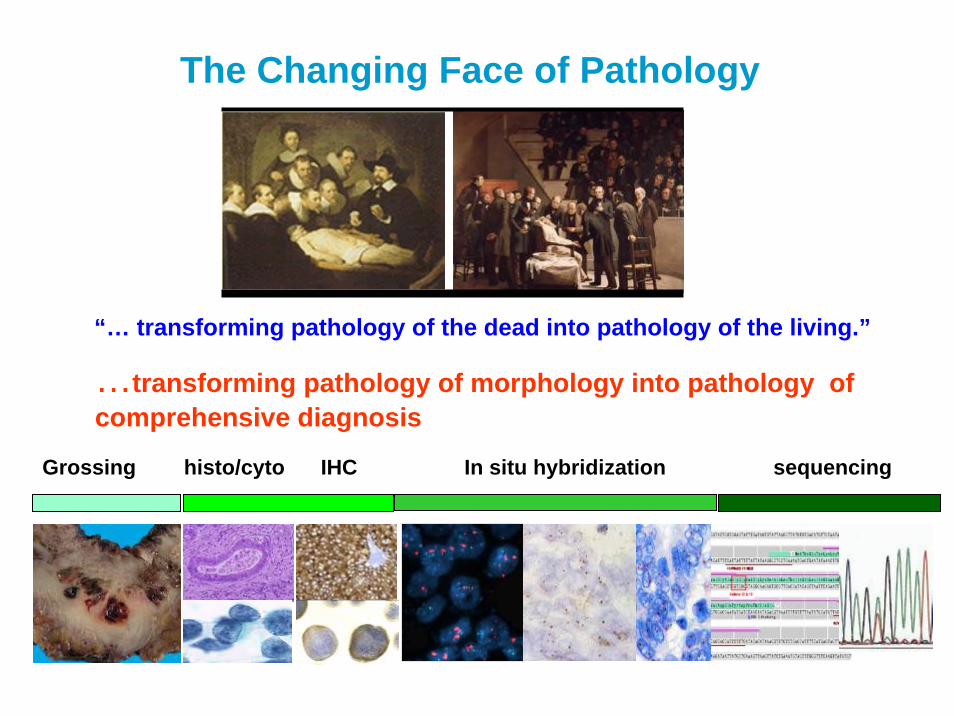
The Changing Face of Pathology
“… transforming pathology of the dead into pathology of the living.”
Grossing histo/cyto IHC In situ hybridization sequencing
…transforming pathology of morphology into pathology of comprehensive diagnosis
Past – Macro/Micro Level• Tests differentiated disease from
non-disease• Disease defined by location, size
and morphology
Today – Molecular Level• Disease defined by individual biology
and /or DNA of tumor• Tests to subcategorize disease:
• predict outcomes of specific therapeutic
• screen for adverse events• monitor disease
Diagnostic technology has improved
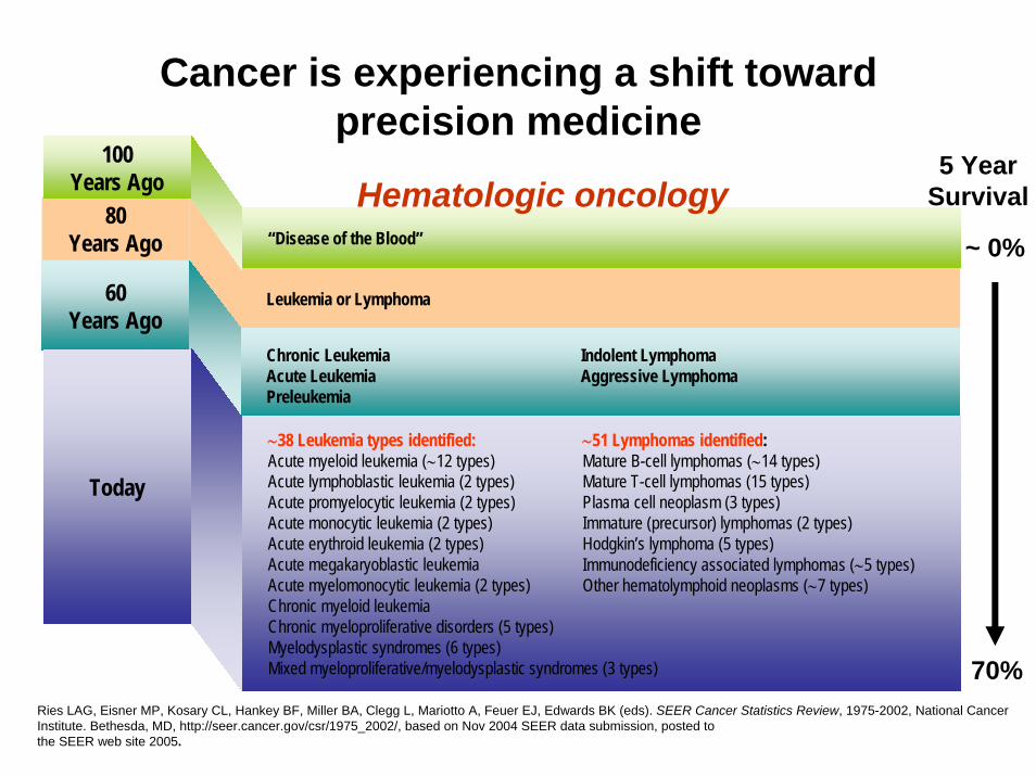
Ries LAG, Eisner MP, Kosary CL, Hankey BF, Miller BA, Clegg L, Mariotto A, Feuer EJ, Edwards BK (eds). SEER Cancer Statistics Review, 1975-2002, National Cancer Institute. Bethesda, MD, http://seer.cancer.gov/csr/1975_2002/, based on Nov 2004 SEER data submission, posted to the SEER web site 2005.
Cancer is experiencing a shift toward precision medicine
Common gene variationsOncogene• a gene with the potential to cause cancer• often mutated or expressed at high levels in tumor cells
Proto-oncogene• a normal gene that can become an oncogene due to mutations or
increased expression• coding for proteins that help to regulate cell growth and
differentiation
anti-oncogene, or tumor suppressor gene• a gene protecting a cell from one step on the path to cancer• when mutated to cause a loss or reduction in its function, the cell can
progress to cancer in combination with other genetic changes
Dominant OncogenesIdentified as transforming genes in viruses with
functions as:• Products involved in:cell cycle / cell division / differentiation• Control of normal cellular growth &
• Intracellular pathways activated• Activation / Repression of various genes
Examples of Oncogenes
abl CML translocation
bcl2 Follicular Lymphoma translocation
erbB-2 Breast/ovarian carcinoma amplification
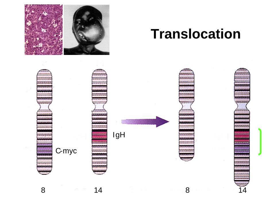
c-myc Burkitt’s lymphoma translocation
ras Thyroid /Colon carcinoma point mutation
ret Thyroid carcinoma rearrangement
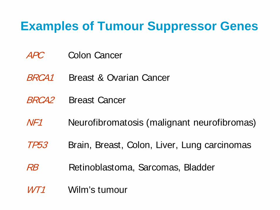
Examples of Tumour Suppressor Genes
APC Colon Cancer
BRCA1 Breast & Ovarian Cancer
BRCA2 Breast Cancer
NF1 Neurofibromatosis (malignant neurofibromas)
TP53 Brain, Breast, Colon, Liver, Lung carcinomas
RB Retinoblastoma, Sarcomas, Bladder
WT1 Wilm’s tumour
Dominant Oncogenes
Tumour Suppressor Genes
Enhanced Reduced
ActivatingGain in function
Dominant
InactivatingLoss of function
Recessive
Changes in tumor cells
Mechanism of Mutations
• Point Mutations
• Amplification
• Translocation / Rearrangements
• Deletions
• Altered Expression
Point Mutation
Change in single base-paire.g. G:C to A:T
SHE HAD ONE MAD CAT AND ONE SAD RAT
SHE HAD ONE BAD CAT AND ONE SAD RAT
8 14 8 14
C-myc
IgH
Translocation
Amplification
N-Myc Gene in NeuroblastomaCerbB2 gene in Breast Cancer
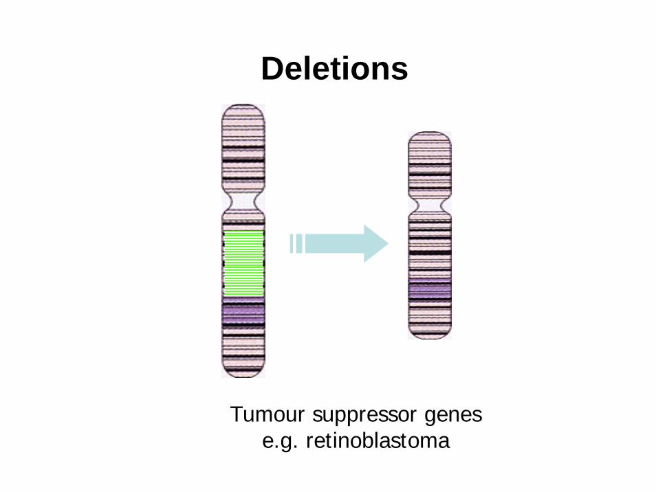
Deletions
Tumour suppressor genese.g. retinoblastoma
Which cell does cancer arise in?
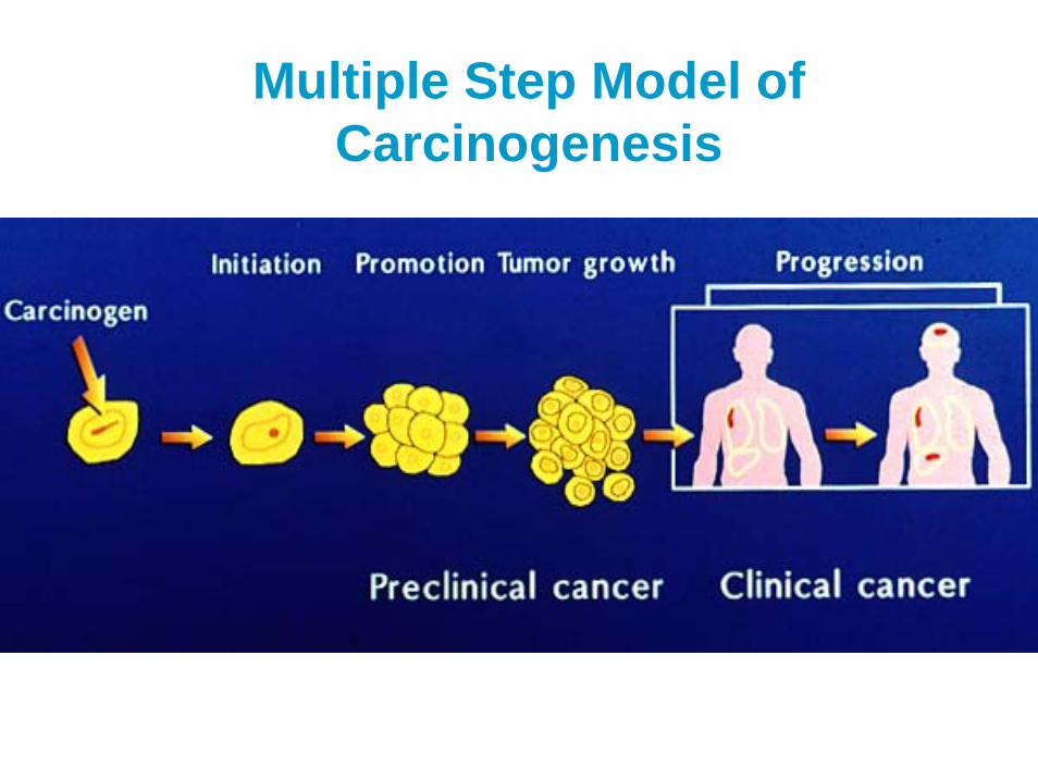
Multiple Step Model of Carcinogenesis
Multi-step model of carcinogenesis of colon cancer
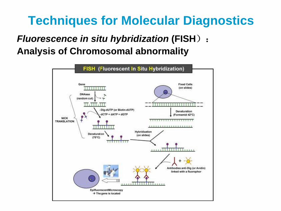
Techniques for Molecular DiagnosticsFluorescence in situ hybridization (FISH):
Analysis of Chromosomal abnormality
HER2 amplified HER2 non-amplified
Gene Amplification
Translocation
Polymerase Chain Reaction (PCR)
DNA Sequenceing
DNA Microarray
aRA
r = 0.91
log 1
0(ra
tio)
, T3-
3
Microarrays
FISH
Laser microscopegenome
Tissue arrays
Tumor Markers
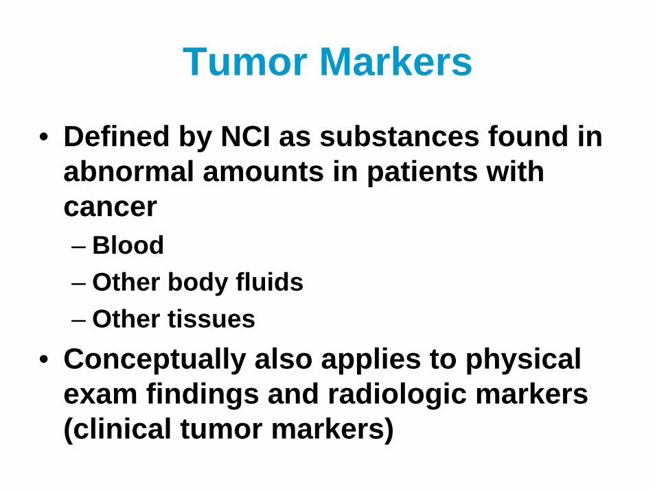
• Defined by NCI as substances found in abnormal amounts in patients with cancer– Blood– Other body fluids– Other tissues
• Conceptually also applies to physical exam findings and radiologic markers (clinical tumor markers)
Tumor Markers
• Used for:– Diagnosis– Monitor response to treatment– Prognosis– Predict response to particular treatments
• Generally cannot be used alone to diagnose cancer – must be used with other methods such as biopsy
How are Tumor Markers selected?
• Basic Research– To show a potential marker exists
• Evidence-based Translational Research– To show the markers add value to healthcare
• Practice Guidelines Organizations– Review the literature and provide standards
Tumor Markers
• Some markers are unique for a cancer type
• Others are found in many types of cancers
• Others may also be found in patients without cancer but that have other diseases
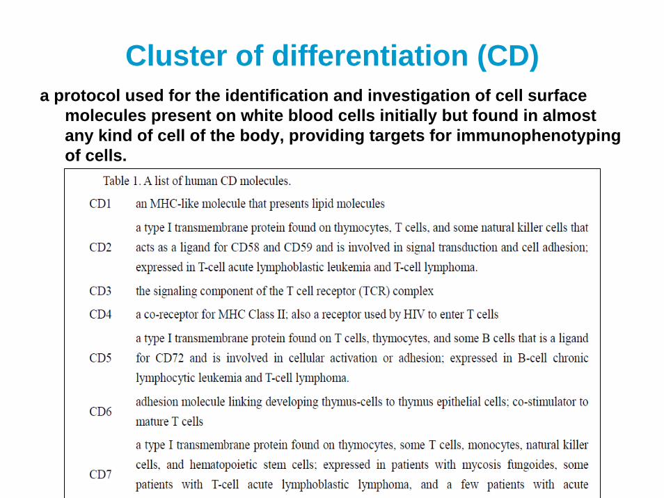
Cluster of differentiation (CD)a protocol used for the identification and investigation of cell surface
molecules present on white blood cells initially but found in almost any kind of cell of the body, providing targets for immunophenotypingof cells.
CD20 in malignant lymphoma
KIT in Gastrointestinal Stromal Sarcoma (GIST)
• GIST – uncommon tumor thought to arise from interstitial cells of Cajal (cells of the autonomic nervous system)
• Most GIST tumors have an activating in a receptor tyrosine kinase– KIT mutation in 50-85% of GIST
• KIT also known as CD117 or stem cell factor receptor• Nearly all KIT mutations in GIST are in exon 11, some in
exon 9 or 13, rarely in exon 17– PDGFR-alpha mutation in 10-20% of GIST
KIT in Gastrointestinal Stromal Sarcoma (GIST)
• Nearly all KIT and PDGFR-alpha mutations in GIST respond to treatment with Gleevec (a tyrosine kinase inhibitor)– Rare exon 17 mutations– Detection by IHC sufficient
• Activating KIT mutations also present in melanoma, mastocytosis, acute myeloid leukemia, myeloproliferative neoplasms– However, these frequently
have exon 17 mutations resistant to Gleevec, so mutation analysis required
PET images before and after 8 weeksof Imatinib for GIST
Oncologist, 2006, 11:9
•Cell membrane tyrosine kinasereceptor•Signals after forming homo- or heterodimers with other EGFR family members•Amplified in ~20% of breast cancers and gastric cancers
•Correlates with - poor prognosis- resistance to endocrine therapy
•Predicts response to - anthracyclines through
linkage to TOPO2 - targeted therapy
(Trastuzumab/Herceptin)
HER2: Human Epidermal Growth Factor Receptor 2
Detection of increased HER2 can be performed in several ways
ProteinWestern blotIHC
RNA Northern hybridizationExpression microarrays
DNA Southern hybridizationDot-blot hybridizationISH
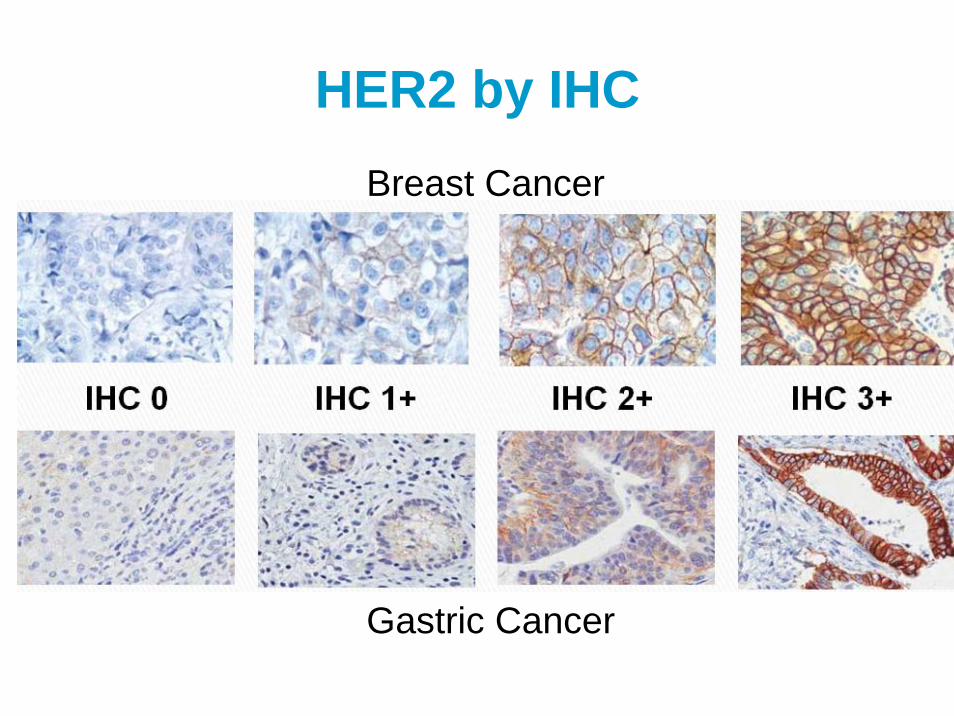
HER2 by IHCBreast Cancer
Gastric Cancer
HER2 by ISH
FISH SISH
Summary• Numbers of new molecular techniques with high
sensitivity and specificity, simple standardized and high speed procedures and falling cost, make cancer diagnosis step into new ear.
• Molecular diagnostics will give a more sensitive and precise diagnosis of cancer, and a guidance toward appropriate and effective treatment options.
• Tumor markers always arise from basic and translational research, clinical practice guidelines, which build on data from tumor registries.
• More and more new tumor markers within tumor signaling pathways and amenable to therapeutic intervention will be added in the future.
Review
• What is oncogene, anti-oncogene? Please give some examples.
• What is biomarkers? Please give some examples to show their significance.
• Do you know any techniques or methods routinely used for molecular diagnosis and biomarkers detection?
复旦大学上海医学院肿瘤学系
Recommended Readings:
Jennifer Hunt. Molecular Oncology, An Issue of Surgical Pathology Clinics. Elsevier Health Science, 2012