International Journal of Molecular Sciences Review Fueling Inflamm-Aging through Mitochondrial Dysfunction: Mechanisms and Molecular Targets Anna Picca 1 , Angela Maria Serena Lezza 2 , Christiaan Leeuwenburgh 3 , Vito Pesce 2 , Riccardo Calvani 1, *, Francesco Landi 1 , Roberto Bernabei 1 and Emanuele Marzetti 1 1 Department of Geriatrics, Neuroscience and Orthopedics, Catholic University of the Sacred Heart School of Medicine, 00168 Rome, Italy; [email protected] (A.P.); [email protected] (F.L.); [email protected] (R.B.); [email protected] (E.M.) 2 Department of Biosciences, Biotechnology and Biopharmaceutics, University of Bari, 70125 Bari, Italy; [email protected] (A.M.S.L.); [email protected] (V.P.) 3 Department of Aging and Geriatric Research, Institute on Aging, Division of Biology of Aging, University of Florida, Gainesville, FL 32611, USA; cleeuwen@ufl.edu * Correspondence: [email protected]; Tel.: +39-06-3015-5559; Fax: +39-06-3051-911 Academic Editor: Mónica De la Fuente Received: 16 March 2017; Accepted: 25 April 2017; Published: 28 April 2017 Abstract: Among the complex determinants of aging, mitochondrial dysfunction has been in the spotlight for a long time. As the hub for many cellular functions, the maintenance of an adequate pool of functional mitochondria is crucial for tissue homeostasis. Their unique role in energy supply makes these organelles essential, especially in those tissues strictly dependent on oxidative metabolism. Mitochondrial quality control (MQC) is ensured by pathways related to protein folding and degradation as well as by processes involving the entire organelle, such as biogenesis, dynamics, and mitophagy. Dysfunctional MQC, oxidative stress and inflammation are hallmarks of senescence and chronic degenerative diseases. One of the consequences of age-related failing MQC and oxidative stress is the release of mitochondria-derived damage-associated molecular patterns (DAMPs). Through their bacterial ancestry, these molecules contribute to mounting an inflammatory response by interacting with receptors similar to those involved in pathogen-associated responses. Mitochondrial DAMPs, especially cell-free mitochondrial DNA, have recently become the subject of intensive research because of their possible involvement in conditions associated with inflammation, such as aging and degenerative diseases. Here, we review the contribution of mitochondrial DAMPs to inflammation and discuss some of the mechanisms at the basis of their generation. Keywords: mitophagy; sterile inflammation; mitochondrial biogenesis; mitochondrial dynamics; TFAM; mitochondrial quality control (MQC); inflammasome; damage-associated molecular patterns (DAMPs) 1. Introduction Aging is a complex and multi-factorial process characterized by increased risk of adverse health outcomes [1]. Understanding the intimate mechanisms of aging is therefore instrumental for contrasting its negative correlates [1]. As initially proposed in the “mitochondrial theory of aging”, mitochondria are deeply involved in the aging process mainly through respiratory dysfunction and oxidant generation [2,3]. Although unique as fueling systems within the cell, mitochondria participate in other essential functions, including heme metabolism, regulation of intracellular calcium homeostasis, modulation of cell proliferation, and integration of apoptotic signaling [4–7]. It is therefore crucial that a pool of healthy and well-functioning organelles is maintained within the cell. To this aim, a comprehensive set of adaptive quality control processes operates via interrelated systems, including Int. J. Mol. Sci. 2017, 18, 933; doi:10.3390/ijms18050933 www.mdpi.com/journal/ijms
Transcript
International Journal of
Molecular Sciences
Review
Fueling Inflamm-Aging through MitochondrialDysfunction: Mechanisms and Molecular Targets
Anna Picca 1, Angela Maria Serena Lezza 2, Christiaan Leeuwenburgh 3, Vito Pesce 2,Riccardo Calvani 1,*, Francesco Landi 1, Roberto Bernabei 1 and Emanuele Marzetti 1
2 Department of Biosciences, Biotechnology and Biopharmaceutics, University of Bari, 70125 Bari, Italy;[email protected] (A.M.S.L.); [email protected] (V.P.)
3 Department of Aging and Geriatric Research, Institute on Aging, Division of Biology of Aging,University of Florida, Gainesville, FL 32611, USA; [email protected]
Academic Editor: Mónica De la FuenteReceived: 16 March 2017; Accepted: 25 April 2017; Published: 28 April 2017
Abstract: Among the complex determinants of aging, mitochondrial dysfunction has been in thespotlight for a long time. As the hub for many cellular functions, the maintenance of an adequatepool of functional mitochondria is crucial for tissue homeostasis. Their unique role in energysupply makes these organelles essential, especially in those tissues strictly dependent on oxidativemetabolism. Mitochondrial quality control (MQC) is ensured by pathways related to proteinfolding and degradation as well as by processes involving the entire organelle, such as biogenesis,dynamics, and mitophagy. Dysfunctional MQC, oxidative stress and inflammation are hallmarks ofsenescence and chronic degenerative diseases. One of the consequences of age-related failing MQCand oxidative stress is the release of mitochondria-derived damage-associated molecular patterns(DAMPs). Through their bacterial ancestry, these molecules contribute to mounting an inflammatoryresponse by interacting with receptors similar to those involved in pathogen-associated responses.Mitochondrial DAMPs, especially cell-free mitochondrial DNA, have recently become the subject ofintensive research because of their possible involvement in conditions associated with inflammation,such as aging and degenerative diseases. Here, we review the contribution of mitochondrial DAMPsto inflammation and discuss some of the mechanisms at the basis of their generation.
Aging is a complex and multi-factorial process characterized by increased risk of adversehealth outcomes [1]. Understanding the intimate mechanisms of aging is therefore instrumentalfor contrasting its negative correlates [1]. As initially proposed in the “mitochondrial theory ofaging”, mitochondria are deeply involved in the aging process mainly through respiratory dysfunctionand oxidant generation [2,3]. Although unique as fueling systems within the cell, mitochondriaparticipate in other essential functions, including heme metabolism, regulation of intracellular calciumhomeostasis, modulation of cell proliferation, and integration of apoptotic signaling [4–7]. It is thereforecrucial that a pool of healthy and well-functioning organelles is maintained within the cell. To this aim,a comprehensive set of adaptive quality control processes operates via interrelated systems, including
Int. J. Mol. Sci. 2017, 18, 933; doi:10.3390/ijms18050933 www.mdpi.com/journal/ijms
pathways pertaining to protein folding and degradation, mitochondrial biogenesis, dynamics, andautophagy (mitophagy) [8,9]. The activation of individual MQC pathways depends on the degree ofmitochondrial damage. Due to these vital responsibilities, disruption of the MQC axis is invoked as amajor pathogenic mechanism in a number of disease conditions (i.e., cancer, cardiovascular disease,diabetes, and neurodegenerative disorders) and aging [8,10,11].
Together with mitochondrial dysfunction, chronic inflammation is another hallmark of both agingand degenerative diseases [12]. Interestingly, emerging evidence suggests that the two phenomenaare related to one another. In particular, circulating cell-free mitochondrial DNA (mtDNA), one ofthe cell damage-associated molecular patterns (DAMPs), has been proposed as a functional linkbetween mitochondrial damage and systemic inflammation [13,14]. Indeed, mtDNA, which is releasedas a result of cellular stress, contains hypomethylated CpG motifs resembling those of bacterialDNA and is therefore able to induce an inflammatory response [15]. These regions bind and activatemembrane or cytoplasmic pattern recognition receptors (PRRs), such as the Toll-like receptor (TLR), thenucleotide-binding oligomerization domain (NOD)-like receptor (NLR) [15], and the cytosolic cyclicGMP-AMP synthase (cGAS)-stimulator of interferon genes (STING) DNA sensing system-mediatedpathways [16]. The mechanisms responsible for the generation of mitochondrial DAMPs as well astheir contribution to the inflammatory milieu that characterizes aging and its associated conditions arenot completely understood.
Here, we provide an overview of major processes of MQC and their changes duringaging. Subsequently, we describe candidate mechanisms responsible for generating and releasingmitochondrial DAMPs. Finally, we summarize the current evidence in support of mitochondrialDAMPs as triggers for age-related chronic inflammation.
2. MQC Processes
2.1. Mitochondrial Proteolytic Quality Control System
The mitochondrial proteolytic quality control system consists of subcompartment-specificproteases (mitoproteases) and the ubiquitin-proteasome system (UPS) that together regulatemitochondrial protein turnover and degrade misfolded or oxidized proteins [17]. Mitochondrialproteases act as the first line of defense against mild mitochondrial damage [8]. Within themitochondrial matrix, protein turnover is controlled by 3 AAA proteases: the soluble Lon and ClpPand the membrane-bound m-AAA [18]. In the inter-membrane space, mitochondrial protein qualityis ensured by the membrane-bound i-AAA Yme1L1, the soluble HtrA2/Omi, the metallopeptidasesOMA1, and the presenilins-associated rhomboid-like protein (PARL) [8]. These mitoproteases canalso affect mitochondrial fate and cell viability by acting on mitochondrial dynamics, mitophagy andapoptosis [19,20].
Mitochondrial protein turnover is also regulated by the cytosolic UPS [21]. A proteomic studyin murine heart identified numerous proteins to be ubiquitinated in the various mitochondrialcompartments [22]. However, the mechanism whereby the cytosolic proteasome degrades integralmitochondrial membrane proteins is still a matter of debate.
Similar to the endoplasmic reticulum (ER), mitochondria possess a stress responsive systemfor protein degradation that shares with its ER analogue some key components, including theAAA ATPase p97 and the cofactor Npl4 [23]. Under stress conditions, when protein degradationpathways become insufficient to restore a normal mitochondrial function, a retrograde signal istriggered, which acts on the nuclear genome. This pathway, named mitochondrial unfolded proteinresponse (UPRmt) [24], up-regulates the expression of nuclear genes encoding mitochondrial stressproteins, including chaperonin 10 and 60, mtDnaJ, ClpP and Yme1 [24]. Through these mediators, theUPRmt promotes mitochondrial proteostasis by improving protein folding and degrading irreversiblydamaged proteins.
Int. J. Mol. Sci. 2017, 18, 933 3 of 15
2.2. Mitochondrial Biogenesis
Mitochondrial biogenesis is a multi-stage process generating new organelles that, in adynamic balance with their degradation, regulate the mitochondrial content within the cell.Mitochondriogenesis is orchestrated through the expression of nuclear and mtDNA-encoded genes.In particular, transcriptional coactivators belonging to the peroxisome proliferator activated receptorgamma coactivator-1 (PGC-1) family (e.g., PGC-1α and PGC-1β), the nuclear respiratory factor (NRF) 1and 2, and the estrogen-related receptor α (ERRα), coordinate the expression of mitochondrial proteinsencoded by nuclear DNA [25]. Subsequently, several mitochondrial proteins are expressed, includingthose binding the mtDNA (e.g., mitochondrial transcription factor A (TFAM), B1 and B2 (TFB1M andTFB2M)), which are then transported into mitochondria by a protein import machinery [26]. Onceentered into the mitochondrion, mtDNA-binding proteins directly activate mtDNA transcription andreplication (Figure 1).
Int. J. Mol. Sci. 2017, 18, 933 3 of 15
2.2. Mitochondrial Biogenesis
Mitochondrial biogenesis is a multi-stage process generating new organelles that, in a dynamic balance with their degradation, regulate the mitochondrial content within the cell. Mitochondriogenesis is orchestrated through the expression of nuclear and mtDNA-encoded genes. In particular, transcriptional coactivators belonging to the peroxisome proliferator activated receptor gamma coactivator-1 (PGC-1) family (e.g., PGC-1α and PGC-1β), the nuclear respiratory factor (NRF) 1 and 2, and the estrogen-related receptor α (ERRα), coordinate the expression of mitochondrial proteins encoded by nuclear DNA [25]. Subsequently, several mitochondrial proteins are expressed, including those binding the mtDNA (e.g., mitochondrial transcription factor A (TFAM), B1 and B2 (TFB1M and TFB2M)), which are then transported into mitochondria by a protein import machinery [26]. Once entered into the mitochondrion, mtDNA-binding proteins directly activate mtDNA transcription and replication (Figure 1).
Figure 1. Schematic representation of the regulation of mitochondrial biogenesis. In response to external stimuli (e.g., skeletal muscle contraction or exercise), the nuclear genome coordinates the expression of nuclear and mitochondrial proteins. This pathway is triggered by the activation of signaling molecules, including AMP-activated protein kinase (AMPK), and converges on the expression of the transcriptional coactivator peroxisome proliferator-activated receptor gamma coactivator-1α (PGC-1α), the master regulator of mitochondrial biogenesis. PGC-1α promotes its own expression as well as that of the nuclear respiratory factor 1 and 2 (NRF-1/2). NRF-1 and 2 bind and up-regulate the expression of nuclear genes encoding mitochondrial proteins as well as the expression of mitochondrial transcription factor A (TFAM), which is subsequently transported into mitochondria. Here, TFAM binds to mitochondrial DNA (mtDNA) and activates the transcription and replication of the mitochondrial genome, a crucial step in the generation of new organelles. TF, transcription factor.
Figure 1. Schematic representation of the regulation of mitochondrial biogenesis. In response toexternal stimuli (e.g., skeletal muscle contraction or exercise), the nuclear genome coordinates theexpression of nuclear and mitochondrial proteins. This pathway is triggered by the activation ofsignaling molecules, including AMP-activated protein kinase (AMPK), and converges on the expressionof the transcriptional coactivator peroxisome proliferator-activated receptor gamma coactivator-1α(PGC-1α), the master regulator of mitochondrial biogenesis. PGC-1α promotes its own expression aswell as that of the nuclear respiratory factor 1 and 2 (NRF-1/2). NRF-1 and 2 bind and up-regulate theexpression of nuclear genes encoding mitochondrial proteins as well as the expression of mitochondrialtranscription factor A (TFAM), which is subsequently transported into mitochondria. Here, TFAM bindsto mitochondrial DNA (mtDNA) and activates the transcription and replication of the mitochondrialgenome, a crucial step in the generation of new organelles. TF, transcription factor.
Int. J. Mol. Sci. 2017, 18, 933 4 of 15
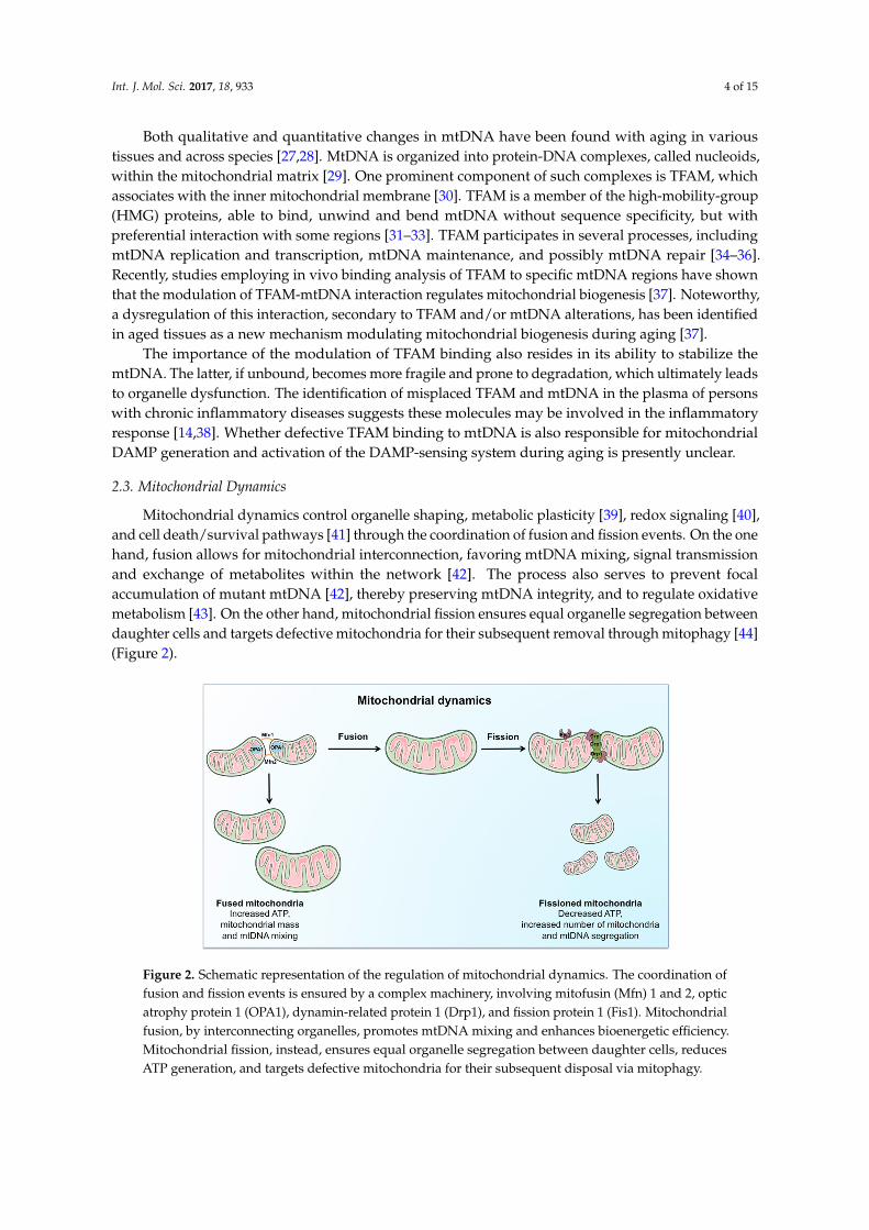
Both qualitative and quantitative changes in mtDNA have been found with aging in varioustissues and across species [27,28]. MtDNA is organized into protein-DNA complexes, called nucleoids,within the mitochondrial matrix [29]. One prominent component of such complexes is TFAM, whichassociates with the inner mitochondrial membrane [30]. TFAM is a member of the high-mobility-group(HMG) proteins, able to bind, unwind and bend mtDNA without sequence specificity, but withpreferential interaction with some regions [31–33]. TFAM participates in several processes, includingmtDNA replication and transcription, mtDNA maintenance, and possibly mtDNA repair [34–36].Recently, studies employing in vivo binding analysis of TFAM to specific mtDNA regions have shownthat the modulation of TFAM-mtDNA interaction regulates mitochondrial biogenesis [37]. Noteworthy,a dysregulation of this interaction, secondary to TFAM and/or mtDNA alterations, has been identifiedin aged tissues as a new mechanism modulating mitochondrial biogenesis during aging [37].
The importance of the modulation of TFAM binding also resides in its ability to stabilize themtDNA. The latter, if unbound, becomes more fragile and prone to degradation, which ultimately leadsto organelle dysfunction. The identification of misplaced TFAM and mtDNA in the plasma of personswith chronic inflammatory diseases suggests these molecules may be involved in the inflammatoryresponse [14,38]. Whether defective TFAM binding to mtDNA is also responsible for mitochondrialDAMP generation and activation of the DAMP-sensing system during aging is presently unclear.
2.3. Mitochondrial Dynamics
Mitochondrial dynamics control organelle shaping, metabolic plasticity [39], redox signaling [40],and cell death/survival pathways [41] through the coordination of fusion and fission events. On the onehand, fusion allows for mitochondrial interconnection, favoring mtDNA mixing, signal transmissionand exchange of metabolites within the network [42]. The process also serves to prevent focalaccumulation of mutant mtDNA [42], thereby preserving mtDNA integrity, and to regulate oxidativemetabolism [43]. On the other hand, mitochondrial fission ensures equal organelle segregation betweendaughter cells and targets defective mitochondria for their subsequent removal through mitophagy [44](Figure 2).
Int. J. Mol. Sci. 2017, 18, 933 4 of 15
Both qualitative and quantitative changes in mtDNA have been found with aging in various tissues and across species [27,28]. MtDNA is organized into protein-DNA complexes, called nucleoids, within the mitochondrial matrix [29]. One prominent component of such complexes is TFAM, which associates with the inner mitochondrial membrane [30]. TFAM is a member of the high-mobility-group (HMG) proteins, able to bind, unwind and bend mtDNA without sequence specificity, but with preferential interaction with some regions [31–33]. TFAM participates in several processes, including mtDNA replication and transcription, mtDNA maintenance, and possibly mtDNA repair [34–36]. Recently, studies employing in vivo binding analysis of TFAM to specific mtDNA regions have shown that the modulation of TFAM-mtDNA interaction regulates mitochondrial biogenesis [37]. Noteworthy, a dysregulation of this interaction, secondary to TFAM and/or mtDNA alterations, has been identified in aged tissues as a new mechanism modulating mitochondrial biogenesis during aging [37].
The importance of the modulation of TFAM binding also resides in its ability to stabilize the mtDNA. The latter, if unbound, becomes more fragile and prone to degradation, which ultimately leads to organelle dysfunction. The identification of misplaced TFAM and mtDNA in the plasma of persons with chronic inflammatory diseases suggests these molecules may be involved in the inflammatory response [14,38]. Whether defective TFAM binding to mtDNA is also responsible for mitochondrial DAMP generation and activation of the DAMP-sensing system during aging is presently unclear.
2.3. Mitochondrial Dynamics
Mitochondrial dynamics control organelle shaping, metabolic plasticity [39], redox signaling [40], and cell death/survival pathways [41] through the coordination of fusion and fission events. On the one hand, fusion allows for mitochondrial interconnection, favoring mtDNA mixing, signal transmission and exchange of metabolites within the network [42]. The process also serves to prevent focal accumulation of mutant mtDNA [42], thereby preserving mtDNA integrity, and to regulate oxidative metabolism [43]. On the other hand, mitochondrial fission ensures equal organelle segregation between daughter cells and targets defective mitochondria for their subsequent removal through mitophagy [44] (Figure 2).
Figure 2. Schematic representation of the regulation of mitochondrial dynamics. The coordination of fusion and fission events is ensured by a complex machinery, involving mitofusin (Mfn) 1 and 2, optic atrophy protein 1 (OPA1), dynamin-related protein 1 (Drp1), and fission protein 1 (Fis1). Mitochondrial fusion, by interconnecting organelles, promotes mtDNA mixing and enhances bioenergetic efficiency. Mitochondrial fission, instead, ensures equal organelle segregation between daughter cells, reduces ATP generation, and targets defective mitochondria for their subsequent disposal via mitophagy.
Figure 2. Schematic representation of the regulation of mitochondrial dynamics. The coordination offusion and fission events is ensured by a complex machinery, involving mitofusin (Mfn) 1 and 2, opticatrophy protein 1 (OPA1), dynamin-related protein 1 (Drp1), and fission protein 1 (Fis1). Mitochondrialfusion, by interconnecting organelles, promotes mtDNA mixing and enhances bioenergetic efficiency.Mitochondrial fission, instead, ensures equal organelle segregation between daughter cells, reducesATP generation, and targets defective mitochondria for their subsequent disposal via mitophagy.
Int. J. Mol. Sci. 2017, 18, 933 5 of 15
The integration of mitochondrial dynamics and mitophagy ensures an efficient MQC process andcontributes to preserving metabolic cellular “fitness” and plasticity [9]. Derangements of fusion-fissionhave been proposed as a mechanism contributing to the formation of structurally abnormal anddysfunctional mitochondria under stress conditions and during senescence [45].
The exposure of cultured cells to subcytotoxic doses of hydrogen peroxide represses the expressionof fission protein 1 (Fis1), thereby promoting the formation of elongated mitochondria with increasedoxidant emission [45]. Excessive activation of fission can also induce mitochondrial dysfunction.Indeed, mitochondrial fragmentation, down-regulation of fusion and impaired bioenergetics havebeen detected in muscle of diabetic persons [46]. On the other hand, depletion of the mitochondrialfusion factor optic atrophy protein 1 (OPA1) disintegrates the mitochondrial network and sensitizescultured cells to apoptosis [47]. Conversely, blockade of Fis1 or dynamin-related protein 1 (Drp1)inhibits mitochondrial fragmentation and the execution of apoptosis in cell culture systems [47].
Imbalanced mitochondrial dynamics toward fission have been found in several diseaseconditions [48] as well as in age-related sarcopenia [49] and cancer cachexia [50]. Conversely, increasedfusion in very old rats has been associated with maintained mtDNA content and seems to contribute tolongevity [51]. However, the molecular mechanisms underlying the relationship among mitochondrialdynamics, senescence and longevity are not fully understood and deserve further investigation.
2.4. Autophagy and Mitophagy
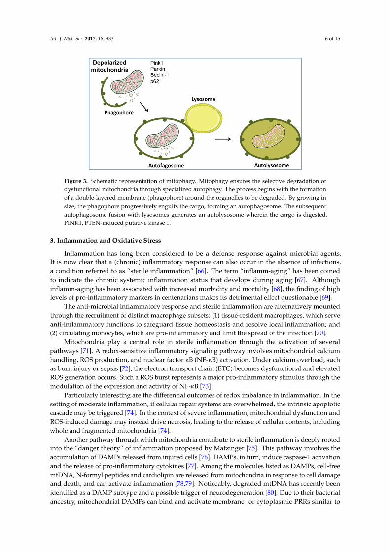
Autophagy is, literally, a cellular self-eating process through which intracellular componentsare degraded within lysosomes during periods of stress, such as nutrient deprivation, as anattempt to adapt and survive [52]. Selective autophagic removal of mitochondria (mitophagy)is triggered by the loss of mitochondrial membrane potential [53] and is aimed at limitingreactive oxygen species (ROS) generation and preserving cell viability through the clearance ofdysfunctional organelles [54]. Mitophagy, however, represents an extreme attempt of the cell tomaintain homeostasis since mitochondria can also take an alternative route to dispose damagedcomponents, before whole-sale organelle degradation is triggered. Indeed, matrix components can beeliminated within vesicles budding from dysfunctional but not yet depolarized mitochondria [55,56].Mitochondrion-derived vesicles (MDVs) serve to eliminate oxidized mitochondrial elements throughthe serine/threonine-protein kinase PTEN-induced putative kinase 1 (PINK1) and the E3 ubiquitinligase Parkin [57]. As part of MQC, the delivery of damaged cargo within MDVs to lysosomes occursas an early response to oxidative stress [55–57]. Conversely, severely damaged mitochondria arefissioned and targeted for elimination through a distinct pathway involving the synergistic activityof the mitochondrial dynamics machinery, PINK1, Parkin, Bnip3L/Nix, and Bnip3 [58] (Figure 3).Indeed, following mitochondrial depolarization, PINK1 accumulates on the mitochondrial surface,leading to the recruitment of Parkin, which ubiquitinates proteins located in the outer mitochondrialmembrane [59]. Ubiquitination of the PARL protease promotes the execution of mitophagy bypreventing PINK1 degradation. Ubiquitin-tagged mitochondria bind to p62, which assists in therecruitment of autophagosomal membranes to mitochondria [60]. Parkin can also interact withactivating molecule in Beclin1-regulated autophagy (AMBRA1), which stimulates the activity of theclass III phosphatidylinositol 3-kinase (PI3K) complex required for phagophore formation [61].
Dysfunctional vesicle trafficking as well as specific molecular patterns recruited in vesicle cargoshas been described under several conditions [14,62]. A senescence-associated secretory phenotypehas also been identified and characterized [63]. Studies have investigated the role of extracellularvesicles as carriers of senescence signals outside the cell [64]. Noticeably, mitochondrial dysfunctionhas been indicated among the mechanisms promoting the development of an aging phenotype [65].However, the molecular identity of the factors involved is presently unknown and is the subject ofactive investigation.
Int. J. Mol. Sci. 2017, 18, 933 6 of 15Int. J. Mol. Sci. 2017, 18, 933 6 of 15
Figure 3. Schematic representation of mitophagy. Mitophagy ensures the selective degradation of dysfunctional mitochondria through specialized autophagy. The process begins with the formation of a double-layered membrane (phagophore) around the organelles to be degraded. By growing in size, the phagophore progressively engulfs the cargo, forming an autophagosome. The subsequent autophagosome fusion with lysosomes generates an autolysosome wherein the cargo is digested. PINK1, PTEN-induced putative kinase 1.
3. Inflammation and Oxidative Stress
Inflammation has long been considered to be a defense response against microbial agents. It is now clear that a (chronic) inflammatory response can also occur in the absence of infections, a condition referred to as “sterile inflammation” [66]. The term “inflamm-aging” has been coined to indicate the chronic systemic inflammation status that develops during aging [67]. Although inflamm-aging has been associated with increased morbidity and mortality [68], the finding of high levels of pro-inflammatory markers in centenarians makes its detrimental effect questionable [69].
The anti-microbial inflammatory response and sterile inflammation are alternatively mounted through the recruitment of distinct macrophage subsets: (1) tissue-resident macrophages, which serve anti-inflammatory functions to safeguard tissue homeostasis and resolve local inflammation; and (2) circulating monocytes, which are pro-inflammatory and limit the spread of the infection [70].
Mitochondria play a central role in sterile inflammation through the activation of several pathways [71]. A redox-sensitive inflammatory signaling pathway involves mitochondrial calcium handling, ROS production, and nuclear factor κB (NF-κB) activation. Under calcium overload, such as burn injury or sepsis [72], the electron transport chain (ETC) becomes dysfunctional and elevated ROS generation occurs. Such a ROS burst represents a major pro-inflammatory stimulus through the modulation of the expression and activity of NF-κB [73].
Particularly interesting are the differential outcomes of redox imbalance in inflammation. In the setting of moderate inflammation, if cellular repair systems are overwhelmed, the intrinsic apoptotic cascade may be triggered [74]. In the context of severe inflammation, mitochondrial dysfunction and ROS-induced damage may instead drive necrosis, leading to the release of cellular contents, including whole and fragmented mitochondria [74].
Another pathway through which mitochondria contribute to sterile inflammation is deeply rooted into the “danger theory” of inflammation proposed by Matzinger [75]. This pathway involves the accumulation of DAMPs released from injured cells [76]. DAMPs, in turn, induce caspase-1 activation and the release of pro-inflammatory cytokines [77]. Among the molecules listed as DAMPs, cell-free mtDNA, N-formyl peptides and cardiolipin are released from mitochondria in response to cell damage and death, and can activate inflammation [78,79]. Noticeably, degraded mtDNA has recently been identified as a DAMP subtype and a possible trigger of neurodegeneration [80]. Due to their bacterial ancestry, mitochondrial DAMPs can bind and activate membrane- or cytoplasmic-PRRs similar to those recognized by pathogen-associated molecular
Figure 3. Schematic representation of mitophagy. Mitophagy ensures the selective degradation ofdysfunctional mitochondria through specialized autophagy. The process begins with the formationof a double-layered membrane (phagophore) around the organelles to be degraded. By growing insize, the phagophore progressively engulfs the cargo, forming an autophagosome. The subsequentautophagosome fusion with lysosomes generates an autolysosome wherein the cargo is digested.PINK1, PTEN-induced putative kinase 1.
3. Inflammation and Oxidative Stress
Inflammation has long been considered to be a defense response against microbial agents.It is now clear that a (chronic) inflammatory response can also occur in the absence of infections,a condition referred to as “sterile inflammation” [66]. The term “inflamm-aging” has been coinedto indicate the chronic systemic inflammation status that develops during aging [67]. Althoughinflamm-aging has been associated with increased morbidity and mortality [68], the finding of highlevels of pro-inflammatory markers in centenarians makes its detrimental effect questionable [69].
The anti-microbial inflammatory response and sterile inflammation are alternatively mountedthrough the recruitment of distinct macrophage subsets: (1) tissue-resident macrophages, which serveanti-inflammatory functions to safeguard tissue homeostasis and resolve local inflammation; and(2) circulating monocytes, which are pro-inflammatory and limit the spread of the infection [70].
Mitochondria play a central role in sterile inflammation through the activation of severalpathways [71]. A redox-sensitive inflammatory signaling pathway involves mitochondrial calciumhandling, ROS production, and nuclear factor κB (NF-κB) activation. Under calcium overload, suchas burn injury or sepsis [72], the electron transport chain (ETC) becomes dysfunctional and elevatedROS generation occurs. Such a ROS burst represents a major pro-inflammatory stimulus through themodulation of the expression and activity of NF-κB [73].
Particularly interesting are the differential outcomes of redox imbalance in inflammation. In thesetting of moderate inflammation, if cellular repair systems are overwhelmed, the intrinsic apoptoticcascade may be triggered [74]. In the context of severe inflammation, mitochondrial dysfunction andROS-induced damage may instead drive necrosis, leading to the release of cellular contents, includingwhole and fragmented mitochondria [74].
Another pathway through which mitochondria contribute to sterile inflammation is deeply rootedinto the “danger theory” of inflammation proposed by Matzinger [75]. This pathway involves theaccumulation of DAMPs released from injured cells [76]. DAMPs, in turn, induce caspase-1 activationand the release of pro-inflammatory cytokines [77]. Among the molecules listed as DAMPs, cell-freemtDNA, N-formyl peptides and cardiolipin are released from mitochondria in response to cell damageand death, and can activate inflammation [78,79]. Noticeably, degraded mtDNA has recently beenidentified as a DAMP subtype and a possible trigger of neurodegeneration [80]. Due to their bacterialancestry, mitochondrial DAMPs can bind and activate membrane- or cytoplasmic-PRRs similar to
Int. J. Mol. Sci. 2017, 18, 933 7 of 15
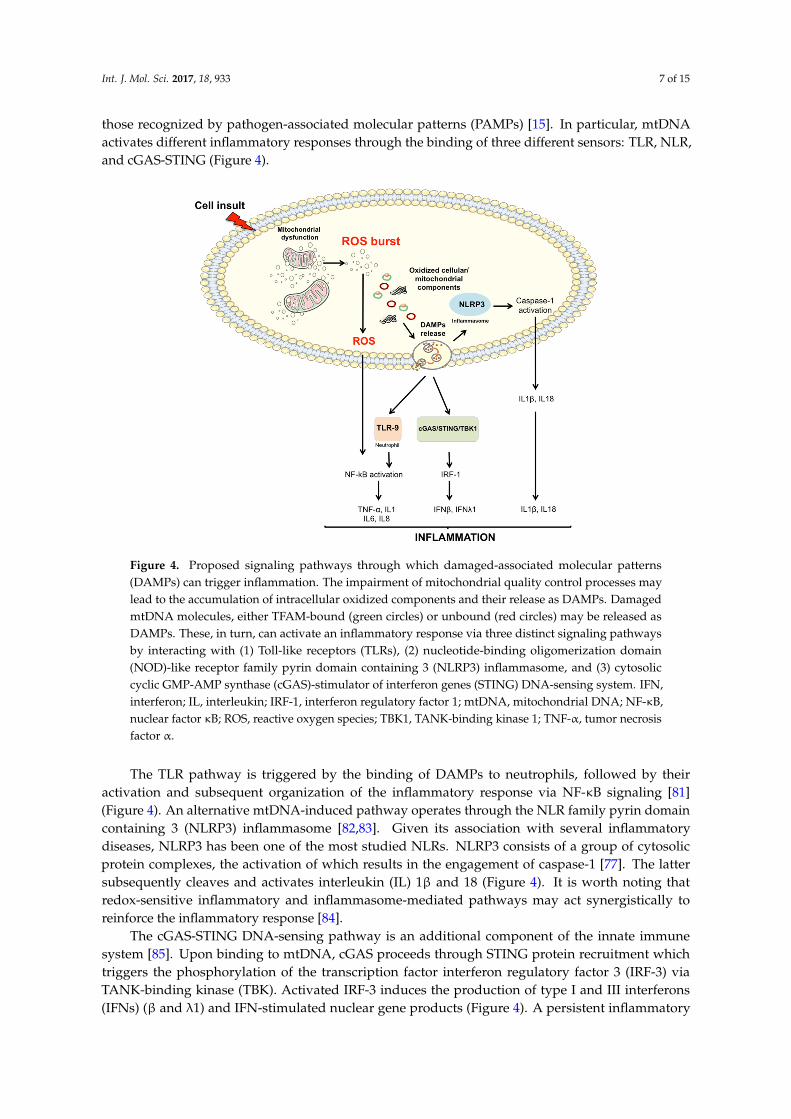
those recognized by pathogen-associated molecular patterns (PAMPs) [15]. In particular, mtDNAactivates different inflammatory responses through the binding of three different sensors: TLR, NLR,and cGAS-STING (Figure 4).
Int. J. Mol. Sci. 2017, 18, 933 7 of 15
patterns (PAMPs) [15]. In particular, mtDNA activates different inflammatory responses through the binding of three different sensors: TLR, NLR, and cGAS-STING (Figure 4).
The TLR pathway is triggered by the binding of DAMPs to neutrophils, followed by their activation and subsequent organization of the inflammatory response via NF-κB signaling [81] (Figure 4). An alternative mtDNA-induced pathway operates through the NLR family pyrin domain containing 3 (NLRP3) inflammasome [82,83]. Given its association with several inflammatory diseases, NLRP3 has been one of the most studied NLRs. NLRP3 consists of a group of cytosolic protein complexes, the activation of which results in the engagement of caspase-1 [77]. The latter subsequently cleaves and activates interleukin (IL) 1β and 18 (Figure 4). It is worth noting that redox-sensitive inflammatory and inflammasome-mediated pathways may act synergistically to reinforce the inflammatory response [84].
The cGAS-STING DNA-sensing pathway is an additional component of the innate immune system [85]. Upon binding to mtDNA, cGAS proceeds through STING protein recruitment which triggers the phosphorylation of the transcription factor interferon regulatory factor 3 (IRF-3) via TANK-binding kinase (TBK). Activated IRF-3 induces the production of type I and III interferons (IFNs) (β and λ1) and IFN-stimulated nuclear gene products (Figure 4). A persistent inflammatory trigger is able to alert circulating immune cells, which, in turn, may mount a systemic response through the activation of mtDNA-induced inflammatory pathways. Cytokines, chemokines, nitric oxide (NO) and ROS released in the circulation by inflammatory cells can induce further mitochondrial damage, thereby establishing a vicious circle which reinforces the whole process.
Figure 4. Proposed signaling pathways through which damaged-associated molecular patterns (DAMPs) can trigger inflammation. The impairment of mitochondrial quality control processes may lead to the accumulation of intracellular oxidized components and their release as DAMPs. Damaged mtDNA molecules, either TFAM-bound (green circles) or unbound (red circles) may be released as DAMPs. These, in turn, can activate an inflammatory response via three distinct signaling pathways by interacting with (1) Toll-like receptors (TLRs), (2) nucleotide-binding oligomerization domain (NOD)-like receptor family pyrin domain containing 3 (NLRP3) inflammasome, and (3) cytosolic cyclic GMP-AMP synthase (cGAS)-stimulator of interferon genes (STING) DNA-sensing system. IFN, interferon; IL, interleukin; IRF-1, interferon regulatory factor 1; mtDNA, mitochondrial DNA; NF-κB, nuclear factor κB; ROS, reactive oxygen species; TBK1, TANK-binding kinase 1; TNF-α, tumor necrosis factor α.
Figure 4. Proposed signaling pathways through which damaged-associated molecular patterns(DAMPs) can trigger inflammation. The impairment of mitochondrial quality control processes maylead to the accumulation of intracellular oxidized components and their release as DAMPs. DamagedmtDNA molecules, either TFAM-bound (green circles) or unbound (red circles) may be released asDAMPs. These, in turn, can activate an inflammatory response via three distinct signaling pathwaysby interacting with (1) Toll-like receptors (TLRs), (2) nucleotide-binding oligomerization domain(NOD)-like receptor family pyrin domain containing 3 (NLRP3) inflammasome, and (3) cytosoliccyclic GMP-AMP synthase (cGAS)-stimulator of interferon genes (STING) DNA-sensing system. IFN,interferon; IL, interleukin; IRF-1, interferon regulatory factor 1; mtDNA, mitochondrial DNA; NF-κB,nuclear factor κB; ROS, reactive oxygen species; TBK1, TANK-binding kinase 1; TNF-α, tumor necrosisfactor α.
The TLR pathway is triggered by the binding of DAMPs to neutrophils, followed by theiractivation and subsequent organization of the inflammatory response via NF-κB signaling [81](Figure 4). An alternative mtDNA-induced pathway operates through the NLR family pyrin domaincontaining 3 (NLRP3) inflammasome [82,83]. Given its association with several inflammatorydiseases, NLRP3 has been one of the most studied NLRs. NLRP3 consists of a group of cytosolicprotein complexes, the activation of which results in the engagement of caspase-1 [77]. The lattersubsequently cleaves and activates interleukin (IL) 1β and 18 (Figure 4). It is worth noting thatredox-sensitive inflammatory and inflammasome-mediated pathways may act synergistically toreinforce the inflammatory response [84].
The cGAS-STING DNA-sensing pathway is an additional component of the innate immunesystem [85]. Upon binding to mtDNA, cGAS proceeds through STING protein recruitment whichtriggers the phosphorylation of the transcription factor interferon regulatory factor 3 (IRF-3) viaTANK-binding kinase (TBK). Activated IRF-3 induces the production of type I and III interferons(IFNs) (β and λ1) and IFN-stimulated nuclear gene products (Figure 4). A persistent inflammatory
Int. J. Mol. Sci. 2017, 18, 933 8 of 15
trigger is able to alert circulating immune cells, which, in turn, may mount a systemic response throughthe activation of mtDNA-induced inflammatory pathways. Cytokines, chemokines, nitric oxide (NO)and ROS released in the circulation by inflammatory cells can induce further mitochondrial damage,thereby establishing a vicious circle which reinforces the whole process.
Beside a generalized pro-oxidant environment, aging is also characterized by a decline in immunecell subtypes and functions, a condition referred to as immunosenescence [86]. Aged immune cells arean additional relevant source of ROS as they use an oxidative burst to carry out their defense functions.Interestingly, these cells are also targets of such molecules. In particular, age-related oxidative stressdamages immune cells even more than others because of their peculiar membrane composition [87].Interestingly, this condition, indicated as oxi-inflamm-aging, has been proposed to act via DAMPsignaling [88].
Ever since Matzinger proposed the “danger theory” of inflammation [75], several conditionscharacterized by an inflammatory response (e.g., trauma, HIV, cancer) have been found to be associatedwith increased levels of circulating mitochondrial DAMPs [13,78,89].
Interestingly, circulating levels of mtDNA molecules increase progressively past the age of50 and correlate with those of pro-inflammatory cytokines, including IL6, tumor necrosis factor α
(TNF-α), Regulated on Activation Normal T Cell Expressed and Secreted (RANTES), and IL1 receptorantagonist [90]. Remarkably, an increase in TNF-α production has been shown to occur when exposingmonocytes to mtDNA concentrations similar to those detected in vivo, suggesting that circulatingmtDNA may contribute to inflamm-aging [90].
TFAM has also been suggested to act as a mitochondrial DAMP [38]. Mouse embryonic fibroblastsexpressing only one TFAM allele show a 50% decrease in mtDNA content associated with constitutiveactivation of the cGAS-STING-IRF-3 pathway [91]. The modulation of TFAM binding to mtDNA seemsto be relevant during inflammation, as suggested by the involvement of TFAM in rerouting oxidizedmtDNA to lysosomes for degradation in neutrophils [14]. Indeed, the extrusion of oxidized nucleoidsby neutrophils in systemic lupus erythematosus is a powerful immune system activator [14]. Moreover,TFAM acts as a specific DAMP-inducing pro-inflammatory and cytotoxic response in in vitro modelsof human brain microglia [38].
General autophagy and mitophagy modulate inflammation by either clearing apoptotic corpsesthrough macrophage activity or inhibiting NLRP3 inflammasome activation. Down-regulation ofmitophagy results in spontaneous inflammasome activation as a consequence of mitochondrial ROSburst [82,92]. Accordingly, autophagy-deficient cells show accumulation of abnormal mitochondriacharacterized by increased levels of ROS and reduced membrane potential [92]. Recently, the activationof caspase-1 by NLRP3 has been shown to block mitophagy, thus reducing the clearance of damagedmitochondria and, as a positive feedback response, enhancing inflammasome activation [93]. Thesefindings strongly support the existence of a pathway in which NLRP3 responds to mitochondrialdysfunction. More specifically, the accumulation of severely damaged mitochondria due to defectivemitophagy could result in the extrusion of components able to induce NLRP3-mediated inflammation(Figure 4).
Cell death has been suggested to be another modulator of immune response throughmitochondrial involvement and inflammasome activation [85]. ATP release following cell deathhas been proposed to function as a danger signal implicated in NLRP3 inflammasome activation [80].Moreover, changes in mtDNA and TFAM content as well as alterations of the expression ofautophagy-related genes have been found in several mitophagy dysfunction-associated conditions.Oxidative modifications occurring at the level of TFAM or mtDNA are indicated as major elementsaffecting TFAM binding and resulting in nucleoid instability. Both cell-free mtDNA and TFAM-boundmtDNA can act as DAMPs and elicit a systemic inflammatory response [14]. Accordingly, oxidizedHMGB1, a member of the high mobility group box family of DNA, released by necrotic cells, is able to
Int. J. Mol. Sci. 2017, 18, 933 9 of 15
trigger a powerful immune response [94]. The potential role of oxidized TFAM both in the modulationof its binding to mtDNA and the inflammatory milieu of aging represents an uninvestigated butinteresting scenario.
Despite the evidence supporting cell-free mtDNA and TFAM-bound mtDNA as DAMPs, the exactmechanisms of mtDNA delivery into the cytosol and then into the circulation are currently unknown.As elegantly reviewed by Safdar et al. [95], one of the mechanisms through which eukaryotic cellscommunicate with each other is a protein-based signaling system relying on exocytosis of proteinscontaining secretion-targeting sequences. Being too labile within the extracellular environment,proteins and other macromolecules (including mtDNA) may be secreted within small membranousextracellular vesicles [96]. A system of vesicles called exosomes is thought to use such a pathway torelease a set of molecules (exerkines) in muscle under endurance exercise [96]. Exerkines contributeto mediating the beneficial effect of exercise by allowing systemic adaptations through autocrine,paracrine and/or endocrine signaling [95]. Interestingly, cell-free mtDNA has been identified amongthe molecules released within exosomes [97]. Although the actual mechanisms generating andreleasing DAMPs is to date still unclear, their accumulation has been shown to activate tissue residentmacrophages and favor tissue leukocyte infiltration [98].
5. Conclusions and Future Perspectives
Population aging poses a tremendous burden on the society. This has instigated intense researchon the mechanisms that make the elderly more susceptible to diseases and disability. Severalprocesses have been identified. Among these, inflamm-aging, a condition of chronic inflammationthat develops independent of infections, has gained special attention. The cellular mechanismsresponsible for inflamm-aging are not fully understood. However, recent studies suggest that a dangercellular-driven response may represent a relevant player. The coexistence of oxidative stress resultingfrom mitochondrial dysfunction and sterile inflammation has been summarized in the concept ofoxy-inflamm-aging that merges the role of inflammation and oxidative stress in the aging process.Specific “danger molecules” generated in an oxidative milieu have been proposed to contribute toinflamm-aging. From this perspective, aging may be envisioned as the result of an “autoimmune-like”process. Given the role played by mitochondrial DAMPs in the activation of sterile inflammation, themechanisms favoring organelle damage, in particular failing MQC processes, represent a relevantmatter to be addressed by future investigations. The elucidation of these mechanisms may provideclinicians with novel therapeutics to counteract inflamm-aging and its negative correlates.
Acknowledgments: This work was supported by Fondazione Roma (NCDs Call for Proposals 2013), InnovativeMedicine Initiative-Joint Undertaking (IMI-JU #115621), an intramural research grant from the Catholic Universityof the Sacred Heart (D3.2 2013 and D3.2 2015), the nonprofit research foundation “Centro Studi Achille e LindaLorenzon”, and the Claude D. Pepper Older Americans Independence Center at the University of Florida’sInstitute on Aging (NIA 1P30AG028740). The figures were drawn using the freely available Servier MedicalArt resource (http://www.servier.com/Powerpoint-image-bank). The authors recognize that not all of theexcellent scientific work in this area could be included or cited because of the vast literature on the subject andspace limitations.
Author Contributions: Anna Picca and Riccardo Calvani conceived the work; Emanuele Marzetti contributedto the study design and drafted the manuscript; Angela Maria Serena Lezza, Christiaan Leeuwenburgh andVito Pesce provided intellectual input and contributed to drafting the manuscript; Francesco Landi and RobertoBernabei provided supervision and revised the manuscript.
Conflicts of Interest: Emanuele Marzetti, Francesco Landi, Roberto Bernabei, and Riccardo Calvani are partnersof the SPRINTT consortium, which is partly funded by the European Federation of Pharmaceutical Industriesand Associations (EFPIA). Emanuele Marzetti served as a consultant for Huron Consulting Group, Genactis, andNovartis; Riccardo Calvani served as a consultant from Novartis. The other authors declare no conflict of interest.
AMBRA1 activating molecule in beclin1-regulated autophagyAMPK AMP-activated protein kinasecGAS cytosolic cyclic GMP-AMP synthaseDAMPs damage-associated molecular patternsDrp1 dynamin-related protein 1ER endoplasmic reticulumETC electron transport chainFis fission protein 1HMG high-mobility-groupIFN interferonIL interleukinIRF-3 interferon regulatory factor 3MDVs mitochondrion-derived vesiclesMfn mitofusinMQC mitochondrial quality controlmtDNA mitochondrial DNANF-kB nuclear factor kBNLR nucleotide-binding oligomerization domain (NOD)-like receptorNLRP3 NLR family pyrin domain containing 3NO nitric oxideNR nuclear respiratory factorOPA1 optic atrophy protein 1PAMPs pathogen-associated molecular patternsPARL presenilins-associated rhomboid-like proteinPGC peroxisome proliferator activated receptor gamma coactivatorPI3K phosphatidylinositol 3-kinasePINK PTEN-induced putative kinase 1PRRs pattern recognition receptorsRANTES Regulated on Activation Normal T Cell Expressed and SecretedROS reactive oxygen speciesSLE systemic lupus erythematosusSTING stimulator of interferon genesTBK TANK-binding kinaseTFAM mitochondrial transcription factor ATFBM mitochondrial transcription factors BTLR Toll-like receptorTNF-α tumor necrosis factor αTWEAK TNF-like weak inducer of apoptosisUPRmt mitochondrial unfolded protein responseUPS ubiquitin-proteasome system
References
1. Seals, D.R.; Justice, J.N.; LaRocca, T.J. Physiological geroscience: Targeting function to increase healthspanand achieve optimal longevity. J. Physiol. 2016, 594, 2001–2024. [CrossRef] [PubMed]
2. Harman, D. Free radical theory of aging: Consequences of mitochondrial aging. Age 1983, 6, 86–94. [CrossRef]3. Miquel, J.; Economos, A.C.; Fleming, J.; Johnson, J.E., Jr. Mitochondrial role in cell aging. Exp. Gerontol. 1980,
15, 575–591. [CrossRef]4. Ajioka, R.S.; Phillips, J.D.; Kushner, J.P. Biosynthesis of heme in mammals. Biochim. Biophys. Acta 2006, 1763,
723–736. [CrossRef] [PubMed]5. De Stefani, D.; Rizzuto, R.; Pozzan, T. Enjoy the trip: Calcium in mitochondria back and forth.
7. Wang, C.; Youle, R.J. The role of mitochondria in apoptosis. Annu. Rev. Genet. 2009, 43, 95–118. [CrossRef][PubMed]
8. Quirós, P.M.; Langer, T.; López-Otín, C. New roles for mitochondrial proteases in health, ageing and disease.Nat. Rev. Mol. Cell Biol. 2015, 16, 345–359. [CrossRef] [PubMed]
9. Twig, G.; Hyde, B.; Shirihai, O.S. Mitochondrial fusion, fission and autophagy as a quality control axis:The bioenergetic view. Biochim. Biophys. Acta 2008, 1777, 1092–1097. [CrossRef] [PubMed]
10. Nunnari, J.; Suomalainen, A. Mitochondria: In sickness and in health. Cell 2012, 148, 1145–1159. [CrossRef][PubMed]
11. Picard, M.; Wallace, D.C.; Burelle, Y. The rise of mitochondria in medicine. Mitochondrion 2016, 30, 105–116.[CrossRef] [PubMed]
12. López-Otín, C.; Blasco, M.A.; Partridge, L.; Serrano, M.; Kroemer, G. The hallmarks of aging. Cell 2013, 153,1194–1217. [CrossRef] [PubMed]
13. Cossarizza, A.; Pinti, M.; Nasi, M.; Gibellini, L.; Manzini, S.; Roat, E.; De Biasi, S.; Bertoncelli, L.;Montagna, J.P.; Bisi, L.; et al. Increased plasma levels of extracellular mitochondrial DNA during HIVinfection: A new role for mitochondrial damage-associated molecular patterns during inflammation.Mitochondrion 2011, 11, 750–755. [CrossRef] [PubMed]
14. Caielli, S.; Athale, S.; Domic, B.; Murat, E.; Chandra, M.; Banchereau, R.; Baisch, J.; Phelps, K.; Clayton, S.;Gong, M.; et al. Oxidized mitochondrial nucleoids released by neutrophils drive type I interferon productionin human lupus. J. Exp. Med. 2016, 213, 697–713. [CrossRef] [PubMed]
15. Collins, L.V.; Hajizadeh, S.; Holme, E.; Jonsson, I.M.; Tarkowski, A. Endogenously oxidized mitochondrialDNA induces in vivo and in vitro inflammatory responses. J. Leukoc. Biol. 2004, 75, 995–1000. [CrossRef][PubMed]
16. Wu, J.; Sun, L.; Chen, X.; Du, F.; Shi, H.; Chen, C.; Chen, Z.J. Cyclic GMP-AMP is an endogenous secondmessenger in innate immune signaling by cytosolic DNA. Science 2013, 339, 826–830. [CrossRef] [PubMed]
17. Baker, B.M.; Haynes, C.M. Mitochondrial protein quality control during biogenesis and aging. Trends Biochem.Sci. 2011, 36, 254–261. [CrossRef] [PubMed]
18. Voos, W. Chaperone-protease networks in mitochondrial protein homeostasis. Biochim. Biophys. Acta 2013,1833, 388–399. [CrossRef] [PubMed]
19. Jin, S.M.; Lazarou, M.; Wang, C.; Kane, L.A.; Narendra, D.P.; Youle, R.J. Mitochondrial membrane potentialregulates PINK1 import and proteolytic destabilization by PARL. J. Cell Biol. 2010, 191, 933–942. [CrossRef][PubMed]
28. Pesce, V.; Nicassio, L.; Fracasso, F.; Musicco, C.; Cantatore, P.; Gadaleta, M.N. Acetyl-L-carnitine activates theperoxisome proliferator-activated receptor-γ coactivators PGC-1α/PGC-1β-dependent signaling cascade ofmitochondrial biogenesis and decreases the oxidized peroxiredoxins content in old rat liver. Rejuvenation Res.2012, 15, 136–139. [CrossRef] [PubMed]
29. Garrido, N.; Griparic, L.; Jokitalo, E.; Wartiovaara, J.; van der Bliek, A.M.; Spelbrink, J.N. Composition anddynamics of human mitochondrial nucleoids. Mol. Biol. Cell 2003, 14, 1583–1596. [CrossRef] [PubMed]
30. Alam, T.I.; Kanki, T.; Muta, T.; Ukaji, K.; Abe, Y.; Nakayama, H.; Takio, K.; Hamasaki, N.; Kang, D. Humanmitochondrial DNA is packaged with TFAM. Nucleic Acids Res. 2003, 31, 1640–1645. [CrossRef] [PubMed]
31. Fisher, R.P.; Clayton, D.A. Purification and characterization of human mitochondrial transcription factor 1.Mol. Cell. Biol. 1988, 8, 3496–3509. [CrossRef] [PubMed]
32. Fisher, R.P.; Lisowsky, T.; Parisi, M.A.; Clayton, D.A. DNA wrapping and bending by a mitochondrial highmobility group-like transcriptional activator protein. J. Biol. Chem. 1992, 267, 3358–3367. [PubMed]
33. Ohgaki, K.; Kanki, T.; Fukuoh, A.; Kurisaki, H.; Aoki, Y.; Ikeuchi, M.; Kim, S.H.; Hamasaki, N.; Kang, D. TheC-terminal tail of mitochondrial transcription factor a markedly strengthens its general binding to DNA.J. Biochem. 2007, 141, 201–211. [CrossRef] [PubMed]
34. Larsson, N.G.; Wang, J.; Wilhelmsson, H.; Oldfors, A.; Rustin, P.; Lewandoski, M.; Barsh, G.S.; Clayton, D.A.Mitochondrial transcription factor A is necessary for mtDNA maintenance and embryogenesis in mice.Nat. Genet. 1998, 18, 231–236. [CrossRef] [PubMed]
35. Maniura-Weber, K.; Goffart, S.; Garstka, H.L.; Montoya, J.; Wiesner, R.J. Transient overexpression ofmitochondrial transcription factor A (TFAM) is sufficient to stimulate mitochondrial DNA transcription,but not sufficient to increase mtDNA copy number in cultured cells. Nucleic Acids Res. 2004, 32, 6015–6027.[CrossRef] [PubMed]
36. Canugovi, C.; Maynard, S.; Bayne, A.C.; Sykora, P.; Tian, J.; de Souza-Pinto, N.C.; Croteau, D.L.; Bohr, V.A.The mitochondrial transcription factor A functions in mitochondrial base excision repair. DNA Repair 2010,9, 1080–1089. [CrossRef] [PubMed]
37. Picca, A.; Pesce, V.; Fracasso, F.; Joseph, A.M.; Leeuwenburgh, C.; Lezza, A.M.S. Aging and calorie restrictionoppositely affect mitochondrial biogenesis through TFAM binding at both origins of mitochondrial DNAreplication in rat liver. PLoS ONE 2013, 8, e74644. [CrossRef] [PubMed]
38. Little, J.P.; Simtchouk, S.; Schindler, S.M.; Villanueva, E.B.; Gill, N.E.; Walker, D.G.; Wolthers, K.R.;Klegeris, A. Mitochondrial transcription factor A (Tfam) is a pro-inflammatory extracellular signalingmolecule recognized by brain microglia. Mol. Cell. Neurosci. 2014, 60, 88–96. [CrossRef] [PubMed]
39. Patten, D.A.; Wong, J.; Khacho, M.; Soubannier, V.; Mailloux, R.J.; Pilon-Larose, K.; MacLaurin, J.G.; Park, D.S.;McBride, H.M.; Trinkle-Mulcahy, L.; et al. OPA1-dependent cristae modulation is essential for cellularadaptation to metabolic demand. EMBO J. 2014, 33, 2676–2691. [CrossRef] [PubMed]
40. Park, J.; Lee, J.; Choi, C. Mitochondrial network determines intracellular ROS dynamics and sensitivity tooxidative stress through switching inter-mitochondrial messengers. PLoS ONE 2011, 6, e23211. [CrossRef][PubMed]
41. Karbowski, M.; Youle, R.J. Dynamics of mitochondrial morphology in healthy cells and during apoptosis.Cell Death Differ. 2003, 10, 870–880. [CrossRef] [PubMed]
42. Ono, T.; Isobe, K.; Nakada, K.; Hayashi, J.I. Human cells are protected from mitochondrial dysfunctionby complementation of DNA products in fused mitochondria. Nat. Genet. 2001, 28, 272–275. [CrossRef][PubMed]
43. Tondera, D.; Grandemange, S.; Jourdain, A.; Karbowski, M.; Mattenberger, Y.; Herzig, S.; Da Cruz, S.; Clerc, P.;Raschke, I.; Merkwirth, C.; et al. SLP-2 is required for stress-induced mitochondrial hyperfusion. EMBO J.2009, 28, 1589–1600. [CrossRef] [PubMed]
44. Twig, G.; Elorza, A.; Molina, A.J.; Mohamed, H.; Wikstrom, J.D.; Walzer, G.; Stiles, L.; Haigh, S.E.; Katz, S.;Las, G.; et al. Fission and selective fusion govern mitochondrial segregation and elimination by autophagy.EMBO J. 2008, 27, 433–446. [CrossRef] [PubMed]
45. Yoon, Y.S.; Yoon, D.S.; Lim, I.K.; Yoon, S.H.; Chung, H.Y.; Rojo, M.; Malka, F.; Jou, M.J.; Martinou, J.C.;Yoon, G. Formation of elongated giant mitochondria in DFO-induced cellular senescence: Involvementof enhanced fusion process through modulation of Fis1. J. Cell. Physiol. 2006, 209, 468–480. [CrossRef][PubMed]
46. Bach, D.; Naon, D.; Pich, S.; Soriano, F.X.; Vega, N.; Rieusset, J.; Laville, M.; Guillet, C.; Boirie, Y.;Wallberg-Henriksson, H.; et al. Expression of Mfn2, the Charcot-Marie-Tooth neuropathy type 2A gene, inhuman skeletal muscle: Effects of type 2 diabetes, obesity, weight loss, and the regulatory role of tumornecrosis factor alpha and interleukin-6. Diabetes 2005, 54, 2685–2693. [CrossRef] [PubMed]
47. Lee, Y.J.; Jeong, S.Y.; Karbowski, M.; Smith, C.L.; Youle, R.J. Roles of the mammalian mitochondrial fissionand fusion mediators Fis1, Drp1, and Opa1 in apoptosis. Mol. Biol. Cell 2004, 15, 5001–5011. [CrossRef][PubMed]
48. Archer, S.L. Mitochondrial dynamics—Mitochondrial fission and fusion in human diseases. N. Engl. J. Med.2013, 369, 2236–2351. [PubMed]
49. Marzetti, E.; Calvani, R.; Lorenzi, M.; Tanganelli, F.; Picca, A.; Bossola, M.; Menghi, A.; Bernabei, R.; Landi, F.Association between myocyte quality control signaling and sarcopenia in old hip-fractured patients: Resultsfrom the Sarcopenia in HIp FracTure (SHIFT) exploratory study. Exp. Gerontol. 2016, 80, 1–5. [CrossRef][PubMed]
50. Marzetti, E.; Lorenzi, M.; Landi, F.; Picca, A.; Rosa, F.; Tanganelli, F.; Galli, M.; Doglietto, G.B.; Pacelli, F.;Cesari, M.; et al. Altered mitochondrial quality control signaling in muscle of old gastric cancer patientswith cachexia. Exp. Gerontol. 2017, 8, 92–99. [CrossRef] [PubMed]
51. Picca, A.; Pesce, V.; Sirago, G.; Fracasso, F.; Leeuwenburgh, C.; Lezza, A.M. “What makes some rats live solong?” The mitochondrial contribution to longevity through balance of mitochondrial dynamics and mtDNAcontent. Exp. Gerontol. 2016, 85, 33–40. [CrossRef] [PubMed]
52. Bento, C.F.; Renna, M.; Ghislat, G.; Puri, C.; Ashkenazi, A.; Vicinanza, M.; Menzies, F.M.; Rubinsztein, D.C.Mammalian autophagy: How does it work? Annu. Rev. Biochem. 2016, 85, 685–713. [CrossRef] [PubMed]
53. Elmore, S.P.; Qian, T.; Grissom, S.F.; Lemasters, J.J. The mitochondrial permeability transition initiatesautophagy in rat hepatocytes. FASEB J. 2001, 15, 2286–2287. [CrossRef] [PubMed]
54. Kurihara, Y.; Kanki, T.; Aoki, Y.; Hirota, Y.; Saigusa, T.; Uchiumi, T.; Kang, D. Mitophagy plays an essentialrole in reducing mitochondrial production of reactive oxygen species and mutation of mitochondrial DNAby maintaining mitochondrial quantity and quality in yeast. J. Biol. Chem. 2012, 287, 3265–3272. [CrossRef][PubMed]
55. Soubannier, V.; McLelland, G.L.; Zunino, R.; Braschi, E.; Rippstein, P.; Fon, E.A.; McBride, H.M. A vesiculartransport pathway shuttles cargo from mitochondria to lysosomes. Curr. Biol. 2012, 22, 135–141. [CrossRef][PubMed]
61. Van Humbeeck, C.; Cornelissen, T.; Hofkens, H.; Mandemakers, W.; Gevaert, K.; De Strooper, B.;Vandenberghe, W. Parkin interacts with Ambra1 to induce mitophagy. J. Neurosci. 2011, 31, 10249–10261.[CrossRef] [PubMed]
62. Domenyuk, V.; Zhong, Z.; Stark, A.; Xiao, N.; O’Neill, H.A.; Wei, X.; Wang, J.; Tinder, T.T.; Tonapi, S.;Duncan, J.; et al. Plasma exosome profiling of cancer patients by a next generation systems biology approach.Sci. Rep. 2017, 7, 42741. [CrossRef] [PubMed]
63. Krtolica, A.; Parrinello, S.; Lockett, S.; Desprez, P.Y.; Campisi, J. Senescent fibroblasts promote epithelialcell growth and tumorigenesis: A link between cancer and aging. Proc. Natl. Acad. Sci. USA 2001, 98,12072–12077. [CrossRef] [PubMed]
66. Chen, G.Y.; Nuñez, G. Sterile inflammation: Sensing and reacting to damage. Nat. Rev. Immunol. 2010, 10,826–837. [CrossRef] [PubMed]
67. Franceschi, C.; Bonafè, M.; Valensin, S.; Olivieri, F.; De Luca, M.; Ottaviani, E.; De Benedictis, G.Inflamm-aging. An evolutionary perspective on immunosenescence. Ann. N. Y. Acad. Sci. 2000, 908,244–254. [CrossRef] [PubMed]
68. Franceschi, C.; Campisi, J. Chronic inflammation (inflammaging) and its potential contribution toage-associated diseases. J. Gerontol. A Biol. Sci. Med. Sci. 2014, 69, S4–S9. [CrossRef] [PubMed]
69. Gangemi, S.; Basile, G.; Merendino, R.A.; Minciullo, P.L.; Novick, D.; Rubinstein, M.; Dinarello, C.A.;Lo Balbo, C.; Franceschi, C.; Basili, S.; et al. Increased circulating Interleukin-18 levels in centenarians withno signs of vascular disease: Another paradox of longevity? Exp. Gerontol. 2003, 38, 669–672. [CrossRef]
71. López-Armada, M.J.; Riveiro-Naveira, R.R.; Vaamonde-García, C.; Valcárcel-Ares, M.N. Mitochondrialdysfunction and the inflammatory response. Mitochondrion 2013, 13, 106–118. [CrossRef] [PubMed]
72. Maass, D.L.; White, J.; Sanders, B.; Horton, J.W. Role of cytosolic vs. mitochondrial Ca2+ accumulation inburn injury-related myocardial inflammation and function. Am. J. Physiol. Heart Circ. Physiol. 2005, 288,H744–H751. [CrossRef] [PubMed]
73. Schreck, R.; Rieber, P.; Baeuerle, P.A. Reactive oxygen intermediates as apparently widely used messengersin the activation of the NF-kappa B transcription factor and HIV-1. EMBO J. 1991, 10, 2247–2258. [PubMed]
74. Fiers, W.; Beyaert, R.; Declercq, W.; Vandenabeele, P. More than one way to die: Apoptosis, necrosis andreactive oxygen damage. Oncogene 1999, 18, 7719–7730. [CrossRef] [PubMed]
75. Matzinger, P. Tolerance, danger, and the extended family. Annu. Rev. Immunol. 1994, 12, 991–1045. [CrossRef][PubMed]
76. Krysko, D.V.; Agostinis, P.; Krysko, O.; Garg, A.D.; Bachert, C.; Lambrecht, B.N.; Vandenabeele, P. Emergingrole of damage-associated molecular patterns derived from mitochondria in inflammation. Trends Immunol.2011, 32, 157–164. [CrossRef] [PubMed]
77. Martinon, F.; Burns, K.; Tschopp, J. The inflammasome: A molecular platform triggering activation ofinflammatory caspases and processing of proIL-beta. Mol. Cell 2002, 10, 417–426. [CrossRef]
79. Peitsch, M.C.; Tschopp, J.; Kress, A.; Isliker, H. Antibody-independent activation of the complement systemby mitochondria is mediated by cardiolipin. Biochem. J. 1988, 249, 495–500. [CrossRef] [PubMed]
80. Mathew, A.; Lindsley, T.A.; Sheridan, A.; Bhoiwala, D.L.; Hushmendy, S.F.; Yager, E.J.; Ruggiero, E.A.;Crawford, D.R. Degraded mitochondrial DNA is a newly identified subtype of the damage associatedmolecular pattern (DAMP) family and possible trigger of neurodegeneration. J. Alzheimers Dis. 2012, 30,617–627. [PubMed]
81. Takeuchi, O.; Akira, S. Pattern recognition receptors and inflammation. Cell 2010, 140, 805–820. [CrossRef][PubMed]
82. Zhou, R.; Yazdi, A.S.; Menu, P.; Tschopp, J. A role for mitochondria in NLRP3 inflammasome activation.Nature 2011, 469, 221–225. [CrossRef] [PubMed]
87. García-Arumí, E.; Andreu, A.L.; López-Hellín, J.; Schwartz, S. Effect of oxidative stress on lymphocytes fromelderly subjects. Clin. Sci. 1998, 94, 447–452. [CrossRef] [PubMed]
88. De la Fuente, M.; Miquel, J. An update of the oxidation-inflammation theory of aging: The involvement ofthe immune system in oxi-inflamm-aging. Curr. Pharm. Des. 2009, 15, 3003–3026. [CrossRef] [PubMed]
89. Kohler, C.; Radpour, R.; Barekati, Z.; Asadollahi, R.; Bitzer, J.; Wight, E.; Bürki, N.; Diesch, C.; Holzgreve, W.;Zhong, X.Y. Levels of plasma circulating cell free nuclear and mitochondrial DNA as potential biomarkersfor breast tumors. Mol. Cancer 2009, 8, 105. [CrossRef] [PubMed]
90. Pinti, M.; Cevenini, E.; Nasi, M.; De Biasi, S.; Salvioli, S.; Monti, D.; Benatti, S.; Gibellini, L.; Cotichini, R.;Stazi, M.A.; et al. Circulating mitochondrial DNA increases with age and is a familiar trait: Implications for“inflamm-aging”. Eur. J. Immunol. 2014, 44, 1552–1562. [CrossRef] [PubMed]
91. West, A.P.; Khoury-Hanold, W.; Staron, M.; Tal, M.C.; Pineda, C.M.; Lang, S.M.; Bestwick, M.; Duguay, B.A.;Raimundo, N.; MacDuff, D.A.; et al. Mitochondrial DNA stress primes the antiviral innate immune response.Nature 2015, 520, 553–557. [CrossRef] [PubMed]
92. Nakahira, K.; Haspel, J.A.; Rathinam, V.A.; Lee, S.J.; Dolinay, T.; Lam, H.C.; Englert, J.A.; Rabinovitch, M.;Cernadas, M.; Kim, H.P.; et al. Autophagy proteins regulate innate immune responses by inhibiting therelease of mitochondrial DNA mediated by the NALP3 inflammasome. Nat. Immunol. 2011, 12, 222–230.[CrossRef] [PubMed]
93. Yu, J.; Nagasu, H.; Murakami, T.; Hoang, H.; Broderick, L.; Hoffman, H.M.; Horng, T. Inflammasomeactivation leads to Caspase-1-dependent mitochondrial damage and block of mitophagy. Proc. Natl. Acad.Sci. USA 2014, 111, 15514–15519. [CrossRef] [PubMed]
94. Scaffidi, P.; Misteli, T.; Bianchi, M.E. Release of chromatin protein HMGB1 by necrotic cells triggersinflammation. Nature 2002, 418, 191–195. [CrossRef] [PubMed]
95. Safdar, A.; Saleem, A.; Tarnopolsky, M.A. The potential of endurance exercise-derived exosomes to treatmetabolic diseases. Nat. Rev. Endocrinol. 2016, 12, 504–517. [CrossRef] [PubMed]
96. Thery, C.; Zitvogel, L.; Amigorena, S. Exosomes: Composition, biogenesis and function. Nat. Rev. Immunol.2002, 2, 569–579. [PubMed]
97. Guescini, M.; Genedani, S.; Stocchi, V.; Agnati, L.F. Astrocytes and Glioblastoma cells release exosomescarrying mtDNA. J. Neural Transm. 2010, 117, 1–4. [CrossRef] [PubMed]
98. Kataoka, H.; Kono, H.; Patel, Z.; Kimura, Y.; Rock, K.L. Evaluation of the contribution of multiple DAMPsand DAMP receptors in cell death-induced sterile inflammatory responses. PLoS ONE 2014, 9, e104741.[CrossRef] [PubMed]