Page 1
Fully denaturing two-dimensional electrophoresis of membrane proteins: a critical update.
Thierry Rabilloud 1,2, Mireille Chevallet 1,2, Sylvie Luche 1,2, Cécile Lelong 1,2
1 CEA-DSV/iRTSV/LBBSI, Biophysique et Biochimie des Systèmes Intégrés, CEA-
Grenoble, 17 rue des martyrs, F-38054 GRENOBLE CEDEX 9, France
2 CNRS UMR 5092, Biophysique et Biochimie des Systèmes Intégrés, CEA-Grenoble, 17
rue des martyrs, F-38054 GRENOBLE CEDEX 9, France
Correspondence :
Thierry Rabilloud, iRTSV/BBSI
CEA-Grenoble, 17 rue des martyrs,
F-38054 GRENOBLE CEDEX 9
Tel (33)-4-38-78-32-12
Fax (33)-4-38-78-44-99
e-mail: Thierry.Rabilloud@ cea.fr
Page 2
Abstract
The quality and ease of proteomics analysis depends on the performance of the analytical
tools used, and thus of the performances of the protein separation tools used to deconvolute
complex protein samples. Among protein samples, membrane proteins are one of the most
difficult sample classes, because of their hydrophobicity and embedment in the lipid bilayers.
This review deals with the recent progresses and advances made in the separation of
membrane proteins by two-dimensional electrophoresis separating only denatured proteins.
Traditional 2D methods, i.e.methods using isoelectric focusing in the first dimension are
compared to methods using only zone electrophoresis in both dimensions, i.e. electrophoresis
in the presence of cationic or anionic detergents. The overall performances and fields of
application of both types of method is critically examined, as are future prospects for this
field
Page 3
1. A historical introduction: the protoproteomics era
Because of their strategic localization at the interface between the cell and its external
environment, which impart for many of their roles (transport, sensing, communication),
membrane proteins have received a lot of attention from the entire field of biochemistry, and
proteomics makes no exception to this rule.
As a matter of facts, the first attempts of separation of membrane proteins by 2D gels quickly
followed the first detailed descriptions of 2D electrophoresis [1]. Several modifications of the
basic 2D electrophoresis protocol were published in the 80's and early 90's, each being
described as "optimized" for membrane proteins, but following the basic constraints in
protein solubilization [2]. As expected from membrane protein chemistry, these protocols
varied mainly by the detergent used in the IEF dimension, as this is a crucial point for the
total 2D electrophoresis protocol. Compared to the initial 2D electrophoresis protocols, which
used NP-40 or Triton X100 in combination with urea as the protein solubilization agent, these
"improved" protocols used a variety of non-ionic or zwitterionic detergents, including
CHAPS [3], linear sulfobetaines [4], amidosulfobetaines [5] or dodecylmaltoside [6].
However, most of these papers were poorly demonstrative, as they just relied on presence of
additional spots in the improved system to claim for solubilization of membrane proteins. It
must be kept in mind that protein identification means at that time were very far from what
they are now, especially for direct protein identification. The most sensitive protein
identification means was protein immunoblotting, but this realizes a targeted identification
(where is protein X) rather than a naive identification (what protein is in this spot).
Despite this important difficulty, some direct evidence of the solubilization of membrane
proteins could already be gained during this period. Demonstration of analysis by 2D
electrophoresis of transferrin receptor [7] or ACTH receptor [8] in complex membrane
samples was obtained by immunoblotting. Conversely, analysis by 2D electrophoresis of
semi-purified membrane preparations allowed to identify some membrane receptors by
classical staining [9-11]. But in a few cases, definitive evidence of poor performance of
classical 2D electrophoresis protocols with well-known membrane proteins could also be
established [12], and the overall situation of the performance of 2D electrophoresis for
membrane proteins using these classical protocols based on urea-detergent as the
solubilization agent in IEF has been reviewed [13].
Page 4
2. The proteomics revolution: taking the measure of the problem
The advent of ultrasensitive protein identification, first by Edman sequencing then by mass
spectrometry (with a further important increase in sensitivity), have had a major impact on
the whole protein biochemistry field. In the field of the wide-scale analysis of membrane
proteins, this revolution was even more dramatic in its impact, as the community became
aware rather fast of an important problem in the analysis of membrane proteins by 2D
electrophoresis. Within a few years, it became obvious that most of the spots visualized on
2D gels of membrane preparations were mostly soluble contaminants, extrinsic proteins, and
that only very few intrinsic membrane proteins, defined as proteins with one or several
transmembrane helices, were present on those 2D gels [14, 15]. Careful examination of the
physico-chemical features of the proteins identified on 2D gels revealed that the general
hydropathy of the polypeptide chain had a major impact on its ability to be seen in 2D gels
[16]. As intrinsic membrane proteins are generally more hydrophobic than classical
intracellular proteins, this explained at least in part why intrinsic membrane proteins were so
poorly represented on classical 2D gels. However, more detailed examination of the features
of membrane proteins separated on 2D gels revealed that the average hydropathy index
(GRAVY) is not the best predictive index for visualization on 2D gels, and that the ratio
between the number of predicted transmembrane domains over molecular weight [13] is a
better predictive index.
It seems rather obvious that the main problem encountered is the solubilization of the
hydrophobic membrane proteins under the conditions prevailing in the IEF dimension (low
ionic strength, no ionic detergent). However, several experiments suggest that besides this
solubilization problem, there is a protein detection problem for membrane proteins. This was
clearly suggested by a publication on brain membrane proteins [17], but this can also be seen
on figure 1.
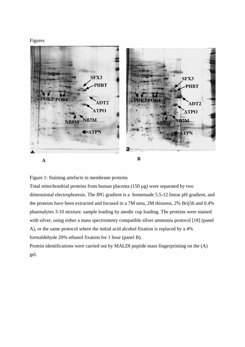
However, in this latter example of inner mitochondrial membrane transporters, there is
clearly a superposition of a detection problem and of a solubilization problem. The detection
problem is highlighted by the fact that some membrane proteins are detection by the MS-
incompatible protocol and not by the MS-compatible protocol. In this case, excision on the
gels stained with the MS-compatible protocol of gel pieces that are unstained but
superposable to stained areas in the gel stained with the MS-incompatible protocol led to
Page 5
positive protein identification (e.g. ADT2, SFX3). This is indicative of a detection problem.
However, excision of other unstained gels pieces, in the areas where other missing
transporters (e.g. phosphate transporter, glutamate-malate shuttle) should be (from their
calculated pI and Mw), did not yield any additional identifications, showing that protein
solubility in 2D gels is still a key issue, and needed to be improved.
3. Improvements of the standard 2D electrophoresis technique
The constraints induced by the IEF separation are rather strict: low ionic strength, no ionic
detergent in the gel, and low amounts of ionic detergent in the sample. Consequently, there
are only two parameters on which the experimentator can play with to increase the solubility
o proteins are the nature and concentrations of chaotropes and the nature and concentrations
of detergents. Both the chaotrope and detergent can be a single compound or a mixture of
various compounds. The historical chaotrope in IEF is urea, as this is an efficient one and the
only one to be compatible with acrylamide polymerization. However, the above-mentioned
corpus of knowledge had shown that urea alone, whatever nonionic detergent it was used
with, was poorly efficient for the solubilization of membrane proteins.
The situation was improved with the introduction of thiourea as an ancillary chaotrope in
addition to urea [19]. While this initial report was purely qualitative and not related to
membrane proteins, dedicated studies investigating several types of detergents in
combination with a urea-thiourea chaotrope were carried out on various membrane systems.
As a matter of facts, the change in chaotrope dramatically affected the solubilizing power of
even "mild" non ionic detergents, as shown by the work carried out on fat globules in human
milk [20]. While the combination of urea and Triton X100 leaded to poor patterns, a
combination of urea-thiourea and Triton X 100 solubilized and focused human butyrophilin,
which is a protein with a transmembrane domain.
This chaotropic combination was tested with various types of detergents, including
amidosulfobetaines [21] and other types of sulfobetaines [22], [23]. For this class of
detergents, a detailed structure-efficiency study was carried out [24], and solubilization of
bona fide intrinsic membrane proteins was demonstrated, including GPCR with seven
transmembrane domains [17], red blood cell membrane proteins with various transmembrane
domains, including the Band III protein with 12 transmembrane domains [22] which was
reluctant to solubilization so far [12], or aquaporins and proton ATPase [21], [23], or
mitochondrial transporters [25].
Page 6
However, sulfobetaines were not the only type of detergent which proved useful. Nonionic
detergents of the oligooxyethylene group proved efficient on the same proteins [26].
Detergents of the glucoside type also proved useful [26], and were able to solubilize
transmembrane proteins from myelin [27]. Finally zwitterionic detergents of the
phosphocholine type were also tested and able to solubilize membrane proteins from muscle
[28].
These positive results prompted several groups to investigate the possibility to analyze
membrane proteins by 2D electrophoresis on more complex systems, either from plants (e.g.
[29]) or on animals (e.g. [30, 31]). However, it is fair to say that the results were generally
considered as disappointing. While it is true that the improved methods did allow to visualize
some membrane proteins (e.g. in [32]), the general situation is that many membrane proteins
are missing [33]. While this impression was first based on previous knowledge (e.g. in [33]),
the concomitant use of proteomics strategies not based on 2D gels, such as shotgun strategies
[34] or strategies based on 1D SDS gels [35], made obvious that many hydrophobic proteins
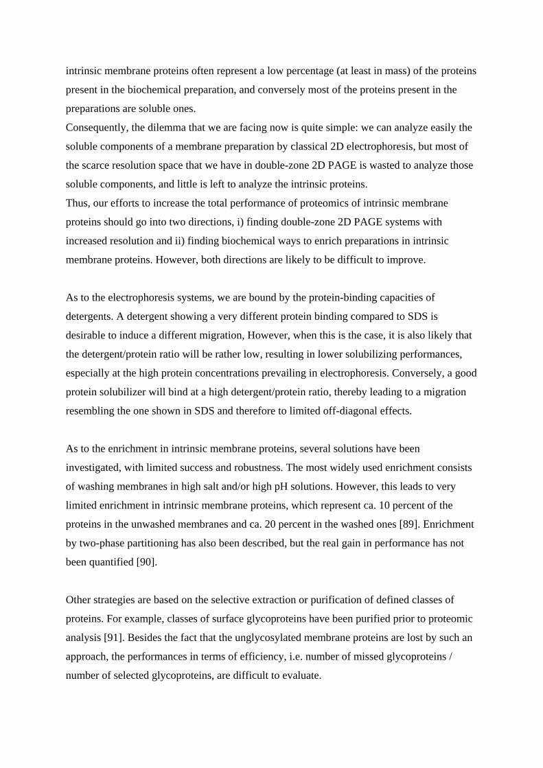
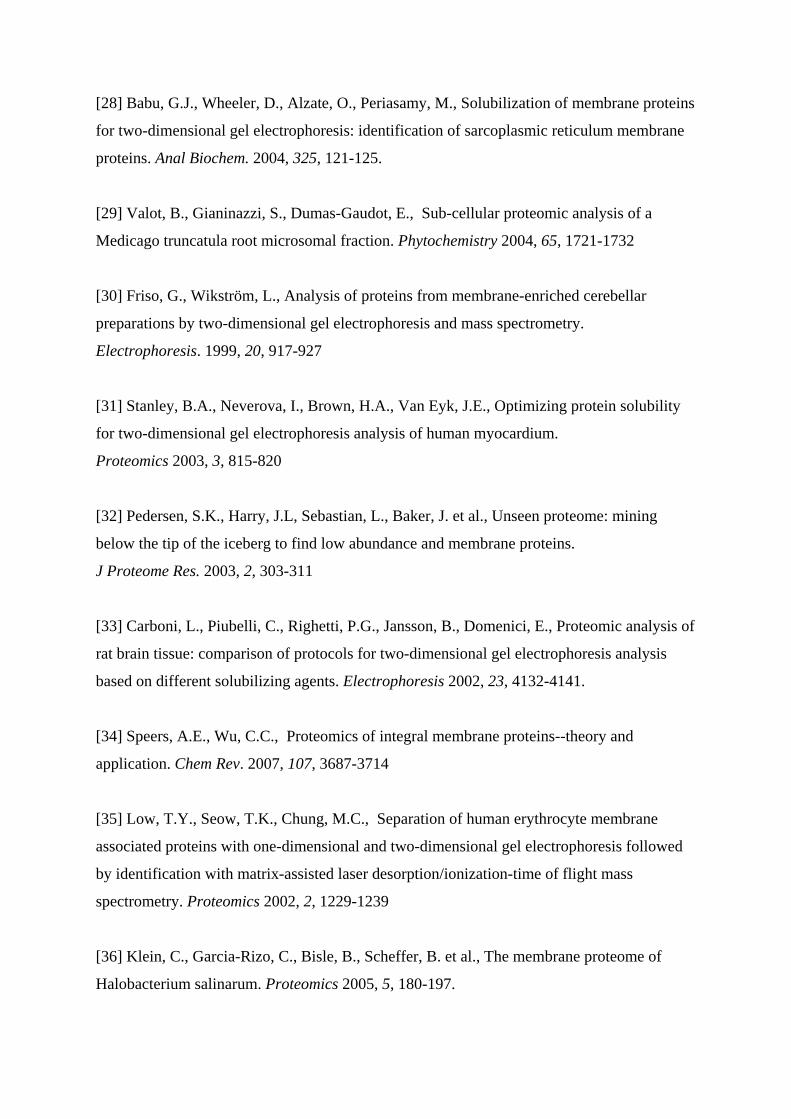
are missing on classical 2D gels [36]. To sum up the situation with the example of the P450
cytochromes, the improvements made on protein solubilization for IEF allowed to go from
none [37] to some (see figure 2), but these are obviously too few.
A happy exception to this rule lies in the bacterial membrane proteins, and especially in the
proteins of the outer membrane (OMPs) of Gram- bacteria [40]. These proteins are
essentially of the porin type, and their transmembrane part is not made of helices but of a beta
barrel. Once properly denatured, these proteins are fairly soluble in the conditions prevailing
in IEF, and can thus be analyzed with high resolution. this is true for bacterial porins [41] but
also for eukaryotic porins [42].
Consequently, proteomics based on 2D gels has been widely applied to bacterial membrane
proteomics. While the success has been limited for Gram+ bacteria [43, 44], where there is
only one membrane with most proteins spanning the membrane by helices, much more work
has been devoted to Gram- bacteria, with greater success such as the identification of a new
OMP [45], the study of E. coli either from the basic microbiology point of view [46] or from
the side pathogenic bacterial strains [47, 48], and more generally the study of various Gram-
organisms of interest in various areas [49-57].
However, this exception shall not mask the general rule, which is that proteins having
Page 7
multiple transmembrane helices generally escape analysis by IEF-based two-dimensional
electrophoresis [58]. The presence of lipids was claimed to be deleterious [59], but
delipidation with organic solvents did not induce a major improvement in the solubilization
of membrane proteins in classical 2D PAGE [33]. It was soon demonstrated that isoelectric
precipitation is the major phenomenon to blame [60], which led to the attempt to solubilize
the isoelectrically-precipitated proteins with hot SDS [61]. Unfortunately, this method did not
really solve the problem, so that other solutions had to be sought.
4. 2D electrophoresis without IEF
As it appears that limited solubility of the membrane proteins is the main limiting factor for
their analysis by classical 2D PAGE, alternate methods must be found. In this respect, the
contrast between the poor solubilizing performances under IEF conditions and the excellent
ones in SDS PAGE gives valuable insights into the directions to be followed for dedicated
solubilization systems for electrophoretic separation of membrane proteins. Of course,
membrane proteins must be separated in the presence of detergents to cover the hydrophobic
parts of the protein. But in addition, the electrostatic repulsion between molecules must be
maximal to prevent aggregation. To this purpose, it is often advantageous to add extraneous
charges to proteins via a charged protein-binding agent. Consequently, two main types of 2D
gels can be used for separating membrane proteins:
In the first type, the first dimension uses native electrophoresis of membrane proteins and/or
membrane complexes, generally with a charge-modifying agent. This concept, which traces
back very early in electrophoresis and has been reviewed previously [62], has been refined
and further developed more recently [63] and will be reviewed in another article of this issue
[64].
In the second type, the first dimension separates denatured proteins, and in this case, the
denaturing agent is most often also the charge transfer agent, and is made of an ionic
detergent. Of course, it would be of little interest to use twice the simple SDS-PAGE
technique, as the proteins would simply lay on the diagonal of the gel. Thus, the optimal
system in the first dimension should offer a separation as different as possible from SDS
PAGE, while keeping the high loading capacity and high solubilizing power of SDS PAGE.
Among the various electrophoretic systems designed to date, the urea-16BAC system
Page 8
originally devised by MacFarlane [65] has these desirable features. It shows a high loading
capacity [66], while also showing a very different migration when compared to SDS [67]. Its
ability to separate bona fide integral membrane proteins and thus its utility in membrane
proteomics was demonstrated rather early [68], and when it became obvious that IEF-based
2D gels did not show adequate solubilization performances, this system became an obvious
choice, as shown for example on bacterial membrane proteins [69]. It therefore received a lot
of applications in various fields, spanning from bacterial proteins [70, 71], to yeast [72], to
mammalian cells or tissues [73, 74] or to subcellular membranes [75].
Thus, this system, as well as closely related ones using other cationic detergents such as
CTAB instead of 16-BAC [76] have gained increased popularity in membrane proteomics.
Compared to the initial description [65], the newest versions do not use the staining between
the two dimensions, but in a simple equilibration in the SDS-containing buffer [76].
However, the cationic system is not as straightforward to use and not as versatile as SDS
PAGE.
For example, the polymerization of gels at low pH cannot be achieved by the classical and
robust TEMED/persulfate system, and the more delicate ascorbate/ferrous ion/hydrogen
peroxide system is often used. Alternatively, the methylene blue-based photopolymerization
system [77], which has been shown to be efficient with other types of acidic, urea-containing
gels [78], can be used to polymerize the acidic first-dimension gels [79].
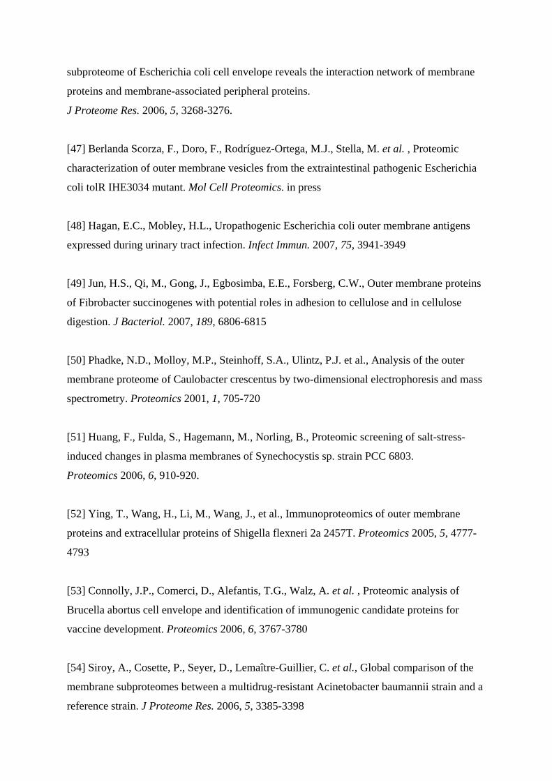
In addition, and oppositely to SDS PAGE, the performance of the urea-cationic detergents
systems is very sensitive to the pH of the separating gels, as shown on figure 3.
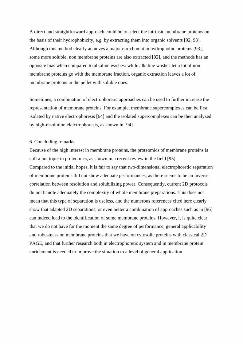
This suggests that the solubilization is not fully driven by the cationic detergent, but rather
both by the native charge of the proteins and the charges added by the detergent. This
suggests in turn that the cationic detergents are less efficient than SDS for the solubilization
of proteins, so that it can be expected that some membrane proteins are soluble in SDS-
containing media and not in cationic detergents-containing media. This assumption has
recently been demonstrated as true [82].
Thus, if optimal membrane protein solubilization is required, SDS must be used in both
dimensions. Consequently, some tricks must be found to reach somewhat different
separations in the two SDS-based separations. This can be achieved to some extent by
changing the buffer system from one dimension to another, as this alters the resolution of the
system [83]. To further enhance this differential mobility effect, nonionic modifyers can be
Page 9
used, such as glycerol [84] or urea [85]. Both approaches have been shown to solubilize
adequately membrane proteins [82], [86], and the urea-based approach has been shown to be
superior to the cationic detergents-based approaches in terms of hydrophobic proteins
solubilization [82].
However, this comparison also showed that the resolution on 2D gels, in terms of spot
spreading on the gels, clearly ranges in the order: double SDS techniques < cationic/anionic
detergent techniques << IEF-based techniques. While this was easily predictable on the basis
of the interdependency of the separation principles used for the two-dimensions of the
system, this is not without consequences on how we can use these electrophoretic systems
optimally in proteomics studies and on how we can improve their use.
5. Future prospects
One of the great strengths of classical, IEF-based 2D electrophoresis is its very high
resolution. It allows to separate many post-translational variants, to separate spots enough so
that image analysis is feasible to follow spots variations between various conditions
investigated, and finally to make the basic assumption that one spot contains most often a
single protein, or at least a single dominating protein, so that the spot volume changes can be
attributed to the change in abundance of this protein.
When switching to 2D systems based only on detergent zone electrophoresis, the loss of
resolution impacts the various features quite differently. While the ability of separating
simple post-translational variants is irremediably lost because of the separation principles at
play, the factors linked to spot crowding are impacted both by the resolution of the gel system
and by the complexity of the sample.
Positional variability, i.e. the difficulties encountered because of gel to gel variations in spot
positions, are of course more severe on crowded gels where only a fraction of the gel space is
used to display proteins. These difficulties can be dealt with by multiplexing, i.e. by labeling
several samples with different fluorescent probes and by comigrating them in a single gel
[87]. this approach has been applied successfully to cationic/anionic 2D PAGE (e.g. in [88]
).
However, the other problem linked to spot crowding, i.e. fusion of several proteins in a
single, average spot, cannot be solved easily, so that we are currently facing a difficult
situation. So-called membrane preparations contain both intrinsic membrane proteins and
more soluble proteins associated to membrane by various mechanisms. As a matter of facts,
Page 10
intrinsic membrane proteins often represent a low percentage (at least in mass) of the proteins
present in the biochemical preparation, and conversely most of the proteins present in the
preparations are soluble ones.
Consequently, the dilemma that we are facing now is quite simple: we can analyze easily the
soluble components of a membrane preparation by classical 2D electrophoresis, but most of
the scarce resolution space that we have in double-zone 2D PAGE is wasted to analyze those
soluble components, and little is left to analyze the intrinsic proteins.
Thus, our efforts to increase the total performance of proteomics of intrinsic membrane
proteins should go into two directions, i) finding double-zone 2D PAGE systems with
increased resolution and ii) finding biochemical ways to enrich preparations in intrinsic
membrane proteins. However, both directions are likely to be difficult to improve.
As to the electrophoresis systems, we are bound by the protein-binding capacities of
detergents. A detergent showing a very different protein binding compared to SDS is
desirable to induce a different migration, However, when this is the case, it is also likely that
the detergent/protein ratio will be rather low, resulting in lower solubilizing performances,
especially at the high protein concentrations prevailing in electrophoresis. Conversely, a good
protein solubilizer will bind at a high detergent/protein ratio, thereby leading to a migration
resembling the one shown in SDS and therefore to limited off-diagonal effects.
As to the enrichment in intrinsic membrane proteins, several solutions have been
investigated, with limited success and robustness. The most widely used enrichment consists
of washing membranes in high salt and/or high pH solutions. However, this leads to very
limited enrichment in intrinsic membrane proteins, which represent ca. 10 percent of the
proteins in the unwashed membranes and ca. 20 percent in the washed ones [89]. Enrichment
by two-phase partitioning has also been described, but the real gain in performance has not
been quantified [90].
Other strategies are based on the selective extraction or purification of defined classes of
proteins. For example, classes of surface glycoproteins have been purified prior to proteomic
analysis [91]. Besides the fact that the unglycosylated membrane proteins are lost by such an
approach, the performances in terms of efficiency, i.e. number of missed glycoproteins /
number of selected glycoproteins, are difficult to evaluate.
Page 11
A direct and straightforward approach could be to select the intrinsic membrane proteins on
the basis of their hydrophobicity, e.g. by extracting them into organic solvents [92, 93].
Although this method clearly achieves a major enrichment in hydrophobic proteins [93],
some more soluble, non membrane proteins are also extracted [92], and the methods has an
opposite bias when compared to alkaline washes: while alkaline washes let a lot of non
membrane proteins go with the membrane fraction, organic extraction leaves a lot of
membrane proteins in the pellet with soluble ones.
Sometimes, a combination of electrophoretic approaches can be used to further increase the
representation of membrane proteins. For example, membrane supercomplexes can be first
isolated by native electrophoresis [64] and the isolated supercomplexes can be then analyzed
by high-resolution elelctrophoresis, as shown in [94]
6. Concluding remarks
Because of the high interest in membrane proteins, the proteomics of membrane proteins is
still a hot topic in proteomics, as shown in a recent review in the field [95]
Compared to the initial hopes, it is fair to say that two-dimensional electrophoretic separation
of membrane proteins did not show adequate performances, as there seems to be an inverse
correlation between resolution and solubilizing power. Consequently, current 2D protocols
do not handle adequately the complexity of whole membrane preparations. This does not
mean that this type of separation is useless, and the numerous references cited here clearly
show that adapted 2D separations, or even better a combination of approaches such as in [96]
can indeed lead to the identification of some membrane proteins. However, it is quite clear
that we do not have for the moment the same degree of performance, general applicability
and robustness on membrane proteins that we have on cytosolic proteins with classical 2D
PAGE, and that further research both in electrophoretic system and in membrane protein
enrichment is needed to improve the situation to a level of general application.
Page 12
References
[1] Ames, G.F., Nikaido, K., Two-dimensional gel electrophoresis of membrane proteins.
Biochemistry 1976, 15, 616-623
[2] Rabilloud, T., Solubilization of proteins for electrophoretic analyses.
Electrophoresis 1996, 17, 813-829
[3] Perdew, G.H., Schaup, H.W., Selivonchick, D.P., The use of a zwitterionic detergent in
two-dimensional gel electrophoresis of trout liver microsomes. Anal Biochem. 1983, 135,
453-455
[4] Gyenes, T., Gyenes, E., Effect of "stacking" on the resolving power of ultrathin-layer
two-dimensional gel electrophoresis. Anal Biochem. 1987, 165, 155-160
[5] Rabilloud, T., Gianazza, E., Cattò, N., Righetti, P.G., Amidosulfobetaines, a family of
detergents with improved solubilization properties: application for isoelectric focusing under
denaturing conditions. Anal Biochem. 1990, 185, 94-102
[6] Witzmann, F., Jarnot, B. and Parker, D., Dodecyl maltoside detergent improves resolution
of hepatic membrane proteins in two-dimensional gels. Electrophoresis 1991 12, 687-688
[7] Felsted, R.L., Gupta, S.K., Glover, C.J., Fischkoff, S.A., Gallagher, R.E., Cell surface
membrane protein changes during the differentiation of cultured human promyelocytic
leukemia HL-60 cells. Cancer Res. 1983, 43, 2754-2761
[8] Lüddens, H., Havsteen, B., Characterization of the porcine ACTH receptor with the aid of
a monoclonal antibody. Biol Chem Hoppe Seyler 1986, 367, 539-547
[9] Sweetnam, P., Nestler, E., Gallombardo, P., Brown, S. et al., Comparison of the
molecular structure of GABA/benzodiazepine receptors purified from rat and human
cerebellum. Brain Res. 1987, 388, 223-233
Page 13
[10] Alla, S.A., Buschko, J., Quitterer, U., Maidhof, A. et al., Structural features of the
human bradykinin B2 receptor probed by agonists, antagonists, and anti-idiotypic antibodies.
J Biol Chem. 1993, 268, 17277-17285
[11] Englund, A.K., Lundahl, P., The isoelectric point of the human red cell glucose
transporter. Biochim Biophys Acta 1991, 1065, 185-194
[12] Rubin, R.W., Milikowski, C., Over two hundred polypeptides resolved from the human
erythrocyte membrane. Biochim Biophys Acta 1978, 509, 100-110
[13] Santoni, V., Molloy, M., Rabilloud, T., Membrane proteins and proteomics: un amour
impossible? Electrophoresis 2000, 21, 1054-1070
[14] Santoni, V., Doumas, P., Rouquié, D., Mansion, M. et al., Large scale characterization
of plant plasma membrane proteins. Biochimie 1999, 81, 655-661
[15] Adessi, C., Miege, C., Albrieux, C., Rabilloud, T., Two-dimensional electrophoresis of
membrane proteins: a current challenge for immobilized pH gradients. Electrophoresis 1997,
18, 127-35
[16] Wilkins, M.R., Gasteiger, E., Sanchez, J.C., Bairoch, A., Hochstrasser, D.F., Two-
dimensional gel electrophoresis for proteome projects: the effects of protein hydrophobicity
and copy number. Electrophoresis 1998, 19, 1501-1505
[17] Henningsen, R., Gale, B.L., Straub, K.M., DeNagel, D.C., Application of zwitterionic
detergents to the solubilization of integral membrane proteins for two-dimensional gel
electrophoresis and mass spectrometry. Proteomics 2002, 2, 1479-1488.
[18] Chevallet, M., Diemer, H., Luche, S., van Dorsselaer, A. et al. , Improved mass
spectrometry compatibility is afforded by ammoniacal silver staining. Proteomics 2006, 6,
2350-2354.
[19] Rabilloud, T., Adessi, C., Giraudel, A., Lunardi, J., Improvement of the solubilization of
proteins in two-dimensional electrophoresis with immobilized pH gradients. Electrophoresis
Page 14
1997, 18, 307-316
[20] Quaranta, S., Giuffrida, M.G., Cavaletto, M., Giunta, C. et al., Human proteome
enhancement: high-recovery method and improved two-dimensional map of colostral fat
globule membrane proteins. Electrophoresis 2001, 22, 1810-1818
[21] Chevallet, M., Santoni, V., Poinas, A., Rouquié, D. et al., New zwitterionic detergents
improve the analysis of membrane proteins by two-dimensional electrophoresis.
Electrophoresis 1998, 19, 1901-1909
[22] Rabilloud, T., Blisnick, T., Heller, M., Luche, S. et al., Analysis of membrane proteins
by two-dimensional electrophoresis: comparison of the proteins extracted from normal or
Plasmodium falciparum-infected erythrocyte ghosts. Electrophoresis 1999, 20, 3603-3610.
[23] Santoni, V., Kieffer, S., Desclaux, D., Masson, F., Rabilloud, T., Membrane proteomics:
use of additive main effects with multiplicative interaction model to classify plasma
membrane proteins according to their solubility and electrophoretic properties.
Electrophoresis 2000, 21, 3329-3344.
[24] Tastet, C., Charmont, S., Chevallet, M., Luche, S., Rabilloud, T., Structure-efficiency
relationships of zwitterionic detergents as protein solubilizers in two-dimensional
electrophoresis. Proteomics 2003, 3, 111-121.
[25] Lescuyer, P., Strub, J.M., Luche, S., Diemer, H. et al., Progress in the definition of a
reference human mitochondrial proteome. Proteomics 2003, 3, 157-167
[26] Luche, S., Santoni, V., Rabilloud, T., Evaluation of nonionic and zwitterionic detergents
as membrane protein solubilizers in two-dimensional electrophoresis.
Proteomics 2003, 3, 249-253.
[27] Taylor, C.M. and Pfeiffer, S.E., Enhanced resolution of glycosylphosphatidylinositol-
anchored and transmembrane proteins from the lipid-rich myelin membrane by two-
dimensional gel electrophoresis. Proteomics 2003, 3, 1303-1312.
Page 15
[28] Babu, G.J., Wheeler, D., Alzate, O., Periasamy, M., Solubilization of membrane proteins
for two-dimensional gel electrophoresis: identification of sarcoplasmic reticulum membrane
proteins. Anal Biochem. 2004, 325, 121-125.
[29] Valot, B., Gianinazzi, S., Dumas-Gaudot, E., Sub-cellular proteomic analysis of a
Medicago truncatula root microsomal fraction. Phytochemistry 2004, 65, 1721-1732
[30] Friso, G., Wikström, L., Analysis of proteins from membrane-enriched cerebellar
preparations by two-dimensional gel electrophoresis and mass spectrometry.
Electrophoresis. 1999, 20, 917-927
[31] Stanley, B.A., Neverova, I., Brown, H.A., Van Eyk, J.E., Optimizing protein solubility
for two-dimensional gel electrophoresis analysis of human myocardium.
Proteomics 2003, 3, 815-820
[32] Pedersen, S.K., Harry, J.L, Sebastian, L., Baker, J. et al., Unseen proteome: mining
below the tip of the iceberg to find low abundance and membrane proteins.
J Proteome Res. 2003, 2, 303-311
[33] Carboni, L., Piubelli, C., Righetti, P.G., Jansson, B., Domenici, E., Proteomic analysis of
rat brain tissue: comparison of protocols for two-dimensional gel electrophoresis analysis
based on different solubilizing agents. Electrophoresis 2002, 23, 4132-4141.
[34] Speers, A.E., Wu, C.C., Proteomics of integral membrane proteins--theory and
application. Chem Rev. 2007, 107, 3687-3714
[35] Low, T.Y., Seow, T.K., Chung, M.C., Separation of human erythrocyte membrane
associated proteins with one-dimensional and two-dimensional gel electrophoresis followed
by identification with matrix-assisted laser desorption/ionization-time of flight mass
spectrometry. Proteomics 2002, 2, 1229-1239
[36] Klein, C., Garcia-Rizo, C., Bisle, B., Scheffer, B. et al., The membrane proteome of
Halobacterium salinarum. Proteomics 2005, 5, 180-197.
Page 16
[37] Galeva, N., Altermann, M., Comparison of one-dimensional and two-dimensional gel
electrophoresis as a separation tool for proteomic analysis of rat liver microsomes:
Cytochromes P450 and other membrane proteins Proteomics 2002, 2, 713-722
[38] Gianazza, E., Celentano, F., Magenes, S., Ettori, C., Righetti, P.G., Formulations for
immobilized pH gradients including pH extremes. Electrophoresis 1989, 10, 806-808.
[39] Richert, S., Luche, S., Chevallet, M., Van Dorsselaer, A. et al. , About the mechanism of
interference of silver staining with peptide mass spectrometry.
Proteomics 2004, 4, 909-916.
[40] Molloy, M.P., Phadke, N.D., Maddock, J.R., Andrews, P.C., Two-dimensional
electrophoresis and peptide mass fingerprinting of bacterial outer membrane proteins.
Electrophoresis 2001, 22, 1686-1696
[41] Molloy, M.P., Herbert, B.R., Slade, M.B., Rabilloud, T. et al., Proteomic analysis of the
Escherichia coli outer membrane. Eur J Biochem. 2000, 267, 2871-2881
[42] Liberatori, S., Canas, B., Tani, C., Bini, L. et al., Proteomic approach to the
identification of voltage-dependent anion channel protein isoforms in guinea pig brain
synaptosomes. Proteomics 2004, 4, 1335-1340
[43] Nandakumar, R., Nandakumar, M.P., Marten, M.R., Ross, J.M., Proteome analysis of
membrane and cell wall associated proteins from Staphylococcus aureus. J Proteome Res.
2005, 4, 250-257.
[44] Gatlin, C.L., Pieper, R., Huang, S.T., Mongodin, E. et al., Proteomic profiling of cell
envelope-associated proteins from Staphylococcus aureus. Proteomics 2006, 6, 1530-1549.
[45] Veith, P.D., Talbo, G.H., Slakeski, N., Reynolds, E.C., Identification of a novel
heterodimeric outer membrane protein of Porphyromonas gingivalis by two-dimensional gel
electrophoresis and peptide mass fingerprinting. Eur J Biochem. 2001, 268, 4748-4757
[46] Huang, C.Z., Lin, X.M., Wu, L.N., Zhang, D.F. et al., Systematic identification of the
Page 17
subproteome of Escherichia coli cell envelope reveals the interaction network of membrane
proteins and membrane-associated peripheral proteins.
J Proteome Res. 2006, 5, 3268-3276.
[47] Berlanda Scorza, F., Doro, F., Rodríguez-Ortega, M.J., Stella, M. et al. , Proteomic
characterization of outer membrane vesicles from the extraintestinal pathogenic Escherichia
coli tolR IHE3034 mutant. Mol Cell Proteomics. in press
[48] Hagan, E.C., Mobley, H.L., Uropathogenic Escherichia coli outer membrane antigens
expressed during urinary tract infection. Infect Immun. 2007, 75, 3941-3949
[49] Jun, H.S., Qi, M., Gong, J., Egbosimba, E.E., Forsberg, C.W., Outer membrane proteins
of Fibrobacter succinogenes with potential roles in adhesion to cellulose and in cellulose
digestion. J Bacteriol. 2007, 189, 6806-6815
[50] Phadke, N.D., Molloy, M.P., Steinhoff, S.A., Ulintz, P.J. et al., Analysis of the outer
membrane proteome of Caulobacter crescentus by two-dimensional electrophoresis and mass
spectrometry. Proteomics 2001, 1, 705-720
[51] Huang, F., Fulda, S., Hagemann, M., Norling, B., Proteomic screening of salt-stress-
induced changes in plasma membranes of Synechocystis sp. strain PCC 6803.
Proteomics 2006, 6, 910-920.
[52] Ying, T., Wang, H., Li, M., Wang, J., et al., Immunoproteomics of outer membrane
proteins and extracellular proteins of Shigella flexneri 2a 2457T. Proteomics 2005, 5, 4777-
4793
[53] Connolly, J.P., Comerci, D., Alefantis, T.G., Walz, A. et al. , Proteomic analysis of
Brucella abortus cell envelope and identification of immunogenic candidate proteins for
vaccine development. Proteomics 2006, 6, 3767-3780
[54] Siroy, A., Cosette, P., Seyer, D., Lemaître-Guillier, C. et al., Global comparison of the
membrane subproteomes between a multidrug-resistant Acinetobacter baumannii strain and a
reference strain. J Proteome Res. 2006, 5, 3385-3398
Page 18
[55] Peng, X., Xu, C., Ren, H., Lin, X. et al., Proteomic analysis of the sarcosine-insoluble
outer membrane fraction of Pseudomonas aeruginosa responding to ampicilin, kanamycin,
and tetracycline resistance. J Proteome Res. 2005, 4, 2257-2265.
[56] Berven, F.S., Karlsen, O.A., Straume, A.H., Flikka, K. et al., Analysing the outer
membrane subproteome of Methylococcus capsulatus (Bath) using proteomics and novel
biocomputing tools. Arch Microbiol. 2006, 184, 362-377
[57] Aivaliotis, M., Haase, W., Karas, M., Tsiotis, G., Proteomic analysis of chlorosome-
depleted membranes of the green sulfur bacterium Chlorobium tepidum. Proteomics 2006, 6,
217-232
[58] Peirce, M.J., Wait, R., Begum, S., Saklatvala, J., Cope, A.P., Expression profiling of
lymphocyte plasma membrane proteins. Mol Cell Proteomics 2004, 3, 56-65
[59] Mastro, R., Hall, M.,Protein delipidation and precipitation by tri-n-butylphosphate,
acetone, and methanol treatment for isoelectric focusing and two-dimensional gel
electrophoresis. Anal Biochem. 1999, 273, 313-315
[60] Coughenour, H.D., Spaulding, R.S., Thompson, C.M., The synaptic vesicle proteome: a
comparative study in membrane protein identification. Proteomics 2004, 4, 3141-3155.
[61] McDonough, J., Marban, E., Optimization of IPG strip equilibration for the basic
membrane protein mABC1 Proteomics 2005, 5, 2892-2895
[62] Hjelmeland, L.M., Chrambach, A., Electrophoresis and electrofocusing in detergent-
containing media: a disussion of basic concepts Electrophoresis 1981, 2, 1-11
[63] Schägger, H., von Jagow G., Blue native electrophoresis for isolation of membrane
protein complexes in enzymatically active form. Anal Biochem. 1991, 199, 223-231.
[64] Schägger, H., et al. this volume
Page 19
[65] MacFarlane, D.E., Use of benzyldimethyl-n-hexadecylammonium chloride ("16-BAC"),
a cationic detergent, in an acidic polyacrylamide gel electrophoresis system to detect base
labile protein methylation in intact cells. Anal Biochem. 1983, 132, 231-235.
[66] MacFarlane, D.E., Two dimensional benzyldimethyl-n-hexadecylammonium chloride-
sodium dodecyl sulfate preparative polyacrylamide gel electrophoresis: a high capacity high
resolution technique for the purification of proteins from complex mixtures. Anal Biochem.
1989, 176, 457-463
[67] Lopez, M.F., Patton, W.F., Utterback, B.L., Chung-Welch, N. et al., Effect of various
detergents on protein migration in the second dimension of two-dimensional gels.
Anal Biochem. 1991, 199, 35-44
[68] Hartinger, J., Stenius, K., Högemann, D., Jahn, R., 16-BAC/SDS-PAGE: a two-
dimensional gel electrophoresis system suitable for the separation of integral membrane
proteins. Anal Biochem. 1996, 240, 126-133.
[69] Bunai, K., Yamane, K., Effectiveness and limitation of two-dimensional gel
electrophoresis in bacterial membrane protein proteomics and perspectives.
J Chromatogr B 2005, 815, 227-236
[70] Bunai, K., Ariga, M., Inoue, T., Nozaki, M. et al., Profiling and comprehensive
expression analysis of ABC transporter solute-binding proteins of Bacillus subtilis membrane
based on a proteomic approach. Electrophoresis 2004, 25, 141-155
[71] Bisle, B., Schmidt, A., Scheibe, B., Klein, C.et al., Quantitative profiling of the
membrane proteome in a halophilic archaeon. Mol Cell Proteomics. 2006, 5, 1543-1558
[72] Navarre, C., Degand, H., Bennett, K.L., Crawford, J.S., et al., Subproteomics:
identification of plasma membrane proteins from the yeast Saccharomyces cerevisiae.
Proteomics 2002, 2, 1706-1714.
[73] Moebius, J., Zahedi, R.P., Lewandrowski, U., Berger, C. et al., The human platelet
membrane proteome reveals several new potential membrane proteins. Mol Cell Proteomics.
Page 20
2005, 4, 1754-1761
[74] Bierczynska-Krzysik, A., Kang, S.U., Silberrring, J., Lubec, G., Mass spectrometrical
identification of brain proteins including highly insoluble and transmembrane proteins.
Neurochem Int. 2006, 49, 245-255
[75] Zahedi, R.P., Meisinger, C., Sickmann, A., Two-dimensional benzyldimethyl-n-
hexadecylammonium chloride/SDS-PAGE for membrane proteomics. Proteomics 2005, 5,
3581-3588.
[76] Helling, S., Schmitt, E., Joppich, C., Schulenborg, T. et al., 2-D differential membrane
proteome analysis of scarce protein samples. Proteomics 2006, 6, 4506-4513.
[77] Lyubimova, T., Caglio, S., Gelfi, C., Righetti, P.G., Rabilloud, T., Photopolymerization
of polyacrylamide gels with methylene blue. Electrophoresis. 1993, 14, 40-50.
[78] Rabilloud, T., Girardot, V., Lawrence, J.J., One- and two-dimensional histone
separations in acidic gels: usefulness of methylene blue-driven photopolymerization.
Electrophoresis. 1996, 17, 67-73.
[79] Buxbaum, E., Cationic electrophoresis and electrotransfer of membrane glycoproteins.
Anal Biochem. 2003, 314, 70-76.
[80] Mócz, G., Bálint, M., Use of cationic detergents for polyacrylamide gel electrophoresis
in multiphasic buffer systems. Anal Biochem. 1984, 143, 283-292.
[81] Paulson, J.R., Mesner, P.W., Delrow, J.J., Mahmoud, N.N., Ciesielski, W.A., Rapid
analysis of mitotic histone H1 phosphorylation by cationic disc electrophoresis at neutral pH
in minigels. Anal Biochem. 1992, 203, 227-234
[82] Burré, J., Beckhaus, T., Schägger, H., Corvey, C. et al., Analysis of the synaptic vesicle
proteome using three gel-based protein separation techniques. Proteomics. 2006, 6, 6250-
6262.
Page 21
[83] Patton, W.F., Chung-Welch, N., Lopez, M.F., Cambria, R.P., et al., Tris-tricine and
Tris-borate buffer systems provide better estimates of human mesothelial cell intermediate
filament protein molecular weights than the standard Tris-glycine system.
Anal Biochem. 1991, 197, 25-33
[84] Williams, T.I., Combs, J.C., Thakur, A.P., Strobel, H.J., Lynn, B.C., A novel Bicine
running buffer system for doubled sodium dodecyl sulfate - polyacrylamide gel
electrophoresis of membrane proteins. Electrophoresis. 2006, 27, 2984-2995
[85] Rais, I., Karas, M., Schägger, H., Two-dimensional electrophoresis for the isolation of
integral membrane proteins and mass spectrometric identification.
Proteomics. 2004, 4, 2567-2571
[86] Williams, T.I., Combs, J.C., Lynn, B.C., Strobel, H.J., Proteomic profile changes in
membranes of ethanol-tolerant Clostridium thermocellum. Appl Microbiol Biotechnol. 2007,
74, 422-432
[87] Unlü, M., Morgan, M.E., Minden, J.S., Difference gel electrophoresis: a single gel
method for detecting changes in protein extracts. Electrophoresis. 1997, 18, 2071-2077.
[88] Burré, J., Beckhaus, T., Corvey, C., Karas, M. et al., Synaptic vesicle proteins under
conditions of rest and activation: analysis by 2-D difference gel electrophoresis.
Electrophoresis 2006, 27, 3488-3496
[89] Schindler, J., Jung, S., Niedner-Schatteburg, G., Friauf, E., Nothwang, H.G., Enrichment
of integral membrane proteins from small amounts of brain tissue.
J Neural Transm. 2006, 113, 995-1013
[90] Everberg, H., Sivars, U., Emanuelsson, C., Persson, C. et al., Protein pre-fractionation in
detergent-polymer aqueous two-phase systems for facilitated proteomic studies of membrane
proteins. J Chromatogr A. 2004, 1029, 113-124
[91] Watarai, H., Hinohara, A., Nagafune, J., Nakayama, T., et al., Plasma membrane-
focused proteomics: dramatic changes in surface expression during the maturation of human
Page 22
dendritic cells. Proteomics. 2005, 5, 4001-4011.
[92] Molloy, M.P., Herbert, B.R., Williams, K.L., Gooley, A.A., Extraction of Escherichia
coli proteins with organic solvents prior to two-dimensional electrophoresis.
Electrophoresis 1999, 20, 701-704
[93] Ferro, M., Seigneurin-Berny, D., Rolland, N., Chapel, A., et al., Organic solvent
extraction as a versatile procedure to identify hydrophobic chloroplast membrane proteins.
Electrophoresis 2000, 21, 3517-3526
[94] Werhahn, W., Braun, H.P. Biochemical dissection of the mitochondrial proteome from Arabidopsis thaliana by three-dimensional gel electrophoresis. Electrophoresis 2002, 23, 640-646.
[95] Braun, R.J., Kinkl, N., Beer, M., Ueffing, M. Two-dimensional electrophoresis of membrane proteins. Anal Bioanal Chem. 2007, 389, 1033-1045.
[96] Aivaliotis, M., Karas, M., Tsiotis, G., An alternative strategy for the membrane
proteome analysis of the green sulfur bacterium Chlorobium tepidum using blue native
PAGE and 2-D PAGE on purified membranes. J Proteome Res. 2007, 6, 1048-1058
Page 23
Figures
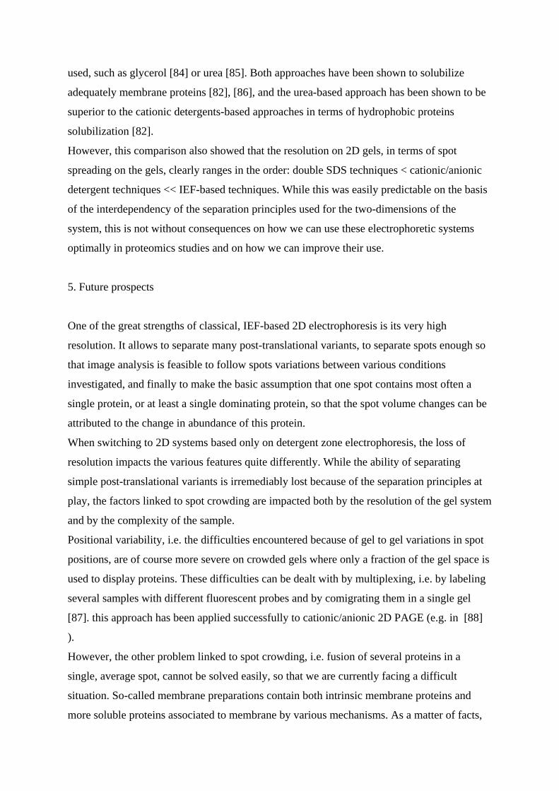
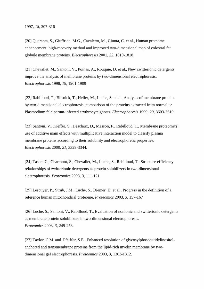
Figure 1: Staining artefacts in membrane proteins
Total mitochondrial proteins from human placenta (150 µg) were separated by two
dimensional electrophoresis. The IPG gradient is a homemade 5.5-12 linear pH gradient, and
the proteins have been extracted and focused in a 7M urea, 2M thiourea, 2% Brij56 and 0.4%
pharmalytes 3-10 mixture. sample loading by anodic cup loading. The proteins were stained
with silver, using either a mass spectrometry compatible silver ammonia protocol [18] (panel
A), or the same protocol where the initial acid alcohol fixation is replaced by a 4%
formaldehyde 20% ethanol fixation for 1 hour (panel B).
Protein identifications were carried out by MALDI peptide mass fingerprinting on the (A)
gel.
Page 24
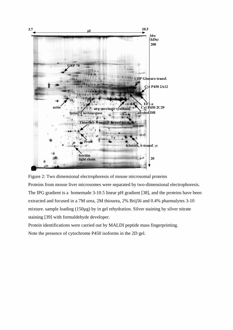
Figure 2: Two dimensional electrophoresis of mouse microsomal proteins
Proteins from mouse liver microsomes were separated by two-dimensional electrophoresis.
The IPG gradient is a homemade 3-10.5 linear pH gradient [38], and the proteins have been
extracted and focused in a 7M urea, 2M thiourea, 2% Brij56 and 0.4% pharmalytes 3-10
mixture. sample loading (150µg) by in gel rehydration. Silver staining by silver nitrate
staining [39] with formaldehyde developer.
Protein identifications were carried out by MALDI peptide mass fingerprinting.
Note the presence of cytochrome P450 isoforms in the 2D gel.
Page 25
Figure 3: cationic/anionic 2D PAGE of bacterial membrane proteins
Membrane proteins from B. subtilis (50µg) were separated by 16BAC/SDS PAGE.
The first dimension, 16BAC gels were run either at pH 2 in the phosphate/glycine system
[65] (panel A), or at pH 5 in the acetate/beta alanine system [80] (panel B), or at pH 7 in the
Hepes/histidine system [81] (panel C). protein detection by silver ammonia staining [18]
Note the increased streaking with increasing running pH