Functional connectivity based parcellation of the human medial temporal lobe Shao-Fang Wang a , Maureen Ritchey a , Laura A. Libby a , Charan Ranganath a,b,⇑ a Center for Neuroscience, University of California, Davis, CA 95618, USA b Department of Psychology, University of California, Davis, CA 95616, USA article info Article history: Received 9 September 2015 Revised 23 December 2015 Accepted 12 January 2016 Available online xxxx Keywords: Functional connectivity Hierarchical clustering algorithm Parcellation Hippocampus Parahippocampal gyrus abstract Regional differences in large-scale connectivity have been proposed to underlie functional specialization along the anterior–posterior axis of the medial temporal lobe (MTL), including the hippocampus (HC) and the parahippocampal gyrus (PHG). However, it is unknown whether functional connectivity (FC) can be used reliably to parcellate the human MTL. The current study aimed to differentiate subregions of the HC and the PHG based on patterns of whole-brain intrinsic FC. FC maps were calculated for each slice along the longitudinal axis of the PHG and the HC. A hierarchical clustering algorithm was then applied to these data in order to group slices according to the similarity of their connectivity patterns. Surprisingly, three discrete clusters were identified in the PHG. Two clusters corresponded to the parahippocampal cortex (PHC) and the perirhinal cortex (PRC), and these regions showed preferential connectivity with previously described posterior-medial and anterior-temporal networks, respectively. The third cluster corresponded to an anterior PRC region previously described as area 36d, and this region exhibited pref- erential connectivity with auditory cortical areas and with a network involved in visceral processing. The three PHG clusters showed different profiles of activation during a memory-encoding task, demonstrat- ing that the FC-based parcellation identified functionally dissociable sub-regions of the PHG. In the hip- pocampus, no sub-regions were identified via the parcellation procedure. These results indicate that connectivity-based methods can be used to parcellate functional regions within the MTL, and they suggest that studies of memory and high-level cognition need to differentiate between PHC, posterior PRC, and anterior PRC. Ó 2016 Elsevier Inc. All rights reserved. 1. Introduction The medial temporal lobe (MTL) region is known to be essential for episodic memory formation (Mishkin, 1978; Scoville & Milner, 1957; Zola-Morgan, Squire, Amaral, & Suzuki, 1989). Studies in humans and animal models have distinguished between memory processes supported by different MTL sub-regions, including the hippocampus (HC) and the adjacent parahippocampal gyrus (PHG) (Aminoff, Kveraga, & Bar, 2013; Brown & Aggleton, 2001; Davachi, 2006; Diana, Yonelinas, & Ranganath, 2007; Eichenbaum, Yonelinas, & Ranganath, 2007). It has further been suggested that the functional differences among the MTL sub- regions are due to their participation in different large-scale brain networks (Kahn, Andrews-Hanna, Vincent, Snyder, & Buckner, 2008; Libby, Ekstrom, Ragland, & Ranganath, 2012; Ranganath & Ritchey, 2012). The perirhinal cortex (PRC), in the anterior PHG, is extensively interconnected with higher-order visual areas (e.g., area TE and area TEO), the insular cortex, the orbitofrontal cortex, and the amygdala. The parahippocampal cortex (PHC) in the posterior PHG, is extensively interconnected with early visual areas (e.g., V4 and V3) in addition to the higher-order visual areas, auditory association areas (e.g. superior temporal gyrus), the retro- splenial cortex, and the posterior parietal cortex. Researchers have also proposed distinctions between the HC regions, given evidence that dorsal/posterior HC is more extensively interconnected with the mammillary bodies, the PHC, and the medial band of the ERC, whereas ventral/anterior HC is more extensively intercon- nected with the amygdala, the medial prefrontal cortex, the PRC, and the lateral band of the ERC (Fanselow & Dong, 2010; Moser & Moser, 1998; Poppenk, Evensmoen, Moscovitch, & Nadel, 2013; Strange, Witter, Lein, & Moser, 2014). Accurately identifying the MTL sub-regions (i.e., the PRC and PHC, posterior and anterior HC) in living human brains is one of the major obstacles in understanding human MTL function. http://dx.doi.org/10.1016/j.nlm.2016.01.005 1074-7427/Ó 2016 Elsevier Inc. All rights reserved. ⇑ Corresponding author at: Center for Neuroscience, 1544 Newton Ct., Davis, CA 95618, USA. E-mail address: [email protected](S.-F. Wang). Neurobiology of Learning and Memory xxx (2016) xxx–xxx Contents lists available at ScienceDirect Neurobiology of Learning and Memory journal homepage: www.elsevier.com/locate/ynlme Please cite this article in press as: Wang, S.-F., et al. Functional connectivity based parcellation of the human medial temporal lobe. Neurobiology of Learning and Memory (2016), http://dx.doi.org/10.1016/j.nlm.2016.01.005
Transcript
Neurobiology of Learning and Memory xxx (2016) xxx–xxx
Contents lists available at ScienceDirect
Neurobiology of Learning and Memory
journal homepage: www.elsevier .com/ locate/ynlme
Functional connectivity based parcellation of the human medialtemporal lobe
http://dx.doi.org/10.1016/j.nlm.2016.01.0051074-7427/� 2016 Elsevier Inc. All rights reserved.
⇑ Corresponding author at: Center for Neuroscience, 1544 Newton Ct., Davis, CA95618, USA.
Please cite this article in press as: Wang, S.-F., et al. Functional connectivity based parcellation of the human medial temporal lobe. Neurobiology of Land Memory (2016), http://dx.doi.org/10.1016/j.nlm.2016.01.005
Shao-Fang Wang a, Maureen Ritchey a, Laura A. Libby a, Charan Ranganath a,b,⇑aCenter for Neuroscience, University of California, Davis, CA 95618, USAbDepartment of Psychology, University of California, Davis, CA 95616, USA
a r t i c l e i n f o a b s t r a c t
Article history:Received 9 September 2015Revised 23 December 2015Accepted 12 January 2016Available online xxxx
Regional differences in large-scale connectivity have been proposed to underlie functional specializationalong the anterior–posterior axis of the medial temporal lobe (MTL), including the hippocampus (HC) andthe parahippocampal gyrus (PHG). However, it is unknown whether functional connectivity (FC) can beused reliably to parcellate the human MTL. The current study aimed to differentiate subregions of the HCand the PHG based on patterns of whole-brain intrinsic FC. FC maps were calculated for each slice alongthe longitudinal axis of the PHG and the HC. A hierarchical clustering algorithm was then applied to thesedata in order to group slices according to the similarity of their connectivity patterns. Surprisingly, threediscrete clusters were identified in the PHG. Two clusters corresponded to the parahippocampal cortex(PHC) and the perirhinal cortex (PRC), and these regions showed preferential connectivity withpreviously described posterior-medial and anterior-temporal networks, respectively. The third clustercorresponded to an anterior PRC region previously described as area 36d, and this region exhibited pref-erential connectivity with auditory cortical areas and with a network involved in visceral processing. Thethree PHG clusters showed different profiles of activation during a memory-encoding task, demonstrat-ing that the FC-based parcellation identified functionally dissociable sub-regions of the PHG. In the hip-pocampus, no sub-regions were identified via the parcellation procedure. These results indicate thatconnectivity-based methods can be used to parcellate functional regions within the MTL, and theysuggest that studies of memory and high-level cognition need to differentiate between PHC, posteriorPRC, and anterior PRC.
� 2016 Elsevier Inc. All rights reserved.
1. Introduction
The medial temporal lobe (MTL) region is known to be essentialfor episodic memory formation (Mishkin, 1978; Scoville & Milner,1957; Zola-Morgan, Squire, Amaral, & Suzuki, 1989). Studies inhumans and animal models have distinguished between memoryprocesses supported by different MTL sub-regions, including thehippocampus (HC) and the adjacent parahippocampal gyrus(PHG) (Aminoff, Kveraga, & Bar, 2013; Brown & Aggleton, 2001;Davachi, 2006; Diana, Yonelinas, & Ranganath, 2007;Eichenbaum, Yonelinas, & Ranganath, 2007). It has further beensuggested that the functional differences among the MTL sub-regions are due to their participation in different large-scale brainnetworks (Kahn, Andrews-Hanna, Vincent, Snyder, & Buckner,2008; Libby, Ekstrom, Ragland, & Ranganath, 2012; Ranganath &
Ritchey, 2012). The perirhinal cortex (PRC), in the anterior PHG,is extensively interconnected with higher-order visual areas (e.g.,area TE and area TEO), the insular cortex, the orbitofrontal cortex,and the amygdala. The parahippocampal cortex (PHC) in theposterior PHG, is extensively interconnected with early visualareas (e.g., V4 and V3) in addition to the higher-order visual areas,auditory association areas (e.g. superior temporal gyrus), the retro-splenial cortex, and the posterior parietal cortex. Researchers havealso proposed distinctions between the HC regions, given evidencethat dorsal/posterior HC is more extensively interconnected withthe mammillary bodies, the PHC, and the medial band of theERC, whereas ventral/anterior HC is more extensively intercon-nected with the amygdala, the medial prefrontal cortex, the PRC,and the lateral band of the ERC (Fanselow & Dong, 2010; Moser& Moser, 1998; Poppenk, Evensmoen, Moscovitch, & Nadel, 2013;Strange, Witter, Lein, & Moser, 2014).
Accurately identifying the MTL sub-regions (i.e., the PRC andPHC, posterior and anterior HC) in living human brains is oneof the major obstacles in understanding human MTL function.
2 S.-F. Wang et al. / Neurobiology of Learning and Memory xxx (2016) xxx–xxx
In animal models, researchers have discriminated between the PRCand the PHC based on cytoarchitectonics, selective lesions, andanatomical connectivity (Baxter & Murray, 2001; Burwell &Amaral 1998a, 1998b; Burwell, Witter, & Amaral, 1995; Lavenex,Suzuki, & Amaral, 2002, 2004; Suzuki & Amaral, 1994a, 1994b;Zola-Morgan et al., 1989). In humans, magnetic resonance imaging(MRI) has been extensively used to understand MTL functionin vivo, and conclusions drawn from structural and functionalMRI studies depend critically on the ability to accurately identifyhomologs of the MTL sub-regions in human subjects. Currentlyused guidelines for distinguishing MTL sub-regions are based onvisible landmarks on MRI images, based on cytoarchitectonic stud-ies from small postmortem samples (Franko, Insausti, Artacho-Perula, Insausti, & Chavoix, 2014; Insausti et al., 1998; Pruessneret al., 2002).
Although landmark-based segmentation protocols have beenhelpful for ROI-based analyses, particularly in high-resolutionimaging studies of the hippocampal subfields (Ding & VanHoesen, 2015; Duvernoy, 2015; Zeineh, Engel, & Bookheimer,2000), these approaches do not account for variability in struc-ture–function mapping among different subject groups. Further-more, these approaches are relatively insensitive to small-scaleanatomical boundaries and transitions in the cytoarchitecturebetween regions in standard MRI images at conventional fieldstrengths. For these and other reasons, visible cortical landmarksidentified in postmortem samples can only coarsely localize func-tionally distinct MTL sub-regions in healthy subjects.
As an alternative to approaches based purely on structural mor-phology, many researchers have begun to use analyses of intrinsicfunctional connectivity (FC) to noninvasively parcellate functionalsubdivisions of the human brain. Many researchers have arguedthat, within the neocortex, functional specialization is determinedlargely, if not entirely, by a region’s unique pattern of connectivity,or ‘‘connectional fingerprint” (Barnes et al., 2010; Cohen et al.,2008; Mishra, Rogers, Chen, & Gore, 2014; Passingham, Stephan,& Kotter, 2002). Therefore, regions that exhibit similar patternsof intrinsic FC could be considered as parts of the same functionalunit. Intrinsic FC is computed by correlating low-frequency fluctu-ations of hemodynamic signals across different voxels in a func-tional magnetic resonance imaging (fMRI) time-series. Theresulting FC patterns reveal brain networks comprised of regionsthat tend to be co-active over time, and this co-activity is thoughtto reflect direct and indirect connections between these structures.Many FC-based parcellation methods have been developed to dif-ferentiate cortical regions or cortical brain networks in humans(Cohen et al., 2008; Gordon et al., 2014; Nelson, McDermott, Wig,Schlaggar, & Petersen, 2013; Wig et al., 2014; Yeo et al., 2011). Afew studies have utilized intrinsic FC to examine connectivity pat-terns for the MTL regions (Kahn et al., 2008; Lacy & Stark, 2012;Libby et al., 2012; Maass, Berron, Libby, Ranganath, & Duzel,2015; Navarro Schroder, Haak, Zaragoza Jimenez, Beckmann, &Doeller, 2015; Poppenk et al., 2013). These studies revealed evi-dence to suggest that MTL sub-regions, defined by structural land-marks visible on MRI, exhibit different patterns of whole-brain FC.However, it is still unclear whether intrinsic FC analyses can beused to accurately and reliably parcellate functionally distinctMTL sub-regions.
In the current report, we addressed this question with a data-driven approach, in which hierarchical clustering analyses ofwhole-brain FC patterns were used to identify functional subdivi-sions of the HC and PHG. Because studies in animal models indicatethat the HC and PHG exhibit functional differentiation along thelongitudinal axis, we identified seed regions in successive coronalslices for these regions. The goal of our hierarchical clusteringanalysis was to identify groups (‘‘clusters”) of slices that exhibitsimilar whole-brain FC, and to test whether these correspond to
Please cite this article in press as: Wang, S.-F., et al. Functional connectivity baseand Memory (2016), http://dx.doi.org/10.1016/j.nlm.2016.01.005
functionally distinct MTL sub-regions. Results revealed new andsurprising evidence to suggest that the PHG could be subdividedinto three sub-regions: one corresponding to the PHC and the othertwo corresponding to posterior and anterior PRC. Notably, the dis-tinction between the anterior and posterior PRC strongly parallelsresults from previous anatomical studies of rodents and monkeys(Burwell, 2001; Burwell & Amaral, 1998b; Lavenex et al., 2004;Suzuki & Amaral, 1994b), but to the best of our knowledge, it hasbeen overlooked in studies of human MTL function. Finally, we fur-ther validated the PHG parcellation by analyzing activity in theseregions during a memory-encoding task. In contrast to the PHG,we did not identify any sub-regions in the HC, but as describedbelow, there was a trend for FC differences between the hippocam-pal head and the hippocampal body and tail.
2. Materials and methods
2.1. Overview
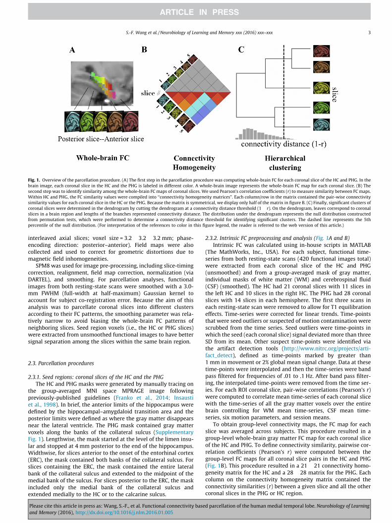
The parcellation scheme aimed to separate the HC and PHG intofunctionally specialized sub-regions according to variations intheir intrinsic FC patterns. Building on the idea that a region’s func-tion is determined by its connectivity, the FC patterns within afunctional region should be homogeneous and the FC patternsamong different functional regions should be heterogeneous. Bydetecting similarities among the FC maps for seed regions of theHC and PHG, we should be able to separate the HC and PHG intodifferent functional clusters. In this report, we began by computingthe functional connectivity between each coronal slice of the HCand PHG (i.e., segments along the longitudinal axis) and all graymatter voxels in the rest of the brain (Fig. 1A). The ‘‘connectivitysimilarity” of two slices was measured by computing the correla-tion (r) between their whole-brain FC maps. The matrix comprisedall the connectivity similarity values for the HC or the PHG was aconnectivity homogeneity matrix (Fig. 1B). A hierarchical cluster-ing algorithm was applied to cluster coronal slices into a dendro-gram according to the dissimilarity of their FC maps, or‘‘connectivity distance” (1 � r) (Fig. 1C). Slices were successivelymerged together in branches representing connectivity distances,and permutation tests were used to identify significant clusters.To further investigate the parcellation, we compared the whole-brain FC maps for each cluster identified via our parcellationscheme. Additionally, we conducted a task-related analysis toinvestigate functional activations of the clusters during a memorytest.
2.2. Image acquisition and pre-processing
The data for this study were drawn from a previously describeddataset (Ritchey, Yonelinas, & Ranganath, 2014) that includedresting-state and task fMRI data from 19 young adults (11 female;ages 19–30). Participants completed a 7-min pre-learning resting-state scan, three 10-min task scans, a 7-min post-learning restingstate scan, and a post-scan behavioral test (see Ritchey et al.,2014 for more details). During the resting state scans, the com-puter screen was black with a white fixation cross at center, andsubjects were instructed to stay awake with their eyes open.
Scanning was performed on a Siemens Skyra 3T scanner systemwith a 32-channel head coil. High-resolution T1-weighted struc-tural images were acquired using a magnetization prepared rapidacquisition gradient echo (MPRAGE) pulse sequence (Field ofview = 25.6 cm, image matrix = 256 � 256, 208 axial slices with1.0 mm thickness). Functional images were acquired using a gradi-ent echo planar imaging (EPI) sequence (TR = 2000 ms; TE = 25 ms;FOV = 20.5 � 21.14 cm; image matrix = 64 � 66; flip angle = 90; 34
d parcellation of the human medial temporal lobe. Neurobiology of Learning
Fig. 1. Overview of the parcellation procedure. (A) The first step in the parcellation procedure was computing whole-brain FC for each coronal slice of the HC and PHG. In thebrain image, each coronal slice in the HC and the PHG is labeled in different color. A whole-brain image represents the whole-brain FC map for each coronal slice. (B) Thesecond step was to identify similarity among the whole-brain FC maps of coronal slices. We used Pearson’s correlation coefficients (r) to measure similarity between FC maps.Within HC and PHG, the FC similarity values were compiled into ‘‘connectivity homogeneity matrices”. Each column/row in the matrix contained the pair-wise connectivitysimilarity values for each coronal slice in the HC or the PHG. Because the matrix is symmetrical, we display only half of the matrix in figure B. (C) Finally, significant clusters ofcoronal slices were determined in the dendrogram by cutting the dendrogram at a connectivity distance threshold (1 � r). On the dendrogram, leaves correspond to coronalslices in a brain region and lengths of the branches represented connectivity distance. The distribution under the dendrogram represents the null distribution constructedfrom permutation tests, which were performed to determine a connectivity distance threshold for identifying significant clusters. The dashed line represents the 5thpercentile of the null distribution. (For interpretation of the references to color in this figure legend, the reader is referred to the web version of this article.)
S.-F. Wang et al. / Neurobiology of Learning and Memory xxx (2016) xxx–xxx 3
interleaved axial slices; voxel size = 3.2 � 3.2 � 3.2 mm; phase-encoding direction: posterior–anterior). Field maps were alsocollected and used to correct for geometric distortions due tomagnetic field inhomogeneities.
SPM8 was used for image pre-processing, including slice-timingcorrection, realignment, field map correction, normalization (viaDARTEL), and smoothing. For parcellation analyses, functionalimages from both resting-state scans were smoothed with a 3.0-mm FWHM (full-width at half-maximum) Gaussian kernel toaccount for subject co-registration error. Because the aim of thisanalysis was to parcellate coronal slices into different clustersaccording to their FC patterns, the smoothing parameter was rela-tively narrow to avoid biasing the whole-brain FC patterns ofneighboring slices. Seed region voxels (i.e., the HC or PHG slices)were extracted from unsmoothed functional images to have bettersignal separation among the slices within the same brain region.
2.3. Parcellation procedures
2.3.1. Seed regions: coronal slices of the HC and the PHGThe HC and PHG masks were generated by manually tracing on
the group-averaged MNI space MPRAGE image followingpreviously-published guidelines (Franko et al., 2014; Insaustiet al., 1998). In brief, the anterior limits of the hippocampus weredefined by the hippocampal–amygdaloid transition area and theposterior limits were defined as where the gray matter disappearsnear the lateral ventricle. The PHG mask contained gray mattervoxels along the banks of the collateral sulcus (SupplementaryFig. 1). Lengthwise, the mask started at the level of the limen insu-lar and stopped at 4 mm posterior to the end of the hippocampus.Widthwise, for slices anterior to the onset of the entorhinal cortex(ERC), the mask contained both banks of the collateral sulcus. Forslices containing the ERC, the mask contained the entire lateralbank of the collateral sulcus and extended to the midpoint of themedial bank of the sulcus. For slices posterior to the ERC, the maskincluded only the medial bank of the collateral sulcus andextended medially to the HC or to the calcarine sulcus.
Please cite this article in press as: Wang, S.-F., et al. Functional connectivity baseand Memory (2016), http://dx.doi.org/10.1016/j.nlm.2016.01.005
2.3.2. Intrinsic FC preprocessing and analysis (Fig. 1A and B)Intrinsic FC was calculated using in-house scripts in MATLAB
(The MathWorks, Inc., USA). For each subject, functional time-series from both resting-state scans (420 functional images total)were extracted from each coronal slice of the HC and PHG(unsmoothed) and from a group-averaged mask of gray matter,individual masks of white matter (WM) and cerebrospinal fluid(CSF) (smoothed). The HC had 21 coronal slices with 11 slices inthe left HC and 10 slices in the right HC. The PHG had 28 coronalslices with 14 slices in each hemisphere. The first three scans ineach resting-state scan were removed to allow for T1 equilibrationeffects. Time-series were corrected for linear trends. Time-pointsthat were seed outliers or suspected of motion contamination werescrubbed from the time series. Seed outliers were time-points inwhich the seed (each coronal slice) signal deviated more than threeSD from its mean. Other suspect time-points were identified viathe artifact detection tools (http://www.nitrc.org/projects/arti-fact_detect), defined as time-points marked by greater than1 mm in movement or 2% global mean signal change. Data at thesetime-points were interpolated and then the time-series were bandpass filtered for frequencies of .01 to .1 Hz. After band pass filter-ing, the interpolated time-points were removed from the time ser-ies. For each ROI coronal slice, pair-wise correlations (Pearson’s r)were computed to correlate mean time-series of each coronal slicewith the time-series of all the gray matter voxels over the entirebrain controlling for WM mean time-series, CSF mean time-series, six motion parameters, and session means.
To obtain group-level connectivity maps, the FC map for eachslice was averaged across subjects. This procedure resulted in agroup-level whole-brain gray matter FC map for each coronal sliceof the HC and PHG. To define connectivity similarity, pairwise cor-relation coefficients (Pearson’s r) were computed between thegroup-level FC maps for all coronal slice pairs in the HC and PHG(Fig. 1B). This procedure resulted in a 21 � 21 connectivity homo-geneity matrix for the HC and a 28 � 28 matrix for the PHG. Eachcolumn on the connectivity homogeneity matrix contained theconnectivity similarities (r) between a given slice and all the othercoronal slices in the PHG or HC region.
d parcellation of the human medial temporal lobe. Neurobiology of Learning
4 S.-F. Wang et al. / Neurobiology of Learning and Memory xxx (2016) xxx–xxx
2.3.3. Hierarchical clustering and dendrograms (Fig. 1C)We used the hierarchical clustering algorithm, UPGMA
(Unweighted Pair Group Method with Arithmetic Mean), imple-mented in MATLAB, to successively merge clusters of the coronalslices based on similarities among their FC maps. Connectivity dis-tance was calculated for each coronal slice pair by one minus con-nectivity similarity (1 � r). Connectivity distance was entered intothe hierarchical clustering algorithm. This procedure resulted intwo respective dendrograms representing the hierarchicalrelationships of the connectivity distance for the coronal slices.On the dendrograms, leaves correspond to coronal slices of a brainregion (i.e. the HC or the PHG) and lengths of the branches repre-sent connectivity distance (1 � r). The longer a given branch was,the more dissimilar the FC patterns between two slices/clusterswere.
To determine significant clusters in the dendrogram, a connec-tivity distance threshold (1 � r) was calculated via permutationtests. The null hypothesis was that the FC patterns for the coronalslices in the HC or the PHG were not heterogeneous enough to sep-arate the coronal slices into different functional regions, and thus,there were no sub-clusters in the HC or the PHG. Permutation testswere used to determine a connectivity distance threshold at whichthe dendrogram was partitioned into disjoint clusters. If the con-nectivity distance between two sets of slices was under thisthreshold, this would mean that their FC patterns were no moredissimilar than what would be expected by chance, in this case,the two sets of slices would be grouped together into one func-tional cluster. In contrast, if the connectivity distance betweentwo sets of slices was above the distance threshold, then their FCpatterns were more dissimilar than what would be expected bychance, in which case the null hypothesis would be rejected andthese two sets of slices would be separated into different func-tional clusters.
The data were permuted 10,000 times by randomly assigning 1or �1 labels to each subject’s FC data each time. On each permuta-tion, after assigning random 1 or �1 weights to the dataset, we cal-culated group-level FC maps and connectivity distances (1 � r) asdescribed above. The mean of the connectivity distances in a givenbrain region (i.e. the HC or the PHG) were calculated to representthe overall degree of heterogeneity in the data for each time. Thecollection of the distance means from all permutations constructedthe null distribution of expected connectivity distances. The dis-tance threshold was defined as the 5th percentile of the null distri-bution, and thus denotes the level of dissimilarity that would beexpected to occur by chance only 5% of the time. Leaves attachedto the branches that intersected the cut-off line were groupedtogether as one functional cluster.
2.4. Intrinsic FC profiles for clusters: t-tests
The purpose for this analysis was to better understand thewhole-brain FC patterns associated with each PHG cluster thatwas identified in the hierarchical clustering analysis. In these anal-yses, whole-brain FC maps associated with each cluster wereobtained by averaging FC maps across all slices within each cluster.These analyses followed the same procedures described above,except that: (1) Instead of calculating intrinsic FC for only graymatter voxels, pair-wise correlations (Pearson’s r) were computedfor all voxels over the entire brain to obtain a smoothly varyinggroup map, and the FC maps were Fisher’s r-to-z transformed. (2)Functional images were smoothed with a 6.0 mm Gaussian kernelfor this analysis. A larger smoothing kernel was used for theseanalyses in order to facilitate accurate correction for multiple com-parisons (i.e., because we conducted statistical analyses to identifyregions that showed supra-threshold FC values for each cluster andsignificant between-cluster differences) and to better account for
Please cite this article in press as: Wang, S.-F., et al. Functional connectivity baseand Memory (2016), http://dx.doi.org/10.1016/j.nlm.2016.01.005
inter-subject anatomical variability. Note that, because the regionsof interest (ROIs) were clusters that had been identified in theprevious parcellation analysis, there was less concern about FCbiasing from neighboring slices.
To determine the connectivity profiles that were associatedwith different clusters, subject-level whole-brain FC maps wereentered into one-sample t-tests. For simplicity, only positive func-tional connectivity values were evaluated and displayed in Fig. 3,although the parcellation procedure incorporated all connectivityvalues across the whole brain. Differences in the intrinsic FCprofiles of different clusters were identified via paired t-tests.The t-maps were thresholded at p < .001, one-tailed, with a38-voxel cluster extent threshold. This combination of thresholdscorresponds to cluster-corrected p < .05 according to simulationsimplemented in 3dClustSim of AFNI (http://afni.nimh.nih.gov/).
2.5. Task activity for clusters: univariate activation analyses
In these analyses, we investigated activations within eachcluster during a memory-encoding task. While in the scanner,participants completed an incidental associative encoding taskusing sentences pairing an object and a fact about either itsappearance, its situational context, or its spatial location. Forexample, one possible sentence might read, ‘‘The apple is in thelecture hall.” The post-scan behavioral test consisted of anassociative memory test for each sentence, and encoding trialswere classified as hits (remembered) or misses (forgotten) basedon subsequent memory performance (see Ritchey et al., 2014 formore details).
Task activation was evaluated through a general linear modelimplemented in SPM8. Functional images from the task scans werenormalized and smoothed with a 6.0-mm Gaussian kernel. Event-related stick-function regressors were used to model trials corre-sponding to one of six trial types: appearance hits, situational hits,spatial hits, appearance misses, situational misses, and spatialmisses. Six motion parameter regressors and a regressor for no-response trials were also included in the model. Nuisance regres-sors were also included to model time-points identified as ART sus-pects. For each subject, contrasts were estimated for each of the sixtrial types, relative to implicit baseline activity. Mean contrast esti-mates were extracted from three PHG clusters. For each of thethree conditions, the misses contrast was subtracted from the hitscontrast to estimate activity related to the difference in memory(Dm). A repeated measures analysis of variance (ANOVA) wasconducted to test the overall differences among memory effects,conditions, and ROIs. A two (memory effects: hit and miss) � three(conditions: appearance, situational context, and spatial loca-tion) � three (ROIs: PHC, postPRC, and antPRC) analysis of variance(ANOVA) was calculated on participants’ contrast estimates.
2.6. PHG clusters connectivity along HC long axis
We next analyzed FC between each of the three PHG clustersand each of the HC coronal slices. Functional images weresmoothed with a 6.0 mm Gaussian kernel. Functional time-seriesfrom both resting-state scans were extracted from each of thethree PHG clusters and each of the HC coronal slices, individualmasks of white matter (WM) and cerebrospinal fluid (CSF). FCpre-processing procedures and parameters were the same as theaforementioned FC procedures. Mean functional time-series foreach of the three PHG clusters were correlated with the meantime-series of each of the HC coronal slices. These proceduresresulted in a 3 by 21 matrix of connectivity between the PHG clus-ters (the PHC cluster, the postPRC cluster, and the antPRC cluster)and each of the HC coronal slices (left HC, 11 slices; right HC, 10slices).
d parcellation of the human medial temporal lobe. Neurobiology of Learning
S.-F. Wang et al. / Neurobiology of Learning and Memory xxx (2016) xxx–xxx 5
3. Results
3.1. PHG parcellation results in three clusters
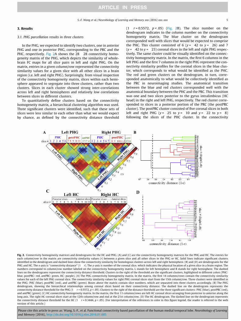
In the PHG, we expected to identify two clusters, one in anteriorPHG and one in posterior PHG, corresponding to the PRC and thePHC, respectively. Fig. 2A shows the 28 � 28 connectivity homo-geneity matrix of the PHG, which depicts the similarity of whole-brain FC maps for all slice pairs in left and right PHG. On thematrix, entries in a given column/row represented the connectivitysimilarity values for a given slice with all other slices in a brainregion (i.e. left and right PHG). Surprisingly, from visual inspectionof the connectivity homogeneity matrix, slices within each hemi-sphere appeared to segregate into three clusters, rather than twoclusters. Slices in each cluster showed strong inter-correlationsacross left and right hemispheres and relatively low correlationsbetween slices in different clusters.
To quantitatively define clusters based on the connectivityhomogeneity matrix, a hierarchical clustering algorithm was used.Three significant clusters were identified, such that these sets ofslices were less similar to each other than what we would expectby chance, as defined by the connectivity distance threshold
Fig. 2. Connectivity homogeneity matrices and dendrograms for the HC and PHG. (A) andeach column/row in the matrix are connectivity similarity values (r) between a givenidentified in the dendrogram and dashed lines show the connectivity similarity for homoPHG and HC. The x-axis is ‘‘connectivity distance” (1 � r). The y-axis is number of the coronumbers correspond to column/row number labeled on the connectivity homogeneity mlines on the dendrograms represent the connectivity distance threshold. Clusters to the riblue, postPRC: red, antPRC: green, HC: purple). (A) The PHG connectivity homogeneity mvalues for each of the left PHG coronal slice. The connectivity similarity values for right Pthe PHG: PHC (blue), postPRC (red), and antPRC (green). Boxes above the matrix contaidendrogram, showing the hierarchical relationships among coronal slices based onconnectivity distance threshold for the PHG (1 � r = 0.5572, p < .05). Clusters to the right oand antPRC (green). (C) HC connectivity homogeneity matrix. In the matrix, the first 11 colong axis. The right HC coronal slices start at the 12th column/row and end at the 21st cothe connectivity distance threshold for the HC (1 � r = 0.3444, p < .05). (For interpretatioversion of this article.)
Please cite this article in press as: Wang, S.-F., et al. Functional connectivity baseand Memory (2016), http://dx.doi.org/10.1016/j.nlm.2016.01.005
(1 � r = 0.5572, p < .05) (Fig. 2B). The slice number on thedendrogram indicates to the column number on the connectivityhomogeneity matrix. The blue cluster on the dendrogramcorresponded well with slices that would be expected to comprisethe PHC. This cluster consisted of 6 (y = �42 to y = �26) and 7(y = �42 to y = �23) coronal slices in the left and right PHG respec-tively. The same cluster could be visually identified on the connec-tivity homogeneity matrix. In the matrix, the first 6 columns in theleft PHG and the first 7 columns in the right PHG represent the con-nectivity similarity profiles for the coronal slices in the blue clus-ter, which corresponds to what would be identified as the PHC.The red and green clusters on the dendrogram, in turn, corre-sponded anatomically to what would be collectively identified asthe PRC in neuroimaging studies. The anatomical transitionbetween the blue and red clusters corresponded well with theanatomical boundary between the PHC and the PRC. This transitionwas one and two slices posterior to the gyrus intralimbicus (HChead) in the right and left PHG, respectively. The red cluster corre-sponded to slices in a posterior portion of the PRC (the postPRCcluster). The postPRC cluster consisted of five coronal slices in bothleft and right PHG (y = �25 to y = �10 and y = �22 to y = �8)following the slices of the PHC cluster. In the connectivity
(C) are the connectivity homogeneity matrices for the PHG and HC. The entries forslice and all other slices in the PHG or HC. Solid lines indicate significant clusterslogous clusters across left and right hemisphere. (B) and (D) are dendrograms for thenal slice, which indicates the physical location of a given slice in a brain region. Theatrix. L stands for left hemisphere and R stands for right hemisphere. The dashed
ght of the threshold are the significant clusters, highlighted in different colors (PHC:atrix. In the matrix, the first 14 columns/rows contain the connectivity similarity
HG coronal slices start from the 15th column/row. Three clusters were identified inn slice numbers, which are separated into three clusters accordingly. (B) The PHGtheir connectivity distance. The dashed line on the dendrogram represents thef the distance threshold are the three significant clusters: PHC (blue), postPRC (red),lumns/rows are left HC coronal slices arranging from posterior to anterior along thelumn/row. (D) The HC dendrogram. The dashed line on the dendrogram representsn of the references to color in this figure legend, the reader is referred to the web
d parcellation of the human medial temporal lobe. Neurobiology of Learning
6 S.-F. Wang et al. / Neurobiology of Learning and Memory xxx (2016) xxx–xxx
homogeneity matrix, the five columns following the columns forthe PHC cluster represent the connectivity similarity profiles forthe coronal slices in the red cluster. Like the PHC cluster, these col-umns could be visually identified as a group based on homogeneityamong their connectivity similarity profiles. Finally, the green clus-ter on the dendrogram consisted of the remaining anterior slices ineach hemisphere, which anatomically corresponded with slices inthe remaining anterior portion of the PRC (the antPRC cluster).The left antPRC cluster consisted of the three most anterior coronalslices (y = �9 to y = �2), whereas the right antPRC cluster consis-tent of the two most anterior slices (y = �7 to y = �2). The transi-tion between the postPRC and antPRC clusters was around thehippocampal–amygdaloid transition area (HATA), at the mostanterior end of the HC. In the left PHG, the transition betweenthese two clusters was two slices posterior to the HATA. In theright PHG, the transition was one slice anterior to the HATA.
We performed a control analysis to confirm that the parcella-tion results were not driven by changes of the PHG masks alongthe longitudinal axis (Supplementary Figs. 1 and 2). We generateda control mask that included only voxels in the lateral half of themedial bank of the collateral sulcus (from the fundus of the collat-eral sulcus to the midpoint of the medial bank of the collateral sul-cus). This region is relatively consistent across the entire PHGlongitudinal axis, and it is included in anatomical definitions ofboth the PRC and PHC. We applied the parcellation schemedescribed above to parcellate the PHG areas covered by this controlmask based on similarity of the FC patterns. The aim was to seewhether this control region could be separated into clusters similarto what we found by using the full PHG mask. Two significant clus-ters were identified in the control region (Supplementary Fig. 2B).One cluster corresponded well with the antPRC cluster mentionedabove (control-antPRC). The other cluster, control-postPHG, con-tained two major sub-clusters, each of which corresponded wellwith the postPRC and PHC clusters. Based on the control analysis,we can affirm that the three PHG clusters identified via the parcel-lation scheme were unlikely driven by differences in the PHG maskalong the longitudinal axis. Finally, additional analyses ruled outthat the parcellation was driven by differences in number of voxelsor signal-to-noise ratio of each coronal slice (Supplementary Figs. 3and 4).
3.2. Networks connected with the three PHG clusters
Following the parcellation results, we examined the underlyingbrain networks that were associated with each cluster. Each clusterwas used as a ROI in a new FC analysis, and a one-sample t-test wasused to identify voxels that showed supra-threshold FC valuesassociated with these ROIs (Fig. 3A–C). The PHC cluster and thepostPRC cluster were associated with networks that have been pre-viously identified in studies that examined FC for anatomically-defined PHC and PRC sub-regions (Kahn et al., 2008; Libby et al.,2012). The PHC cluster showed strong connectivity with the poste-rior cingulate cortex (BA23 and BA31), the retrosplenial cortex(BA30), the precuneus (BA7), the inferior temporal gyrus (BA20),early visual areas (e.g. V1, V2, V3), and the thalamus (Fig. 3A).The PHC cluster also showed significant FC with voxels in the ante-rior cingulate gyrus (BA32 and BA24), the ventromedial superiorfrontal gyrus (BA10), the ventromedial prefrontal cortex (BA10),the dorsal lateral prefrontal cortex (BA9), the premotor cortex(BA6), the frontal eye field (BA8), the angular gyrus (BA39), thefusiform gyrus, and the superior temporal gyrus (BA22).
The postPRC cluster strongly connected to voxels in the anteriorcingulate cortex (BA24), the rostromedial prefrontal cortex (BA10),and the orbitofrontal cortex (BA11) (Fig. 3B). The postPRC clusteralso showed significant FC with voxels in the posterior cingulate(BA31 and BA23), the retrosplenial cortex (BA30), the visual cortex
Please cite this article in press as: Wang, S.-F., et al. Functional connectivity baseand Memory (2016), http://dx.doi.org/10.1016/j.nlm.2016.01.005
V3, the angular gyrus (BA39), the premotor cortex (BA6), and thefrontal eye field (BA8). In addition, the postPRC cluster had exten-sive temporal lobe connections, including the superior temporalgyrus (BA22), the middle temporal gyrus (BA21), the inferior tem-poral gyrus (BA20), and the fusiform gyrus (BA37).
Finally, the antPRC cluster exhibited high FC with a distinctfronto-insular network (Fig. 3C) consisting of the insula, the pla-num temporale, the orbitofrontal cortex (BA47), the postcentralgyrus (BA43), and the ventrolateral prefrontal cortex (BA45). TheantPRC cluster also connected to ventral part of the premotor cor-tex (BA6), the amygdala, the anterior HC, and the ventral temporallobe (BA20).
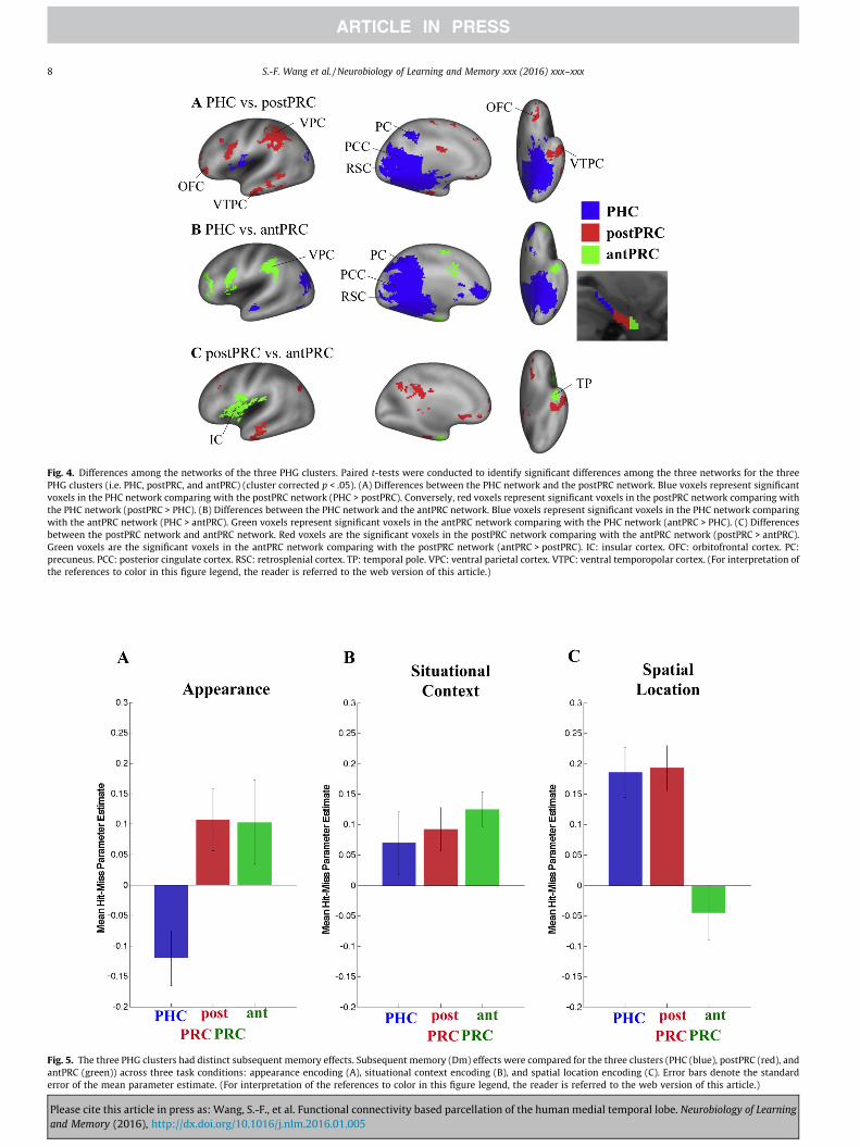
Paired t-tests (cluster corrected p < .05) were conducted toidentify regions that showed preferential FC with each cluster(Fig. 4). As in the previous studies, voxels in the posterior-medialnetwork, including the posterior cingulate, the retrosplenial cortex,and the precuneus, showed stronger connectivity with the PHCcluster than with the postPRC and antPRC clusters(Fig. 4A and B). Voxels in orbitofrontal areas and ventral tem-poropolar cortex, in turn, exhibited stronger FC with the postPRCthan with the PHC and the antPRC clusters (Fig. 4A and C). Voxelsin the insula and temporal pole showed stronger connectivity withthe antPRC than with the PHC and the postPRC clusters(Fig. 4B and C). The PRC clusters (i.e. the postPRC and the antPRCclusters) had stronger connectivity with the ventral parietal cortex,the premotor cortex, the orbitofrontal areas, and the ventral tem-poropolar cortex. The antPRC cluster was more connected withthe insular areas than the postPRC cluster. In the temporal lobe,both the postPRC and the antPRC clusters connected to the amyg-dala, the anterior HC, and the ventral temporal lobe. However, theirconnections to these regions followed their topographical relation-ships such that the postPRC cluster connected to more posteriorportion of these regions and the antPRC cluster connected to rela-tively more anterior portion of these regions.
3.3. The three PHG clusters had distinct subsequent memory effects
The previous analyses identified three regions that exhibitedmarkedly different patterns of FC during the resting-state scans.Although the result is consistent with the idea that the PHG couldbe subdivided into three regions, it is unclear whether these FC dis-tinctions are functionally meaningful or informative. To addressthis question, we interrogated activity in the three PHG clustersduring a memory-encoding task (Fig. 5) that was performed bythe same participants between the two resting state scans. Duringthe task, participants incidentally encoded sentences describing anobject’s appearance, situational context, or spatial location (seeRitchey et al., 2014 for further details), and following the scan ses-sion, they were tested on memory for each association. Results ofthe clustering analysis described above were used to identify bilat-eral ROIs for the antPRC, postPRC, and PHC clusters, and activity ineach ROI was separately examined for subsequently rememberedassociations and forgotten association. Results, summarized in(Fig. 5), revealed that the three PHG clusters showed different pat-terns of activation during memory encoding. A three-way ANOVA(Memory (hit, miss) � Condition (appearance, situational context,spatial location) � ROI (PHC, postPRC, antPRC), p < .05) analysisrevealed that there was a significant two-way interaction betweenCondition and ROI (F(4,72) = 18.0551, p < .001) and a significantthree-way interaction between Memory, Condition, and ROI(F(4,72) = 4.3469, p = 0.003). Putting the results together, we cansee three different profiles of activation during memory encoding:the PHC cluster was preferentially involved in encoding of spatialassociations, the antPRC cluster was preferentially involved inencoding of item-appearance associations, and the postPRC cluster
d parcellation of the human medial temporal lobe. Neurobiology of Learning
Fig. 3. Networks connected with the three PHG clusters and the one HC cluster. One-sample t-tests were conducted to identify voxels that showed supra-threshold FC valuesassociated with different clusters (voxel-wise p < .001, cluster-corrected p < .05). (A) Significant voxels connected with the PHC cluster (blue). (B) Significant voxels connectedwith the postPRC cluster (red). (C) Significant voxels connected with the antPRC cluster (green). (D) Significant voxels connected with the HC cluster (purple). (Forinterpretation of the references to color in this figure legend, the reader is referred to the web version of this article.)
S.-F. Wang et al. / Neurobiology of Learning and Memory xxx (2016) xxx–xxx 7
was involved in encoding both spatial and item-appearanceassociations.
3.4. HC parcellation results in a single cluster
The same parcellation procedure was applied to identify func-tionally different clusters along the longitudinal axis of the HC. A21 � 21 connectivity homogeneity matrix was generated repre-senting connectivity similarity among the whole-brain FC mapsof all the coronal slices in the HC (Fig. 2C). In contrast to thePHG, the connectivity patterns were relatively homogeneousacross the longitudinal axis of the HC. When we applied the hier-archical clustering algorithm to quantitatively determine signifi-cant clusters, all left and right HC slices were grouped into asingle cluster. The connectivity distance threshold (1 � r = 0.3444,p < .05) was larger than, but close to, the largest connectivity dis-tance on the HC dendrogram (Fig. 2D). This result indicated thatthe FC maps of the HC coronal slices were not different enoughto justify separating the slices into distinct clusters. Fig. 3Dillustrates the whole-brain FC map connected to the HC cluster(one-sample t-test, cluster corrected p < .05). Like the PHCposterior-medial network, the HC showed high FC with voxels in theposterior cingulate cortex (BA23 and BA31) and the retrosplenialcortex (BA30). In the anterior-medial part of the brain, the HC clusterhad connections with the anterior cingulate gyrus (BA12 and BA32),the ventromedial superior frontal gyrus (BA10), and the medialprefrontal cortex (BA10 and BA11). The HC cluster also connectedto the superior temporal gyrus (BA22), the middle temporal gyrus,the angular gyrus (BA39), and part of the primary auditory cortex.
Please cite this article in press as: Wang, S.-F., et al. Functional connectivity baseand Memory (2016), http://dx.doi.org/10.1016/j.nlm.2016.01.005
Although we did not identify any significant sub-clusters in theHC, there were two major branches dividing the HC into an ante-rior and a posterior part on the dendrogram (Fig. 2D). Slices inthe anterior part of the HC corresponded anatomically to the hip-pocampal head, and slices in the posterior part of the HC corre-sponded to the hippocampal body and tail according topreviously-published parcellation guidelines (Duvernoy, 2015;Franko et al., 2014). We did another set of one-sample t-test anal-yses to identify the associative brain networks for the anterior andposterior HC (Fig. 6A). Basically, the results revealed that both ofthe anterior and posterior HC showed high FC with the same brainnetwork described above, with subtle topographic differences. TheFC maps for the anterior HC appeared to show a larger extent ofsupra-threshold voxels in the anterior part of the brain, includingthe anterior-medial superior frontal gyrus (BA10) and the dorsallateral prefrontal cortex (BA9), as well as in the insula andthe superior and middle temporal gyrus (BA21 and BA22). Theposterior HC, in turn, appeared to show a larger extent of supra-threshold voxels in the posterior cingulate cortex (BA23).Altogether, in contrast to the three PHG clusters, the anterior andposterior HC clusters appeared to be more similar to one anotherthan they were different.
3.5. FC of PHG clusters along long axis of the hippocampus
Although the whole-brain FC maps for the anterior and poste-rior HC were generally similar, prior work has shown that theintrinsic connectivity between the PHG and the HC might beheterogeneous along the HC longitudinal axis (Libby et al., 2012;
d parcellation of the human medial temporal lobe. Neurobiology of Learning
Fig. 4. Differences among the networks of the three PHG clusters. Paired t-tests were conducted to identify significant differences among the three networks for the threePHG clusters (i.e. PHC, postPRC, and antPRC) (cluster corrected p < .05). (A) Differences between the PHC network and the postPRC network. Blue voxels represent significantvoxels in the PHC network comparing with the postPRC network (PHC > postPRC). Conversely, red voxels represent significant voxels in the postPRC network comparing withthe PHC network (postPRC > PHC). (B) Differences between the PHC network and the antPRC network. Blue voxels represent significant voxels in the PHC network comparingwith the antPRC network (PHC > antPRC). Green voxels represent significant voxels in the antPRC network comparing with the PHC network (antPRC > PHC). (C) Differencesbetween the postPRC network and antPRC network. Red voxels are the significant voxels in the postPRC network comparing with the antPRC network (postPRC > antPRC).Green voxels are the significant voxels in the antPRC network comparing with the postPRC network (antPRC > postPRC). IC: insular cortex. OFC: orbitofrontal cortex. PC:precuneus. PCC: posterior cingulate cortex. RSC: retrosplenial cortex. TP: temporal pole. VPC: ventral parietal cortex. VTPC: ventral temporopolar cortex. (For interpretation ofthe references to color in this figure legend, the reader is referred to the web version of this article.)
Fig. 5. The three PHG clusters had distinct subsequent memory effects. Subsequent memory (Dm) effects were compared for the three clusters (PHC (blue), postPRC (red), andantPRC (green)) across three task conditions: appearance encoding (A), situational context encoding (B), and spatial location encoding (C). Error bars denote the standarderror of the mean parameter estimate. (For interpretation of the references to color in this figure legend, the reader is referred to the web version of this article.)
8 S.-F. Wang et al. / Neurobiology of Learning and Memory xxx (2016) xxx–xxx
Please cite this article in press as: Wang, S.-F., et al. Functional connectivity based parcellation of the human medial temporal lobe. Neurobiology of Learningand Memory (2016), http://dx.doi.org/10.1016/j.nlm.2016.01.005
Fig. 6. The MTL and whole-brain connectivity for the HC. (A) Networks connected with the anterior and posterior HC. A one-sample t-test was conducted to identify voxelsthat showed supra-threshold FC values associated with different anterior (pink) and posterior (purple) HC (voxel-wise p < .001, cluster-corrected p < .05). Voxels significantlyconnected with both anterior and posterior HC are in magenta. (B) FC between the three PHG clusters and the HC coronal slices. Entries in the matrix are FC values (r). Rowsrepresent each of the three clusters in the PHG (PHC, postPRC, and antPRC). Columns represent each of the coronal slices in the left and right HC ranging from posterior toanterior. (For interpretation of the references to color in this figure legend, the reader is referred to the web version of this article.)
S.-F. Wang et al. / Neurobiology of Learning and Memory xxx (2016) xxx–xxx 9
Maass et al., 2015). We therefore investigated FC between thethree PHG clusters and the HC coronal slices (Fig. 6B). Consistentwith previous results, the PHC cluster exhibited high FC with theentire HC, whereas the adjacent postPRC cluster showed preferen-tial connectivity with the anterior part of the HC. Although both ofthe two PRC clusters preferentially connected to the anterior partof the HC, the antPRC cluster had strong FC limited to the mostanterior portion of the HC and the postPRC had a relatively broaderanterior HC connectivity. Finally, the homologous brain regionsshared similar HC connectivity across left and right hemisphere.
4. Discussion
In this study, we used FC-based parcellation to characterize thefunctional organization of the human PHG and hippocampus. Sur-prisingly, our results revealed that the PHG was subdivided intothree clusters that could be distinguished on the basis of whole-brain FC and on the basis of activation profiles during memoryencoding. Two of these clusters corresponded closely to the PRCand PHC, but the third antPRC cluster has not been previously con-sidered in theoretical accounts of human MTL function. In contrastto the PHG, we did not observe significant evidence that the HCcould be functionally subdivided, although we did observe a trendfor differences between anterior and posterior HC regions. Below,we consider the implications of these findings for understandingMTL contributions to cognition.
4.1. Three sub-regions of the human PHG
The main finding in this study was that the PHG could be sub-divided into three sub-regions along the longitudinal axis based onconnectivity differences. The most posterior cluster exhibited highfunctional connectivity with a posterior-medial network thatincluded the posterior cingulate cortex, the retrosplenial cortex,the precuneus, and occipital areas. This pattern of results corre-sponds well to previous studies that examined resting-state FC
Please cite this article in press as: Wang, S.-F., et al. Functional connectivity baseand Memory (2016), http://dx.doi.org/10.1016/j.nlm.2016.01.005
profiles for the posterior PHC, as defined on the basis of structurallandmarks (Kahn et al., 2008; Libby et al., 2012). A second,‘‘postPRC” cluster exhibited strong connectivity with an anterior-temporal network that included the orbitofrontal cortex, the supe-rior temporal gyrus, the middle temporal gyrus, and the inferiortemporal gyrus. This connectivity pattern corresponds well to whatwould be expected based on the anatomical connectivity of thePRC (Suzuki & Naya, 2014), and to some extent, with previous anal-yses of FC profiles for the PRC (Kahn et al., 2008; Libby et al., 2012).The third, ‘‘antPRC” cluster exhibited strong connectivity with afronto-insular network that included the insula, the planum tem-porale, the orbitofrontal cortex, the postcentral region, and theventrolateral prefrontal cortex. Critically, we found that FC mapsfor the left and right homologous clusters were much more similarthan for non-homologous clusters, serving as an internal replica-tion of the results.
To validate the FC-based parcellation, we examined activationprofiles in the three PHG regions during a memory-encoding task.Results revealed dissociable profiles of encoding-related activityacross the three regions. Whereas activity in the antPRC clusterwas specifically associated with successful encoding of appearanceassociations, activity in the PHC cluster was associated with suc-cessful encoding of spatial location associations. Finally, activityin the postPRC cluster was associated with both appearance andspatial location encoding. During situational context encoding,we saw progressive changes along the longitudinal axis of thePHG, with the antPRC cluster showing the largest memory effectamong the three clusters. The PHC cluster showed the smallestmemory effect. The memory effect for the postPRC cluster was inbetween that of the antPRC and PHC clusters. These results demon-strate that the FC differences between the three clusters are indica-tive of meaningful functional differences. These differences wouldnot have been apparent in a traditional analysis of activity instructurally-defined PRC and PHC sub-regions, and as such, theresults highlight the added value of the FC-based parcellationapproach introduced here.
d parcellation of the human medial temporal lobe. Neurobiology of Learning
10 S.-F. Wang et al. / Neurobiology of Learning and Memory xxx (2016) xxx–xxx
4.2. The antPRC cluster: a homologue of area 36d?
Given the traditional view that the MTL neocortex can be sub-divided into the PHC and the PRC, it might be tempting to dismissthe current results by assuming that the antPRC module corre-sponds to ‘‘temporopolar cortex”, a poorly understood region thatis sometimes treated as separate from the PRC (Kondo, Saleem, &Price, 2003; Olson, Plotzker, & Ezzyat, 2007). A closer look at theanatomy of rodent and monkey PRC, however, indicates that theantPRC cluster exhibits compelling parallels with a region knownas area 36d. Anatomical studies of rats and monkeys have consis-tently distinguished between area 36d, located in the anterior–dor-sal part of the PRC, and adjacent PRC regions (i.e., the rest of thearea 36 and area 35). In rats, cytoarchitectonic and histochemicalanalyses revealed unique radially oriented cells and deep layersof myelinated fibers that distinguished area 36d from the rest ofthe area 36 (Burwell, 2001). Further, a clustering analysis dividedretrograde tracer injection sites in the rat area 36 into a dorsaland a ventral clusters based on their neocortical connectivity.The injection sites in the dorsal cluster, approximately correspond-ing to the rat area 36d, show strong connections with the primaryauditory regions and the ventral temporal association areas(Burwell, 2001; Burwell & Amaral, 1998b). Other unique intrinsicand extrinsic connection patterns of the rat dorsal area 36 also sep-arated it from the rest of the PRC, including their strong connectiv-ity with the PHC, and the dorsal–ventral connectivity gradient withthe ERC and with itself (Burwell & Amaral, 1998a, 1998b). In non-human primates, area 36d has strong internal connectivity but lim-ited connections with the rest of the PRC (Lavenex et al., 2004). Inmonkeys, the major cortical inputs to the area 36d originated fromthe rostral superior temporal gyrus, an auditory processing area(Suzuki & Amaral, 1994b). In contrast, the rest of the monkeyPRC has strong cortical inputs from the visual areas TE and TEOand has weaker connections with the dorsal bank of the superiortemporal sulcus (STS), the insular cortex, and the orbitofrontalareas.
In sum, the human antPRC cluster identified in the currentstudy may be the homologous region of the area 36d in animals.First, the physical location of the antPRC cluster was close to theanterior PRC in monkeys and dorsal PRC in rats, which are wherearea 36d locates in both animals. Further, features of intrinsic,whole-brain, and PHC connectivity for the antPRC corresponds wellwith the connectivity patterns of monkey area 36d and the rat dor-sal area 36 (Burwell & Amaral, 1998a, 1998b; Kondo et al., 2003;Lavenex et al., 2004; Suzuki & Amaral, 1994b).
The present results have implications for understanding thefunctions of human PRC. Almost every model of PRC functionemphasizes its role in memory for objects, with some models plac-ing more emphasis on visual object perception, and others broad-ening the functions to encompass representations of ‘‘items” or‘‘entities” (Brown & Aggleton, 2001; Brown, Warburton, &Aggleton, 2010; Bussey, Saksida, & Murray, 2002; Graham,Barense, & Lee, 2010; Meunier, Bachevalier, Mishkin, & Murray,1993; Murray & Richmond, 2001). Based on the present results,one could speculate that these descriptions only apply to thepostPRC, whereas the antPRC might instead encode informationrelated to inputs conveyed by auditory association areas, intero-ceptive information conveyed by the insula, and information aboutgoals and task context conveyed by regions in lateral prefrontalcortex (Murray & Richmond, 2001; Petrides, 2005). Alternatively,it is possible that an ‘‘item” is separately and differentially pro-cessed by antPRC and postPRC might, with postPRC preferentiallyemphasizing visual properties and antPRC preferentiallyemphasizing auditory properties, personal significance, and rele-vance for action selection (Belin, Zatorre, & Ahad, 2002; Belin,Zatorre, Lafaille, Ahad, & Pike, 2000; Munoz-Lopez, Insausti,
Please cite this article in press as: Wang, S.-F., et al. Functional connectivity baseand Memory (2016), http://dx.doi.org/10.1016/j.nlm.2016.01.005
Mohedano-Moriano, Mishkin, & Saunders, 2015; Olson et al.,2007; Petkov et al., 2008). These ideas are of course speculative,and further research is needed to better understand how theantPRC, postPRC, and PHC separately and collectively encode theattributes of an event.
4.3. Differences in the HC MTL and whole-brain connectivity
Despite strong evidence for anatomical and functional differ-ences between the dorsal and ventral HC in rodents (Fanselow &Dong, 2010; Moser & Moser, 1998; Strange et al., 2014), we didnot observe strong evidence for a parallel connectivity-based dis-sociation in humans. Although we only identified a single clusterin the HC, there was a weak trend for differences between anteriorand posterior HC (Fig. 2D). The pattern of HC connectivity withneocortical areas within and outside of the MTL reflected thisambiguity (Fig. 6). In the MTL, the two PRC clusters heavily con-nected to the anterior part of the HC whereas the PHC cluster heav-ily connected to the entire HC with a preference in thehippocampal head and tail (Fig. 6B). In contrast, FC with neocorti-cal areas outside of the MTL was relatively homogeneous along thelongitudinal axis of the HC (Fig. 6A). Both the anterior and poste-rior HC showed high connectivity with regions in the default net-work, including the posterior cingulate cortex, the ventralanterior cingulate cortex, and the ventromedial prefrontal cortex.Consistent with findings in rodents (Jones & Witter, 2007), therewere minor connectivity differences, such that the anterior HCshowed more extensive FC with voxels in the dorsal prefrontal cor-tex and lateral temporal lobe, whereas the posterior HC showedslightly more extensive HC with voxels in the posterior cingulatecortex and the precuneus (Fig. 6A). These differences were rela-tively small, however, relative to the visually apparent distinctionsin FC profiles associated with the three PHG clusters.
Although our study did not reveal strong evidence for functionalheterogeneity in the HC, there are several reasons why one shouldbe careful in interpreting this null result. First, neuroanatomy stud-ies suggest that functional differences along the longitudinal axisof the HC should differ across subfields (Chase et al., 2015;Malykhin, Lebel, Coupland, Wilman, & Carter, 2010), with thestrongest gradients to be seen in CA1 and subiculum. This possibil-ity could be assessed in an analysis of high-resolution fMRI datausing an approach that would allow parcellation both along thelongitudinal and transverse axes of the hippocampus. Alterna-tively, it is possible that, during the resting state, hippocampalregions function in unison, but during performance of tasks thatdifferentially engage targets of anterior and posterior HC, differ-ences would become apparent. For instance, one might see largeFC differences between anterior and posterior HC during naviga-tion in empty spatial contexts or during processing of emotionallyarousing objects (Poppenk et al., 2013; Strange et al., 2014), in con-trast to the more homogenous pattern of FC seen during rest.
4.4. Limitations
There are some limitations to the current study. First, the par-cellation reported here, like most previously reported cortical par-cellations (Cohen et al., 2008; Gordon et al., 2014; Nelson et al.,2013; Wig et al., 2014; Yeo et al., 2011), is based on a group-level analysis. Researchers are starting to develop parcellationschemes for identifying cortical systems at single subject level(Gordon, Laumann, Adeyemo, & Petersen, 2015; Wang et al.,2015), but single-subject parcellation requires a large amount ofresting-state data. For instance, Wang et al. (2015) collected anhour of resting-scan data to evaluate intrinsic functional connec-tivity in single-subjects. It is also worth noting that these studies
d parcellation of the human medial temporal lobe. Neurobiology of Learning
S.-F. Wang et al. / Neurobiology of Learning and Memory xxx (2016) xxx–xxx 11
used previously identified group-level cortical networks in order toguide the single subject analysis.
As we mentioned above, the hippocampus could not be subdi-vided into multiple clusters. In the current study, hippocampalsubfields were collapsed within each coronal slice, and thus, theproportions of each subfield in each coronal slice varied. The intrin-sic FC patterns we obtained for each coronal slice should be amixed result combining connectivity patterns for different sub-fields at different longitudinal levels. Thus, high-resolution fMRIdata might be necessary to identify functional subdivisions in thehippocampus, and the parcellation would be best performed atthe level of voxels, rather than using coronal slices as seed regions.
5. Conclusion
In the current report, three functionally different clusters wereidentified via the parcellation procedure in the PHG. Our resultssuggest that the PHC, postPRC, and antPRC each affiliates with dif-ferent large-scale neocortical association networks, providing apossible substrate for their role in associating different kinds ofinformation during memory formation. The hippocampus, in turn,is positioned to integrate information across the three networksand to modulate the flow of information within each network.Although further research is needed to better understand how FCis related to the anatomical and functional organization of theMTL, our results are sufficient to establish the feasibility and valid-ity of FC-based parcellation of the MTL. Furthermore, by revealingnew information about the distinction between antPRC andpostPRC, the present study indicates that the use of FC in combina-tion with anatomy could be a more effective means of identifyingMTL subdivisions than traditional approaches based solely onanatomical landmarks. This is an important advance because theability to accurately and noninvasively identify human MTL sub-regions is a prerequisite for understanding the neural basis ofmemory and cognition in healthy individuals and clinicalpopulations.
Acknowledgments
Wewould like to thank Szu-Wen Fang for assistance with figurepreparation. Funding was provided by the National Institutes ofHealth Grant R01MH083734 to C.R., K99MH103401 to M.R., andby a National Security Science and Engineering Faculty Fellowshipto C.R. (Office of Naval Research Grant N00014-15-1-0033). Anyopinions, findings, and conclusions or recommendations expressedin this material are those of the author(s) and do not necessarilyreflect the views of the National Institutes of Health, the Office ofNaval Research, or the U.S. Department of Defense.
Appendix A. Supplementary material
Supplementary data associated with this article can be found, inthe online version, at http://dx.doi.org/10.1016/j.nlm.2016.01.005.
References
Aminoff, E. M., Kveraga, K., & Bar, M. (2013). The role of the parahippocampal cortexin cognition. Trends in Cognitive Sciences, 17(8), 379–390. http://dx.doi.org/10.1016/j.tics.2013.06.009.
Barnes, K. A., Cohen, A. L., Power, J. D., Nelson, S. M., Dosenbach, Y. B., Miezin, F. M., ...Schlaggar, B. L. (2010). Identifying Basal Ganglia divisions in individuals usingresting-state functional connectivity MRI. Frontiers in Systems Neuroscience, 4,18. http://dx.doi.org/10.3389/fnsys.2010.00018.
Baxter, M. G., & Murray, E. A. (2001). Opposite relationship of hippocampal andrhinal cortex damage to delayed nonmatching-to-sample deficits in monkeys.Hippocampus, 11(1), 61–71. http://dx.doi.org/10.1002/1098-1063(2001)11:1<61::AID-HIPO1021>3.0.CO;2-Z.
Please cite this article in press as: Wang, S.-F., et al. Functional connectivity baseand Memory (2016), http://dx.doi.org/10.1016/j.nlm.2016.01.005
Belin, P., Zatorre, R. J., & Ahad, P. (2002). Human temporal-lobe response to vocalsounds. Brain Research. Cognitive Brain Research, 13(1), 17–26.
Belin, P., Zatorre, R. J., Lafaille, P., Ahad, P., & Pike, B. (2000). Voice-selective areas inhuman auditory cortex. Nature, 403(6767), 309–312. http://dx.doi.org/10.1038/35002078.
Brown, M. W., & Aggleton, J. P. (2001). Recognition memory: What are the roles ofthe perirhinal cortex and hippocampus? Nature Reviews Neuroscience, 2(1),51–61. http://dx.doi.org/10.1038/35049064.
Brown, M. W., Warburton, E. C., & Aggleton, J. P. (2010). Recognition memory:Material, processes, and substrates. Hippocampus, 20(11), 1228–1244. http://dx.doi.org/10.1002/hipo.20858.
Burwell, R. D. (2001). Borders and cytoarchitecture of the perirhinal and postrhinalcortices in the rat. Journal of Comparative Neurology, 437(1), 17–41.
Burwell, R. D., & Amaral, D. G. (1998a). Cortical afferents of the perirhinal,postrhinal, and entorhinal cortices of the rat. Journal of Comparative Neurology,398(2), 179–205.
Burwell, R. D., & Amaral, D. G. (1998b). Perirhinal and postrhinal cortices of the rat:Interconnectivity and connections with the entorhinal cortex. Journal ofComparative Neurology, 391(3), 293–321.
Burwell, R. D., Witter, M. P., & Amaral, D. G. (1995). Perirhinal and postrhinalcortices of the rat: A review of the neuroanatomical literature and comparisonwith findings from the monkey brain. Hippocampus, 5(5), 390–408. http://dx.doi.org/10.1002/hipo.450050503.
Bussey, T. J., Saksida, L. M., & Murray, E. A. (2002). Perirhinal cortex resolves featureambiguity in complex visual discriminations. European Journal of Neuroscience,15(2), 365–374.
Chase, H. W., Clos, M., Dibble, S., Fox, P., Grace, A. A., Phillips, M. L., & Eickhoff, S. B.(2015). Evidence for an anterior–posterior differentiation in the humanhippocampal formation revealed by meta-analytic parcellation of fMRIcoordinate maps: Focus on the subiculum. Neuroimage, 113, 44–60. http://dx.doi.org/10.1016/j.neuroimage.2015.02.069.
Cohen, A. L., Fair, D. A., Dosenbach, N. U., Miezin, F. M., Dierker, D., Van Essen, D. C., ...Petersen, S. E. (2008). Defining functional areas in individual human brainsusing resting functional connectivity MRI. Neuroimage, 41(1), 45–57. http://dx.doi.org/10.1016/j.neuroimage.2008.01.066.
Davachi, L. (2006). Item, context and relational episodic encoding in humans.Current Opinion in Neurobiology, 16, 693–700. http://dx.doi.org/10.1016/j.conb.2006.10.012.
Diana, R. A., Yonelinas, A. P., & Ranganath, C. (2007). Imaging recollection andfamiliarity in the medial temporal lobe: A three-component model. Trends inCognitive Sciences, 11(9), 379–386. http://dx.doi.org/10.1016/j.tics.2007.08.001.
Ding, S. L., & Van Hoesen, G. W. (2015). Organization and detailed parcellation ofhuman hippocampal head and body regions based on a combined analysis ofCyto- and chemoarchitecture. Journal of Comparative Neurology, 523(15),2233–2253. http://dx.doi.org/10.1002/cne.23786.
Duvernoy, H. M. (2015). The human hippocampus: Functional anatomy,vascularization and serial sections with MRI. Berlin, Heidelberg: Springer Verlag.
Eichenbaum, H., Yonelinas, A. P., & Ranganath, C. (2007). The medial temporal lobeand recognition memory. Annual Review of Neuroscience, 30, 123–152. http://dx.doi.org/10.1146/annurev.neuro.30.051606.094328.
Fanselow, M. S., & Dong, H. W. (2010). Are the dorsal and ventral hippocampusfunctionally distinct structures? Neuron, 65(1), 7–19. http://dx.doi.org/10.1016/j.neuron.2009.11.031.
Franko, E., Insausti, A. M., Artacho-Perula, E., Insausti, R., & Chavoix, C. (2014).Identification of the human medial temporal lobe regions on magneticresonance images. Human Brain Mapping, 35(1), 248–256. http://dx.doi.org/10.1002/hbm.22170.
Gordon, E. M., Laumann, T. O., Adeyemo, B., Huckins, J. F., Kelley, W. M., & Petersen, S.E. (2014). Generation and evaluation of a cortical area parcellation from resting-state correlations. Cerebral Cortex. http://dx.doi.org/10.1093/cercor/bhu239.
Gordon, E. M., Laumann, T. O., Adeyemo, B., & Petersen, S. E. (2015). Individualvariability of the system-level organization of the human brain. Cerebral Cortex.bhv239-14.
Graham, K. S., Barense, M. D., & Lee, A. C. (2010). Going beyond LTM in the MTL: Asynthesis of neuropsychological and neuroimaging findings on the role of themedial temporal lobe in memory and perception. Neuropsychologia, 48(4),831–853. http://dx.doi.org/10.1016/j.neuropsychologia.2010.01.001.
Insausti, R., Juottonen, K., Soininen, H., Insausti, A. M., Partanen, K., Vainio, P., ...Pitkanen, A. (1998). MR volumetric analysis of the human entorhinal, perirhinal,and temporopolar cortices. AJNR. American Journal of Neuroradiology, 19(4),659–671.
Jones, B. F., & Witter, M. P. (2007). Cingulate cortex projections to theparahippocampal region and hippocampal formation in the rat. Hippocampus,17(10), 957–976. http://dx.doi.org/10.1002/hipo.20330.
Kahn, I., Andrews-Hanna, J. R., Vincent, J. L., Snyder, A. Z., & Buckner, R. L. (2008).Distinct cortical anatomy linked to subregions of the medial temporal loberevealed by intrinsic functional connectivity. Journal of Neurophysiology, 100(1),129–139. http://dx.doi.org/10.1152/jn.00077.2008.
Kondo, H., Saleem, K. S., & Price, J. L. (2003). Differential connections of the temporalpole with the orbital and medial prefrontal networks in macaque monkeys.Journal of Comparative Neurology, 465(4), 499–523. http://dx.doi.org/10.1002/cne.10842.
Lacy, J. W., & Stark, C. E. (2012). Intrinsic functional connectivity of the humanmedial temporal lobe suggests a distinction between adjacent MTL cortices andhippocampus. Hippocampus, 22(12), 2290–2302. http://dx.doi.org/10.1002/hipo.22047.
d parcellation of the human medial temporal lobe. Neurobiology of Learning
12 S.-F. Wang et al. / Neurobiology of Learning and Memory xxx (2016) xxx–xxx
Lavenex, P., Suzuki, W. A., & Amaral, D. G. (2002). Perirhinal and parahippocampalcortices of the macaque monkey: Projections to the neocortex. Journal ofComparative Neurology, 447(4), 394–420. http://dx.doi.org/10.1002/cne.10243.
Lavenex, P., Suzuki, W. A., & Amaral, D. G. (2004). Perirhinal and parahippocampalcortices of the macaque monkey: Intrinsic projections and interconnections.Journal of Comparative Neurology, 472(3), 371–394. http://dx.doi.org/10.1002/cne.20079.
Libby, L. A., Ekstrom, A. D., Ragland, J. D., & Ranganath, C. (2012). Differentialconnectivity of perirhinal and parahippocampal cortices within humanhippocampal subregions revealed by high-resolution functional imaging.Journal of Neuroscience, 32(19), 6550–6560. http://dx.doi.org/10.1523/JNEUROSCI.3711-11.2012.
Maass, A., Berron, D., Libby, L. A., Ranganath, C., & Duzel, E. (2015). Functionalsubregions of the human entorhinal cortex. Elife, 4. http://dx.doi.org/10.7554/eLife.06426.
Malykhin, N. V., Lebel, R. M., Coupland, N. J., Wilman, A. H., & Carter, R. (2010). Invivo quantification of hippocampal subfields using 4.7 T fast spin echoimaging. Neuroimage, 49(2), 1224–1230. http://dx.doi.org/10.1016/j.neuroimage.2009.09.042.
Meunier, M., Bachevalier, J., Mishkin, M., & Murray, E. A. (1993). Effects on visualrecognition of combined and separate ablations of the entorhinal and perirhinalcortex in rhesus monkeys. Journal of Neuroscience, 13(12), 5418–5432.
Mishkin, M. (1978). Memory in monkeys severely impaired by combined but not byseparate removal of amygdala and hippocampus. Nature, 273(5660), 297–298.http://dx.doi.org/10.1038/273297a0.
Mishra, A., Rogers, B. P., Chen, L. M., & Gore, J. C. (2014). Functional connectivity-based parcellation of amygdala using self-organized mapping: A data drivenapproach. Human Brain Mapping, 35(4), 1247–1260. http://dx.doi.org/10.1002/hbm.22249.
Moser, M. B., & Moser, E. I. (1998). Functional differentiation in the hippocampus.Hippocampus, 8(6), 608–619. http://dx.doi.org/10.1002/(SICI)1098-1063(1998)8:6<608::AID-HIPO3>3.0.CO;2-7.
Munoz-Lopez, M., Insausti, R., Mohedano-Moriano, A., Mishkin, M., & Saunders, R. C.(2015). Anatomical pathways for auditory memory II: Information from rostralsuperior temporal gyrus todorsolateral temporal pole andmedial temporal cortex.Frontiers in Neuroscience, 9, 158. http://dx.doi.org/10.3389/fnins.2015.00158.
Murray, E. A., & Richmond, B. J. (2001). Role of perirhinal cortex in object perception,memory, and associations. Current Opinion in Neurobiology, 11(2), 188–193.
Navarro Schroder, T., Haak, K. V., Zaragoza Jimenez, N. I., Beckmann, C. F., & Doeller,C. F. (2015). Functional topography of the human entorhinal cortex. Elife, 4.http://dx.doi.org/10.7554/eLife.06738.
Nelson, S. M., McDermott, K. B., Wig, G. S., Schlaggar, B. L., & Petersen, S. E. (2013).The critical roles of localization and physiology for understanding parietalcontributions to memory retrieval. Neuroscientist, 19(6), 578–591. http://dx.doi.org/10.1177/1073858413492389.
Olson, I. R., Plotzker, A., & Ezzyat, Y. (2007). The enigmatic temporal pole: A reviewof findings on social and emotional processing. Brain, 130(Pt 7), 1718–1731.http://dx.doi.org/10.1093/brain/awm052.
Passingham, R. E., Stephan, K. E., & Kotter, R. (2002). The anatomical basis offunctional localization in the cortex. Nature Reviews Neuroscience, 3(8),606–616. http://dx.doi.org/10.1038/nrn893.
Petkov, C. I., Kayser, C., Steudel, T., Whittingstall, K., Augath, M., & Logothetis, N. K.(2008). A voice region in the monkey brain. Nature Neuroscience, 11(3),367–374. http://dx.doi.org/10.1038/nn2043.
Please cite this article in press as: Wang, S.-F., et al. Functional connectivity baseand Memory (2016), http://dx.doi.org/10.1016/j.nlm.2016.01.005
Petrides, M. (2005). Lateral prefrontal cortex: Architectonic and functionalorganization. Philosophical Transactions of the Royal Society of London. Series B,Biological Sciences, 360(1456), 781–795. http://dx.doi.org/10.1098/rstb.2005.1631.
Poppenk, J., Evensmoen, H. R., Moscovitch, M., & Nadel, L. (2013). Long-axisspecialization of the human hippocampus. Trends in Cognitive Sciences, 17(5),230–240. http://dx.doi.org/10.1016/j.tics.2013.03.005.
Pruessner, J. C., Kohler, S., Crane, J., Pruessner, M., Lord, C., Byrne, A., ... Evans, A. C.(2002). Volumetry of temporopolar, perirhinal, entorhinal andparahippocampal cortex from high-resolution MR images: Considering thevariability of the collateral sulcus. Cerebral Cortex, 12(12), 1342–1353.
Ranganath, C., & Ritchey, M. (2012). Two cortical systems for memory-guidedbehaviour. Nature Reviews Neuroscience, 13(10), 713–726. http://dx.doi.org/10.1038/nrn3338.
Ritchey, M., Yonelinas, A. P., & Ranganath, C. (2014). Functional connectivityrelationships predict similarities in task activation and pattern informationduring associative memory encoding. Journal of Cognitive Neuroscience, 26(5),1085–1099. http://dx.doi.org/10.1162/jocn_a_00533.
Scoville, W. B., & Milner, B. (1957). Loss of recent memory after bilateralhippocampal lesions. Journal of Neurology, Neurosurgery and Psychiatry, 20(1),11–21.
Strange, B. A., Witter, M. P., Lein, E. S., & Moser, E. I. (2014). Functional organizationof the hippocampal longitudinal axis. Nature Reviews Neuroscience, 15(10),655–669. http://dx.doi.org/10.1038/nrn3785.
Suzuki, W. A., & Amaral, D. G. (1994a). Perirhinal and parahippocampal cortices ofthe macaque monkey: Cortical afferents. Journal of Comparative Neurology, 350(4), 497–533. http://dx.doi.org/10.1002/cne.903500402.
Suzuki, W. A., & Amaral, D. G. (1994b). Topographic organization of the reciprocalconnections between the monkey entorhinal cortex and the perirhinal andparahippocampal cortices. Journal of Neuroscience, 14(3 Pt 2), 1856–1877.
Suzuki, W. A., & Naya, Y. (2014). The perirhinal cortex. Annual Review ofNeuroscience, 37, 39–53. http://dx.doi.org/10.1146/annurev-neuro-071013-014207.
Wang, D., Buckner, R. L., Fox, M. D., Holt, D. J., Holmes, A. J., Stoecklein, S., ... Liu, H.(2015). Parcellating cortical functional networks in individuals. NatureNeuroscience, 18(12). http://dx.doi.org/10.1038/nn.4164.
Wig, G. S., Laumann, T. O., Cohen, A. L., Power, J. D., Nelson, S. M., Glasser, M. F., ...Petersen, S. E. (2014). Parcellating an individual subject’s cortical andsubcortical brain structures using snowball sampling of resting-statecorrelations. Cerebral Cortex, 24(8), 2036–2054. http://dx.doi.org/10.1093/cercor/bht056.
Yeo, B. T., Krienen, F. M., Sepulcre, J., Sabuncu, M. R., Lashkari, D., Hollinshead, M., ...Buckner, R. L. (2011). The organization of the human cerebral cortex estimatedby intrinsic functional connectivity. Journal of Neurophysiology, 106(3),1125–1165. http://dx.doi.org/10.1152/jn.00338.2011.
Zeineh, M. M., Engel, S. A., & Bookheimer, S. Y. (2000). Application of corticalunfolding techniques to functional MRI of the human hippocampal region.Neuroimage, 11(6 Pt 1), 668–683. http://dx.doi.org/10.1006/nimg.2000.0561.
Zola-Morgan, S., Squire, L. R., Amaral, D. G., & Suzuki, W. A. (1989). Lesions ofperirhinal and parahippocampal cortex that spare the amygdala andhippocampal formation produce severe memory impairment. Journal ofNeuroscience, 9(12), 4355–4370.
d parcellation of the human medial temporal lobe. Neurobiology of Learning