Functional endoscopic sinus surgery (FESS) Aim of the surgical procedure: This surgical procedure aims at widening the naturally present normal ostium of the nasal sinuses, with conservation of normal nasal and sinus mucosa. This surgical procedure also helps in normalising the mucosal ciliary wave pattern. Indications of FESS: 1. Chronic bacterial sinusitis unresponsive/ resistant to adequate medical treatment. 2. Recurrent acute bacterial sinusitis. 3. Diffuse nasal polyposis. 4. Fungal sinusitis. 5. Antrochoanal polyp. 6. Mucocele of fronto-ethmoid/ sphenoid sinus. 7. Control of epistaxis by endoscopic cautery. 8. Removal of deep seated foreign body from nose/ paranasal sinuses. 9. Endoscopic septoplasty. 10. Removal of benign tumours (papilloma/ angiofibroma). 11. Management of orbital abscess/ orbital cellulitis. 12. DCR (Dacryocystorhinostomy). 13. Repair of CSF leak in case of CSF Rhinorrhoea. 14. Pituitary surgery. 15. Optic nerve decompression. 16. Orbital decompression in case of Grave’s disease. 17. Choanal atresia.
Transcript
Functional endoscopic sinus surgery (FESS)
Aim of the surgical procedure:
This surgical procedure aims at widening the naturally present normal
ostium of the nasal sinuses, with conservation of normal nasal and sinus
mucosa. This surgical procedure also helps in normalising the mucosal
ciliary wave pattern.
Indications of FESS:
1. Chronic bacterial sinusitis unresponsive/ resistant to adequate
medical treatment.
2. Recurrent acute bacterial sinusitis.
3. Diffuse nasal polyposis.
4. Fungal sinusitis.
5. Antrochoanal polyp.
6. Mucocele of fronto-ethmoid/ sphenoid sinus.
7. Control of epistaxis by endoscopic cautery.
8. Removal of deep seated foreign body from nose/ paranasal sinuses.
9. Endoscopic septoplasty.
10. Removal of benign tumours (papilloma/ angiofibroma).
11. Management of orbital abscess/ orbital cellulitis.
12. DCR (Dacryocystorhinostomy).
13. Repair of CSF leak in case of CSF Rhinorrhoea.
14. Pituitary surgery.
15. Optic nerve decompression.
16. Orbital decompression in case of Grave’s disease.
17. Choanal atresia.
Relevant anatomy of the lateral nasal wall:
Nasal turbinates:
The turbinates are the most prominent feature of the lateral nasal
wall. Turbinates appear as delicate scrolls of bone covered by
ciliated columnar epithelium.
These turbinates sometimes may contain an air cell, in which case it
is termed as a concha.
These turbinates project from the lateral wall of the nose. Out of these turbinates the following are present in all individuals:
1. Superior turbinate, 2. Middle turbinate and 3. Inferior turbinates. 4. A small supreme turbinate may be present in some individuals.
Among these turbinates the superior and the middle turbinates are components of the ethmodial complex where as the inferior turbinate is a separate bone.
Commonly a prominence may be seen at the anterior attachment of the middle turbinate. This prominence is known as the agger nasi cell.
These agger nasi cells overlie the lacrimal sac, separated from it just by a thin layer of bone.
When the anterior attachments of the inferior and middle turbinates are removed, the lacrimal drainage system and sinus drainage system can be clearly seen.
Lateral wall of the nose after removal of inferior and middle turbinates
Inferior meatus:
The inferior meatus is present between the inferior turbianate and the lateral nasal wall.
The nasal opening of the naso lacrimal duct opens in the anterior third of the inferior meatus.
This opening is covered by a mucosal valve known as the Hassner's valve.
The course of the naso lacrimal duct from the lacrimal sac lies under the agger nasi cell.
Middle meatus:
The middle meatus lie between the middle turbinate and the lateral nasal wall.
The middle turbinate is part of the ethmoidal complex.
The sinuses have been divided into the anterior and posterior groups:
The anterior group of sinuses are frontal, maxillary and anterior ethmoidal sinuses.
The posterior group of sinuses are posterior ethmoidal and sphenoid sinuses.
Uncinate process:
The uncinate process is a wing or boomerang shaped piece of bone. It attaches anteriorly to the posterior edge of the lacrimal bone,
and inferiorly to the superior edge of the inferior turbinate.
Superior attachement of the uncinate process is highly variable, may be attached to the lamina papyracea, or the roof of the ethmoidal sinus or sometimes to the middle turbinate.
The uncinate process can be classified into 3 types depending on its superior attachment:
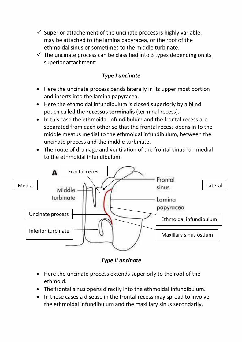
Type I uncinate
Here the uncinate process bends laterally in its upper most portion and inserts into the lamina papyracea.
Here the ethmoidal infundibulum is closed superiorly by a blind pouch called the recessus terminalis (terminal recess).
In this case the ethmoidal infundibulum and the frontal recess are separated from each other so that the frontal recess opens in to the middle meatus medial to the ethmoidal infundibulum, between the uncinate process and the middle turbinate.
The route of drainage and ventilation of the frontal sinus run medial to the ethmoidal infundibulum.
Type II uncinate
Here the uncinate process extends superiorly to the roof of the ethmoid.
The frontal sinus opens directly into the ethmoidal infundibulum.
In these cases a disease in the frontal recess may spread to involve the ethmoidal infundibulum and the maxillary sinus secondarily.
Uncinate process
Inferior turbinate Maxillary sinus ostium
Ethmoidal infundibulum
Frontal recess
Medial Lateral
Type III uncinate
Here the superior end of the uncinate process turns medially to get attached to the middle turbinate.
Here also the frontal sinus drains directly into the ethmoidal infundibulum, causing same problem as in type 2 uncinate.
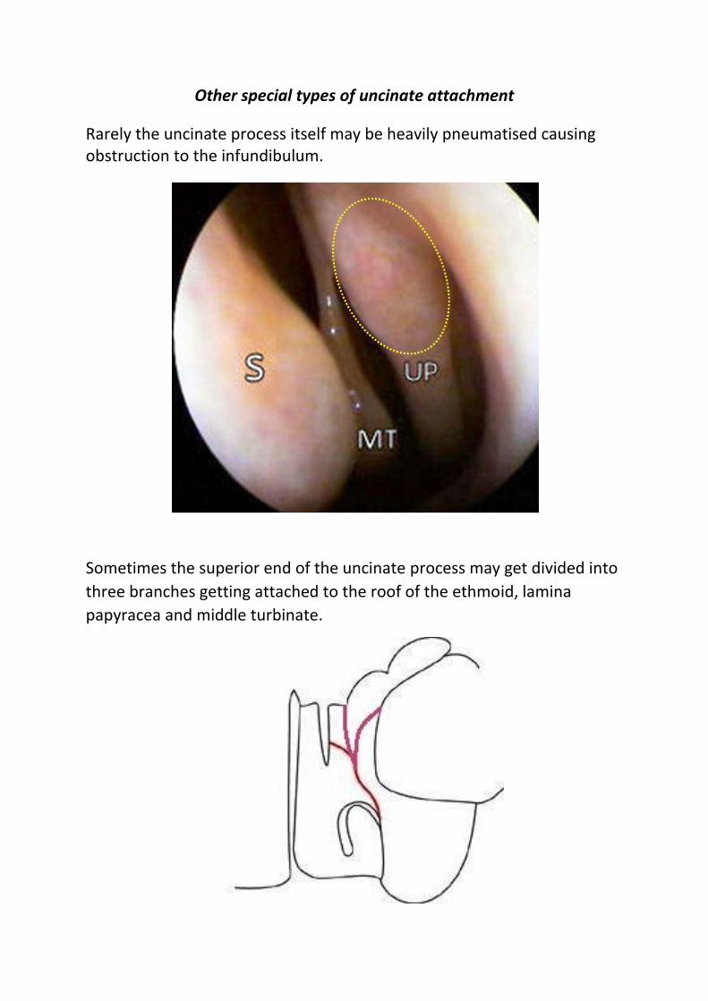
Other special types of uncinate attachment
Rarely the uncinate process itself may be heavily pneumatised causing obstruction to the infundibulum.
Sometimes the superior end of the uncinate process may get divided into
three branches getting attached to the roof of the ethmoid, lamina
papyracea and middle turbinate.
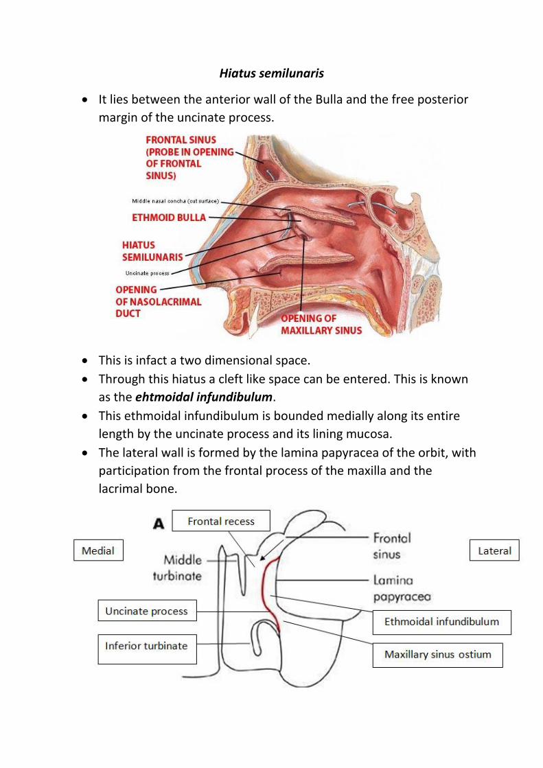
Hiatus semilunaris
It lies between the anterior wall of the Bulla and the free posterior
margin of the uncinate process.
This is infact a two dimensional space.
Through this hiatus a cleft like space can be entered. This is known
as the ehtmoidal infundibulum.
This ethmoidal infundibulum is bounded medially along its entire
length by the uncinate process and its lining mucosa.
The lateral wall is formed by the lamina papyracea of the orbit, with
participation from the frontal process of the maxilla and the
lacrimal bone.
The anterior group of sinuses drain into this area. Infact this area
acts as a cess pool for all the secretions from the anterior group of
sinuses.
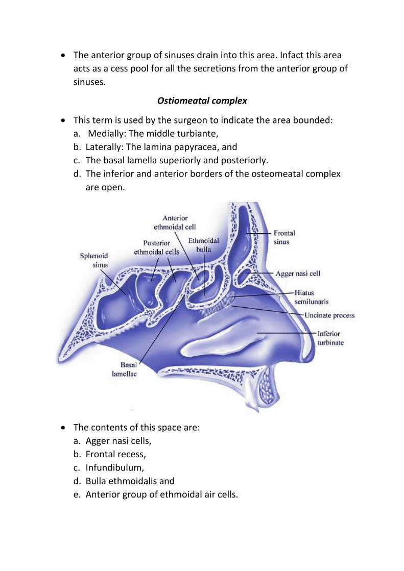
Ostiomeatal complex
This term is used by the surgeon to indicate the area bounded:
a. Medially: The middle turbiante,
b. Laterally: The lamina papyracea, and
c. The basal lamella superiorly and posteriorly.
d. The inferior and anterior borders of the osteomeatal complex
are open.
The contents of this space are:
a. Agger nasi cells,
b. Frontal recess,
c. Infundibulum,
d. Bulla ethmoidalis and
e. Anterior group of ethmoidal air cells.
Some authors divide this osteomeatal complex into anterior and
posterior.
The classic osteomeatal complex described already has been
described as the anterior osteomeatal complex, while the space
behind the basal lamella containing the posterior ethmoidal cells
is referred to as the posterior ethmoidal complex, thus recognising
the importance of basal lamella as an anatomical landmark to the
posterior ethmoidal system.
Hence the anterior and the posterior osteomeatal complex have
separate drainage systems, so when the disease is limited to the
anterior compartment of the osteomeatal complex, the ethmoid
cells can be opened and diseased tissue removed as far as the basal
lamella, leaving the basal lamella undisturbed minimising the risk
during surgery.
Pathologic basis of FESS:
As described above, the paranasal sinuses can be divided into
anterior and posterior groups.
The anterior group is always involved during sinus infections
before the posterior group of sinuses.
Infact the infected anterior group of sinuses predispose to posterior
sinus infections. Commonly the anterior ethmoidal sinuses are
involved first, causing a block in the drainage channels of other
sinuses.
The clearance of the disease should hence start from the anterior
ethmoidal group and proceed further posteriorly.
Anaesthesia:
This surgery is performed under local / general anesthesia using a rigid
nasal endoscope.
About endoscopes used in FESS:
Rigid nasal endoscope is nothing but an optical telescope of 4 mm
diameter. Telescopes which are capable of angular vision are also
available (30, 45, 70, 90 degrees). These angled telescopes can be used
to visualise the crevices inside the nasal cavity.
Procedure:
The nasal cavities are decongested using 4% xylocaine mixed with 1 in
10,000 adrenaline soaked cotton pledgets. The aim of decongesting the
nasal mucosa is:
a. To make the nasal cavity roomy hence facilitating endoscopic
visualisation.
b. To reduce bleeding during the surgical procedure.
General anesthesia is preferred in most of the cases. Administration of
hypotensive anesthesia using Nitroglycerine infusion will help in
reduction of bleeding during surgery, thus improving visualisation.
Steps of surgery:
1. The middle turbinate is gently medialised using a septal elevator. This
procedure helps to open up the middle meatal area thus facilitating
better visualisation.
2. Removal of the uncinate process:
This is the most important step of FESS surgery.
The incision can be made in the most anterior portion of the
uncinate process.
The uncinate bone is thin and soft when compared to the thicker
and firmer lacrimal bone.
The uncinate process should be removed completely. Incomplete
removal of uncinate process is cited to be most common cause of
failure of FESS.
A sickle knife can be used to make the incision.
A Blakesley forceps/ a back biting forceps can be used to totally
remove the incised uncinate process.
While performing uncinectomy care should be taken not to injure
the mucosa over adjacent middle turbinate, because it could
cause bleeding making visibility difficult.
Video link: http://www.sinusvideos.com/videos/69/uncinectomy-procedure-with-sickle-knife
3. Identification of natural ostium of maxillary sinus:
The natural ostium of maxillary sinus should be identified next. It is
typically present at the level of the inferior edge of the middle
turbinate, about 1/3 of the way back.
It becomes visible after resection of the uncinate process.
If it is not visible even after uncinectomy then it could either be
closed by diseased mucosa or may be hidden behind the posterior
remnant of the uncinate process. This ostium should be widened.
Ideal size of natural ostium is 1 cm.
Video link: http://www.youtube.com/watch?v=7OYqR_gBFY0
4. Opening of Bulla ethmoidalis:
Bulla should be opened in its inferior and medial aspect using a J
curette. A suction tip can also be used to open the bulla.
Anterior ethmoidal cells are cleared gradually.
Video link: http://www.youtube.com/watch?v=K3RmC_8hC48
5. To clear posterior ethmoids and sphenoid sinus the basal lamina