Functional Proteomics of Arabidopsis thaliana Guard Cells Uncovers New Stomatal Signaling Pathways W OA Zhixin Zhao, a Wei Zhang, a Bruce A. Stanley, b and Sarah M. Assmann a,1 1 Biology Department, Penn State University, University Park, Pennsylvania, 16802 b Section of Research Resources, Pennsylvania State University College of Medicine, Hershey, Pennsylvania 17033 We isolated a total of 3 3 10 8 guard cell protoplasts from 22,000 Arabidopsis thaliana plants and identified 1734 unique proteins using three complementary proteomic methods: protein spot identification from broad and narrow pH range two- dimensional (2D) gels, and 2D liquid chromatography–matrix assisted laser desorption/ionization multidimensional protein identification technology. This extensive single-cell-type proteome includes 336 proteins not previously represented in transcriptome analyses of guard cells and 52 proteins classified as signaling proteins by Gene Ontology analysis, of which only two have been previously assessed in the context of guard cell function. THIOGLUCOSIDE GLUCOHYDROLASE1 (TGG1), a myrosinase that catalyzes the production of toxic isothiocyanates from glucosinolates, showed striking abundance in the guard cell proteome. tgg1 mutants were hyposensitive to abscisic acid (ABA) inhibition of guard cell inward K + channels and stomatal opening, revealing that the glucosinolate-myrosinase system, previously identified only as a defense against biotic invaders, is required for key ABA responses of guard cells. Our results also suggest a mechanism whereby exposure to abiotic stresses may enhance plant defense against subsequent biotic stressors and exemplify how enhanced knowledge of the signaling networks of a specific cell type can be gained by proteomics approaches. INTRODUCTION Multicellular organisms develop specialized cell types, each with unique functions with regard to its specific role in the organism. The importance of single-cell-type transcriptomics studies in elucidating the functions of specialized cell types is uncontested (Dinneny et al., 2008). Single-cell-type proteomics studies are also essential to unravel the functions of specialized cells, particularly for cell types, like the guard cell (GC), where essential responses to stimuli can occur within seconds (Assmann and Grantz, 1990) and thus are unlikely to be mediated by tran- scriptomic changes. However, there have been very few single- cell-type proteomics studies in either plant or metazoan systems to date, in part owing to the greater complexity of the proteome and the greater technical challenges of proteomic methodolo- gies (Tyers and Mann, 2003). The most common subjects for single-cell-type proteomic studies have been cultured mammalian cell lines, where material is not limiting for proteomic analyses (Schirle et al., 2003; Diks and Peppelenbosch, 2004). In addition, studies have been done on human red blood cells (Pasini et al., 2006) and mouse red blood cells (Pasini et al., 2008), where 593 and 668 proteins were identified. By contrast, for plant cells, only the proteomes of Arabidopsis thaliana and tobacco (Nicotiana tabacum) trichomes (Wienkoop et al., 2004; Amme et al., 2005), Arabidopsis epider- mal cells (Wienkoop et al., 2004), and soybean (Glycine max) root hairs (Wan et al., 2005) have been assessed, and in each case only a handful of proteins were identified: 63 from Arabidopsis trichomes, 35 from tobacco trichomes, 26 from Arabidopsis epidermal cells, and 36 from root hairs. Arabidopsis pollen (a tricell microspore) has been more widely studied using proteo- mics because of the relative ease of isolation; however, only 135 (Holmes-Davis et al., 2005) and 121 (Noir et al., 2005) proteins have been identified from these studies. In contrast with previ- ously published studies that reported at most hundreds of proteins in single-cell proteomes from a multicellular organism, here we report the identification of 1734 proteins in the GC proteome of Arabidopsis. Carbon dioxide (CO 2 ) uptake for photosynthesis, as well as water vapor loss, occur through stomatal pores and are con- trolled by GC movements, regulated by changes in the turgor pressure and volume of GCs. The study of GC function is particularly topical given that climate change models predict that global warming will result in more frequent and more severe droughts (Breshears et al., 2005; Schroter et al., 2005) and that stomata have been implicated in physiological forcing of the global water cycle (Hetherington and Woodward, 2003; Betts et al., 2007). In addition to the central importance of GC function to terrestrial vegetation, the GC system has also become a model system in plant cell biology. GCs respond autonomously, di- rectly, rapidly, and reversibly to diverse environmental cues, including light, CO 2 , oxidative stress, humidity, and pathogens (Blatt, 2000; Fan et al., 2004, 2008; Israelsson et al., 2006; Pandey et al., 2007; Shimazaki et al., 2007; Acharya and Assmann, 2008). Stomatal apertures can be easily observed under the microscope, and direct measurement of ion channel 1 Address correspondence to [email protected]. The author responsible for distribution of materials integral to the findings presented in this article in accordance with the policy described in the Instructions for Authors (www.plantcell.org) is: Sarah M. Assmann ([email protected]). W Online version contains Web-only data. OA Open Access articles can be viewed online without a subscription. www.plantcell.org/cgi/doi/10.1105/tpc.108.063263 This article is a Plant Cell Advance Online Publication. The date of its first appearance online is the official date of publication. The article has been edited and the authors have corrected proofs, but minor changes could be made before the final version is published. Posting this version online reduces the time to publication by several weeks. The Plant Cell Preview, www.aspb.org ã 2008 American Society of Plant Biologists 1 of 17
Transcript
Functional Proteomics of Arabidopsis thaliana Guard CellsUncovers New Stomatal Signaling Pathways W OA
Zhixin Zhao,a Wei Zhang,a Bruce A. Stanley,b and Sarah M. Assmanna,1
1 Biology Department, Penn State University, University Park, Pennsylvania, 16802b Section of Research Resources, Pennsylvania State University College of Medicine, Hershey, Pennsylvania 17033
We isolated a total of 3 3 108 guard cell protoplasts from 22,000 Arabidopsis thaliana plants and identified 1734 unique
proteins using three complementary proteomic methods: protein spot identification from broad and narrow pH range two-
dimensional (2D) gels, and 2D liquid chromatography–matrix assisted laser desorption/ionization multidimensional protein
identification technology. This extensive single-cell-type proteome includes 336 proteins not previously represented in
transcriptome analyses of guard cells and 52 proteins classified as signaling proteins by Gene Ontology analysis, of which
only two have been previously assessed in the context of guard cell function. THIOGLUCOSIDE GLUCOHYDROLASE1
(TGG1), a myrosinase that catalyzes the production of toxic isothiocyanates from glucosinolates, showed striking
abundance in the guard cell proteome. tgg1 mutants were hyposensitive to abscisic acid (ABA) inhibition of guard cell
inward K+ channels and stomatal opening, revealing that the glucosinolate-myrosinase system, previously identified only as
a defense against biotic invaders, is required for key ABA responses of guard cells. Our results also suggest a mechanism
whereby exposure to abiotic stresses may enhance plant defense against subsequent biotic stressors and exemplify how
enhanced knowledge of the signaling networks of a specific cell type can be gained by proteomics approaches.
INTRODUCTION
Multicellular organisms develop specialized cell types, each with
unique functions with regard to its specific role in the organism.
The importance of single-cell-type transcriptomics studies in
elucidating the functions of specialized cell types is uncontested
(Dinneny et al., 2008). Single-cell-type proteomics studies are
also essential to unravel the functions of specialized cells,
particularly for cell types, like the guard cell (GC), where essential
responses to stimuli can occur within seconds (Assmann and
Grantz, 1990) and thus are unlikely to be mediated by tran-
scriptomic changes. However, there have been very few single-
cell-type proteomics studies in either plant or metazoan systems
to date, in part owing to the greater complexity of the proteome
and the greater technical challenges of proteomic methodolo-
gies (Tyers and Mann, 2003).
The most common subjects for single-cell-type proteomic
studies have been cultured mammalian cell lines, where material
is not limiting for proteomic analyses (Schirle et al., 2003; Diks
and Peppelenbosch, 2004). In addition, studies have been done
on human red blood cells (Pasini et al., 2006) and mouse red
blood cells (Pasini et al., 2008), where 593 and 668 proteins were
identified. By contrast, for plant cells, only the proteomes of
Arabidopsis thaliana and tobacco (Nicotiana tabacum) trichomes
(Wienkoop et al., 2004; Amme et al., 2005), Arabidopsis epider-
mal cells (Wienkoop et al., 2004), and soybean (Glycinemax) root
hairs (Wan et al., 2005) have been assessed, and in each case
only a handful of proteins were identified: 63 from Arabidopsis
trichomes, 35 from tobacco trichomes, 26 from Arabidopsis
epidermal cells, and 36 from root hairs. Arabidopsis pollen (a
tricell microspore) has been more widely studied using proteo-
mics because of the relative ease of isolation; however, only 135
(Holmes-Davis et al., 2005) and 121 (Noir et al., 2005) proteins
have been identified from these studies. In contrast with previ-
ously published studies that reported at most hundreds of
proteins in single-cell proteomes from a multicellular organism,
here we report the identification of 1734 proteins in the GC
proteome of Arabidopsis.
Carbon dioxide (CO2) uptake for photosynthesis, as well as
water vapor loss, occur through stomatal pores and are con-
trolled by GC movements, regulated by changes in the turgor
pressure and volume of GCs. The study of GC function is
particularly topical given that climate change models predict
that global warming will result in more frequent and more severe
droughts (Breshears et al., 2005; Schroter et al., 2005) and that
stomata have been implicated in physiological forcing of the
global water cycle (Hetherington and Woodward, 2003; Betts
et al., 2007). In addition to the central importance of GC function
to terrestrial vegetation, theGCsystemhas also become amodel
system in plant cell biology. GCs respond autonomously, di-
rectly, rapidly, and reversibly to diverse environmental cues,
including light, CO2, oxidative stress, humidity, and pathogens
(Blatt, 2000; Fan et al., 2004, 2008; Israelsson et al., 2006;
Pandey et al., 2007; Shimazaki et al., 2007; Acharya and
Assmann, 2008). Stomatal apertures can be easily observed
under the microscope, and direct measurement of ion channel
1 Address correspondence to [email protected] author responsible for distribution of materials integral to thefindings presented in this article in accordance with the policy describedin the Instructions for Authors (www.plantcell.org) is: Sarah M. Assmann([email protected]).WOnline version contains Web-only data.OAOpen Access articles can be viewed online without a subscription.www.plantcell.org/cgi/doi/10.1105/tpc.108.063263
This article is a Plant Cell Advance Online Publication. The date of its first appearance online is the official date of publication. The article has been
edited and the authors have corrected proofs, but minor changes could be made before the final version is published. Posting this version online
reduces the time to publication by several weeks.
The Plant Cell Preview, www.aspb.org ã 2008 American Society of Plant Biologists 1 of 17
and pump activity can be attained by electrophysiological
assays. Ions (Ca2+, K+, Cl2, malate22, and NO32), signaling
elements (e.g., G-proteins, phospholipase C [PLC], phospholi-
pase D [PLD], inositol 1,4,5-trisphosphate, nitric oxide [NO], and
reactive oxygen species [ROS]), and plant hormones (e.g.,
abscisic acid [ABA], auxin, and ethylene) regulate stomatal
movements. With the advent of systems biology techniques, a
dynamic model for induction of stomatal closure by the drought
and stress hormone, ABA, has been developed (Li et al., 2006).
However, despite these achievements, basic questions re-
main unanswered: Are there proteins or sets of proteins that are
preferentially expressed in GCs? How does the GC proteome
compare with that of other plant cell types, and, importantly, how
can proteomics inform studies of GC signaling and reveal new
functions and relationships? These questions are addressed by
this study.
RESULTS
Proteomic Methods and GC Proteins
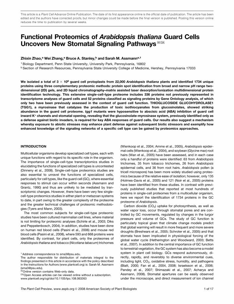
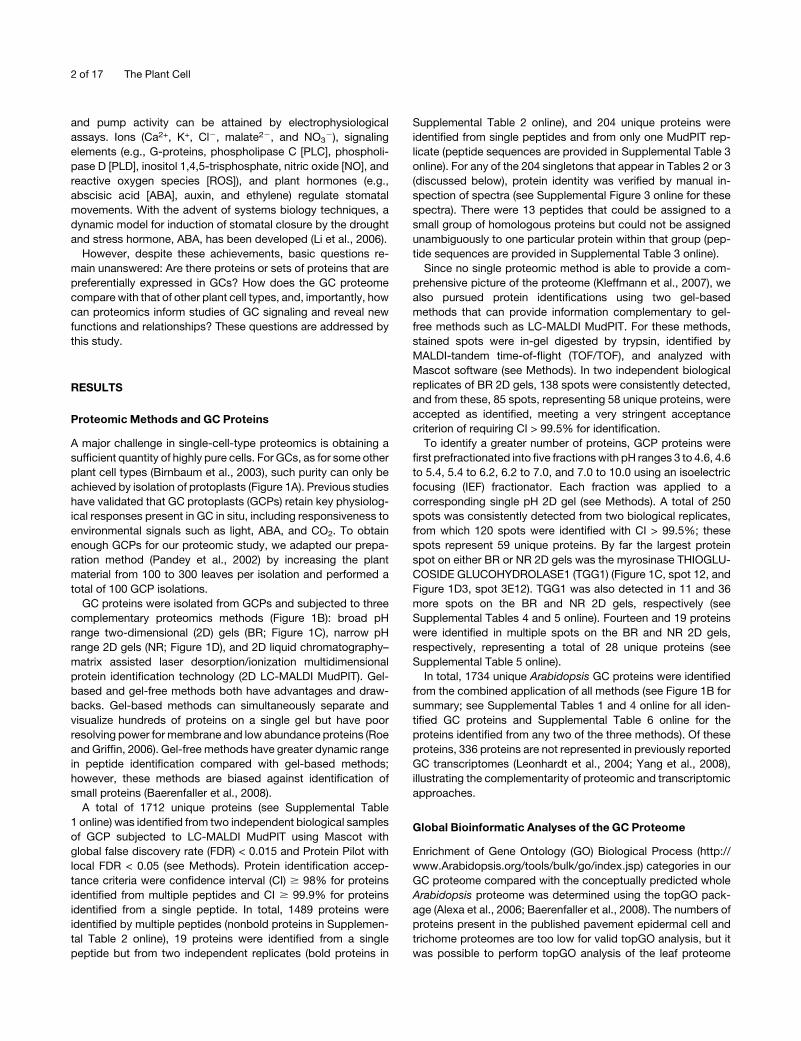
A major challenge in single-cell-type proteomics is obtaining a
sufficient quantity of highly pure cells. For GCs, as for some other
plant cell types (Birnbaum et al., 2003), such purity can only be
achieved by isolation of protoplasts (Figure 1A). Previous studies
have validated that GC protoplasts (GCPs) retain key physiolog-
ical responses present in GC in situ, including responsiveness to
environmental signals such as light, ABA, and CO2. To obtain
enough GCPs for our proteomic study, we adapted our prepa-
ration method (Pandey et al., 2002) by increasing the plant
material from 100 to 300 leaves per isolation and performed a
total of 100 GCP isolations.
GC proteins were isolated from GCPs and subjected to three
(B) A total of 1712, 58, and 59 unique proteins were identified from 2D LC-MALDI MudPIT, BR, and NR methods, respectively; 19 proteins were
identified by all three methods. For each method, two independent biological samples were analyzed.
(C) A 2D gel image from the broad pH range method. The first dimension was run using a 24-cm, pH 3 to 10 IPG strip. In total, 138 protein spots were
detected via Coomassie blue staining. Twelve spots were identified as TGG1. Identifications of numbered spots can be found in Supplemental Table 5
online.
(D) 2D gel images from the narrow pH range method. Proteins were first fractionated into five fractions, and each protein fraction was separated on a
narrow pH range IPG strip. From B1 to B5, the pH ranges are 3 to 6, 4.5 to 5.5, 5.3 to 6.3, 6.1 to 7.1, and 6 to 10 respectively. Thirty-seven spots were
identified as TGG1. Identifications of numbered spots can be found in Supplemental Table 5 online.
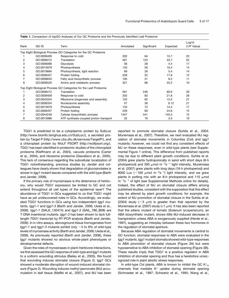
4 of 17 The Plant Cell
TGG1 is predicted to be a cytoplasmic protein by SubLoc
(http://www.bioinfo.tsinghua.edu.cn/SubLoc/), a secreted pro-
tein by Target P (http://www.cbs.dtu.dk/services/TargetP/), and
a chloroplast protein by WoLF PSORT (http://wolfpsort.org/).
TGG1 has been identified in proteomic studies of the chloroplast
proteome (Kleffmann et al., 2004), vacuole proteome (Carter
et al., 2004), and ribosome proteome (Giavalisco et al., 2005).
This lack of consensus regarding the subcellular localization of
TGG1 notwithstanding, previous studies by Jander and col-
leagues have clearly shown that degradation of glucosinolates is
slower in tgg1mutant leaves compared with the wild type (Barth
and Jander, 2006).
If the primary role of myrosinases is the deterrence of herbiv-
ory, why would TGG1 expression be limited to GC and not
extend throughout all cell types of the epidermal layer? The
abundance of TGG1 in GCs suggested to us that TGG1 might
have as yet undiscovered roles in GCs. Accordingly, we evalu-
ated TGG1 functions in GCs using two independent tgg1 mu-
tants, tgg1-1 and tgg1-3 (Barth and Jander, 2006; Ueda et al.,
2006). tgg1-1 (SALK_130474) and tgg1-3 (SAIL_786_B08) are
T-DNA insertional mutants. tgg1-3 has been shown to lack full-
length TGG1 transcript by RT-PCR analysis (Barth and Jander,
2006). In in vitro assays, aboveground tissue homogenates from
tgg1-1 and tgg1-3 mutants exhibit only ;5 to 8% of wild-type
levels ofmyrosinase activity (Barth and Jander, 2006; Ueda et al.,
2006). As previously reported (Barth and Jander, 2006), these
tgg1 mutants showed no obvious whole-plant phenotypes or
developmental defects.
Given the roles of myrosinases in plant–herbivore interactions,
we first assessed theGC response ofwild-type and tgg1mutants
to a uniform wounding stimulus (Bailey et al., 2005). We found
that wounding induces stomatal closure (Figure 2). tgg1 GCs
showed a moderate disruption of wound-induced stomatal clo-
At5g16590 Leu-rich repeat transmembrane protein kinase
At5g19390 Similar to pleckstrin homology domain-containing protein
At5g39500 Pattern formation protein
At5g53320 Leu-rich repeat transmembrane protein kinase
(Continued)
Functional Proteomics of Arabidopsis Guard Cells 7 of 17
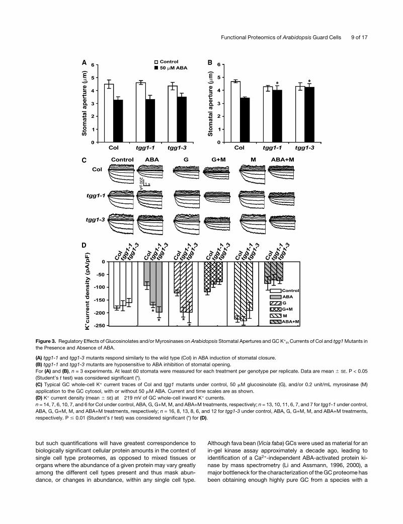
2001) (Figures 3C and 3D). In the absence of ABA treatment, tgg1
mutant GC had similar K+in current amplitudes and kinetics as
Col; however, ABA inhibition of K+in current was abolished in
both independent tgg1mutants (Figures 3C and 3D). Consistent
with the fact that tgg1mutants show awild-type response in ABA
promotion of stomatal closure, outward K+ currents, which
mediate K+ efflux during stomatal closure, were statistically
identical in Col versus tgg1 mutants either with or without ABA
treatment (Figure 3C; see Supplemental Figure 2 online).
Alterations in the glucosinolate profiles of tgg mutants have
already been characterized at the whole-leaf level, with signifi-
cant increases in aliphatic and indole glucosinolates primarily
observed in tgg1 tgg2 double mutants (Barth and Jander, 2006).
Since myrosinases hydrolyze glucosinolates, one key question
that arises is, what is functioning in the ABA inhibition of K+in
channels: myrosinase itself, glucosinolates, or the hydrolyzed
products of glucosinolates? As a first step toward addressing
this question, glucosinolates, myrosinase, or a combination of
glucosinolates and myrosinase was directly applied to the cyto-
sol of Col and tgg1 mutant GCs via the patch pipette solution
(Figures 3C and 3D). Whole-cell patch clamp data showed that
glucosinolate administration resulted in inhibition of K+in chan-
nels in Col GCs but not inGCs of tgg1mutants, indicating that the
glucosinolates themselves do not suffice to inhibit channel
activity. By contrast, coadministration of glucosinolates and
myrosinase resulted in a similar extent of inhibition of K+in
channels in both Col and tgg1 mutants, suggesting that it is the
hydrolyzed products of glucosinolates that evoke ion channel
inhibition. However, myrosinase addition alone had no effect on
K+in channels in either Col or tgg1 mutants, suggesting that, in
the absence of an appropriate triggering event, glucosinolate
substrates are not available for myrosinase action. Most impor-
tantly, even though myrosinase application alone had no effect
on K+in currents, application of myrosinase restored K+
in sensi-
tivity to ABA in tgg1 GCs (Figures 3C and 3D). These results
indicate that the hydrolysis of glucosinolates by myrosinases is
induced in some manner by ABA in Arabidopsis GCs (see Figure
4 and Discussion) and is a necessary component of ABA-
mediated K+in channel inhibition.
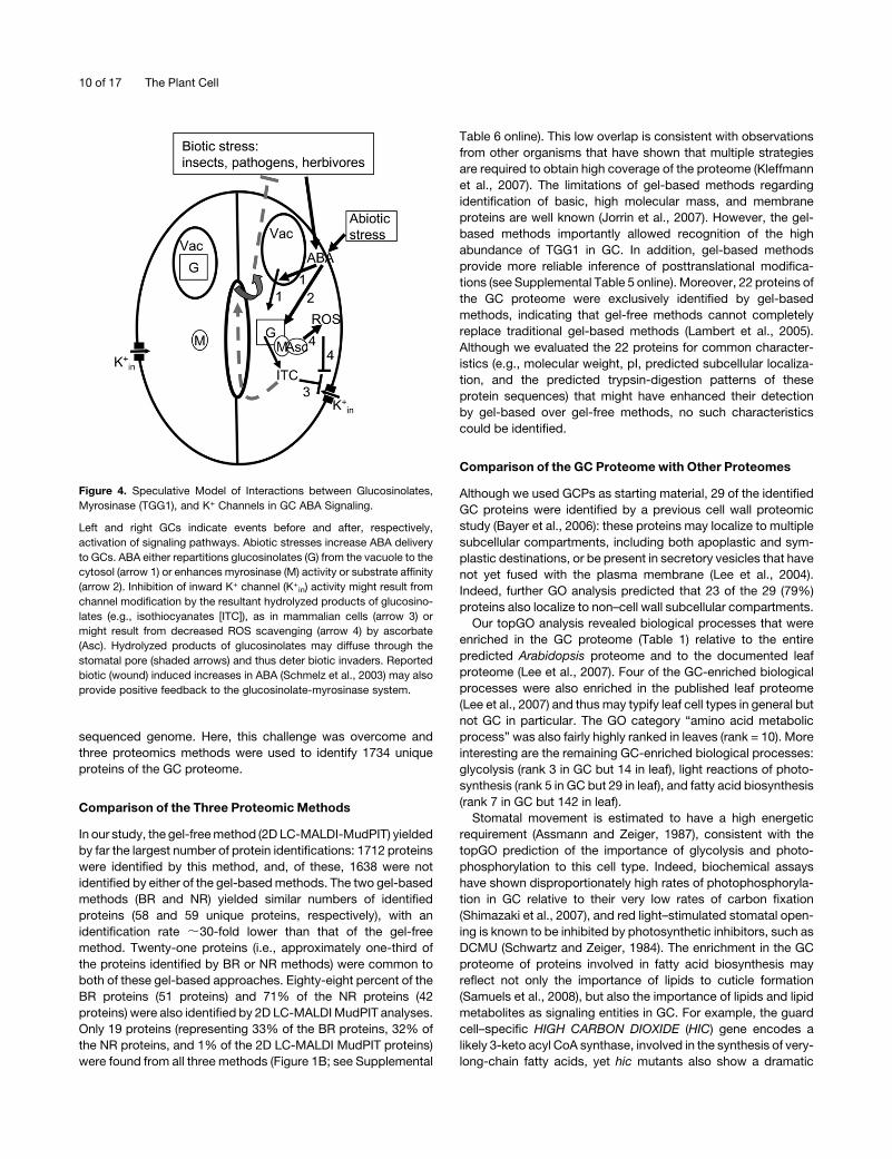
DISCUSSION
Proteomics, an important postgenomic approach, has been ap-
plied tomany fields (e.g., identification of protein expression pro-
file changes under stress conditions) (Hashimoto and Komatsu,
2007), analysis of posttranslational modifications (Kwon et al.,
2006), and determination of protein–protein interactions (Parrish
et al., 2007). Quantitative proteomic methods are also emerging,
Table 3. (continued).
AGI Name
At5g58440 Phox domain–containing protein
At5g60600 GCPE
At5g63310 NDPK2
At5g67030 ABA1
Proteins in boldface were shown to play a role in GC function in previous studies. Proteins in italics were identified by a single peptide from one
MudPIT replicate. Corresponding peptide sequences and spectra for these proteins are provided in Supplemental Table 3 and Supplemental Figure 3
online, respectively.
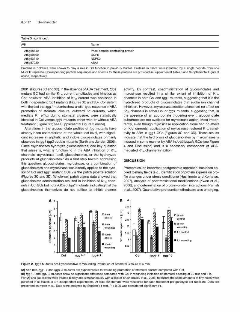
Figure 2. tgg1 Mutants Are Hyposensitive to Wounding Promotion of Stomatal Closure at 5 min.
(A) At 5 min, tgg1-1 and tgg1-3 mutants are hyposensitive to wounding promotion of stomatal closure compared with Col.
(B) tgg1-1 and tgg1-3 mutants show no significant difference compared with Col in wounding inhibition of stomatal opening at 30 min and 1 h.
For (A) and (B), leaves were treated blindly and simultaneously with a slicker brush (Bailey et al., 2005) to ensure the same amounts of tiny holes were
punched in all leaves. n = 4 independent experiments. At least 60 stomata were measured for each treatment per genotype per replicate. Data are
presented as mean 6 SE. Data were analyzed by Student’s t test, P < 0.05 was considered significant (*).
8 of 17 The Plant Cell
but such quantifications will have greatest correspondence to
biologically significant cellular protein amounts in the context of
single cell type proteomes, as opposed to mixed tissues or
organs where the abundance of a given protein may vary greatly
among the different cell types present and thus mask abun-
dance, or changes in abundance, within any single cell type.
Although fava bean (Vicia faba) GCs were used as material for an
in-gel kinase assay approximately a decade ago, leading to
identification of a Ca2+-independent ABA-activated protein ki-
nase by mass spectrometry (Li and Assmann, 1996, 2000), a
major bottleneck for the characterization of theGCproteome has
been obtaining enough highly pure GC from a species with a
Figure 3. Regulatory Effects of Glucosinolates and/or Myrosinases on Arabidopsis Stomatal Apertures and GC K+in Currents of Col and tgg1Mutants in
the Presence and Absence of ABA.
(A) tgg1-1 and tgg1-3 mutants respond similarly to the wild type (Col) in ABA induction of stomatal closure.
(B) tgg1-1 and tgg1-3 mutants are hyposensitive to ABA inhibition of stomatal opening.
For (A) and (B), n = 3 experiments. At least 60 stomata were measured for each treatment per genotype per replicate. Data are mean 6 SE. P < 0.05
(Student’s t test) was considered significant (*).
(C) Typical GC whole-cell K+ current traces of Col and tgg1 mutants under control, 50 mM glucosinolate (G), and/or 0.2 unit/mL myrosinase (M)
application to the GC cytosol, with or without 50 mM ABA. Current and time scales are as shown.
(D) K+ current density (mean 6 SE) at �219 mV of GC whole-cell inward K+ currents.
n = 14, 7, 6, 10, 7, and 6 for Col under control, ABA, G, G+M, M, and ABA+M treatments, respectively; n = 13, 10, 11, 6, 7, and 7 for tgg1-1 under control,
ABA, G, G+M, M, and ABA+M treatments, respectively; n = 16, 8, 13, 8, 6, and 12 for tgg1-3 under control, ABA, G, G+M, M, and ABA+M treatments,
respectively. P # 0.01 (Student’s t test) was considered significant (*) for (D).
Functional Proteomics of Arabidopsis Guard Cells 9 of 17
sequenced genome. Here, this challenge was overcome and
three proteomics methods were used to identify 1734 unique
proteins of the GC proteome.
Comparison of the Three Proteomic Methods
In our study, the gel-freemethod (2D LC-MALDI-MudPIT) yielded
by far the largest number of protein identifications: 1712 proteins
were identified by this method, and, of these, 1638 were not
identified by either of the gel-basedmethods. The two gel-based
methods (BR and NR) yielded similar numbers of identified
proteins (58 and 59 unique proteins, respectively), with an
identification rate ;30-fold lower than that of the gel-free
method. Twenty-one proteins (i.e., approximately one-third of
the proteins identified by BR or NR methods) were common to
both of these gel-based approaches. Eighty-eight percent of the
BR proteins (51 proteins) and 71% of the NR proteins (42
proteins) were also identified by 2D LC-MALDIMudPIT analyses.
Only 19 proteins (representing 33% of the BR proteins, 32% of
the NR proteins, and 1% of the 2D LC-MALDI MudPIT proteins)
were found from all three methods (Figure 1B; see Supplemental
Table 6 online). This low overlap is consistent with observations
from other organisms that have shown that multiple strategies
are required to obtain high coverage of the proteome (Kleffmann
et al., 2007). The limitations of gel-based methods regarding
identification of basic, high molecular mass, and membrane
proteins are well known (Jorrin et al., 2007). However, the gel-
based methods importantly allowed recognition of the high
abundance of TGG1 in GC. In addition, gel-based methods
provide more reliable inference of posttranslational modifica-
tions (see Supplemental Table 5 online). Moreover, 22 proteins of
the GC proteome were exclusively identified by gel-based
methods, indicating that gel-free methods cannot completely
replace traditional gel-based methods (Lambert et al., 2005).
Although we evaluated the 22 proteins for common character-