HAL Id: hal-00814288 https://hal.archives-ouvertes.fr/hal-00814288 Submitted on 17 Jul 2013 HAL is a multi-disciplinary open access archive for the deposit and dissemination of sci- entific research documents, whether they are pub- lished or not. The documents may come from teaching and research institutions in France or abroad, or from public or private research centers. L’archive ouverte pluridisciplinaire HAL, est destinée au dépôt et à la diffusion de documents scientifiques de niveau recherche, publiés ou non, émanant des établissements d’enseignement et de recherche français ou étrangers, des laboratoires publics ou privés. Functionalized nanomaterials: their use as contrast agents in bioimaging : mono- and multimodal approaches Quentin Le Trequesser, Hervé Seznec, Marie-Hélène Delville To cite this version: Quentin Le Trequesser, Hervé Seznec, Marie-Hélène Delville. Functionalized nanomaterials: their use as contrast agents in bioimaging: mono- and multimodal approaches. Nanotechnology Reviews, 2013, 2 (2), pp.125-169. <10.1515/ntrev-2012-0080>. <hal-00814288>
Transcript
HAL Id: hal-00814288https://hal.archives-ouvertes.fr/hal-00814288
Submitted on 17 Jul 2013
HAL is a multi-disciplinary open accessarchive for the deposit and dissemination of sci-entific research documents, whether they are pub-lished or not. The documents may come fromteaching and research institutions in France orabroad, or from public or private research centers.
L’archive ouverte pluridisciplinaire HAL, estdestinée au dépôt et à la diffusion de documentsscientifiques de niveau recherche, publiés ou non,émanant des établissements d’enseignement et derecherche français ou étrangers, des laboratoirespublics ou privés.
Functionalized nanomaterials : their use as contrastagents in bioimaging : mono- and multimodal approaches
Quentin Le Trequesser, Hervé Seznec, Marie-Hélène Delville
To cite this version:Quentin Le Trequesser, Hervé Seznec, Marie-Hélène Delville. Functionalized nanomaterials : their useas contrast agents in bioimaging : mono- and multimodal approaches. Nanotechnology Reviews, 2013,2 (2), pp.125-169. <10.1515/ntrev-2012-0080>. <hal-00814288>
cardiovascular, pulmonary, and tumor imaging, as well as for pharmacokinetic evaluation
[161].
3.1 Radionuclide Labeled Nanoparticles
Common isotopes that can be chelated on to or incorporated within NPs (in an analogous way
to the gadolinium ions used for MRI) include 18F, 11C, 15O, 13N, 64Cu, 124I, 68Ga, 82Rb, and 86Y. PET imaging using 18F, which is the most widespread radionuclide probe used in this
field, has become an established clinical tool for whole-body imaging. In light its short half-
life, its quick conjugation into the probe with a high reaction yield is necessary to improve its
efficiency and to reduce cost. A general synthesis strategy for such 18F-labeled rare-earth
nanoparticles was developed through a facile inorganic reaction between rare-earth cations
(Y3+ and Gd3+) and fluoride ions [165]. The 18F-labeled rare-earth NPs were further evaluated
by PET imaging, for their in vivo distribution and their application in lymph monitoring.
There are, however, other types of probes and the main nanoparticle-based PET ones and their
labeling radionuclide are reported in Table 2.
Table 2. Labeling Strategies and Specific Activities of PET Radionuclides Labeled Nanoparticles and the Nuclear Characteristics of the corresponding PET Radionuclides. Adapted from Ref. [164] and up-dated.
NPs labeling strategy radionuclide T1/2 β energy (KeV) main photon KeV (%)
Ref.
decay (%) max. Mean.
QDs nucleophilic substitution
18F 109.7 min
β+ (96.7) EC (0.1)
634 245 511 (193.5) [166]
DOTA 64Cu 12.7 h β+ (17) EC (44)
653 278 511 (34.8) [167]
DO3A 64Cu [168] Iron oxide Click chemistry 18F [34,
169] DOTA 64Cu [170]
[171] [172]
dithiocarbamatebisphosphonate
64Cu [173]
Direct labeling 68Ga 67.7 min
β+ (89) EC (11)
1899 829 511 (178.3) [174]
Chelation 64Cu [175] NOTA 68Ga [176] Tyrosine 124I 4.18 d β+ (23)
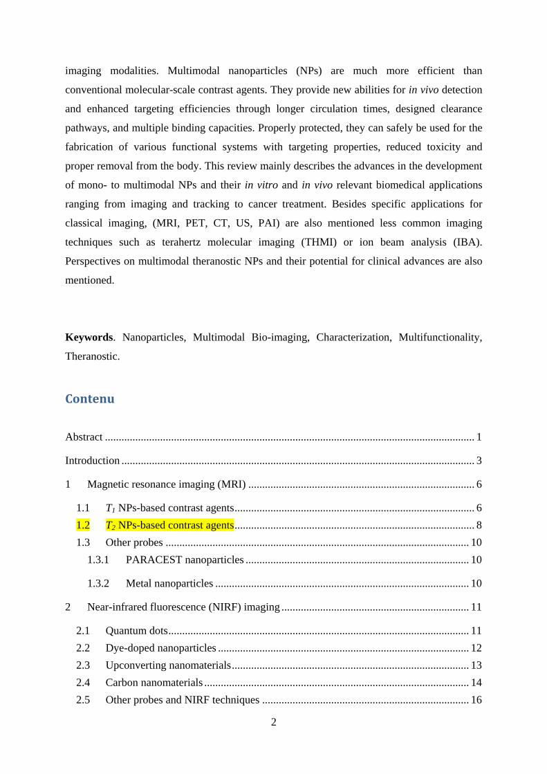
The pharmacokinetic and in vivo cancer targeting issues of 64Cu2+ ions functionalized gold
nanocages (NCs) (64Cu-DOTA-PEGAuNCs) when followed by PET imaging in normal
rodents revealed Au NCs size dependent [161]. 30 nm Au NCs showed much-improved in
vivo pharmacokinetics with decreased RES system uptake and enhanced blood circulation as
compared to 55 nm ones. The PET/CT imaging demonstrated rapid accumulation and
centralized distribution of the 30 nm Au NCs in tumors and, more importantly, high tumor-to-
muscle ratios. PET/CT images (Figure 4) clearly showed this rapid localization of the 30 nm 64Cu-DOTAPEG-Au NCs in tumors at 1 h post-injection even only with the administration of
a trace amount (23.8 fmol).
Figure 4 here
Most of the time PET tracers are incorporated with another modality in NPs, most notably CT
[199-202]. Figure 5 illustrates the use 18F-doped cross-linked iron oxide modified tri-modal
NPs (18F-CLIO) to image the liver and blood pool of a mouse. The in vivo dynamic PET
imaging showed very high signal-to-noise ratios for injected 18F-CLIO. The nanoparticle had
a vascular half-life of 5.8 h in mice and was internalized into macrophages of liver, spleen,
19
and phagocytic cells of other lymphatic organs. The NP is additionally biodegradable and
breaks down into elemental components within months. PET/CT allows concentrations at 2-4
orders of magnitude lower than those required for MR imaging which likely makes PET
imaging an important platform for clinical molecular.
Figure 5 here
Biocompatible inorganic NPs such as hydroxyapatite NPs also revealed as useful PET/CT
probes [178]. They showed particularly avid and stable binding of 18F-fluoride in various
biological media. The in vivo behaviour of the 18F-labelled hydroxyapatite particles
determined by PET-CT imaging in mice showed that hydroxyapatite was stable in circulation
but its accumulation in liver via reticuloendothelial clearance was followed by gradual
degradation and release of 18F-fluoride (over a period of 4 h) which then accumulated in bone.
3.2 Coupling with other contrast agents
Among the other modalities that have been combined with PET, should be mentioned ultra-
small cancer-selective silica particles grafted with iodine and dye-doped polymers which were
recently approved by FDA for in-human clinical trials [203], NIRF agents within the silica
NPs [204] or QDs [168, 205, 206] and MRI [199] agents in conjunction with iron oxide
nanomaterials [35, 171]. A bifunctional chelator (dithiocarbamate bisphosphonate, (dtcbp))
containing a dithiocarbamate group for binding the PET isotope 64Cu, and a bisphosphonate
group for strong binding to Fe3O4 was elaborated. Dtcbp efficiently binds 64Cu to form the
[64Cu(dtcbp)2] complex which is then grafted on the iron oxide NPs leading to a PET-MR
dual modality imaging capabilities of which in vivo accumulates in draining lymph nodes
(Figure 6) [173].
Figure 6 here
However, the ratio between PET tracer and MRI contrast agent must be carefully controlled,
because PET is extremely sensitive whereas MRI is not.
Single-photon emission computed tomography (SPECT), a similar technique which can also
detect nanomoles of tracer. SPECT is based on the detection of lower energy γ-emitting
radioisotopes such as 99mTc, 111In, 123I, and 131I [14, 207-209]. As compared to PET, SPECT
has the advantage to be more sensitive and versatile, it is cheaper and more widely available
20
as it does not rely on a local cyclotron for production of isotopes even if it is an order of
magnitude less sensitive than PET.
Covalent functionalization of radionuclide-filled single-walled carbon nanotubes were used as
radio-probes [210]. The intravenous administration of these 125I loaded SWNTs was tracked
in vivo using SPECT. Specific tissue accumulation (here lung) coupled with high in vivo
stability prevented leakage of radionuclide to high-affinity organs (thyroid/stomach) or
excretion, and resulted in ultrasensitive imaging and delivery of unprecedented radio-dose
density. The nano-encapsulation of iodide within SWNTs enabled its biodistribution to be
completely redirected from tissue with innate affinity (thyroid) to lung (Figure 7).
Figure 7 here
4 X-ray imaging and computed tomography (CT)
X-ray computed tomography (CT) is one of the most powerful noninvasive diagnostic
imaging techniques in modern medicine. It has been a clinical tool for more than half a
century and the first widespread clinical use of NPs as X-ray contrast agents in humans were
3 to 10-nm thorium dioxide nanoparticles [12, 211]. However due to the long-term radiation
effects and significant carcinogenicity of the 232Th, its clinical application was rapidly given
up.
Iodinated molecules were then used as CT contrast agents in the clinics. They however have
relatively short in vivo circulation times, which significantly restrict the applications of this
technique in target-specific imaging and angiography. The use of large dose of these agents,
which may induce serious adverse effects as well as a hypersensitivity to iodine of some
patients, led researchers to address these issues. Over the past decade, advances in
nanoscience brought some solutions thanks to the unique properties of nanomaterials, such as
their prolonged circulating half-life, passive accumulation at the tumor sites, facile surface
modification, and integration of multiple functions into a single particle, make them
advantageous for in vivo applications [212].
4.1 Iodinated nanoparticles
The widespread clinical use of iodinated compounds has encouraged the development of
iodinated nanomaterials. Research essentially focuses [213, 214] on the incorporation of
iodinated organic compounds into NPs, with designs ranging from emulsions, [215-218]
21
liposomes, [214, 219-223] and lipoproteins, [224, 225] to insoluble nano objects, [214, 226-
228] and polymeric NPs, [215, 229-233]; many of them have been successfully applied in
vivo [28, 56]. The purpose of these nanomaterials is to locally increase iodine concentrations,
resulting in higher local contrast compared with conventional water-soluble CT contrast
agents. A key feature of many of these NPs is their pharmacokinetics, which are often
markedly different from those of small iodinated molecules in clinical use. They have
increased circulation time with the subsequent implications for targeting because longer
circulation times increase the chance of interaction and binding of the contrast agent to a
target.
Some multimodal nanoparticles were doped with an iodinated compound and used to enhance
X-ray contrast [216, 224, 234, 235] as discussed in later on in the review. Iodine has a lower
atomic number than gold and bismuth, it however exhibits higher elemental mass attenuation
coefficient and incident X-ray energies so that when compared to gold NPs under conditions
used for coronary angiography, iodinated contrast agents had equivalent performance [236].
When a contrast agent was developed by combining the two radio-dense elements iodine and
gold within a single PAMAM dendrimer, it was demonstrated that the incorporation of both
Au NPs and iodine-containing small molecules resulted in a significant cooperative enhancing
effect in X-ray attenuation [237].
4.2 Gold nanoparticles
In addition to the clinically used iodine, the element gold has received much attention due to
its higher atomic number than iodine, and thus, a larger contribution of photoelectron effect to
X-ray attenuation generating a substantial interest in gold NP-based contrast agents for in vivo
X-ray CT. Gold nano-objects contain a large number of the contrast element (Au) as opposed
to iodine-based nanoparticles in which iodinated molecules are often only covalently grafted
onto the NP surface thereby lowering the concentration of the agent. Various gold, [10, 61,
238-240] gold@dielectric hybrids, [10, 241-246] and multimodal materials, [96, 246-248]
have been fabricated and their in vivo functionality as X-ray CT contrast agents for cancer,
tissue-specific, and blood-pool imaging. Gold nanomaterials are currently being explored in
multiple clinical trials and they constitute a promising next generation candidate for X-ray
contrast materials, radiotherapy [249-251] and cancer therapy [252].
4.3 Other contrast materials
22
Other CT molecular imaging agents have also been studied besides iodinated and gold-based
ones. These NPs consist of bismuth sulfide and composite ceramics containing iron oxide and
lanthanide materials. Bismuth sulfide NPs have recently been shown to have superior
performance to iodine on a molar basis [253-255]. Even if bismuth displays similar mass
attenuation coefficients to that of gold and a higher k-edge transition, its toxicity may prevent
bismuth-based nanomaterials clinical use as CT agents. Other types of CT contrast agents, are
those based on iron oxide [256] and have recently been reviewed [94, 257, 258]. A facile
approach for fabrication of Fe3O4@Au NPs as a dual mode contrast agent for both magnetic
resonance (MR) and computed tomography (CT) imaging applications has been performed
via the combination of a LbL (layer by layer) self-assembly process and dendrimer chemistry
[259]. The use of Fe3O4@Au NPs as a contrast agent for dual mode MR/CT imaging has been
demonstrated not only for in vitro imaging of cancer cells, but also for in vivo liver imaging
via MR and subcutaneous tissue imaging via CT.
The high atomic weight and large number of unpaired electron makes Gadolinium an
excellent contrasting agent both for MRI and CT imaging [12, 260-262]. Therefore, most of
the time, NPs are at least dual probe: CT/MRI or CT/NIRF [261, 263, 264]. As an example
Gd3+ complex-modified NaLuF4-based upconversion nanophosphors were success fully
applied for UCL, MR and CT multi-modal imaging, by integrating NIR-to-NIR UCL, X-ray
attenuation and paramagnetic function in one single nanoparticle. The property of NIR-to-
NIR UCL enhances the high signal-to-noise ratio of in vivo imaging of small animal.
Moreover, Gd3+ grafting on the surface of NPs generates high r1 relaxivity for the core–shell
nanoparticles which become suitable for T1 MR imaging. In addition, this nanoparticle with
the core of Yb3+ and Tm3+-doped NaLuF4 shows a high X-ray attenuation and are good for CT
imaging. The SiO2 shell reduces the toxicity of these lanthanide-based nanoparticles for small
animals, which was confirmed by MTT assays and histological analyses. The incorporation of
different lanthanide ions of Lu3+, Yb3+, Tm3+ and Gd3+ into one particle,
NaLuF4:Yb3+,Tm3+@SiO2-GdDTPA, provides a facile design strategy to fabricate
multimodality imaging agents [261].
Yb-based NPs (NaYbF4:Er) which already revealed to be efficient upconversion
nanophosphors (UCNPs) have also recently been identified as CT contrasts agents [265].
When encapsulated in a polymer (PEG) shell they showed low cytotoxicity and long
circulation time in vivo. And a much higher efficacy as compared to the clinical iodinate
agents commonly used. This improvement was attributed to the K-edge energy of Yb that
locates within the higher energy region of the X-ray spectrum. Furthermore, a gadolinium
23
doping in these nanoparticles endowed them with enhanced fluorescence as well as NMR
imaging capabilities providing clearance issues are solved.
By virtue of its high atomic number and this well-positioned K-edge, Yb provides excellent
spectral CT contrast both in vitro and in vivo.[266] To partly solve bioelimination and
preliminary biodistribution issues Yb nanocolloids were used as spectral CT contrast agents.
The synthetic approach involved an organically soluble organometallic Yb(III)-2,4-
pentadionate complex to produce polysorbate encapsulated nanocolloids of Yb incorporating
a high density of Yb (>500K/nanoparticle) into a stable metal particle. Such high payloads of
the Yb in the form of hydrophobic small molecule metal complexes could be obtained and
stably concentrated into lipid-encapsulated nanocolloids and provide novel molecular imaging
for use for spectral“multicolor” computed tomography (CT).
5 Ultrasound (US)
US is also a well-established clinical imaging modality. In particular, it is routinely used to
characterize lesions in liver, urogenital tract, head and neck and soft tissues. Its main
advantages are (i) the ability to extract molecular information (ii) portability, (iii) cost-
effectiveness, (iv) absence of ionizing irradiation, (v) high spatial and temporal resolution
(real-time examination) and (vi) global availability. It is based on the pulse–echo principle,
with emitted and received wave’s frequencies higher than 20 kHz. US clinical application
involves sound waves in the range of 2–3 MHz for pediatric imaging and 5–12 MHz for adult
imaging, providing spatial resolution in the range 0.2 to 1 mm [28].
Contrast in US is provided by the variable ability of sound to propagate through media,
resulting in reflection and refraction of the sound waves. Reflection and refraction depend on
the sound itself but also on the nature of the medium and of its density. Therefore several
microbubble-based contrast agents have been developed and are applied clinically to enhance
the echogenicity of vasculature and organ-specific regions [267-272]. These microbubbles
composed of surfactant, protein, and/or polymer shells containing gas cores, for example air,
perfluorocarbons, or nitrogen should have an optimum size of 2-3 μm for common imaging
practice [273]. Contrast agents for ultrasound imaging echogenic liposomes, [274-276]
perfluorocarbon droplets, [277] and other materials such as gold particles, which have a
density and compressibility substantially different from that of blood and tissue.
24
The use of nanobubbles [29, 278-286] as US contrast agents is in constant progress. Even if
named nanobubbles they are typically in the range 150 to 1000-nm in diameter. They are
generally composed of a perfluorocarbon gas encapsulated by a surfactant, protein, and/or
polymer shell. The clinical significance of ultrasound as well as the labeling advantages of
nanomaterials prompted a continued interest in developing smaller ultrasound contrast agents.
[287-289] Despite this increasing interest, one should mention that due to lower scattering
cross sections and often low mechanical properties of the shell, the performance of these
nanomaterials is often inferior to that of microbubbles.
As for other imaging techniques, there is a general trend to combine both imaging and
therapy. A solution was proposed which consisted in a medical imaging contrast agent
combining both NPs and microbubbles for imaging and therapy applications in a single agent
resulting in more accurate diagnosis and local treatment of diseased tissue. Various silica-
coated NPs (e.g., CdSe/ZnS QDs, Au NRs, Fe3O4, and Gd-loaded silica NPs) were
incorporated into highly monodisperse, microfluidic-generated compressible protein-lipid-
coated, perfluorobutane microbubbles (with size control down to 3 μm) [290]. When diluted
in saline the NP-incorporated microbubbles are detectable using low-pressure ultrasound.
They can be produced at high-throughput, sufficient for in vivo usage (106 MB/s).
Polymeric micelles and perfluorocarbon nano/microbubble systems that encapsulated a drug,
which can be released locally within tumor cells were also obtained; US was then used to
determine the efficacy of this drug therapy [278, 279]. Designed redox polymer NPs were
shown to reduce intra-cerebral hemorrhage induced by 1-MHz focused ultrasound sonication
coupled with microbubble treatment. Sonication coupled with redox polymer NPs loaded
microbubbles produced intra-cerebral hemorrhage but the incorporated redox polymer
nanoparticles had a significant neuro-protective on the intra-cerebral hemorrhage-induced
brain [291].
Microbubbles can potentially be used as carriers for nanoscale US contrast agents and the
incorporation of multimodal probes with potential therapeutic applications: sonodynamic
area and chemistry) [495] as well as toxicity assays. It has generally been assumed that in
vitro toxicity tests designed for soluble chemicals are appropriate for nanomaterials. However
extrapolation of in vitro toxicology findings to humans is not so obvious when the mode of
action and/or metabolic conditions in the cell culture model may not be relevant in humans
40
[496-498]. This is not an easy task, since the standardized established tests should work for
multiple particle types, despite the fact that these NPs have different characteristics and
behaviors (fluorescence, magnetism, metallic nature…) but it should definitely be undertaken.
When such NPs are used in bio-imaging , it is also crucial to have accurate characterization to
fully understand (i) their structure–function relationship such as particle number and dose
administered so as to make the right balance between safety and good SNRs, (ii) their
behavior in the biological environment (e.g., dispersibility or aggregation), and (iii) the
interactions between the functionalized surface of the NP and the target of interest (i.e.,
binding kinetics and thermodynamics).
11 Perspectives: Multimodal Theranostic NPs
To successfully translate multimodal NPs into clinical treatment, several issues have to be
taken into consideration such as (i) reasonable blood half-life; (ii) favorable physiological
behavior with minimal off-target; (iii) any possible metabolism to clearable components; (iv)
effective clearance of NPs from the human body; and (v) their potential toxicity for living
subjects and humans. So when adequately modified multimodal nanoprobes can also become
theranostic NPs. They are then essentially found in cancer research where they provide the
diagnostic capability using an imaging modality to detect a tumor, while supplying the
component for therapy against this specific tumor type, commonly utilizing photothermal
ablation (PTA) or photodynamic therapy (PDT). PTA works by exciting a NP with a large
absorption cross section (e.g., gold), which causes a localized heating that then kills the tumor
cells into which the NPs have been injected [337]. Once more gold-based nanoobjects are
numerous: gold nanoshells and nanocages surrounding a silica core have been used in photo-
ablative therapies [329, 345-347, 356, 499-503] even if PTA has also been used with SWNTs
[504].
PDT uses photosensitizers that, when excited by light, react with molecular oxygen in the
biological environment to produce ROS, which are cytotoxic to cells. Multifunctional NPs
used for multimodal imaging and theranostic applications have been reviewed [8, 126, 150,
388-391, 416, 481, 505-508]. One material that may prove useful in combining a dual
imaging and therapy is mesoporous silica NPs; with their large surface areas and pore
41
volumes, one or two modalities (optical imaging agent and an anticancer drug) can be
incorporated into the silica matrix while loading the other modality into its pores. [509-511]
Encapsulating drug payloads in NPs can prevent exposure of healthy cells to the cytotoxic
drug and may prove more beneficial (e.g., lower toxicity and fewer side effects) at lower
doses than the free drug. However, this can reveal much more complex than the use of simple
small-molecule drugs that are easily characterized. In addition, the NP must remain intact
until reaching the tumor site and then release the drug controllably through its desired
mechanism—issues that will require further research and development.
Due to their nanoscale dimensions and high aspect ratio, single-walled carbon nanotubes
(SWCNTs) have been used as a high drug loading transporter for anti-cancer drugs, as they
are capable of penetrating mammalian cell membranes. The triple functionalization of
oxidized SWCNTs with the anti-cancer drug doxorubicin, a monoclonal antibody, and a
fluorescent marker at non-competing binding sites allowed for targeted delivery of drug to
cancer cells and visualization of their cellular uptake by confocal microscopy. An intracellular
release of doxorubicin (DOX).was observed which then translocated to the nucleus while the
nanotubes remain in the cytoplasm [512].
Other important areas in which multimodal NPs can be beneficial include (i) tumor imaging
for guided surgery, [323, 513-516] imaging of gene expression in vivo to elucidate disease
development, [287, 288, 296, 370, 517-520] drug delivery [471] and efficacy of anti-cancer
drugs [234, 337, 429, 498, 521-525].
In the next future, NPs will not only be used as contrast agents not simply to find and
delineate tumors, but also aim at elucidating the biological processes and cellular mechanisms
so as to understand and hopefully cure other diseases than cancer, such as Alzheimer’s,
Parkinson’s, multiple sclerosis, rheumatoid arthritis, and diabetes.
Major developments of the future should then concern diagnosis as well as treatment with
therapeutic NPs. These new agents to be developed from a societal point of view will have to
prove as highly superior to any currently existing system with the same function. Therefore,
the development of CAs incorporating an additional functionality (i.e., therapeutic agent,
measure of disease progression, or evaluation of treatment effectiveness) with the classical in-
vivo imaging modality will most likely be expanded, especially if based on some already-
approved material.
Additional types of NP contrast agents may see development in the future. These smart
responsive probes that turn “on” or “off” when exposed to the target or given conditions are
42
being developed. Two fluorochromes (Cy5.5) cleavable by proteases, and (Cy7) which serves
as an internal standard were grafted on superparamagnetic iron oxide NPs leading to a dual
fluorochrome optical probe which reacted to the presence of protease enzymes in its
environment with a change in signal (700% increase in fluorescence) [526].
Tumor cells have a more acidic internal environment compared with normal cells (most
cancer tissues have lower extracellular pH values (pH 6.0–7.0) than normal tissues (pH 7.4),
and the pH drops further in tumor cells, especially inside endosomes (4.5–5.5), which
provides a high possibility to control the drug release behaviors through the use of pH-
sensitive vehicles. Many systems exploiting this pH modification are under scrutiny [527].
Well-defined core@shell (MCNC@PAA) nanospheres based on a 100 nm sized magnetic
colloid nanocrystal cluster core and across-linked poly(acrylic acid) (PAA) shell were for
example loaded with of doxorubicin (DOX). [528] The experimental results showed that (i)
the MCNC@PAA NPS could achieve a high drug loading content and entrapment efficiency;
(ii) a synergistic pH-responsive effect derived from the entrapped DOX and PAA network
was found to effectively manipulate the drug releasing behavior at 37 °C. In fact the
premature release was highly restricted at a pH of 7.4, while upon more acidic ones pH (from
7.4 to 5.0 or 4.0), a large amount of the drug was rapidly released. The in vitro cellular
cytotoxicity test proved they are highly biocompatible and suitable for use as a drug carrier in
CDDS and that MCNC/PAA–DOX show a higher cytotoxicity compared with that of free
DOX to HeLa cells. Other pH-sensitive based NPs have been used such as chitosan [529]
polymers, [530] core@shell NPs [527], nanogels [531]. Among stimuli sensitive NPs are light
sensitive one. For example, light-stimulated remote release of nucleic acid has been attempted
by utilizing the tunable optical properties and moderating Au–S bond strength of various gold
nanomaterials [532, 533]. For example, temporally and spatially controlled delivery of siRNA
using (NIR)-sensitive gold nanoshell-siRNA conjugates was explored [534]. Pulsed NIR laser
irradiation, after their easy cellular uptake by TAT-lipid attached on the gold nanoshell,
triggered siRNA release and resulted in efficient gene silencing in vitro. A remote optical
switch for localized and selective control of RNA interference was also achieved using gold
nanorods conjugated with thiol-modified sense strand of double stranded oligonucleotides
[535]. Some of these NPs are dual probes with both photo- and pH- responsive properties
[536].
A recent review by Kwon et al. on stimuli-responsive polymers and nanomaterials for gene
delivery and imaging applications came out in which they introduces the recent advances in
tackling the key challenges in achieving efficient, targeted, and safe non-viral gene delivery
43
using various nucleic acid-containing nanomaterials that are designed to respond to various
extra- and intracellular biological stimuli (pH, redox potential, and enzyme) as well as
external artificial triggers (light and ultrasound). Nanomaterials platform for combined
imaging and gene therapy, nanotheranostics, using stimuli-responsive materials was also
highlighted in this review. It is clear that developing novel multifunctional l vectors, which
transform their physico-chemical properties in response to various stimuli in a timely and
spatially controlled manner, is highly desired to translate the promise of gene therapy for the
clinical success. Temperature-sensitive NPs have been identified many of them including heat
activable iron oxide and thermo-sensitive polymers, [537-547] silica NPs, [542, 544, 548]
liposomes, [548-550] micelles, [551] multifunctional nanoparticles containing both CdTe
quantum dots (QDs) and Fe3O4 magnetic particles [552].
These nanomaterials CAs will certainly find more than numerous applications as tunable,
remotely controlled platforms for drug delivery, hyperthermia cancer treatment, and various
other biomedical applications. The basis for the interest lies in their unique properties
achieved at the nanoscale that can be accessed via remote stimuli. These properties could then
be exploited to simultaneously activate secondary systems that are not remotely actuatable
[440, 535, 536, 553-561]. Despite all the work already performed, Richard Feynman is still
more than right: “There is still Plenty of Room at the Bottom”.
Acknowledgments:
The authors are grateful to the financial support from the CNRS, IN2P3/CNRS, University of
Bordeaux, the Région Aquitaine and the French National Research Institution (ANR
CES2010, TITANIUMS).
REFERENCES
[1] Suetens P. Fundamentals of Medical Imaging. 2nd edition ed. New York, NY, USA,: Cambridge University Press; 2009. [2] Naz S, Qadir MI, Ali M, Janbaz KH. Nanotechnology for imaging and drug delivery in cancer. J. Chem. Soc. Pak. 2012, 34, 107-111. [3] Chi X, Huang D, Zhao Z, Zhou Z, Yin Z, Gao J. Nanoprobes for in vitro diagnostics of cancer and infectious diseases. Biomaterials 2012, 33, 189-206.
44
[4] Dykman L, Khlebtsov N. Gold nanoparticles in biomedical applications: recent advances and perspectives. Chemical Society Reviews 2012, 41, 2256-2282. [5] Lee D-E, Koo H, Sun I-C, Ryu JH, Kim K, Kwon IC. Multifunctional nanoparticles for multimodal imaging and theragnosis. Chemical Society Reviews 2012, 41, 2656-2672. [6] Re F, Moresco R, Masserini M. Nanoparticles for neuroimaging. J. Phys. D: Appl. Phys. 2012, 45, 073001/073001-073001/073012. [7] Saha K, Agasti SS, Kim C, Li X, Rotello VM. Gold Nanoparticles in Chemical and Biological Sensing. Chemical Reviews 2012, 112, 2739-2779. [8] Smith L, Kuncic Z, Ostrikov K, Kumar S. Nanoparticles in cancer imaging and therapy. J. Nanomater. 2012, 891318, 891317 pp. [9] Cormode DP, Klink A, Fayad ZA, Mulder WJM. Nanoparticle contrast agents for cardiovascular medical imaging. Science Publishers, Inc.; 2012. p. 3-24. [10] Kojima C, Cho S-H, Higuchi E. Gold nanoparticle-loaded PEGylated dendrimers for theragnosis. Res. Chem. Intermed. 2012, 38, 1279-1289. [11] Li K, Liu B. Polymer encapsulated conjugated polymer nanoparticles for fluorescence bioimaging. Journal of Materials Chemistry 2012, 22, 1257-1264. [12] Jakhmola A, Anton N, Vandamme TF. Inorganic Nanoparticles Based Contrast Agents for X-ray Computed Tomography. Advanced Healthcare Materials 2012, 1, 413-431. [13] Taylor A, Wilson KM, Murray P, Fernig DG, Levy R. Long-term tracking of cells using inorganic nanoparticles as contrast agents: are we there yet? Chem. Soc. Rev. 2012, 41, 2707-2717. [14] Patel V, Papineni RVL, Gupta S, Stoyanova R, Ahmed MM. A realistic utilization of nanotechnology in molecular imaging and targeted radiotherapy of solid tumors. Radiat Res 2012, 177, 483-495. [15] Lucas M, Riedo E. Combining scanning probe microscopy with optical spectroscopy for applications in biology and materials science. Rev. Sci. Instrum. 2012, 83, 061101/061101-061101/061135. [16] Peti-Peterdi J, Burford JL, Hackl MJ. The first decade of using multiphoton microscopy for high-power kidney imaging. Am. J. Physiol. 2012, 302, F227-F233. [17] Johnston LJ. Fluorescence imaging on the nanoscale: bioimaging using near-field scanning optical microscopy. Photochemistry 2011, 39, 191-210. [18] Ntziachristos V. Going deeper than microscopy: the optical imaging frontier in biology. Nat. Methods 2010, 7, 603-614. [19] Huff TB, Shi Y, Fu Y, Wang H, Cheng J-X. Multimodal nonlinear optical microscopy and applications to central nervous system imaging. IEEE J. Sel. Top. Quantum Electron. 2008, 14, 4-9. [20] Wabuyele MB, Vo-Dinh T. Nanoimaging of biomolecules using near-field scanning optical microscopy. CRC Press LLC; 2007. p. 12/11-12/13. [21] Kawata S, Inouye Y, Ichimura T. Near-field optics and spectroscopy for molecular nano-imaging. Sci. Prog. (St. Albans, U. K.) 2004, 87, 25-49. [22] Bragas AV, Scarpettini AF, Masip M. Optical nanoimaging with plasmonic probes. American Chemical Society; 2010. p. PHYS-713. [23] Sokolov K, Follen M, Aaron J, Pavlova I, Malpica A, Lotan R, Richards-Kortum R. Real-time vital optical imaging of precancer using anti-epidermal growth factor receptor antibodies conjugated to gold nanoparticles. Cancer Res. 2003, 63, 1999-2004. [24] Elliott AM, Stafford RJ, Schwartz J, Wang J, Shetty AM, Bourgoyne C, O'Neal P, Hazle JD. Laser-induced thermal response and characterization of nanoparticles for cancer treatment using magnetic resonance thermal imaging. Med. Phys. 2007, 34, 3102-3108. [25] Gao J, Gu H, Xu B. Multifunctional Magnetic Nanoparticles: Design, Synthesis, and Biomedical Applications. Accounts of Chemical Research 2009, 42, 1097-1107.
45
[26] Xu W, Kattel K, Park JY, Chang Y, Kim TJ, Lee GH. Paramagnetic nanoparticle T1 and T2 MRI contrast agents. Phys. Chem. Chem. Phys. 2012, 14, 12687-12700. [27] Wallnofer EA, Thurner GC, Abdelmoez AA, Rohr I, Klammsteiner N, Talasz H, Kremser C, Jaschke W, Debbage P. MRI molecular imaging with nanoparticles: a technical platform for early diagnosis of cancer. Int J Clin Pharmacol Ther 2011, 49, 73-74. [28] Kircher MF, Willmann JK. Molecular body imaging: MR imaging, CT, and US. part I. principles. Radiology 2012, 263, 633-643. [29] Wang C-H, Huang Y-F, Yeh C-K. Aptamer-Conjugated Nanobubbles for Targeted Ultrasound Molecular Imaging. Langmuir 2011, 27, 6971-6976. [30] Ke H, Wang J, Dai Z, Jin Y, Qu E, Xing Z, Guo C, Yue X, Liu J. Gold-Nanoshelled Microcapsules: A Theranostic Agent for Ultrasound Contrast Imaging and Photothermal Therapy. Angew. Chem., Int. Ed. 2011, 50, 3017-3021, S3017/3011-S3017/3015. [31] Deshpande N, Willmann JK. Microparticle- anmd nanoparticle-based contrast-enhanced ultrasound imaging contrast-enhanced ultrasound imaging. John Wiley & Sons, Inc.; 2011. p. 233-262. [32] Wang Y-H, Liao A-H, Chen J-H, Lee Y-H, Wang C-R, Li P-C. Thermotherapy with a photoacoustic/ultrasound dual-modality agent. Proc. SPIE 2011, 7899, 78993V/78991-78993V/78995. [33] Caissie A, Karshafian R, Hynynen K, Czarnota GJ. Ultrasound contrast microbubbles: in vivo imaging and potential therapeutic applications. Pan Stanford Ser. Biomed. Nanotechnol. 2011, 2, 267-291. [34] Devaraj NK, Keliher EJ, Thurber GM, Nahrendorf M, Weissleder R. 18F Labeled Nanoparticles for in Vivo PET-CT Imaging. Bioconjugate Chem. 2009, 20, 397-401. [35] Werner MK, Schmidt H, Schwenzer NF. MR/PET: A New Challenge in Hybrid Imaging. American Journal of Roentgenology 2012, 199, 272-277. [36] Lee H-Y, Li Z, Chen K, Hsu AR, Xu C, Xie J, Sun S, Chen X. PET/MRI dual-modality tumor imaging using arginine-glycine-aspartic (RGD)-conjugated radiolabeled iron oxide nanoparticles. J. Nucl. Med. 2008, 49, 1371-1379. [37] Phelps ME. Positron emission tomography provides molecular imaging of biological processes. Proc. Natl. Acad. Sci. U. S. A. 2000, 97, 9226-9233. [38] Sensale-Rodriguez B, Yan R, Kelly MM, Fang T, Tahy K, Hwang WS, Jena D, Liu L, Xing HG. Broadband graphene terahertz modulators enabled by intraband transitions. Nat Commun 2012, 3, 780. [39] Patil RR, Yu J, Banerjee SR, Ren Y, Leong D, Jiang X, Pomper M, Tsui B, Kraitchman DL, et al. Probing In Vivo Trafficking of Polymer/DNA Micellar Nanoparticles Using SPECT/CT Imaging. Mol. Ther. 2011, 19, 1626-1635. [40] Kryza D, Taleb J, Janier M, Marmuse L, Miladi I, Bonazza P, Louis Cd, Perriat P, Roux Sp, et al. Biodistribution Study of Nanometric Hybrid Gadolinium Oxide Particles as a Multimodal SPECT/MR/Optical Imaging and Theragnostic Agent. Bioconjugate Chemistry 2011, 22, 1145-1152. [41] Yelin D, Oron D, Thiberge S, Moses E, Silberberg Y. Multiphoton plasmon-resonance microscopy. Opt Express 2003, 11, 1385-1391. [42] Wang C, Kim J, Jin CT, Leong PHW, McEwan A. Near infrared spectroscopy in optical coherence tomography. J. Near Infrared Spectrosc. 2012, 20, 237-247. [43] Rodriguez-Lorenzo L, Fabris L, Alvarez-Puebla RA. Multiplex optical sensing with surface-enhanced Raman scattering: A critical review. Anal. Chim. Acta 2012, 745, 10-23. [44] Hankus ME, Cullum BM. SERS nano-imaging probes for characterizing extracellular surfaces. Proc. SPIE-Int. Soc. Opt. Eng. 2007, 6759, 675908/675901-675908/675910.
46
[45] Hankus ME, Cullum BM. SERS probes for the detection and imaging of biochemical species on the nanoscale. Proc. SPIE-Int. Soc. Opt. Eng. 2006, 6380, 638004/638001-638004/638012. [46] Vo-Dinh T, Wang H-N, Scaffidi J. Plasmonic nanoprobes for SERS biosensing and bioimaging. J. Biophotonics 2010, 3, 89-102. [47] Kiser JB, Cullum BM, Porterfield DM, Booksh KS. Optical cross-talk and surface characterization of SERS nanoimaging bundle substrates. Proc. SPIE 2010, 7674, 76740D/76741-76740D/76748. [48] Zaman RT, Diagaradjane P, Wang JC, Schwartz J, Rajaram N, Gill-Sharp KL, Cho SH, Rylander HG, III, Payne JD, et al. In vivo detection of gold nanoshells in tumors using diffuse optical spectroscopy. IEEE J. Sel. Top. Quantum Electron. 2007, 13, 1715-1720. [49] Majumdar D, Peng X-H, Shin DM. The medicinal chemistry of theragnostics, multimodality imaging and applications of nanotechnology in cancer. Curr. Top. Med. Chem. (Sharjah, United Arab Emirates) 2010, 10, 1211-1226. [50] Sosnovik D, Weissleder R. Magnetic resonance and fluorescence based molecular imaging technologies Imaging in Drug Discovery and Early Clinical Trials. In: Herrling PL, Matter A, Rudin M, editors.: Birkhäuser Basel, 2005, pp. 83-115. [51] Baker M. Whole-animal imaging: The whole picture. Nature (London, U. K.) 2010, 463, 977-980. [52] Jeynes C, Bailey MJ, Bright NJ, Christopher ME, Grime GW, Jones BN, Palitsin VV, Webb RP. “Total IBA” – Where are we? Nuclear Instruments and Methods in Physics Research Section B: Beam Interactions with Materials and Atoms 2012, 271, 107-118. [53] Breese MBH, Landsberg JP, King PJC, Grime GW, Watt F. Applications of scanning transmission ion microscopy. Nuclear Instruments and Methods in Physics Research Section B: Beam Interactions with Materials and Atoms 1992, 64, 505-511. [54] Massoud TF, Gambhir SS. Molecular imaging in living subjects: seeing fundamental biological processes in a new light. Genes Dev. 2003, 17, 545-580. [55] Debbage P, Jaschke W. Molecular imaging with nanoparticles: giant roles for dwarf actors. Histochem. Cell Biol. 2008, 130, 845-875. [56] Kircher MF, Willmann JK. Molecular body imaging: MR imaging, CT, and US. Part II. Applications. Radiology 2012, 264, 349-368. [57] Terreno E, Delli CD, Viale A, Aime S. Challenges for Molecular Magnetic Resonance Imaging. Chem. Rev. (Washington, DC, U. S.) 2010, 110, 3019-3042. [58] Na HB, Hyeon T. Nanostructured T1 MRI contrast agents. J. Mater. Chem. 2009, 19, 6267-6273. [59] Bourlinos AB, Bakandritsos A, Kouloumpis A, Gournis D, Krysmann M, Giannelis EP, Polakova K, Safarova K, Hola K, et al. Gd(iii)-doped carbon dots as a dual fluorescent-MRI probe. Journal of Materials Chemistry 2012, 22, 23327-23330. [60] Manus LM, Mastarone DJ, Waters EA, Zhang X-Q, Schultz-Sikma EA, MacRenaris KW, Ho D, Meade TJ. Gd(III)-Nanodiamond Conjugates for MRI Contrast Enhancement. Nano Letters 2009, 10, 484-489. [61] Alric C, Taleb J, Le DG, Mandon C, Billotey C, Le M-HA, Brochard T, Vocanson F, Janier M, et al. Gadolinium Chelate Coated Gold Nanoparticles As Contrast Agents for Both X-ray Computed Tomography and Magnetic Resonance Imaging. J. Am. Chem. Soc. 2008, 130, 5908-5915. [62] Voisin P, Ribot EJ, Miraux S, Bouzier-Sore A-K, Lahitte J-F, Bouchaud V, Mornet S, Thiaudiere E, Franconi J-M, et al. Use of Lanthanide-Grafted Inorganic Nanoparticles as Effective Contrast Agents for Cellular Uptake Imaging. Bioconjugate Chem. 2007, 18, 1053-1063.
47
[63] Ribot EJ, Miraux S, Konsman JP, Bouchaud V, Pourtau L, Delville M-H, Franconi J-M, Thiaudiere E, Voisin PJ. In vivo MR tracking of therapeutic microglia to a human glioma model. NMR Biomed 2011, 24, 1361-1368. [64] Pinho SLC, Faneca H, Geraldes CFGC, Delville M-H, Carlos LD, Rocha J. Lanthanide-DTPA grafted silica nanoparticles as bimodal-imaging contrast agents. Biomaterials 2012, 33, 925-935. [65] Pinho SLC, Faneca H, Geraldes CFGC, Rocha J, Carlos LD, Delville M-H. Silica Nanoparticles for Bimodal MRI–Optical Imaging by Grafting Gd3+ and Eu3+/Tb3+ Complexes. European Journal of Inorganic Chemistry 2012, 2012, 2828-2837. [66] Taylor KML, Kim JS, Rieter WJ, An H, Lin W, Lin W. Mesoporous Silica Nanospheres as Highly Efficient MRI Contrast Agents. J. Am. Chem. Soc. 2008, 130, 2154-2155. [67] Ribot E, Bouzier-Sore AK, Bouchaud V, Miraux S, Delville MH, Franconi JM, Voisin P. Microglia used as vehicles for both inducible thymidine kinase gene therapy and MRI contrast agents for glioma therapy. Cancer Gene Ther. 2007, 14, 724-737. [68] Hahn M, Singh A, Sharma P, Brown S, Moudgil B. Nanoparticles as contrast agents for in-vivo bioimaging: current status and future perspectives. Analytical and Bioanalytical Chemistry 2011, 399, 3-27. [69] Lux F, Roux S, Perriat P, Tillement O. Biomedical applications of nanomaterials containing gadolinium. Curr. Inorg. Chem. 2011, 1, 117-129. [70] Kattel K, Park JY, Xu W, Kim HG, Lee EJ, Bony BA, Heo WC, Lee JJ, Jin S, et al. A Facile Synthesis, In vitro and In vivo MR Studies of d-Glucuronic Acid-Coated Ultrasmall Ln2O3 (Ln = Eu, Gd, Dy, Ho, and Er) Nanoparticles as a New Potential MRI Contrast Agent. ACS Applied Materials & Interfaces 2011, 3, 3325-3334. [71] Na HB, Lee JH, An K, Park YI, Park M, Lee IS, Nam D-H, Kim ST, Kim S-H, et al. Development of a T1 Contrast Agent for Magnetic Resonance Imaging Using MnO Nanoparticles. Angewandte Chemie International Edition 2007, 46, 5397-5401. [72] Baek MJ, Park JY, Xu W, Kattel K, Kim HG, Lee EJ, Patel AK, Lee JJ, Chang Y, et al. Water-Soluble MnO Nanocolloid for a Molecular T1 MR Imaging: A Facile One-Pot Synthesis, In vivo T1 MR Images, and Account for Relaxivities. ACS Applied Materials & Interfaces 2010, 2, 2949-2955. [73] Shin J, Anisur RM, Ko MK, Im GH, Lee JH, Lee IS. Hollow manganese oxide nanoparticles as multifunctional agents for magnetic resonance imaging and drug delivery. Angew. Chem., Int. Ed. 2009, 48, 321-324. [74] Lee JE, Lee N, Kim T, Kim J, Hyeon T. Multifunctional Mesoporous Silica Nanocomposite Nanoparticles for Theranostic Applications. Accounts of Chemical Research 2011, 44, 893-902. [75] Kim T, Momin E, Choi J, Yuan K, Zaidi H, Kim J, Park M, Lee N, McMahon MT, et al. Mesoporous Silica-Coated Hollow Manganese Oxide Nanoparticles as Positive T1 Contrast Agents for Labeling and MRI Tracking of Adipose-Derived Mesenchymal Stem Cells. Journal of the American Chemical Society 2011, 133, 2955-2961. [76] Kim BH, Lee N, Kim H, An K, Park YI, Choi Y, Shin K, Lee Y, Kwon SG, et al. Large-Scale Synthesis of Uniform and Extremely Small-Sized Iron Oxide Nanoparticles for High-Resolution T1 Magnetic Resonance Imaging Contrast Agents. Journal of the American Chemical Society 2011, 133, 12624-12631. [77] Zeng L, Ren W, Zheng J, Cui P, Wu A. Ultrasmall water-soluble metal-iron oxide nanoparticles as T1-weighted contrast agents for magnetic resonance imaging. Phys. Chem. Chem. Phys. 2012, 14, 2631-2636. [78] Corot C, Robert P, Idee J-M, Port M. Recent advances in iron oxide nanocrystal technology for medical imaging. Adv. Drug Delivery Rev. 2006, 58, 1471-1504.
48
[79] Na HB, Song IC, Hyeon T. Inorganic Nanoparticles for MRI Contrast Agents. Advanced Materials 2009, 21, 2133-2148. [80] Rosen JE, Chan L, Shieh D-B, Gu FX. Iron oxide nanoparticles for targeted cancer imaging and diagnostics. Nanomedicine: Nanotechnology, Biology and Medicine 2012, 8, 275-290. [81] Rosen JE, Yoffe S, Meerasa A, Verma M, Gu FX. Nanotechnology and diagnostic imaging: new advances in contrast agent technology. J. Nanomed. Nanotechnol. 2011, 2, 1000115. [82] Laurent S, Forge D, Port M, Roch A, Robic C, Vander Elst L, Muller RN. Magnetic iron oxide nanoparticles: synthesis, stabilization, vectorization, physicochemical characterizations, and biological applications. Chem Rev 2008, 108, 2064-2110. [83] Semelka RC, Helmberger TK. Contrast agents for MR imaging of the liver. Radiology 2001, 218, 27-38. [84] Kiessling F. Noninvasive cell tracking. Handb Exp Pharmacol 2008, 305-321. [85] Berthault P, Huber G, Desvaux H. Biosensing using laser-polarized xenon NMR/MRI. Prog. Nucl. Magn. Reson. Spectrosc. 2009, 55, 35-60. [86] Tang TY, Muller KH, Graves MJ, Li ZY, Walsh SR, Young V, Sadat U, Howarth SPS, Gillard JH. Iron Oxide Particles for Atheroma Imaging. Arterioscler., Thromb., Vasc. Biol. 2009, 29, 1001-1008. [87] Cooper KL, Meng Y, Harnan S, Ward SE, Fitzgerald P, Papaioannou D, Wyld L, Ingram C, Wilkinson ID, et al. Positron emission tomography (PET) and magnetic resonance imaging (MRI) for the assessment of axillary lymph node metastases in early breast cancer: systematic review and economic evaluation. Health Technol Assess 2011, 15, iii-iv, 1-134. [88] Harnan SE, Cooper KL, Meng Y, Ward SE, Fitzgerald P, Papaioannou D, Ingram C, Lorenz E, Wilkinson ID, et al. Magnetic resonance for assessment of axillary lymph node status in early breast cancer: a systematic review and meta-analysis. Eur J Surg Oncol 2011, 37, 928-936. [89] Mattei A, Danuser H. Contemporary imaging analyses of pelvic lymph nodes in the prostate cancer patient. Curr Opin Urol 2011, 21, 211-218. [90] Skotland T. Molecular imaging: challenges of bringing imaging of intracellular targets into common clinical use. Contrast Media Mol. Imaging 2012, 7, 1-6. [91] Sosnovik DE, Nahrendorf M, Weissleder R. Magnetic nanoparticles for MR imaging: agents, techniques and cardiovascular applications. Basic Res. Cardiol. 2008, 103, 122-130. [92] Rosenblum LT, Kosaka N, Mitsunaga M, Choyke PL, Kobayashi H. In vivo molecular imaging using nanomaterials: General in vivo characteristics of nano-sized reagents and applications for cancer diagnosis (Review). Mol. Membr. Biol. 2010, 27, 274-285. [93] Park JY, Choi HJ, Nam G-E, Cho K-S, Son J-H. In vivo dual-modality terahertz/magnetic resonance imaging using superparamagnetic iron oxide nanoparticles as a dual contrast agent. IEEE Trans. Terahertz Sci. Technol. 2012, 2, 93-98. [94] Goodwill PW, Saritas EU, Croft LR, Kim TN, Krishnan KM, Schaffer DV, Conolly SM. X-Space MPI: Magnetic Nanoparticles for Safe Medical Imaging. Adv. Mater. (Weinheim, Ger.) 2012, 24, 3870-3877. [95] Hogemann-Savellano D, Bos E, Blondet C, Sato F, Abe T, Josephson L, Weissleder R, Gaudet J, Sgroi D, et al. The transferrin receptor: a potential molecular imaging marker for human cancer. Neoplasia 2003, 5, 495-506. [96] Narayanan S, Sathy BN, Mony U, Koyakutty M, Nair SV, Menon D. Biocompatible Magnetite/Gold Nanohybrid Contrast Agents via Green Chemistry for MRI and CT Bioimaging. ACS Appl. Mater. Interfaces 2012, 4, 251-260.
49
[97] Jun Y-w, Huh Y-M, Choi J-s, Lee J-H, Song H-T, Kim S, Yoon S, Kim K-S, Shin J-S, et al. Nanoscale size effect of magnetic nanocrystals and their utilization for cancer diagnosis via magnetic resonance imaging. J. Am. Chem. Soc. 2005, 127, 5732-5733. [98] Lu AH, Salabas EL, Schueth F. Magnetic nanoparticles: synthesis, protection, functionalization, and application. Angew. Chem., Int. Ed. 2007, 46, 1222-1244. [99] Pinho SLC, Laurent S, Rocha J, Roch A, Delville M-H, Mornet S, Carlos LD, Vander EL, Muller RN, et al. Relaxometric Studies of γ-Fe2O3@SiO2 Core Shell Nanoparticles: When the Coating Matters. J. Phys. Chem. C 2012, 116, 2285-2291. [100] Pinho SLC, Pereira GA, Voisin P, Kassem J, Bouchaud V, Etienne L, Peters JA, Carlos L, Mornet S, et al. Fine Tuning of the Relaxometry of γ-Fe2O3@SiO2 Nanoparticles by Tweaking the Silica Coating Thickness. ACS Nano 2010, 4, 5339-5349. [101] Suzuki Y, Cunningham CH, Noguchi K-i, Chen IY, Weissman IL, Yeung AC, Robbins RC, Yang PC. In vivo serial evaluation of superparamagnetic iron-oxide labeled stem cells by off-resonance positive contrast. Magnetic Resonance in Medicine 2008, 60, 1269-1275. [102] Cunningham CH, Arai T, Yang PC, McConnell MV, Pauly JM, Conolly SM. Positive contrast magnetic resonance imaging of cells labeled with magnetic nanoparticles. Magn. Reson. Med. 2005, 53, 999-1005. [103] Senpan A, Caruthers SD, Rhee I, Mauro NA, Pan D, Hu G, Scott MJ, Fuhrhop RW, Gaffney PJ, et al. Conquering the Dark Side: Colloidal Iron Oxide Nanoparticles. ACS Nano 2009, 3, 3917-3926. [104] Kattel K, Park JY, Xu W, Kim HG, Lee EJ, Bony BA, Heo WC, Jin S, Baeck JS, et al. Paramagnetic dysprosium oxide nanoparticles and dysprosium hydroxide nanorods as T2 MRI contrast agents. Biomaterials 2012, 33, 3254-3261. [105] Das GK, Johnson NJJ, Cramen J, Blasiak B, Latta P, Tomanek B, van Veggel FCJM. NaDyF4 Nanoparticles as T2 Contrast Agents for Ultrahigh Field Magnetic Resonance Imaging. The Journal of Physical Chemistry Letters 2012, 3, 524-529. [106] Lee J-H, Huh Y-M, Jun Y-w, Seo J-w, Jang J-t, Song H-T, Kim S, Cho E-J, Yoon H-G, et al. Artificially engineered magnetic nanoparticles for ultra-sensitive molecular imaging. Nat Med 2007, 13, 95-99. [107] Winter PM. Magnetic resonance chemical exchange saturation transfer imaging and nanotechnology. Wiley Interdisciplinary Reviews: Nanomedicine and Nanobiotechnology 2012, 4, 389-398. [108] Cai K, Kiefer GE, Caruthers SD, Wickline SA, Lanza GM, Winter PM. Quantification of water exchange kinetics for targeted PARACEST perfluorocarbon nanoparticles. NMR in Biomedicine 2012, 25, 279-285. [109] Evbuomwan OM, Merritt ME, Kiefer GE, Dean SA. Nanoparticle-based PARACEST agents: the quenching effect of silica nanoparticles on the CEST signal from surface-conjugated chelates. Contrast Media Mol. Imaging 2012, 7, 19-25. [110] Bouchard L-S, Anwar MS, Liu GL, Hann B, Xie ZH, Gray JW, Wang X, Pines A, Chen FF. Picomolar sensitivity MRI and photoacoustic imaging of cobalt nanoparticles. Proc. Natl. Acad. Sci. U. S. A. 2009, 106, 4085-4089. [111] Yang H, Zhou H, Zhang C, Li X, Hu H, Wu H, Yang S. Water-soluble magnetic CoO nanocrystals functionalized with surfactants as T2-weighed MRI contrast agents in vitro. Dalton Trans. 2011, 40, 3616-3621. [112] He X, Wang K, Cheng Z. In vivo near-infrared fluorescence imaging of cancer with nanoparticle-based probes. Wiley Interdiscip. Rev.: Nanomed. Nanobiotechnol. 2010, 2, 349-366. [113] Altinoglu EI, Adair JH. Near infrared imaging with nanoparticles. Wiley Interdiscip. Rev.: Nanomed. Nanobiotechnol. 2010, 2, 461-477.
50
[114] Wittenberg NJ, Haynes CL. Using nanoparticles to push the limits of detection. Wiley Interdisciplinary Reviews: Nanomedicine and Nanobiotechnology 2009, 1, 237-254. [115] Valizadeh A, Mikaeili H, Samiei M, Farkhani SM, Zarghami N, Kouhi M, Akbarzadeh A, Davaran S. Quantum dots: synthesis, bioapplications, and toxicity. Nanoscale Res Lett 2012, 7, 7-480. [116] Hama Y, Koyama Y, Urano Y, Choyke PL, Kobayashi H. Simultaneous two-color spectral fluorescence lymphangiography with near infrared quantum dots to map two lymphatic flows from the breast and the upper extremity. Breast cancer research and treatment 2007, 103, 23-28. [117] Kobayashi H, Hama Y, Koyama Y, Barrett T, Regino CAS, Urano Y, Choyke PL. Simultaneous Multicolor Imaging of Five Different Lymphatic Basins Using Quantum Dots. Nano Lett. 2007, 7, 1711-1716. [118] Liu C-P, Cheng S-H, Chen N-T, Lo L-W. Intra/Inter-Particle Energy Transfer of Luminescence Nanocrystals for Biomedical Applications. Journal of Nanomaterials 2012, 2012, 9. [119] So M-K, Xu C, Loening AM, Gambhir SS, Rao J. Self-illuminating quantum dot conjugates for in vivo imaging. Nat Biotech 2006, 24, 339-343. [120] Clift MJD, Stone V. Quantum Dots: An Insight and Perspective of Their Biological Interaction and How This Relates to Their Relevance for Clinical Use Theranostics 2012, 2, 668-680. [121] Kim S, Lim YT, Soltesz EG, De GAM, Lee J, Nakayama A, Parker JA, Mihaljevic T, Laurence RG, et al. Near-infrared fluorescent type II quantum dots for sentinel lymph node mapping. Nat. Biotechnol. 2004, 22, 93-97. [122] Ye L, Yong K-T, Liu L, Roy I, Hu R, Zhu J, Cai H, Law W-C, Liu J, et al. A pilot study in non-human primates shows no adverse response to intravenous injection of quantum dots. Nat Nano 2012, 7, 453-458. [123] Quan B, Choi K, Kim Y-H, Kang KW, Chung DS. Near infrared dye indocyanine green doped silica nanoparticles for biological imaging. Talanta 2012, 99, 387-393. [124] Huang X, Zhang F, Lee S, Swierczewska M, Kiesewetter DO, Lang L, Zhang G, Zhu L, Gao H, et al. Long-term multimodal imaging of tumor draining sentinel lymph nodes using mesoporous silica-based nanoprobes. Biomaterials 2012, 33, 4370-4378. [125] Bonacchi S, Genovese D, Juris R, Montalti M, Prodi L, Rampazzo E, Sgarzi M, Zaccheroni N. Luminescent Chemosensors Based on Silica Nanoparticles. In: Prodi L, Montalti M, Zaccheroni N, editors.: Springer Berlin / Heidelberg, 2011, pp. 93-138. [126] Kuo W-S, Chang Y-T, Cho K-C, Chiu K-C, Lien C-H, Yeh C-S, Chen S-J. Gold nanomaterials conjugated with indocyanine green for dual-modality photodynamic and photothermal therapy. Biomaterials 2012, 33, 3270-3278. [127] Altinoglu EI, Russin TJ, Kaiser JM, Barth BM, Eklund PC, Kester M, Adair JH. Near-Infrared Emitting Fluorophore-Doped Calcium Phosphate Nanoparticles for In Vivo Imaging of Human Breast Cancer. ACS Nano 2008, 2, 2075-2084. [128] Kobayashi H, Ogawa M, Alford R, Choyke PL, Urano Y. New Strategies for Fluorescent Probe Design in Medical Diagnostic Imaging. Chemical Reviews 2009, 110, 2620-2640. [129] Kim HM, Cho BR. Two-Photon Probes for Intracellular Free Metal Ions, Acidic Vesicles, And Lipid Rafts in Live Tissues. Accounts of Chemical Research 2009, 42, 863-872. [130] Bachmann PK, Hummel H, Juestel T, Merikhi J, Ronda CR, Weiler V. Near-infrared luminescent nanomaterials for in-vivo optical imaging. Journal of Nanophotonics 2008, 2, 021920-021920.
51
[131] Zhou J, Liu Z, Li F. Upconversion nanophosphors for small-animal imaging. Chem. Soc. Rev. 2012, 41, 1323-1349. [132] Zeng S, Tsang M-K, Chan C-F, Wong K-L, Fei B, Hao J. Dual-modal fluorescent/magnetic bioprobes based on small sized upconversion nanoparticles of amine-functionalized BaGdF5:Yb/Er. Nanoscale 2012, 4, 5118-5124. [133] Yu X-F, Chen L-D, Li M, Xie M-Y, Zhou L, Li Y, Wang Q-Q. Highly efficient fluorescence of NdF3/SiO2 core/shell nanoparticles and the applications for in vivo NIR detection. Adv. Mater. 2008, 20, 4118-4123. [134] Soga K, Nagasaki Y. Polyscale technology for developing near infrared fluorescence bioimaging system based on novel synthese approaches for rare-earth doped nanophospors. Mater. Res. Innovations 2010, 14, 51-55. [135] Chatterjee DK, Rufaihah AJ, Zhang Y. Upconversion fluorescence imaging of cells and small animals using lanthanide doped nanocrystals. Biomaterials 2008, 29, 937-943. [136] Soga K, Tokuzen K, Tsuji K, Yamano T, Venkatachalam N, Hyodo H, Kishimoto H, Jiang S, Digonnet MJF, et al. Application of ceramic phosphors for near infrared biomedical imaging technologies. Proc. SPIE 2010, 7598, 759807/759801-759807/759809. [137] Zhou J, Yu M, Sun Y, Zhang X, Zhu X, Wu Z, Wu D, Li F. Fluorine-18-labeled Gd3+/Yb3+/Er3+ co-doped NaYF4 nanophosphors for multimodality PET/MR/UCL imaging. Biomaterials 2011, 32, 1148-1156. [138] Hilderbrand SA, Shao F, Salthouse C, Mahmood U, Weissleder R. Upconverting luminescent nanomaterials: application to in vivo bioimaging. Chemical Communications 2009, 4188-4190. [139] Zako T, Hyodo H, Tsuji K, Tokuzen K, Kishimoto H, Ito M, Kaneko K, Maeda M, Soga K. Development of Near Infrared-Fluorescent Nanophosphors and Applications for Cancer Diagnosis and Therapy. Journal of Nanomaterials 2010, 2010. [140] Venkatachalam N, Okumura Y, Soga K, Fukuda R, Tsuji T. Bioimaging of M1 cells using ceramic nanophosphors: Synthesis and toxicity assay of Y 2 O 3 nanoparticles. Journal of Physics: Conference Series 2009, 191, 012002. [141] Wang L, Zhang Y, Zhu Y. One-pot synthesis and strong near-infrared upconversion luminescence of poly(acrylic acid)-functionalized YF3:Yb3+/Er3+ nanocrystals. Nano Res. 2010, 3, 317-325. [142] Chen G, Shen J, Ohulchanskyy TY, Patel NJ, Kutikov A, Li Z, Song J, Pandey RK, Ågren H, et al. (α-NaYbF4:Tm3+)/CaF2 Core/Shell Nanoparticles with Efficient Near-Infrared to Near-Infrared Upconversion for High-Contrast Deep Tissue Bioimaging. ACS Nano 2012, 6, 8280-8287. [143] Welsher K, Liu Z, Sherlock SP, Robinson JT, Chen Z, Daranciang D, Dai H. A route to brightly fluorescent carbon nanotubes for near-infrared imaging in mice. Nat Nano 2009, 4, 773-780. [144] Huang H, Zou M, Xu X, Liu F, Li N, Wang X. Near-infrared fluorescence spectroscopy of single-walled carbon nanotubes and its applications. TrAC, Trends Anal. Chem. 2011, 30, 1109-1119. [145] Liu Z, Yang K, Lee S-T. Single-walled carbon nanotubes in biomedical imaging. J. Mater. Chem. 2011, 21, 586-598. [146] Bandaru NM, Voelcker NH. Glycoconjugate-functionalized carbon nanotubes in biomedicine. Journal of Materials Chemistry 2012, 22, 8748-8758. [147] Esteves da Silva JCG, Goncalves HMR. Analytical and bioanalytical applications of carbon dots. TrAC, Trends Anal. Chem. 2011, 30, 1327-1336. [148] Dong Y, Wang R, Li G, Chen C, Chi Y, Chen G. Polyamine-Functionalized Carbon Quantum Dots as Fluorescent Probes for Selective and Sensitive Detection of Copper Ions. Analytical Chemistry 2012, 84, 6220-6224.
52
[149] Liu J-M, Lin L-p, Wang X-X, Lin S-Q, Cai W-L, Zhang L-H, Zheng Z-Y. Highly selective and sensitive detection of Cu2+ with lysine enhancing bovine serum albumin modified-carbon dots fluorescent probe. Analyst 2012, 137, 2637-2642. [150] Huang P, Lin J, Wang X, Wang Z, Zhang C, He M, Wang K, Chen F, Li Z, et al. Light-Triggered Theranostics Based on Photosensitizer-Conjugated Carbon Dots for Simultaneous Enhanced-Fluorescence Imaging and Photodynamic Therapy. Advanced Materials 2012, 24, 5104-5110. [151] Cao L, Yang S, Wang X, Luo P, Liu J, Sahu S, Liu Y, Sun Y. Competitive Performance of Carbon “Quantum” Dots in Optical Bioimaging. . Theranostics 2012, 2, 295-301. [152] Sun Y-P, Zhou B, Lin Y, Wang W, Fernando KAS, Pathak P, Meziani MJ, Harruff BA, Wang X, et al. Quantum-Sized Carbon Dots for Bright and Colorful Photoluminescence. Journal of the American Chemical Society 2006, 128, 7756-7757. [153] Yang S-T, Wang X, Wang H, Lu F, Luo PG, Cao L, Meziani MJ, Liu J-H, Liu Y, et al. Carbon Dots as Nontoxic and High-Performance Fluorescence Imaging Agents. J. Phys. Chem. C 2009, 113, 18110-18114. [154] Hui YY, Cheng C-L, Chang H-C. Nanodiamonds for optical bioimaging. J. Phys. D: Appl. Phys. 2010, 43, 374021/374021-374021/374011. [155] Mochalin VN, Shenderova O, Ho D, Gogotsi Y. The properties and applications of nanodiamonds. Nat Nano 2012, 7, 11-23. [156] Barnard AS. Diamond standard in diagnostics: nanodiamond biolabels make their mark. Analyst (Cambridge, U. K.) 2009, 134, 1751-1764. [157] Xing Y, Dai L. Nanodiamonds for nanomedicine. Nanomedicine 2009, 4, 207-218. [158] Shen J, Zhu Y, Yang X, Li C. Graphene quantum dots: emergent nanolights for bioimaging, sensors, catalysis and photovoltaic devices. J Chem. Soc. Chem. Commun. 2012, 48, 3686-3699. [159] Zhu S, Tang S, Zhang J, Yang B. Control the size and surface chemistry of graphene for the rising fluorescent materials. Chemical Communications 2012, 48, 4527-4539. [160] Park J-H, Gu L, von MG, Ruoslahti E, Bhatia SN, Sailor MJ. Biodegradable luminescent porous silicon nanoparticles for in vivo applications. Nat. Mater. 2009, 8, 331-336. [161] Wang Y, Liu Y, Luehmann H, Xia X, Brown P, Jarreau C, Welch M, Xia Y. Evaluating the Pharmacokinetics and In Vivo Cancer Targeting Capability of Au Nanocages by Positron Emission Tomography Imaging. ACS Nano 2012, 6, 5880-5888. [162] Miller PW, Long NJ, Vilar R, Gee AD. Synthesis of 11C, 18F, 15O, and 13N radiolabels for positron emission tomography. Angew. Chem., Int. Ed. 2008, 47, 8998-9033. [163] Sioka C, Fotopoulos A, Kyritsis A. Recent advances in PET imaging for evaluation of Parkinson’s disease. Eur J Nucl Med Mol Imaging 2010, 37, 1594-1603. [164] Liu Y, Welch MJ. Nanoparticles Labeled with Positron Emitting Nuclides: Advantages, Methods, and Applications. Bioconjugate Chem. 2012, 23, 671-682. [165] Sun Y, Yu M, Liang S, Zhang Y, Li C, Mou T, Yang W, Zhang X, Li B, et al. Fluorine-18 labeled rare-earth nanoparticles for positron emission tomography (PET) imaging of sentinel lymph node. Biomaterials 2011, 32, 2999-3007. [166] Duconge F, Pons T, Pestourie C, Herin L, Theze B, Gombert K, Mahler B, Hinnen F, Kuhnast B, et al. Fluorine-18-labeled phospholipid quantum dot micelles for in vivo multimodal imaging from whole body to cellular scales Bioconjugate Chem. 2008, 19, 1921. [167] Schipper ML, Iyer G, Koh AL, Cheng Z, Ebenstein Y, Aharoni A, Keren S, Bentolila LA, Li J, et al. Particle size, surface coating, and PEGylation influence the biodistribution of quantum dots in living mice. Small 2009, 5, 126. [168] Tu C, Ma X, House A, Kauzlarich SM, Louie AY. PET imaging and biodistribution of silicon quantum dots in
53
mice. ACS Med. Chem. Lett. 2011, 2, 285. [169] Nahrendorf M, Keliher E, Marinelli B, Leuschner F, Robbins CS, Gerszten RE, Pittet MJ, Swirski FK, Weissleder R. Detection of Macrophages in Aortic Aneurysms by Nanoparticle Positron Emission Tomography-Computed Tomography. Arterioscler., Thromb., Vasc. Biol. 2011, 31, 750-757. [170] Glaus C, Rossin R, Welch MJ, Bao G. In vivo evaluation of (64)Cu-labeled magnetic nanoparticles as a dual modalityPET/MR imaging agent. Bioconjugate Chem. 2010, 21, 715. [171] Xie J, Chen K, Huang J, Lee S, Wang J, Gao J, Li X, Chen X. PET/NIRF/MRI triple functional iron oxide nanoparticles. Biomaterials 2010, 31, 3016-3022. [172] Patel D, Kell A, Simard B, Xiang B, Lin HY, Tian G. The cell labeling efficacy, cytotoxicity and relaxivity of copper-activated MRI/PET imaging contrast agents. Biomaterials 2011, 32, 1167-1176. [173] Martin dRRT, Tavare R, Paul RL, Jauregui-Osoro M, Protti A, Glaria A, Varma G, Szanda I, Blower PJ. Synthesis of 64CuII-Bis(dithiocarbamatebisphosphonate) and Its Conjugation with Superparamagnetic Iron Oxide Nanoparticles: In Vivo Evaluation as Dual-Modality PET-MRI Agent. Angew. Chem., Int. Ed. 2011, 50, 5509-5513, S5509/5501-S5509/5524. [174] Stelter L, Pinkernelle JG, Michel R, Schwartlander R, Raschzok N, Morgul MH, Koch M, Denecke T, Ruf J, et al. Modification of aminosilanized superparamagnetic nanoparticles: feasibility of multimodal etection using 3T MRI, small animal PET, and fluorescence imaging. Mol. Imaging Biol. 2010, 12, 25. [175] Yang X, Hong H, Grailer JJ, Rowland IJ, Javadi A, Hurley SA, Xiao Y, Yang Y, Zhang Y, et al. cRGD-functionalized, DOX-conjugated, and 64Cu-labeled superparamagnetic iron oxide nanoparticles for targeted anticancer drug delivery and PET/MR imaging. Biomaterials 2011, 32, 4151-4160. [176] Hwang DW, Ko HY, Kim S-K, Kim D, Lee DS, Kim S. Development of a Quadruple Imaging Modality by Using Nanoparticles. Chem.--Eur. J. 2009, 15, 9387-9393, S9387/9381-S9387/9386. [177] Choi JS, Park JC, Nah H, Woo S, Oh J, Kim KM, Cheon GJ, Chang Y, Yoo J, et al. A hybrid nanoparticle probe for dual-modality positron emission tomography and magnetic esonance imaging. Angew. Chem., Int. Ed. Engl. 2008, 47, 6259. [178] Jauregui-Osoro M, Williamson PA, Glaria A, Sunassee K, Charoenphun P, Green MA, Mullen GED, Blower PJ. Biocompatible inorganic nanoparticles for [18F]-fluoride binding with applications in PET imaging. Dalton Trans. 2011, 40, 6226-6237. [179] Liu Q, Sun Y, Li C, Zhou J, Yang T, Zhang X, Yi T, Wu D, Li F. (18)F-labeled magnetic-upconversion nanophosphors via rare-earth cation-assisted ligand assembly. ACS Nano 2011, 5, 3146. [180] Xie H, Wang ZJ, Bao A, Goins B, Phillips WT. In vivo PET imaging and biodistribution of radiolabeled gold nanoshells in rats with tumor xenografts. Int. J. Pharm. 2010, 395, 324. [181] Xie H, Wang ZJ, Bao A, Goins B, Phillips WT. Radiolabeled gold nanoshells for in vivo imaging: example of methodology for initial evaluation of biodistribution of a novel nanoparticle. Pan Stanford Ser. Biomed. Nanotechnol. 2011, 2, 213-224. [182] Cartier R, Kaufner L, Paulke BR, Wustneck R, Pietschmann S, Michel R, Bruhn H, Pison U. Latex nanoparticles for multimodal imaging and detection in vivo. Nanotechnology 2007, 18, 195102. [183] Petersen AL, Binderup T, Rasmussen P, Henriksen JR, Elema DR, Kjaer A, Andresen TL. 64Cu loaded liposomes as positron emission tomography imaging agents. Biomaterials 2011, 32, 2334.
54
[184] Seo JW, Mahakian LM, Kheirolomoom A, Zhang H, Meares CF, Ferdani R, Anderson CJ, Ferrara KW. Liposomal Cu-64 labeling method using bifunctional chelators: poly(ethylene glycol) spacer and chelator effects. Bioconjugate Chem. 2010, 21, 1206. [185] Seo JW, Zhang H, Kukis DL, Meares CF, Ferrara KW. A novel method to label preformed liposomes with 64Cu for positron emission tomography (PET) imaging. Bioconjugate Chem. 2008, 19, 2577. [186] Oku N, Yamashita M, Katayama Y, Urakami T, Hatanaka K, Shimizu K, Asai T, Tsukada H, Akai S, et al. PET imaging of brain cancer with positron emitter-labeled liposomes. Int. J. Pharm. 2011, 403, 170-177. [187] Rygh CB, Qin S, Seo JW, Mahakian LM, Zhang H, Adamson R, Chen JQ, Borowsky AD, Cardiff RD, et al. Longitudinal Investigation of Permeability and Distribution of Macromolecules in Mouse Malignant Transformation Using PET. Clin. Cancer Res. 2011, 17, 550-559. [188] Marik J, Tartis MS, Zhang H, Fung JY, Kheirolomoom A, Sutcliffe JL, Ferrara KW. Long-circulating liposomes radiolabeled with [18F]fluorodipalmitin ([18F]FDP). Nucl. Med. Biol. 2007, 34, 165. [189] Helbok A, Decristoforo C, Dobrozemsky G, Rangger C, Diederen E, Stark B, Prassl R, von Guggenberg E. Radiolabeling of lipid-based nanoparticles for diagnostics and therapeutic applications: a comparison using different radiometals. J. Liposome Res. 2009, 20, 219. [190] Andreozzi E, Seo JW, Ferrara K, Louie A. Novel method to label solid lipid nanoparticles with (64)cu for positron emission tomography imaging. Bioconjugate Chem. 2011, 22, 808. [191] Almutairi A, Rossin R, Shokeen M, Hagooly A, Ananth A, Capoccia B, Guillaudeu S, Abendschein D, Anderson CJ, et al. Biodegradable dendritic positron-emitting nanoprobes for the noninvasive imaging of ngiogenesis. Proc. Natl. Acad. Sci. U. S. A. 2009, 106, 685. [192] Sun G, Xu J, Hagooly A, Rossin R, Li Z, Moore DA, Hawker CJ, Welch MJ, Wooley KL. Strategies for optimized radiolabeling of nanoparticles for in vivo PET imaging. Adv. Mater. 2007, 19, 3157. [193] Herth MM, Barz M, Moderegger D, Allmeroth M, Jahn M, Thews O, Zentel R, Rosch F. Radioactive labeling of defined HPMA-based polymeric structures using [18F]FETos for in vivo imaging by positron emission tomography. Biomacromolecules 2009, 10, 1697. [194] Simone EA, Zern BJ, Chacko A-M, Mikitsh JL, Blankemeyer ER, Muro S, Stan RV, Muzykantov VR. Endothelial targeting of polymeric nanoparticles stably labeled with the PET imaging radioisotope iodine-124. Biomaterials 2012, 33, 5406-5413. [195] Liu Z, Cai W, He L, Nakayama N, Chen K, Sun X, Chen X, Dai H. In vivo biodistribution and highly efficient tumour targeting of carbon nanotubes in mice. Nat. Nanotechnol. 2007, 2, 47. [196] Ruggiero A, Villa CH, Holland JP, Sprinkle SR, May C, Lewis JS, Scheinberg DA, McDevitt MR. Imaging and treating tumor vasculature with targeted radiolabeled carbon nanotubes. Int. J. Nanomed. 2010, 5, 783. [197] McDevitt MR, Chattopadhyay D, Jaggi JS, Finn RD, Zanzonico PB, Villa C, Rey D, Mendenhall J, Batt CA, et al. PET imaging of soluble yttrium-86-labeled carbon nanotubes in mice. PLoS One 2007, 2, e907. [198] Hong H, Zhang Y, Engle JW, Nayak TR, Theuer CP, Nickles RJ, Barnhart TE, Cai W. In vivo targeting and positron emission tomography imaging of tumor vasculature with 66Ga-labeled nano-graphene. Biomaterials 2012, 33, 4147-4156. [199] Huang W-Y, Davis JJ. Multimodality and nanoparticles in medical imaging. Dalton Trans. 2011, 40, 6087-6103.
55
[200] Sharma P, Singh A, Brown S, Bengtsson N, Walter G, Grobmyer S, Iwakuma N, Santra S, Scott E, et al. Multimodal Nanoparticulate Bioimaging Contrast Agents. In: Grobmyer SR, Moudgil BM, editors. Cancer Nanotechnology: Humana Press, 2010, pp. 67-81. [201] Jarzyna PA, Gianella A, Skajaa T, Knudsen G, Deddens LH, Cormode DP, Fayad ZA, Mulder WJM. Multifunctional imaging nanoprobes. Wiley Interdiscip. Rev.: Nanomed. Nanobiotechnol. 2010, 2, 138-150. [202] Nahrendorf M, Zhang H, Hembrador S, Panizzi P, Sosnovik DE, Aikawa E, Libby P, Swirski FK, Weissleder R. Nanoparticle PET-CT Imaging of Macrophages in Inflammatory Atherosclerosis. Circulation 2008, 117, 379-387. [203] Benezra M, Penate-Medina O, Zanzonico PB, Schaer D, Ow H, Burns A, DeStanchina E, Longo V, Herz E, et al. Multimodal silica nanoparticles are effective cancer-targeted probes in a model of human melanoma. J Clin Invest 2011, 121, 2768-2780. [204] Kumar R, Roy I, Ohulchanskky TY, Vathy LA, Bergey EJ, Sajjad M, Prasad PN. In Vivo Biodistribution and Clearance Studies Using Multimodal Organically Modified Silica Nanoparticles. ACS Nano 2010, 4, 699-708. [205] Chen K, Li Z-B, Wang H, Cai W, Chen X. Dual-modality optical and positron emission tomography imaging of vascular endothelial growth factor receptor on tumor vasculature using quantum dots. Eur J Nucl Med Mol Imaging 2008, 35, 2235-2244. [206] Lee YK, Jeong JM, Hoigebazar L, Yang BY, Lee Y-S, Lee BC, Youn H, Lee DS, Chung J-K, et al. Nanoparticles modified by encapsulation of ligands with a long alkyl chain to affect multispecific and multimodal imaging. J. Nucl. Med. 2012, 53, 1462-1470. [207] Psimadas D, Georgoulias P, Valotassiou V, Loudos G. Molecular nanomedicine towards cancer: 111In-labeled nanoparticles. J Pharm Sci 2012, 101, 2271-2280. [208] Chen HH, Josephson L, Sosnovik DE. Imaging of apoptosis in the heart with nanoparticle technology. Wiley Interdiscip. Rev.: Nanomed. Nanobiotechnol. 2011, 3, 86-99. [209] Makino A, Kimura S. Preparation of peptide- and protein-based molecular assemblies and their utilizations as nanocarriers for tumor imaging. Reactive and Functional Polymers 2011, 71, 272-279. [210] Hong SY, Tobias G, Al-Jamal KT, Ballesteros B, Ali-Boucetta H, Lozano-Perez S, Nellist PD, Sim RB, Finucane C, et al. Filled and glycosylated carbon nanotubes for in vivo radio-emitter localization and imaging. Nat. Mater. 2010, 9, 485-490. [211] Becker N, Liebermann D, Wesch H, Van Kaick G. Mortality among Thorotrast-exposed patients and an unexposed comparison group in the German Thorotrast study. European Journal of Cancer 2008, 44, 1259-1268. [212] Liu Y, Ai K, Lu L. Nanoparticulate X-ray Computed Tomography Contrast Agents: From Design Validation to in Vivo Applications. Accounts of Chemical Research 2012, 45, 1817-1827. [213] Galperin A, Margel D, Baniel J, Dank G, Biton H, Margel S. Radiopaque iodinated polymeric nanoparticles for X-ray imaging applications. Biomaterials 2007, 28, 4461-4468. [214] Hallouard F, Anton N, Choquet P, Constantinesco A, Vandamme T. Iodinated blood pool contrast media for preclinical X-ray applications - A review. Biomaterials 2010, 31, 6249-6268. [215] de Vries A, Custers E, Lub J, van dBS, Nicolay K, Gruell H. Block-copolymer-stabilized iodinated emulsions for use as CT contrast agents. Biomaterials 2010, 31, 6537-6544. [216] Kong WH, Lee WJ, Cui ZY, Bae KH, Park TG, Kim JH, Park K, Seo SW. Nanoparticulate carrier containing water-insoluble iodinated oil as a multifunctional contrast agent for computed tomography imaging. Biomaterials 2007, 28, 5555-5561.
56
[217] Bakan DA, Weichert JP, Longino MA, Counsell RE. Polyiodinated triglyceride lipid emulsions for use as hepatoselective contrast agents in CT: effects of physicochemical properties on biodistribution and imaging profiles. Invest. Radiol. 2000, 35, 158-169. [218] Hallouard F, Briancon S, Anton N, Li X, Vandamme T, Fessi H. Iodinated nano-emulsions as contrast agents for preclinical X-ray imaging, impact of the free surfactants on the pharmacokinetics. Eur J Pharm Biopharm 2012. [219] Badea CT, Athreya KK, Espinosa G, Clark D, Ghafoori AP, Li Y, Kirsch DG, Johnson GA, Annapragada A, et al. Computed tomography imaging of primary lung cancer in mice using a liposomal-iodinated contrast agent. PLoS One 2012, 7, e34496. [220] Elrod DB, Partha R, Danila D, Casscells SW, Conyers JL. An iodinated liposomal computed tomographic contrast agent prepared from a diiodophosphatidylcholine lipid. Nanomedicine (N. Y., NY, U. S.) 2009, 5, 42-45. [221] Dunne M, Zheng J, Rosenblat J, Jaffray DA, Allen C. APN/CD13-targeting as a strategy to alter the tumor accumulation of liposomes. J. Controlled Release 2011, 154, 298-305. [222] Montet X, Pastor CM, Vallee J-P, Becker CD, Geissbuhler A, Morel DR, Meda P. Improved Visualization of Vessels and Hepatic Tumors by Micro-Computed Tomography (CT) Using Iodinated Liposomes. Invest. Radiol. 2007, 42, 652-658. [223] Samei E, Saunders RS, Badea CT, Ghaghada KB, Hedlund LW, Qi Y, Yuan H, Bentley RC, Mukundan S, Jr. Micro-CT imaging of breast tumors in rodents using a liposomal, nanoparticle contrast agent. Int. J. Nanomed. 2009, 4, 277-282. [224] Skajaa T, Cormode DP, Falk E, Mulder WJM, Fisher EA, Fayad ZA. High-Density Lipoprotein-Based Contrast Agents for Multimodal Imaging of Atherosclerosis. Arterioscler., Thromb., Vasc. Biol. 2010, 30, 169-176. [225] Hill ML, Corbin IR, Levitin RB, Cao W, Mainprize JG, Yaffe MJ, Zheng G. In vitro assessment of poly-iodinated triglyceride reconstituted low-density lipoprotein: initial steps toward CT molecular imaging. Acad Radiol 2010, 17, 1359-1365. [226] Gazelle GS, Wolf GL, McIntire GL, Bacon ER, Na G, Halpern EF, Toner JL. Hepatic imaging with iodinated nanoparticles: a comparison with iohexol in rabbits. Acad Radiol 1995, 2, 700-704. [227] McIntire GL, Bacon ER, Toner JL, Cornacoff JB, Losco PE, Illig KJ, Nikula KJ, Muggenburg BA, Ketai L. Pulmonary Delivery of Nanoparticles of Insoluble, Iodinated CT X-ray Contrast Agents to Lung Draining Lymph Nodes in Dogs. J. Pharm. Sci. 1998, 87, 1466-1470. [228] Hyafil F, Cornily J-C, Feig JE, Gordon R, Vucic E, Amirbekian V, Fisher EA, Fuster V, Feldman LJ, et al. Noninvasive detection of macrophages using a nanoparticulate contrast agent for computed tomography. Nat. Med. (N. Y., NY, U. S.) 2007, 13, 636-641. [229] deKrafft KE, Xie Z, Cao G, Tran S, Ma L, Zhou OZ, Lin W. Iodinated Nanoscale Coordination Polymers as Potential Contrast Agents for Computed Tomography. Angew. Chem., Int. Ed. 2009, 48, 9901-9904, S9901/9901-S9901/9918. [230] Aviv H, Bartling S, Kieslling F, Margel S. Radiopaque iodinated copolymeric nanoparticles for X-ray imaging applications. Biomaterials 2009, 30, 5610-5616. [231] Cai Q-Y, Kim SH, Choi KS, Kim SY, Byun SJ, Kim KW, Park SH, Juhng SK, Yoon K-H. Colloidal Gold Nanoparticles as a Blood-Pool Contrast Agent for X-ray Computed Tomography in Mice. Invest. Radiol. 2007, 42, 797-806. [232] Xu C, Tung GA, Sun S. Size and Concentration Effect of Gold Nanoparticles on X-ray Attenuation As Measured on Computed Tomography. Chem. Mater. 2008, 20, 4167-4169. [233] Margel S, Galperin A, Aviv H, Bartling S, Kiessling F. Radiopaque polymeric nanoparticles for X-ray medical imaging. Wiley-VCH Verlag GmbH & Co. KGaA; 2011. p. 343-364.
57
[234] Oh KS, Lee S, Na JH, Kim J-Y, Kim D-E, Kim K, Kwon IC, Yuk SH, Jeong SY. Blood-pool multifunctional nanoparticles formed by temperature-induced phase transition for cancer-targeting therapy and molecular imaging. Int. J. Pharm. (Amsterdam, Neth.) 2012, 437, 192-202. [235] Park J-C, Yu M-K, An G-I, Park S-I, Oh J-M, Kim H-J, Kim J-H, Wang E-K, Hong I-H, et al. Facile Preparation of a Hybrid Nanoprobe for Triple-Modality Optical/PET/MR Imaging. Small 2010, 6, 2863-2868. [236] Jackson PA, Rahman WNWA, Wong CJ, Ackerly T, Geso M. Potential dependent superiority of gold nanoparticles in comparison to iodinated contrast agents. European Journal of Radiology 2010, 75, 104-109. [237] Peng C, Li K, Cao X, Xiao T, Hou W, Zheng L, Guo R, Shen M, Zhang G, et al. Facile formation of dendrimer-stabilized gold nanoparticles modified with diatrizoic acid for enhanced computed tomography imaging applications. Nanoscale 2012, 4, 6768-6778. [238] Popovtzer R, Agrawal A, Kotov NA, Popovtzer A, Balter J, Carey TE, Kopelman R. Targeted Gold Nanoparticles Enable Molecular CT Imaging of Cancer. Nano Lett. 2008, 8, 4593-4596. [239] Kim D, Park S, Lee JH, Jeong YY, Jon S. Antibiofouling Polymer-Coated Gold Nanoparticles as a Contrast Agent for in Vivo X-ray Computed Tomography Imaging. J. Am. Chem. Soc. 2007, 129, 7661-7665. [240] Sun I-C, Eun D-K, Na JH, Lee S, Kim I-J, Youn I-C, Ko C-Y, Kim H-S, Lim D, et al. Heparin-Coated Gold Nanoparticles for Liver-Specific CT Imaging. Chem.--Eur. J. 2009, 15, 13341-13347, S13341/13341-S13341/13345. [241] Park Y-S, Kasuya A, Dmytruk A, Yasuto N, Takeda M, Ohuchi N, Sato Y, Tohji K, Uo M, et al. Concentrated colloids of silica-encapsulated gold nanoparticles: colloidal stability, cytotoxicity, and X-ray absorption. J. Nanosci. Nanotechnol. 2007, 7, 2690-2695. [242] Park Y-S, Liz-Marzan LM, Kasuya A, Kobayashi Y, Nagao D, Konno M, Mamykin S, Dmytruk A, Takeda M, et al. X-ray absorption of gold nanoparticles with thin silica shell. J. Nanosci. Nanotechnol. 2006, 6, 3503-3506. [243] Guo R, Wang H, Peng C, Shen M, Zheng L, Zhang G, Shi X. Enhanced X-ray attenuation property of dendrimer-entrapped gold nanoparticles complexed with diatrizoic acid. J. Mater. Chem. 2011, 21, 5120-5127. [244] Guo R, Wang H, Peng C, Shen M, Pan M, Cao X, Zhang G, Shi X. X-ray Attenuation Property of Dendrimer-Entrapped Gold Nanoparticles. J. Phys. Chem. C 2010, 114, 50-56. [245] Beija M, Li Y, Duong HT, Laurent S, Vander EL, Muller RN, Lowe AB, Davis TP, Boyer C. Polymer-gold nanohybrids with potential use in bimodal MRI/CT: enhancing the relaxometric properties of Gd(iii) complexes. J. Mater. Chem. 2012, 22, 21382-21386. [246] Luo T, Huang P, Gao G, Shen G, Fu S, Cui D, Zhou C, Ren Q. Mesoporous silica-coated gold nanorods with embedded indocyanine green for dual mode X-ray CT and NIR fluorescence imaging. Opt. Express 2011, 19, 17030-17039. [247] Xiao M, Nyagilo J, Arora V, Kulkarni P, Xu D, Sun X, Dave DP. Gold nanotags for combined multi-colored Raman spectroscopy and X-ray computed tomography. Nanotechnology 2010, 21, 035101/035101-035101/035108. [248] Nyagilo J, Xiao M, Sun X-K, Dave DP. Gold nanoprobes for multi-modality tumor imaging. Proc. SPIE 2010, 7576, 75760X/75761-75760X/75764. [249] Hossain M, Su M. Nanoparticle Location and Material-Dependent Dose Enhancement in X-ray Radiation Therapy. J. Phys. Chem. C 2012, 116, 23047-23052. [250] Kannan R, Zambre A, Chanda N, Kulkarni R, Shukla R, Katti K, Upendran A, Cutler C, Boote E, et al. Functionalized radioactive gold nanoparticles in tumor therapy. Wiley Interdiscip. Rev.: Nanomed. Nanobiotechnol. 2012, 4, 42-51.
58
[251] Rahman WN, Wong CJ, Ackerly T, Yagi N, Geso M. Polymer gels impregnated with gold nanoparticles implemented for measurements of radiation dose enhancement in synchrotron and conventional radiotherapy type beams. Australas Phys Eng Sci Med 2012, 35, 301-309. [252] Jain S, Hirst DG, O'Sullivan JM. Gold nanoparticles as novel agents for cancer therapy. Br J Radiol 2012, 85, 101-113. [253] Rabin O, Manuel PJ, Grimm J, Wojtkiewicz G, Weissleder R. An X-ray computed tomography imaging agent based on long-circulating bismuth sulphide nanoparticles. Nat. Mater. 2006, 5, 118-122. [254] Kinsella JM, Jimenez RE, Karmali PP, Rush AM, Kotamraju VR, Gianneschi NC, Ruoslahti E, Stupack D, Sailor MJ. X-Ray Computed Tomography Imaging of Breast Cancer by using Targeted Peptide-Labeled Bismuth Sulfide Nanoparticles. Angew. Chem., Int. Ed. 2011, 50, 12308-12311, S12308/12301-S12308/12315. [255] Mongan J, Rathnayake S, Fu Y, Wang R, Jones EF, Gao D-W, Yeh BM. In Vivo Differentiation of Complementary Contrast Media at Dual-Energy CT. Radiology 2012, 265, 267-272. [256] Lee N, Cho HR, Oh MH, Lee SH, Kim K, Kim BH, Shin K, Ahn T-Y, Choi JW, et al. Multifunctional Fe3O4/TaOx Core/Shell Nanoparticles for Simultaneous Magnetic Resonance Imaging and X-ray Computed Tomography. Journal of the American Chemical Society 2012, 134, 10309-10312. [257] Ajeesh M, Francis BF, Annie J, Varma PRH. Nano iron oxide-hydroxyapatite composite ceramics with enhanced radiopacity. J. Mater. Sci.: Mater. Med. 2010, 21, 1427-1434. [258] Mahmoudi M, Serpooshan V, Laurent S. Engineered nanoparticles for biomolecular imaging. Nanoscale 2011, 3, 3007-3026. [259] Cai H, Li K, Shen M, Wen S, Luo Y, Peng C, Zhang G, Shi X. Facile assembly of Fe3O4@Au nanocomposite particles for dual mode magnetic resonance and computed tomography imaging applications. J. Mater. Chem. 2012, 22, 15110-15120. [260] Torres AS, Bonitatibus PJ, Jr., Colborn RE, Goddard GD, FitzGerald PF, Lee BD, Marino ME. Biological Performance of a Size-Fractionated Core-Shell Tantalum Oxide Nanoparticle X-Ray Contrast Agent. Invest. Radiol. 2012, 47, 578-587. [261] Xia A, Chen M, Gao Y, Wu D, Feng W, Li F. Gd3+ complex-modified NaLuF4-based upconversion nanophosphors for trimodality imaging of NIR-to-NIR upconversion luminescence, X-Ray computed tomography and magnetic resonance. Biomaterials 2012, 33, 5394-5405. [262] Cheung ENM, Alvares RDA, Oakden W, Chaudhary R, Hill ML, Pichaandi J, Mo GCH, Yip C, MacDonald PM, et al. Polymer-Stabilized Lanthanide Fluoride Nanoparticle Aggregates as Contrast Agents for Magnetic Resonance Imaging and Computed Tomography. Chem. Mater. 2010, 22, 4728-4739. [263] Xing H, Bu W, Ren Q, Zheng X, Li M, Zhang S, Qu H, Wang Z, Hua Y, et al. A NaYbF4: Tm3+ nanoprobe for CT and NIR-to-NIR fluorescent bimodal imaging. Biomaterials 2012, 33, 5384-5393. [264] Zhang G, Liu Y, Yuan Q, Zong C, Liu J, Lu L. Dual modal in vivo imaging using upconversion luminescence and enhanced computed tomography properties. Nanoscale 2011, 3, 4365-4371. [265] Liu Y, Ai K, Liu J, Yuan Q, He Y, Lu L. A High-Performance Ytterbium-Based Nanoparticulate Contrast Agent for In Vivo X-Ray Computed Tomography Imaging. Angew. Chem., Int. Ed. 2012, 51, 1437-1442, S1437/1431-S1437/1415.
59
[266] Pan D, Schirra CO, Senpan A, Schmieder AH, Stacy AJ, Roessl E, Thran A, Wickline SA, Proska R, et al. An Early Investigation of Ytterbium Nanocolloids for Selective and Quantitative "Multicolor" Spectral CT Imaging. ACS Nano 2012, 6, 3364-3370. [267] Dayton PA, Rychak JJ. Molecular ultrasound imaging using microbubble contrast agents. Front. Biosci. 2007, 12, 5124-5142. [268] Gessner R, Dayton PA. Advances in molecular imaging with ultrasound. Mol Imaging 2010, 9, 117-127. [269] Choyke PL. Science to practice: angiogenic marker expression during tumor growth--can targeted US microbubbles help monitor molecular changes in the microvasculature? Radiology 2011, 258, 655-656. [270] Zheng Y, Zhang Y, Ao M, Zhang P, Zhang H, Li P, Qing L, Wang Z, Ran H. Hematoporphyrin encapsulated PLGA microbubble for contrast enhanced ultrasound imaging and sonodynamic therapy. J. Microencapsulation 2012, 29, 437-444. [271] Prajapati JV, Agrawal YK. Synthesis, characterization and application of microbubbles: a review. Int. J. Pharm. Sci. Res. 2012, 3, 1532-1543. [272] Anderson DR, Duryee MJ, Garvin RP, Boska MD, Thiele GM, Klassen LW. A method for the making and utility of gadolinium-labeled albumin microbubbles. Magn. Reson. Imaging 2012, 30, 96-103. [273] Mukdadi OM, Kim H-B, Hertzberg J, Shandas R. Numerical modeling of microbubble backscatter to optimize ultrasound particle image velocimetry imaging: initial studies. Ultrasonics 2004, 42, 1111-1121. [274] Alkan-Onyuksel H, Demos SM, Lanza GM, Vonesh MJ, Klegerman ME, Kane BJ, Kuszak J, McPherson DD. Development of inherently echogenic liposomes as an ultrasonic contrast agent. J Pharm Sci 1996, 85, 486-490. [275] Tiukinhoy-Laing SD, Buchanan K, Parikh D, Huang S, MacDonald RC, McPherson DD, Klegerman ME. Fibrin targeting of tissue plasminogen activator-loaded echogenic liposomes. Journal of drug targeting 2007, 15, 109-114. [276] Klegerman ME, Zou Y, McPherson DD. Fibrin targeting of echogenic liposomes with inactivated tissue plasminogen activator. Journal of liposome research 2008, 18, 95-112. [277] Marsh JN, Partlow KC, Abendschein DR, Scott MJ, Lanza GM, Wickline SA. Molecular imaging with targeted perfluorocarbon nanoparticles: quantification of the concentration dependence of contrast enhancement for binding to sparse cellular epitopes. Ultrasound in medicine & biology 2007, 33, 950-958. [278] Rapoport N. Phase-shift, stimuli-responsive perfluorocarbon nanodroplets for drug delivery to cancer. Wiley Interdiscip. Rev.: Nanomed. Nanobiotechnol. 2012, 4, 492-510. [279] Rapoport N, Nam K-H. Droplet-to-bubble transition in phase-shift nanoemulsions for tumor chemotherapy. Int. J. Transp. Phenom. 2011, 12, 51-62. [280] Sheeran PS, Luois SH, Mullin LB, Matsunaga TO, Dayton PA. Design of ultrasonically-activatable nanoparticles using low boiling point perfluorocarbons. Biomaterials 2012, 33, 3262-3269. [281] Xu RX. Multifunctional microbubbles and nanobubbles for photoacoustic imaging. Contrast Media Mol. Imaging 2011, 6, 401-411. [282] Hwang T-L, Lin Y-K, Chi C-H, Huang T-H, Fang J-Y. Development and evaluation of perfluorocarbon nanobubbles for apomorphine delivery. J. Pharm. Sci. 2009, 98, 3735-3747. [283] Cavalli R, Bisazza A, Giustetto P, Civra A, Lembo D, Trotta G, Guiot C, Trotta M. Preparation and characterization of dextran nanobubbles for oxygen delivery. Int. J. Pharm. 2009, 381, 160-165. [284] Cavalli R, Bisazza A, Rolfo A, Balbis S, Madonnaripa D, Caniggia I, Guiot C. Ultrasound-mediated oxygen delivery from chitosan nanobubbles. Int. J. Pharm. 2009, 378, 215-217.
60
[285] Lee C-H, Lin C-AJ, Rajendram R, Chang WH. Use of Microbubbles and Nanobubbles for Diagnostic Vascular Molecular Imaging and Therapeutic Applications. Science Publishers, Inc.; 2012. p. 303-323. [286] Lapotko D. Plasmonic nanobubbles as tunable cellular probes for cancer theranostics. Cancers 2011, 3, 802-840. [287] Watanabe Y, Horie S, Funaki Y, Kikuchi Y, Yamazaki H, Ishii K, Mori S, Vassaux G, Kodama T. Delivery of Na/I symporter gene into skeletal muscle using nanobubbles and ultrasound: visualization of gene expression by PET. J. Nucl. Med. 2010, 51, 951-958. [288] Horie S, Watanabe Y, Ono M, Mori S, Kodama T. Evaluation of antitumor effects following tumor necrosis factor-α gene delivery using nanobubbles and ultrasound. Cancer Sci. 2011, 102, 2082-2089. [289] Akif TM. Transcranial Doppler ultrasound in neurovascular diseases: diagnostic and therapeutic aspects. J Neurochem 2012, 123 Suppl 2, 39-51. [290] Seo M, Gorelikov I, Williams R, Matsuura N. Microfluidic Assembly of Monodisperse, Nanoparticle-Incorporated Perfluorocarbon Microbubbles for Medical Imaging and Therapy. Langmuir 2010, 26, 13855-13860. [291] Chonpathompikunlert P, Fan C-H, Ozaki Y, Yoshitomi T, Yeh C-K, Nagasaki Y. Redox nanoparticle treatment protects against neurological deficit in focused ultrasound-induced intracerebral hemorrhage. Nanomedicine (London, U. K.) 2012, 7, 1029-1043. [292] Tachibana K, Feril LB, Ikeda-Dantsuji Y. Sonodynamic therapy. Ultrasonics 2008, 48, 253-259. [293] Xu B, Lu R, Dou H, Tao K, Sun K, Qiu Y, Ding J, Zhang D, Li J, et al. Exploring the structure-property relationships of ultrasonic/MRI dual imaging magnetite/PLA microbubbles: magnetite@Cavity versus magnetite@Shell systems. Colloid Polym. Sci. 2012, 290, 1617-1626. [294] Owen J, Pankhurst Q, Stride E. Magnetic targeting and ultrasound mediated drug delivery: Benefits, limitations and combination. Int. J. Hyperthermia 2012, 28, 362-373. [295] Mannell H, Pircher J, Fochler F, Stampnik Y, Raethel T, Gleich B, Plank C, Mykhaylyk O, Dahmani C, et al. Site directed vascular gene delivery in vivo by ultrasonic destruction of magnetic nanoparticle coated microbubbles. Nanomedicine (New York, NY, U. S.) 2012, 8, 1309-1318. [296] Mannell H, Pircher J, Raethel T, Schilberg K, Zimmermann K, Pfeifer A, Mykhaylyk O, Gleich B, Pohl U, et al. Targeted Endothelial Gene Delivery by Ultrasonic Destruction of Magnetic Microbubbles Carrying Lentiviral Vectors. Pharm. Res. 2012, 29, 1282-1294. [297] Lin C-Y, Li J-R, Tseng H-C, Wu M-F, Lin W-L. Enhancement of focused ultrasound with microbubbles on the treatments of anticancer nanodrug in mouse tumors. Nanomedicine (New York, NY, U. S.) 2012, 8, 900-907. [298] He W, Yang F, Wu Y, Wen S, Chen P, Zhang Y, Gu N. Microbubbles with surface coated by superparamagnetic iron oxide nanoparticles. Mater. Lett. 2012, 68, 64-67. [299] Cai X, Yang F, Gu N. Applications of magnetic microbubbles for theranostics. Theranostics 2012, 2, 103-112. [300] Wang X, Chen H, Zheng Y, Ma M, Chen Y, Zhang K, Zeng D, Shi J. Au-nanoparticle coated mesoporous silica nanocapsule-based multifunctional platform for ultrasound mediated imaging, cytoclasis and tumor ablation. Biomaterials 2012, http://dx.doi.org/10.1016/j.biomaterials.2012.11.044,. [301] Yao J, Wang LV. Photoacoustic tomography: fundamentals, advances and prospects. Contrast Media Mol. Imaging 2011, 6, 332-345. [302] Xu M, Wang LV. Photoacoustic imaging in biomedicine. Review of Scientific Instruments 2006, 77, 041101-041122.
[303] Wang LV. Multiscale photoacoustic microscopy and computed tomography. Nat. Photonics 2009, 3, 503-509. [304] Yang X, Stein EW, Ashkenazi S, Wang LV. Nanoparticles for photoacoustic imaging. Wiley Interdiscip. Rev.: Nanomed. Nanobiotechnol. 2009, 1, 360-368. [305] Yang X, Skrabalak SE, Li Z-Y, Xia Y, Wang LV. Photoacoustic Tomography of a Rat Cerebral Cortex in vivo with Au Nanocages as an Optical Contrast Agent. Nano Lett. 2007, 7, 3798-3802. [306] Wang Y, Xie X, Wang X, Ku G, Gill KL, O'Neal DP, Stoica G, Wang LV. Photoacoustic Tomography of a Nanoshell Contrast Agent in the in Vivo Rat Brain. Nano Lett. 2004, 4, 1689-1692. [307] Wang X, Pang Y, Ku G, Xie X, Stoica G, Wang LV. Noninvasive laser-induced photoacoustic tomography for structural and functional in vivo imaging of the brain. Nat. Biotechnol. 2003, 21, 803-806. [308] Wells PNT, Liang H-D, Young TP. Ultrasonic imaging technologies in perspective. J Med Eng Technol 2011, 35, 289-299. [309] Wang L, Wu H. Biomedical optics: Principles and imaging. Wiley-In 2007. [310] Kruger RA, Reinecke DR, Kruger GA. Thermoacoustic computed tomography--technical considerations. Med Phys 1999, 26, 1832-1837. [311] Qin H, Xu D, Yang S. Dextran-coated Fe3O4 magnetic nanoparticles as a contrast agent in thermoacoustic tomography for hepatocellular carcinoma detection. J. Phys.: Conf. Ser. 2011, 277, 012028/012021-012028/012027, 012010.011088/011742-016596/012277/012021/012028. [312] Meissner KE, Majithiaa R, Brown RA, Wang LV, Maffeis TGG. Microwaves and nanoparticles: from synthesis to imaging. Proc. SPIE 2011, 7909, 79091E/79091-79091E/79096. [313] Nie L, Ou Z, Yang S, Xing D. Thermoacoustic molecular tomography with magnetic nanoparticle contrast agents for targeted tumor detection. Med. Phys. 2010, 37, 4193-4200. [314] Zhou T, Wu B, Xing D. Bio-modified Fe3O4 core/Au shell nanoparticles for targeting and multimodal imaging of cancer cells. J. Mater. Chem. 2012, 22, 470-477. [315] Nam SY, Ricles LM, Sokolov K, Suggs LJ, Emelianov SY. Ultrasound and photoacoustic imaging to monitor mesenchymal stem cells labeled with gold nanoparticles. Proc. SPIE 2011, 7899, 78991Z/78991-78991Z/78997. [316] Sharma R, Sharma A. Gold nanoparticles and emerging applications in imaging. CRC Press; 2010. p. 73-76. [317] Kim C, Cho EC, Chen J, Song KH, Au L, Favazza CP, Zhang Q, Cobley CM, Xia Y, et al. Molecular photoacoustic imaging using gold nanoparticles as a contrast agent. Proc. SPIE 2010, 7564, 75641V/75641-75641V/75645. [318] Mallidi S, Larson T, Tam J, Joshi PP, Karpiouk A, Sokolov K, Emelianov S. Multiwavelength Photoacoustic Imaging and Plasmon Resonance Coupling of Gold Nanoparticles for Selective Detection of Cancer. Nano Lett. 2009, 9, 2825-2831. [319] Zhang Q, Iwakuma N, Sharma P, Moudgil BM, Wu C, McNeill J, Jiang H, Grobmyer SR. Gold nanoparticles as a contrast agent for in vivo tumor imaging with photoacoustic tomography. Nanotechnology 2009, 20, 395102. [320] Sharma P, Brown SC, Bengtsson N, Zhang Q, Walter GA, Grobmyer SR, Santra S, Jiang H, Scott EW, et al. Gold-Speckled Multimodal Nanoparticles for Noninvasive Bioimaging. Chem. Mater. 2008, 20, 6087-6094. [321] Manohar S, Ungureanu C, Van LTG. Gold nanorods as molecular contrast agents in photoacoustic imaging: the promises and the caveats. Contrast Media Mol. Imaging 2011, 6, 389-400.
62
[322] Ito T, Kusaka E, Isobe Y, Nishimoto S-i. Synthesis of gold nanoparticles coated with pH-responsive polymers and evaluation of the cellular uptake. MRS Online Proc. Libr. 2011, 1416, No pp. given. [323] Chen Y-S, Frey W, Kim S, Homan K, Kruizinga P, Sokolov K, Emelianov S. Enhanced thermal stability of silica-coated gold nanorods for photoacoustic imaging and image-guided therapy. Opt. Express 2010, 18, 8867-8877. [324] Pan D, Pramanik M, Senpan A, Wickline SA, Wang LV, Lanza GM. A facile synthesis of novel self-assembled gold nanorods designed for near-infrared imaging. J. Nanosci. Nanotechnol. 2010, 10, 8118-8123. [325] Chen L-C, Wei C-W, Souris JS, Cheng S-H, Chen C-T, Yang C-S, Li P-C, Lo L-W. Enhanced photoacoustic stability of gold nanorods by silica matrix confinement. J. Biomed. Opt. 2010, 15, 016010/016011-016010/016016. [326] Cho EC, Kim C, Zhou F, Cobley CM, Song KH, Chen J, Li Z-Y, Wang LV, Xia Y. Measuring the Optical Absorption Cross Sections of Au-Ag Nanocages and Au Nanorods by Photoacoustic Imaging. J. Phys. Chem. C 2009, 113, 9023-9028. [327] Li P-C, Wang C-RC, Shieh D-B, Wei C-W, Liao C-K, Poe C, Jhan S, Ding A-A, Wu Y-N. In vivo photoacoustic molecular imaging with simultaneous multiple selective targeting using antibody-conjugated gold nanorods. Opt. Express 2008, 16, 18605-18615. [328] Kim C, Song H-M, Cai X, Yao J, Wei A, Wang LV. In vivo photoacoustic mapping of lymphatic systems with plasmon-resonant nanostars. J. Mater. Chem. 2011, 21, 2841-2844. [329] Au L, Cobley CM, Chen J, Xia Y. Gold nanocages: A multifunctional platform for molecular optical imaging and photothermal treatment. John Wiley & Sons, Inc.; 2011. p. 615-638. [330] Xia Y, Li W, Cobley CM, Chen J, Xia X, Zhang Q, Yang M, Cho EC, Brown PK. Gold Nanocages: From Synthesis to Theranostic Applications. Acc. Chem. Res. 2011, 44, 914-924. [331] Li W, Brown PK, Wang LV, Xia Y. Gold nanocages as contrast agents for photoacoustic imaging. Contrast Media Mol. Imaging 2011, 6, 370-377. [332] Cai X, Li W, Kim C-H, Yuan Y, Wang LV, Xia Y. In Vivo Quantitative Evaluation of the Transport Kinetics of Gold Nanocages in a Lymphatic System by Noninvasive Photoacoustic Tomography. ACS Nano 2011, 5, 9658-9667. [333] Song KH, Kim C, Cobley CM, Xia Y, Wang LV. Near-Infrared Gold Nanocages as a New Class of Tracers for Photoacoustic Sentinel Lymph Node Mapping on a Rat Model. Nano Lett. 2009, 9, 183-188. [334] Kim C, Cho EC, Chen J, Song KH, Au L, Favazza C, Zhang Q, Cobley CM, Gao F, et al. In Vivo Molecular Photoacoustic Tomography of Melanomas Targeted by Bioconjugated Gold Nanocages. ACS Nano 2010, 4, 4559-4564. [335] Lu W, Huang Q, Ku G, Wen X, Zhou M, Guzatov D, Brecht P, Su R, Oraevsky A, et al. Photoacoustic imaging of living mouse brain vasculature using hollow gold nanospheres. Biomaterials 2010, 31, 2617-2626. [336] Li M-L, Wang JC, Schwartz JA, Gill-Sharp KL, Stoica G, Wang LV. In-vivo photoacoustic microscopy of nanoshell extravasation from solid tumor vasculature. Journal of Biomedical Optics 2009, 14, 010507-010507. [337] Liu H, Liu T, Wu X, Li L, Tan L, Chen D, Tang F. Targeting Gold Nanoshells on Silica Nanorattles: a Drug Cocktail to Fight Breast Tumors via a Single Irradiation with Near-Infrared Laser Light. Adv. Mater. (Weinheim, Ger.) 2012, 24, 755-761. [338] Pan D, Pramanik M, Wickline SA, Wang LV, Lanza GM. Recent advances in colloidal gold nanobeacons for molecular photoacoustic imaging. Contrast Media Mol. Imaging 2011, 6, 378-388.
63
[339] Pan D, Pramanik M, Senpan A, Ghosh S, Wickline SA, Wang LV, Lanza GM. Near infrared photoacoustic detection of sentinel lymph nodes with gold nanobeacons. Biomaterials 2010, 31, 4088-4093. [340] Kalele S, Gosavi SW, Urban J, Kulkarni SK. Nanoshell particles: synthesis, properties and applications. Curr. Sci. 2006, 91, 1038-1052. [341] Huang X, Neretina S, El-Sayed MA. Gold Nanorods: From Synthesis and Properties to Biological and Biomedical Applications. Adv. Mater. (Weinheim, Ger.) 2009, 21, 4880-4910. [342] Ji X, Shao R, Elliott AM, Stafford RJ, Esparza-Coss E, Bankson JA, Liang G, Luo Z-P, Park K, et al. Bifunctional Gold Nanoshells with a Superparamagnetic Iron Oxide-Silica Core Suitable for Both MR Imaging and Photothermal Therapy. J. Phys. Chem. C 2007, 111, 6245-6251. [343] Huang G, Yuan Y, Xing D. Antibiofouling polymer coated gold nanoparticles as a dual modal contrast agent for X-ray and photoacoustic imaging. J. Phys.: Conf. Ser. 2011, 277, 012012/012011-012012/012016, 012010.011088/011742-016596/012277/012011/012012. [344] Chen Y-S, Frey W, Kim S, Kruizinga P, Homan K, Emelianov S. Silica-coated gold nanorods as photoacoustic signal nanoamplifiers. Nano Lett. 2011, 11, 348-354. [345] Zhang JZ. Biomedical Applications of Shape-Controlled Plasmonic Nanostructures: A Case Study of Hollow Gold Nanospheres for Photothermal Ablation Therapy of Cancer. J. Phys. Chem. Lett. 2010, 1, 686-695. [346] Choi J, Yang J, Jang E, Suh J-S, Huh Y-M, Lee K, Haam S. Gold nanostructures as photothermal therapy agent for cancer. Anti-Cancer Agents Med. Chem. 2011, 11, 953-964. [347] Lu W, Melancon MP, Li C. Theranostic applications of gold core - shell structured nanoparticles. John Wiley & Sons, Inc.; 2011. p. 683-708. [348] Hu M, Chen J, Li Z-Y, Au L, Hartland GV, Li X, Marquez M, Xia Y. Gold nanostructures: engineering their plasmonic properties for biomedical applications. Chem. Soc. Rev. 2006, 35, 1084-1094. [349] Eghtedari M, Oraevsky A, Copland JA, Kotov NA, Conjusteau A, Motamedi M. High Sensitivity of In Vivo Detection of Gold Nanorods Using a Laser Optoacoustic Imaging System. Nano Lett. 2007, 7, 1914-1918. [350] Song KH, Kim C, Maslov K, Wang LV. Noninvasive in vivo spectroscopic nanorod-contrast photoacoustic mapping of sentinel lymph nodes. Eur J Radiol 2009, 70, 227-231. [351] Jokerst JV, Thangaraj M, Kempen PJ, Sinclair R, Gambhir SS. Photoacoustic Imaging of Mesenchymal Stem Cells in Living Mice via Silica-Coated Gold Nanorods. ACS Nano 2012, 6, 5920-5930. [352] Alkilany AM, Murphy CJ. Toxicity and cellular uptake of gold nanoparticles: what we have learned so far? J. Nanopart. Res. 2010, 12, 2313-2333. [353] Alkilany AM, Thompson LB, Boulos SP, Sisco PN, Murphy CJ. Gold nanorods: Their potential for photothermal therapeutics and drug delivery, tempered by the complexity of their biological interactions. Adv. Drug Delivery Rev. 2012, 64, 190-199. [354] Thakor AS, Jokerst J, Zaveleta C, Massoud TF, Gambhir SS. Gold Nanoparticles: A Revival in Precious Metal Administration to Patients. Nano Lett. 2011, 11, 4029-4036. [355] Murphy CJ, Gole AM, Stone JW, Sisco PN, Alkilany AM, Goldsmith EC, Baxter SC. Gold Nanoparticles in Biology: Beyond Toxicity to Cellular Imaging. Acc. Chem. Res. 2008, 41, 1721-1730. [356] Cole JR, Mirin NA, Knight MW, Goodrich GP, Halas NJ. Photothermal Efficiencies of Nanoshells and Nanorods for Clinical Therapeutic Applications. J. Phys. Chem. C 2009, 113, 12090-12094. [357] Foldvari M, Bagonluri M. Carbon nanotubes as functional excipients for nanomedicines: I. Pharmaceutical properties. Nanomedicine (N. Y., NY, U. S.) 2008, 4, 173-182.
64
[358] Foldvari M, Bagonluri M. Carbon nanotubes as functional excipients for nanomedicines: II. Drug delivery and biocompatibility issues. Nanomedicine (N. Y., NY, U. S.) 2008, 4, 183-200. [359] Hong H, Gao T, Cai W. Molecular imaging with single-walled carbon nanotubes. Nano Today 2009, 4, 252-261. [360] Depan D, Misra RDK. Hybrid nanoparticle architecture for cellular uptake and bioimaging: direct crystallization of a polymer immobilized with magnetic nanoparticles on carbon nanotubes. Nanoscale 2012, 4, 6325-6335. [361] Khandare JJ, Jalota-Badhwar A, Satavalekar SD, Bhansali SG, Aher ND, Kharas F, Banerjee SS. PEG-conjugated highly dispersive multifunctional magnetic multi-walled carbon nanotubes for cellular imaging. Nanoscale 2012, 4, 837-844. [362] Hu Z, Pantos GD, Kuganathan N, Arrowsmith RL, Jacobs RMJ, Kociok-Koehn G, O'Byrne J, Jurkschat K, Burgos P, et al. Interactions between Amino acid-Tagged Naphthalenediimide and Single Walled Carbon Nanotubes for the Design and Construction of New Bioimaging Probes. Adv. Funct. Mater. 2012, 22, 503-518. [363] de la Zerda A, Bodapati S, Teed R, May SY, Tabakman SM, Liu Z, Khuri-Yakub BT, Chen X, Dai H, et al. Family of Enhanced Photoacoustic Imaging Agents for High-Sensitivity and Multiplexing Studies in Living Mice. ACS Nano 2012, 6, 4694-4701. [364] Avti PK, Hu S, Favazza C, Mikos AG, Jansen JA, Shroyer KR, Wang LV, Sitharaman B. Detection, mapping, and quantification of single walled carbon nanotubes in histological specimens with photoacoustic microscopy. PLoS One 2012, 7, e35064. [365] Cai X, Paratala BS, Hu S, Sitharaman B, Wang LV. Multiscale Photoacoustic Microscopy of Single-Walled Carbon Nanotube-Incorporated Tissue Engineering Scaffolds. Tissue Eng., Part C 2012, 18, 310-317. [366] Wang C, Ma X, Ye S, Cheng L, Yang K, Guo L, Li C, Li Y, Liu Z. Protamine Functionalized Single-Walled Carbon Nanotubes for Stem Cell Labeling and In Vivo Raman/Magnetic Resonance/Photoacoustic Triple-Modal Imaging. Adv. Funct. Mater. 2012, 22, 2363-2375. [367] Zhou F, Wu S, Yuan Y, Chen WR, Xing D. Mitochondria-Targeting Photoacoustic Therapy Using Single-Walled Carbon Nanotubes. Small 2012, 8, 1543-1550. [368] Zanganeh S, Aguirre A, Biswal NC, Pavlik C, Smith MB, Alqasemi U, Li H, Zhu Q. Hypoxia targeted carbon nanotubes as a sensitive contrast agent for photoacoustic imaging of tumors. Proc. SPIE 2011, 7899, 78991S/78991-78991S/78996. [369] Cai X, Hu S, Paratala B, Sitharaman B, Wang LV. Dual-mode photoacoustic microscopy of carbon nanotube incorporated scaffolds in blood and biological tissues. Proc. SPIE 2011, 7899, 78992I/78991-78992I/78996. [370] Khodakovskaya MV, de SK, Nedosekin DA, Dervishi E, Biris AS, Shashkov EV, Galanzha EI, Zharov VP. Complex genetic, photothermal, and photoacoustic analysis of nanoparticle-plant interactions. Proc. Natl. Acad. Sci. U. S. A. 2011, 108, 1028-1033, S1028/1021-S1028/1011. [371] de la Zerda A, Liu Z, Bodapati S, Teed R, Vaithilingam S, Khuri-Yakub BT, Chen X, Dai H, Gambhir SS. Ultrahigh Sensitivity Carbon Nanotube Agents for Photoacoustic Molecular Imaging in Living Mice. Nano Lett. 2010, 10, 2168-2172. [372] Lee IYS, Hayama Y, Suzuki H, Osawa T. Photoacoustic Sensitization and Laser-Induced Cavitation in Polymer Solutions by Carbon Nanotubes. J. Phys. Chem. C 2010, 114, 22392-22397. [373] Pramanik M, Swierczewska M, Green D, Sitharaman B, Wang LV. Single-walled carbon nanotubes as a multimodal-thermoacoustic and photoacoustic-contrast agent. J. Biomed. Opt. 2009, 14, 034018/034011-034018/034018.
65
[374] O'Connell MJ, Bachilo SM, Huffman CB, Moore VC, Strano MS, Haroz EH, Rialon KL, Boul PJ, Noon WH, et al. Band gap fluorescence from individual single-walled carbon nanotubes. Science 2002, 297, 593-596. [375] Berciaud S, Cognet L, Poulin P, Weisman RB, Lounis B. Absorption spectroscopy of individual single-walled carbon nanotubes. Nano Lett 2007, 7, 1203-1207. [376] De La Zerda A, Zavaleta C, Keren S, Vaithilingam S, Bodapati S, Liu Z, Levi J, Smith BR, Ma T-J, et al. Carbon nanotubes as photoacoustic molecular imaging agents in living mice. Nat. Nanotechnol. 2008, 3, 557-562. [377] Xiang L, Yuan Y, Xing D, Ou Z, Yang S, Zhou F. Photoacoustic molecular imaging with antibody-functionalized single-walled carbon nanotubes for early diagnosis of tumor. J Biomed Opt 2009, 14, 021008. [378] Pramanik M, Song KH, Swierczewska M, Green D, Sitharaman B, Wang LV. In vivo carbon nanotube-enhanced non-invasive photoacoustic mapping of the sentinel lymph node. Physics in Medicine and Biology 2009, 54, 3291. [379] de la Zerda A, Kim J-W, Galanzha EI, Gambhir SS, Zharov VP. Advanced contrast nanoagents for photoacoustic molecular imaging, cytometry, blood test and photothermal theranostics. Contrast Media Mol. Imaging 2011, 6, 346-369. [380] Kim J-W, Galanzha EI, Shashkov EV, Moon H-M, Zharov VP. Golden carbon nanotubes as multimodal photoacoustic and photothermal high-contrast molecular agents. Nat. Nanotechnol. 2009, 4, 688-694. [381] Algar WR, Prasuhn DE, Stewart MH, Jennings TL, Blanco-Canosa JB, Dawson PE, Medintz IL. The Controlled Display of Biomolecules on Nanoparticles: A Challenge Suited to Bioorthogonal Chemistry. Bioconjugate Chemistry 2011, 22, 825-858. [382] Galanzha EI, Shashkov EV, Kelly T, Kim J-W, Yang L, Zharov VP. In vivo magnetic enrichment and multiplex photoacoustic detection of circulating tumor cells. Nat. Nanotechnol. 2009, 4, 855-860. [383] Yang K, Hu L, Ma X, Ye S, Cheng L, Shi X, Li C, Li Y, Liu Z. Multimodal Imaging Guided Photothermal Therapy using Functionalized Graphene Nanosheets Anchored with Magnetic Nanoparticles. Advanced Materials 2012, 24, 1868-1872. [384] Sharma P, Brown S, Walter G, Santra S, Moudgil B. Nanoparticles for bioimaging. Adv. Colloid Interface Sci. 2006, 123-126, 471-485. [385] Akers WJ, Kim C, Berezin M, Guo K, Fuhrhop R, Lanza GM, Fischer GM, Daltrozzo E, Zumbusch A, et al. Noninvasive Photoacoustic and Fluorescence Sentinel Lymph Node Identification using Dye-Loaded Perfluorocarbon Nanoparticles. ACS Nano 2011, 5, 173-182. [386] Kohl Y, Kaiser C, Bost W, Stracke F, Thielecke H, Wischke C, Lendlein A, Kratz K, Lemor R. Near-infrared dye-loaded PLGA nanoparticles prepared by spray drying for photoacoustic applications. Int. J. Artif. Organs 2011, 34, 249-254. [387] Ray A, Wang X, Koo LY-E, Hah HJ, Kim G, Chen T, Orrienger D, Sagher O, Kopelman R. Photo-acoustic imaging of blue nanoparticle targeted brain tumor for intra-operative glioma delineation. Proc. SPIE 2011, 8089, 808906/808901-808906/808906. [388] Chouikrat R, Seve A, Vanderesse R, Benachour H, Barberi-Heyob M, Richeter S, Raehm L, Durand JO, Verelst M, et al. Non polymeric nanoparticles for photodynamic therapy applications: recent developments. Curr. Med. Chem. 2012, 19, 781-792. [389] Cheng S-H, Lo L-W. Inorganic nanoparticles for enhanced photodynamic cancer therapy. Curr. Drug Discovery Technol. 2011, 8, 269-276. [390] Mazzaglia A. Photodynamic tumor therapy with cyclodextrin nanoassemblies. John Wiley & Sons, Inc.; 2011. p. 343-361,, 346 plates. [391] Nann T. Nanoparticles in photodynamic therapy. Nano Biomed. Eng. 2011, 3, 137-143.
66
[392] Wang X, Ku G, Wegiel MA, Bornhop DJ, Stoica G, Wang LV. Noninvasive photoacoustic angiography of animal brains in vivo with near-infrared light and an optical contrast agent. Opt Lett 2004, 29, 730-732. [393] Qu M, Kim S, Mehrmohammadi M, Mallidi S, Joshi P, Homan K, Chen Y-S, Emelianov S, Oraevsky AA, et al. Combined photoacoustic and magneto-motive ultrasound imaging. Proc. SPIE 2010, 7564, 756433/756431-756433/756437. [394] Qu M, Mehrmohammadi M, Emelianov S. Detection of Nanoparticle Endocytosis Using Magneto-Photoacoustic Imaging. Small 2011, 7, 2858-2862. [395] Alwi R, Telenkov S, Mandelis A, Leshuk T, Gu F, Oladepo S, MichAlian K. Silica-coated super paramagnetic iron oxide nanoparticles (SPION) as biocompatible contrast agent in biomedical photoacoustics. Biomed. Opt. Express 2012, 3, 2500-2509. [396] Doiron AL, Homan KA, Emelianov S, Brannon-Peppas L. Poly(Lactic-co-Glycolic) Acid as a Carrier for Imaging Contrast Agents. Pharm. Res. 2009, 26, 674-682. [397] Huynh E, Lovell JF, Helfield BL, Jeon M, Kim C, Goertz DE, Wilson BC, Zheng G. Porphyrin Shell Microbubbles with Intrinsic Ultrasound and Photoacoustic Properties. Journal of the American Chemical Society 2012, 134, 16464-16467. [398] Oh SJ, Choi J, Maeng I, Park JY, Lee K, Huh Y-M, Suh J-S, Haam S, Son J-H. Molecular imaging with terahertz waves. Opt. Express 2011, 19, 4009-4016. [399] Oh SJ, Kang J, Maeng I, Suh J-S, Huh Y-M, Haam S, Son J-H. Nanoparticle-enabled terahertz imaging for cancer diagnosis. Opt. Express 2009, 17, 3469-3475. [400] Lee D-K, Kim H, Kim T, Cho B, Lee K, Son J-H. Characteristics of Gadolinium Oxide Nanoparticles as Contrast Agents for Terahertz Imaging. J Infrared Milli Terahz Waves 2011, 32, 506-512. [401] Larue C, Khodja H, Herlin-Boime N, Brisset F, Flank AM, Fayard B, Chaillou S, Carriere M. Investigation of titanium dioxide nanoparticles toxicity and uptake by plants. J. Phys.: Conf. Ser. 2011, 304, 012057/012051-012057/012057. [402] Homma-Takeda S, Nishimura Y, Watanabe Y, Yukawa M, Ueno S. Lobe-specific changes in zinc levels in the prostate of rats exposed to tributyltin chloride. Int. J. PIXE 2005, 15, 131-138. [403] Morawski M, Reinert T, Meinecke C, Arendt T, Butz T. Antibody meets the microbeam - or how to find neurofibrillary tangles. Nucl. Instrum. Methods Phys. Res., Sect. B 2005, 231, 229-233. [404] Harada S, Tamakawa Y, Ishii K, Tanaka A, Satoh T, Matsuyama S, Yamazaki H, Komori Y, Kamiya T, et al. The kinetics of Fe and Ca for the development of radiation-induced apoptosis by micro-PIXE imaging. Nucl. Instrum. Methods Phys. Res., Sect. B 2003, 210, 383-387. [405] Homma-Takeda S, Nishimura Y, Watanabe Y, Imaseki H, Yukawa M. Elemental imaging of rat epididymis by micro-PIXE analysis. Nucl. Instrum. Methods Phys. Res., Sect. B 2003, 210, 368-372. [406] Pinheiro T, Alves LC, Barreiros A, Araujo F, Bohic S, Simionovici A. Imaging and quantification of trace metals in thin biological specimens using microprobe techniques: Synchrotron induced X-ray fluorescence microprobe and nuclear microprobe. J. Phys. IV 2003, 104, 321-324. [407] Yukawa M, Imaseki H. Imaging of elemental distribution in the small area of biological samples -micro-PIXE analysis. Biomed. Res. Trace Elem. 2003, 14, 11-16. [408] Harada S, Tamakawa Y, Ishii K, Tanaka A, Satoh T, Matsuyama S, Yamazaki H, Kamiya T, Sakai T, et al. The kinetics of Fe and Ca for the development of radiation-induced apoptosis by micro-PIXE imaging. Nucl. Instrum. Methods Phys. Res., Sect. B 2002, 189, 437-442.
67
[409] Gontier E, Ynsa M-D, Bíró T, Hunyadi J, Kiss B, Gáspár K, Pinheiro T, Silva J-N, Filipe P, et al. Is there penetration of titania nanoparticles in sunscreens through skin? A comparative electron and ion microscopy study. Nanotoxicology 2008, 2, 218-231. [410] Simon M, Barberet P, Moretto P, Bacqueville D, Mavon A, Seznec H. The skin barrier function: a micro-PIXE study. X-Ray Spectrometry 2009, 38, 132-137. [411] Menzel F, Reinert T, Vogt J, Butz T. Investigations of percutaneous uptake of ultrafine TiO2 particles at the high energy ion nanoprobe LIPSION. Nucl. Instrum. Methods Phys. Res., Sect. B 2004, 219-220, 82-86. [412] Sakai N, Matsui Y, Yamamoto S, Sera K, Fujimaki H, Uchiyama I. Biodistribution of ultrafine particles of titanium dioxide by intratracheal administration to mice. J. UOEH 2008, 30, 27-38. [413] Abe S, Koyama C, Uo M, Akasaka T, Kuboki Y, Watari F. Time-dependence and visualization of TiO2 and Pt particle biodistribution in mice. J. Nanosci. Nanotechnol. 2009, 9, 4988-4991. [414] Kim J-K, Seo S-J, Kim K-H, Kim T-J, Chung M-H, Kim K-R, Yang T-K. Therapeutic application of metallic nanoparticles combined with particle-induced X-ray emission effect. Nanotechnology 2010, 21, 425102/425101-425102/425110. [415] Bradley DA, Farquharson MJ, Gundogdu O, Al-Ebraheem A, Che IE, Kaabar W, Bunk O, Pfeiffer F, Falkenberg G, et al. Applications of condensed matter understanding to medical tissues and disease progression: Elemental analysis and structural integrity of tissue scaffolds. Radiat. Phys. Chem. 2009, 79, 162-175. [416] Jiang X-M, Wang L-M, Wang J, Chen C-Y. Gold Nanomaterials: Preparation, Chemical Modification, Biomedical Applications and Potential Risk Assessment. Appl. Biochem. Biotechnol. 2012, 166, 1533-1551. [417] Szikszai Z, Kertesz Z, Bodnar E, Major I, Borbiro I, Kiss AZ, Hunyadi J. Nuclear microprobe investigation of the penetration of ultrafine zinc oxide into intact and tape-stripped human skin. Nucl. Instrum. Methods Phys. Res., Sect. B 2010, 268, 2160-2163. [418] Lekki J, Stachura Z, Dabros W, Stachura J, Menzel F, Reinert T, Butz T, Pallon J, Gontier E, et al. On the follicular pathway of percutaneous uptake of nanoparticles: Ion microscopy and autoradiography studies. Nucl. Instrum. Methods Phys. Res., Sect. B 2007, 260, 174-177. [419] Deves G, Isaure MP, Le LP, Bourguignon J, Ortega R. Fully quantitative imaging of chemical elements in Arabidopsis thaliana tissues using STIM, PIXE and RBS. Nucl. Instrum. Methods Phys. Res., Sect. B 2005, 231, 117-122. [420] Kertesz Z, Szikszai Z, Gontier E, Moretto P, Surleve-Bazeille JE, Kiss B, Juhasz I, Hunyadi J, Kiss AZ. Nuclear microprobe study of TiO2-penetration in the epidermis of human skin xenografts. Nucl. Instrum. Methods Phys. Res., Sect. B 2005, 231, 280-285. [421] Simon M, Barberet P, Delville M-H, Moretto P, Seznec H. Titanium dioxide nanoparticles induced intracellular calcium homeostasis modification in primary human keratinocytes. Towards an in vitro explanation of titanium dioxide nanoparticles toxicity. Nanotoxicology 2011, 5, 125-139. [422] Barberet P, Vianna F, Karamitros M, Brun T, Gordillo N, Moretto P, Incerti S, Seznec H. Monte-Carlo dosimetry on a realistic cell monolayer geometry exposed to alpha particles. Physics in Medicine and Biology 2012, 57, 2189. [423] Le Trequesser Q, Saez G, Deves G, Barberet P, Delville M, Seznec H. unpublished work. [424] Sharma P, Singh A, Brown SC, Walter GA, Santra S, Grobmyer SR, Scott EW, Moudgil BM. The emergence of magnetic and fluorescent multimodal nanoparticles as contrast agents in bioimaging. CRC Press; 2009. p. 353-392.
68
[425] Cherry SR, Louie AY, Jacobs RE. The integration of positron emission tomography with magnetic resonance imaging. Proc. IEEE 2008, 96, 416-438. [426] Catana C, Wu Y, Judenhofer MS, Qi J, Pichler BJ, Cherry SR. Simultaneous acquisition of multislice PET and MR images: initial results with a MR-compatible PET scanner. J Nucl Med 2006, 47, 1968-1976. [427] Schlemmer HP, Pichler BJ, Schmand M, Burbar Z, Michel C, Ladebeck R, Jattke K, Townsend D, Nahmias C, et al. Simultaneous MR/PET imaging of the human brain: feasibility study. Radiology 2008, 248, 1028-1035. [428] Abe M. Nanomedicine: molecular imaging for in vivo diagnostics. MTA Dialog 2009, 10, 994-997. [429] Hwang JY, Park J, Kang BJ, Lubow DJ, Chu D, Farkas DL, Shung KK, Medina-Kauwe LK. Multimodality imaging in vivo for preclinical assessment of tumor-targeted doxorubicin nanoparticles. PLoS One 2012, 7, e34463. [430] Kim J, Piao Y, Hyeon T. Multifunctional nanostructured materials for multimodal imaging, and simultaneous imaging and therapy. Chem. Soc. Rev. 2009, 38, 372-390. [431] Louie A. Multimodality Imaging Probes: Design and Challenges. Chem. Rev. (Washington, DC, U. S.) 2010, 110, 3146-3195. [432] Masotti A. Multifuntional nanoparticles: preparation and applications in biomedicine and in non-invasive bioimaging. Recent Pat. Nanotechnol. 2010, 4, 53-62. [433] Masotti A. Multifunctional nanoparticles, nanocages and degradable polymers as a potential novel generation of non-invasive molecular and cellular imaging systems. Recent Pat. Nanotechnol. 2011, 5, 163-177. [434] McCarthy JR, Weissleder R. Multimodal imaging and therapy with magnethofluorescent nanoparticles. John Wiley & Sons, Inc.; 2011. p. 593-613. [435] Wang L, O'Donoghue MB, Tan W. Nanoparticles for multiplex diagnostics and imaging. Nanomedicine (London, U. K.) 2006, 1, 413-426. [436] Xing H, Bu W, Zhang S, Zheng X, Li M, Chen F, He Q, Zhou L, Peng W, et al. Multifunctional nanoprobes for upconversion fluorescence, MR and CT trimodal imaging. Biomaterials 2012, 33, 1079-1089. [437] Yong K-T, Roy I, Swihart MT, Prasad PN. Multifunctional nanoparticles as biocompatible targeted probes for human cancer diagnosis and therapy. J. Mater. Chem. 2009, 19, 4655-4672. [438] Zrazhevskiy P, Sena M, Gao X. Designing multifunctional quantum dots for bioimaging, detection, and drug delivery. Chem. Soc. Rev. 2010, 39, 4326-4354. [439] Jennings LE, Long NJ. 'Two is better than one'--probes for dual-modality molecular imaging. Chemical communications (Cambridge, England) 2009, 3511-3524. [440] Chen P-J, Hu S-H, Hsiao C-S, Chen Y-Y, Liu D-M, Chen S-Y. Multifunctional magnetically removable nanogated lids of Fe3O4-capped mesoporous silica nanoparticles for intracellular controlled release and MR imaging. J. Mater. Chem. 2011, 21, 2535-2543. [441] Gindy ME, Prud'homme RK. Multifunctional nanoparticles for imaging, delivery and targeting in cancer therapy. Expert Opin. Drug Delivery 2009, 6, 865-878. [442] Sharma R, Sharma A, Chen CJ. State of art on bioimaging by nanoparticles in hyperthermia and thermometry: visualization of tissue protein targeting. Open Nanomed. J. 2010, 3, 10-23. [443] Cherry SR. Fundamentals of positron emission tomography and applications in preclinical drug development. J. Clin. Pharmacol. 2001, 41, 482-491. [444] Cherry SR. The 2006 henry N. wagner lecture: of mice and men (and positrons)-advances in PET imaging technology. J. Nucl. Med. 2006, 47, 1735-1745.
69
[445] Kircher MF, de la Zerda A, Jokerst JV, Zavaleta CL, Kempen PJ, Mittra E, Pitter K, Huang R, Campos C, et al. A brain tumor molecular imaging strategy using a new triple-modality MRI-photoacoustic-Raman nanoparticle. Nat Med 2012, 18, 829-834. [446] Kim D, Yu MK, Lee TS, Park JJ, Jeong YY, Jon S. Amphiphilic polymer-coated hybrid nanoparticles as CT/MRI dual contrast agents. Nanotechnology 2011, 22, 155101/155101-155101/155107, S155101/155101-S155101/155112. [447] Janczewski D, Zhang Y, Das GK, Yi DK, Padmanabhan P, Bhakoo KK, Tan TTY, Selvan ST. Bimodal magnetic-fluorescent probes for bioimaging. Microsc. Res. Tech. 2011, 74, 563-576. [448] Chen F, Huang P, Zhu Y-J, Wu J, Zhang C-L, Cui D-X. The photoluminescence, drug delivery and imaging properties of multifunctional Eu3+/Gd3+ dual-doped hydroxyapatite nanorods. Biomaterials 2011, 32, 9031-9039. [449] Singh MP, Atkins TM, Muthuswamy E, Kamali S, Tu C, Louie AY, Kauzlarich SM. Development of Iron-Doped Silicon Nanoparticles As Bimodal Imaging Agents. ACS Nano 2012, 6, 5596-5604. [450] Lim YT, Cho MY, Kim JK, Hwangbo S, Chung BH. Plasmonic magnetic nanostructure for bimodal imaging and photonic-based therapy of cancer cells. ChemBioChem 2007, 8, 2204-2209. [451] Medarova Z, Kumar M, Ng S-w, Moore A. Development and application of a dual-purpose nanoparticle platform for delivery and imaging of siRNA in tumors. Methods Mol. Biol. (Totowa, NJ, U. S.) 2009, 555, 1-13. [452] Wu S, Zhang L, Zhong J, Zhang Z. Dual contrast magnetic resonance imaging tracking of iron-labeled cells in vivo. Cytotherapy 2010, 12, 859-869. [453] Pichler BJ, Wehrl HF, Kolb A, Judenhofer MS. Positron emission tomography/magnetic resonance imaging: the next generation of multimodality imaging? Semin Nucl Med 2008, 38, 199-208. [454] Zaidi H, Del Guerra A. An outlook on future design of hybrid PET/MRI systems. Med Phys 2011, 38, 5667-5689. [455] Uppal R, Ciesienski KL, Chonde DB, Loving GS, Caravan P. Discrete Bimodal Probes for Thrombus Imaging. Journal of the American Chemical Society 2012, 134, 10799-10802. [456] Tran TD, Caruthers SD, Hughes M, Marsh JN, Cyrus T, Winter PM, Neubauer AM, Wickline SA, Lanza GM. Clinical applications of perfluorocarbon nanoparticles for molecular imaging and targeted therapeutics. Int. J. Nanomed. 2007, 2, 515-526. [457] Oh MH, Lee N, Kim H, Park SP, Piao Y, Lee J, Jun SW, Moon WK, Choi SH, et al. Large-Scale Synthesis of Bioinert Tantalum Oxide Nanoparticles for X-ray Computed Tomography Imaging and Bimodal Image-Guided Sentinel Lymph Node Mapping. J. Am. Chem. Soc. 2011, 133, 5508-5515. [458] Agarwal A, Huang SW, O'Donnell M, Day KC, Day M, Kotov N, Ashkenazi S. Targeted gold nanorod contrast agent for prostate cancer detection by photoacoustic imaging. Journal of Applied Physics 2007, 102, 064701-064704. [459] Park JM, Gambhir SS. Multimodality Radionuclide, Fluorescence, and Bioluminescence Small-Animal Imaging. Proceedings of the IEEE 2005, 93, 771-783. [460] Lin C-AJ, Chuang W-K, Huang Z-Y, Kang S-T, Chang C-Y, Chen C-T, Li J-L, Li JK, Wang H-H, et al. Rapid Transformation of Protein-Caged Nanomaterials into Microbubbles As Bimodal Imaging Agents. ACS Nano 2012, 6, 5111-5121. [461] Zhu X, Zhou J, Chen M, Shi M, Feng W, Li F. Core-shell Fe3O4@NaLuF4:Yb,Er/Tm nanostructure for MRI, CT and upconversion luminescence tri-modality imaging. Biomaterials 2012, 33, 4618-4627. [462] Barnett BP, Ruiz-Cabello J, Hota P, Ouwerkerk R, Shamblott MJ, Lauzon C, Walczak P, Gilson WD, Chacko VP, et al. Use of perfluorocarbon nanoparticles for non-invasive
70
multimodal cell tracking of human pancreatic islets. Contrast Media Mol. Imaging 2011, 6, 251-259. [463] John R, Nguyen FT, Kolbeck KJ, Chaney EJ, Marjanovic M, Suslick KS, Boppart SA. Targeted multifunctional multimodal protein-shell microspheres as cancer imaging contrast agents. Mol Imaging Biol 2012, 14, 17-24. [464] Koole R, Mulder WJM, van SMM, Strijkers GJ, Meijerink A, Nicolay K. Magnetic quantum dots for multimodal imaging. Wiley Interdiscip. Rev.: Nanomed. Nanobiotechnol. 2009, 1, 475-491. [465] Erathodiyil N, Ying JY. Functionalization of Inorganic Nanoparticles for Bioimaging Applications. Acc. Chem. Res. 2011, 44, 925-935. [466] Park G, Lee KG, Lee SJ, Park TJ, Wi R, Wang KW, Kim DH. Ultrasound-aided formation of gold nanoparticles on multi-walled carbon nanotubes functionalized with mercatobenzene moieties. J. Nanosci. Nanotechnol. 2011, 11, 6222-6226. [467] Frey NA, Peng S, Cheng K, Sun S. Magnetic nanoparticles: synthesis, functionalization, and applications in bioimaging and magnetic energy storage. Chem. Soc. Rev. 2009, 38, 2532-2542. [468] Tiwari PM, Vig K, Dennis VA, Singh SR. Functionalized gold nanoparticles and their biomedical applications. Nanomaterials 2011, 1, 31-63. [469] Subbiah R, Veerapandian M, Yun KS. Nanoparticles: functionalization and multifunctional applications in biomedical sciences. Curr Med Chem 2010, 17, 4559-4577. [470] Thanh NTK, Green LAW. Functionalisation of nanoparticles for biomedical applications. Nano Today 2010, 5, 213-230. [471] Liu Y, Miyoshi H, Nakamura M. Nanomedicine for drug delivery and imaging: a promising avenue for cancer therapy and diagnosis using targeted functional nanoparticles. Int J Cancer 2007, 120, 2527-2537. [472] Kong W-H, Bae K-H, Hong C-A, Lee Y-H, Hahn S-K, Park T-G. Multimerized siRNA Cross-linked by Gold Nanoparticles. Bioconjugate Chem. 2011, 22, 1962-1969. [473] Hu R, Zhang X-B, Kong R-M, Zhao X-H, Jiang J, Tan W. Nucleic acid-functionalized nanomaterials for bioimaging applications. J. Mater. Chem. 2011, 21, 16323-16334. [474] Koba W, Kim K, Lipton ML, Jelicks L, Das B, Herbst L, Fine E. Imaging devices for use in small animals. Semin Nucl Med 2011, 41, 151-165. [475] Kong R-M, Zhang X-B, Chen Z, Tan W. Aptamer-Assembled Nanomaterials for Biosensing and Biomedical Applications. Small 2011, 7, 2428-2436. [476] Lee JH, Yigit MV, Mazumdar D, Lu Y. Molecular diagnostic and drug delivery agents based on aptamer-nanomaterial conjugates. Advanced Drug Delivery Reviews 2010, 62, 592-605. [477] Chen Y-S, Hung Y-C, Liau I, Huang GS. Assessment of the in vivo toxicity of gold nanoparticles. Nanoscale Res. Lett. 2009, 4, 858-864. [478] Ferro-Flores G, Ramirez FdM, Melendez-Alafort L, Santos-Cuevas CL. Peptides for in vivo target-specific cancer imaging. Mini-Rev. Med. Chem. 2010, 10, 87-97. [479] Filfil R, Jin A, Tolkatchev D, Tonan K, Gingras R, Barber PA, Ni F. Development of polyvalent peptide-conjugated magnetic nanoparticles for targeted in-vivo imaging of micro-thrombi. Adv. Exp. Med. Biol. 2009, 611, 415-416. [480] Haidar ZS. Bio-inspired/-functional colloidal core-shell polymeric-based nanosystems: technology promise in tissue engineering, bioimaging and nanomedicine. Polymers (Basel, Switz.) 2010, 2, 323-352. [481] Koo H, Huh MS, Sun I-C, Yuk SH, Choi K, Kim K, Kwon IC. In Vivo Targeted Delivery of Nanoparticles for Theranosis. Acc. Chem. Res. 2011, 44, 1018-1028. [482] Li J, Wu D, Miao Z, Zhang Y. Preparation of quantum dot bioconjugates and their applications in bio-imaging. Curr. Pharm. Biotechnol. 2010, 11, 662-671.
71
[483] Cormode DP, Skajaa T, van Schooneveld MM, Koole R, Jarzyna P, Lobatto ME, Calcagno C, Barazza A, Gordon RE, et al. Nanocrystal Core High-Density Lipoproteins: A Multimodality Contrast Agent Platform. Nano Letters 2008, 8, 3715-3723. [484] Wells MA, Abid A, Kennedy IM, Barakat AI. Serum proteins prevent aggregation of Fe2O3 and ZnO nanoparticles. Nanotoxicology 2012, 6, 837-846. [485] Volden S, Lystvet SM, Halskau O, Glomm WR. Generally applicable procedure for in situ formation of fluorescent protein-gold nanoconstructs. RSC Adv. 2012, 2, 11704-11711. [486] Tong S, Cradick TJ, Ma Y, Dai ZF, Bao G. Engineering imaging probes and molecular machines for nanomedicine. Sci. China: Life Sci. 2012, 55, 843-861. [487] Aleksenko SS, Shmykov AY, Oszwaldowski S, Timerbaev AR. Interactions of tumour-targeting nanoparticles with proteins: potential of using capillary electrophoresis as a direct probe. Metallomics 2012, 4, 1141-1148. [488] Mansur A, Mansur H, Gonzalez J. Enzyme-polymers conjugated to quantum-dots for sensing applications. Sensors 2011, 11, 9951-9972. [489] Sun X, Zhao Y, Lin VSY, Slowing II, Trewyn BG. Luciferase and Luciferin Co-immobilized Mesoporous Silica Nanoparticle Materials for Intracellular Biocatalysis. J. Am. Chem. Soc. 2011, 133, 18554-18557. [490] Lee D-E, Kim AY, Saravanakumar G, Koo H, Kwon IC, Choi K, Park JH, Kim K. Hyaluronidase-sensitive SPIONs for MR/optical dual imaging nanoprobes. Macromol. Res. 2011, 19, 861-867. [491] Yang Y, Aw J, Chen K, Liu F, Padmanabhan P, Hou Y, Cheng Z, Xing B. Enzyme-Responsive Multifunctional Magnetic Nanoparticles for Tumor Intracellular Drug Delivery and Imaging. Chem.--Asian J. 2011, 6, 1381-1389. [492] Eck W, Nicholson AI, Zentgraf H, Semmler W, Bartling S. Anti-CD4-targeted Gold Nanoparticles Induce Specific Contrast Enhancement of Peripheral Lymph Nodes in X-ray Computed Tomography of Live Mice. Nano Lett. 2010, 10, 2318. [493] Khlebtsov N, Dykman L. Biodistribution and toxicity of engineered gold nanoparticles: a review of in vitro and in vivo studies. Chemical Society Reviews 2011, 40, 1647-1671. [494] Liang F, Chen B. A review on biomedical applications of single-walled carbon nanotubes. Curr. Med. Chem. 2010, 17, 10-24. [495] Oberdorster G, Stone V, Donaldson K. Toxicology of nanoparticles: A historical perspective. Nanotoxicology 2007, 1, 2-25. [496] Park MVDZ, Lankveld DPK, van Loveren H, de Jong WH. The status of in vitro toxicity studies in the risk assessment of nanomaterials. Nanomedicine 2009, 4, 669-685. [497] Han X, Corson N, Wade-Mercer P, Gelein R, Jiang J, Sahu M, Biswas P, Finkelstein JN, Elder A, et al. Assessing the relevance of in vitro studies in nanotoxicology by examining correlations between in vitro and in vivo data. Toxicology 2012, 297, 1-9. [498] Sharma A, Madhunapantula SRV, Robertson GP. Toxicological considerations when creating nanoparticle-based drugs and drug delivery systems. Expert Opin. Drug Metab. Toxicol. 2012, 8, 47-69. [499] Bardhan R, Lal S, Joshi A, Halas NJ. Theranostic Nanoshells: From Probe Design to Imaging and Treatment of Cancer. Acc. Chem. Res. 2011, 44, 936-946. [500] Melancon MP, Zhou M, Li C. Cancer Theranostics with Near-Infrared Light-Activatable Multimodal Nanoparticles. Acc. Chem. Res. 2011, 44, 947-956. [501] von MG, Park J-H, Agrawal A, Bandaru NK, Das SK, Sailor MJ, Bhatia SN. Computationally Guided Photothermal Tumor Therapy Using Long-Circulating Gold Nanorod Antennas. Cancer Res. 2009, 69, 3892-3900. [502] Melancon MP, Lu W, Yang Z, Zhang R, Cheng Z, Elliot AM, Stafford J, Olson T, Zhang JZ, et al. In vitro and in vivo targeting of hollow gold nanoshells directed at epidermal
72
growth factor receptor for photothermal ablation therapy. Mol. Cancer Ther. 2008, 7, 1730-1739. [503] Gobin AM, Lee MH, Halas NJ, James WD, Drezek RA, West JL. Near-Infrared Resonant Nanoshells for Combined Optical Imaging and Photothermal Cancer Therapy. Nano Lett. 2007, 7, 1929-1934. [504] Kam NWS, O'Connell M, Wisdom JA, Dai H. Carbon nanotubes as multifunctional biological transporters and near-infrared agents for selective cancer cell destruction. Proc. Natl. Acad. Sci. U. S. A. 2005, 102, 11600-11605. [505] Ishikawa M, Biju V. Luminescent quantum dots, making invisibles visible in bioimaging. Prog. Mol. Biol. Transl. Sci. 2011, 104, 53-99,, 54 plates. [506] Azzazy HME, Mansour MMH, Kazmierczak SC. From diagnostics to therapy: Prospects of quantum dots. Clinical Biochemistry 2007, 40, 917-927. [507] O'Neal DP, Hirsch LR, Halas NJ, Payne JD, West JL. Photo-thermal tumor ablation in mice using near infrared-absorbing nanoparticles. Cancer Lett. (Amsterdam, Neth.) 2004, 209, 171-176. [508] Nagao T, Takahashi M, Matsuzaki K, Minamitani H. Application of bioimaging techniques to mechanistic studies on photodynamic therapy. Bioimages 2003, 11, 53-60. [509] Couleaud P, Morosini V, Frochot C, Richeter S, Raehm L, Durand J-O. Silica-based nanoparticles for photodynamic therapy applications. Nanoscale 2010, 2, 1083-1095. [510] Bae SW, Tan W, Hong J-I. Fluorescent dye-doped silica nanoparticles: new tools for bioapplications. Chemical Communications 2012, 48, 2270-2282. [511] Li W, Lu W, Fan Z, Zhu X, Reed A, Newton B, Zhang Y, Courtney S, Tiyyagura PT, et al. Enhanced photodynamic selectivity of nano-silica-attached porphyrins against breast cancer cells. Journal of Materials Chemistry 2012, 22, 12701-12708. [512] Heister E, Neves V, Tîlmaciu C, Lipert K, Beltrán VS, Coley HM, Silva SRP, McFadden J. Triple functionalisation of single-walled carbon nanotubes with doxorubicin, a monoclonal antibody, and a fluorescent marker for targeted cancer therapy. Carbon 2009, 47, 2152-2160. [513] Fernandez-Fernandez A, Manchanda R, McGoron AJ. Theranostic Applications of Nanomaterials in Cancer: Drug Delivery, Image-Guided Therapy, and Multifunctional Platforms. Appl. Biochem. Biotechnol. 2011, 165, 1628-1651. [514] Huang P, Bao L, Zhang C, Lin J, Luo T, Yang D, He M, Li Z, Gao G, et al. Folic acid-conjugated silica-modified gold nanorods for X-ray/CT imaging-guided dual-mode radiation and photo-thermal therapy. Biomaterials 2011, 32, 9796-9809. [515] Liu Z, Lammers T, Ehling J, Fokong S, Bornemann J, Kiessling F, Gaetjens J. Iron oxide nanoparticle-containing microbubble composites as contrast agents for MR and ultrasound dual-modality imaging. Biomaterials 2011, 32, 6155-6163. [516] Kim S, Chen Y-S, Luke GP, Emelianov SY. In vivo three-dimensional spectroscopic photoacoustic imaging for monitoring nanoparticle delivery. Biomed Opt Express 2011, 2, 2540-2550. [517] Ghaghada KB, Badea CT, Karumbaiah L, Fettig N, Bellamkonda RV, Johnson GA, Annapragada A. Evaluation of tumor microenvironment in an animal model using a nanoparticle contrast agent in computed tomography imaging. Acad Radiol 2011, 18, 20-30. [518] Byers RJ, Hitchman ER. Quantum dots brighten biological imaging. Prog Histochem Cytochem 2011, 45, 201-237. [519] Zicha D. Quantitative imaging in metastasis research. Int. Drug Discovery 2010, 5, 68-71. [520] Jacobsen B, Ploug M. The urokinase receptor and its structural homologue C4.4A in human cancer: expression, prognosis and pharmacological inhibition. Curr. Med. Chem. 2008, 15, 2559-2573.
73
[521] Chen K-J, Tang L, Garcia MA, Wang H, Lu H, Lin W-Y, Hou S, Yin Q, Shen CKF, et al. The therapeutic efficacy of camptothecin-encapsulated supramolecular nanoparticles. Biomaterials 2012, 33, 1162-1169. [522] Tsai C-C, Chang C-H, Chen L-C, Chang Y-J, Lan K-L, Wu Y-H, Hsu C-W, Liu IH, Ho C-L, et al. Biodistribution and pharmacokinetics of 188Re-liposomes and their comparative therapeutic efficacy with 5-fluorouracil in C26 colonic peritoneal carcinomatosis mice. Int. J. Nanomed. 2011, 6, 2607-2619. [523] Wu L, Wu X. Current application of nanotechnology in cancer prevention and treatment. Zhonghua Zhongliu Fangzhi Zazhi 2007, 14, 1740-1743. [524] Rotomskis R, Streckyte G, Karabanovas V. Nanoparticles in diagnostics and therapy: towards nanomedicine. Medicina (Kaunas) 2006, 42, 542-558. [525] Sukhanova A, Venteo L, Cohen JHM, Pluot M, Nabiev I. Nano-biocaptures for research and diagnostics in inflammation diseases and cancer. Ann. Pharm. Fr. 2006, 64, 125-134. [526] Kircher MF, Weissleder R, Josephson L. A Dual Fluorochrome Probe for Imaging Proteases. Bioconjugate Chem. 2004, 15, 242-248. [527] Yu MK, Jeong YY, Park J, Park S, Kim JW, Min JJ, Kim K, Jon S. Drug-loaded superparamagnetic iron oxide nanoparticles for combined cancer imaging and therapy in vivo. Angew. Chem., Int. Ed. 2008, 47, 5362-5365. [528] Ma W-F, Wu K-Y, Tang J, Li D, Wei C, Guo J, Wang S-L, Wang C-C. Magnetic drug carrier with a smart pH-responsive polymer network shell for controlled delivery of doxorubicin. J. Mater. Chem. 2012, 22, 15206-15214. [529] Aydin RST, Pulat M. 5-Fluorouracil encapsulated chitosan nanoparticles for pH-stimulated drug delivery: evaluation of controlled release kinetics. J. Nanomater. 2012, 313961, 313910 pp. [530] Santra S, Kaittanis C, Perez JM. Biodegradable hyperbranched polyester: a new building block in the construction of multifunctional nanoparticles and nanocomposites for targeted cancer therapy and imaging. Polym. Prepr. (Am. Chem. Soc., Div. Polym. Chem.) 2010, 51, 212-213. [531] Oishi M, Nagasaki Y. pH-responsive PEGylated nanogels as smart nanodevice for cancer diagnosis and therapy. Polym. Prepr. (Am. Chem. Soc., Div. Polym. Chem.) 2008, 49, 461-462. [532] Shim MS, Kwon YJ. Stimuli-responsive polymers and nanomaterials for gene delivery and imaging applications. Advanced Drug Delivery Reviews 2012, 64, 1046-1059. [533] Wijaya A, Schaffer SB, Pallares IG, Hamad-Schifferli K. Selective Release of Multiple DNA Oligonucleotides from Gold Nanorods. ACS Nano 2008, 3, 80-86. [534] Braun GB, Pallaoro A, Wu G, Missirlis D, Zasadzinski JA, Tirrell M, Reich NO. Laser-Activated Gene Silencing via Gold Nanoshell-siRNA Conjugates. ACS Nano 2009, 3, 2007-2015. [535] Lee SE, Liu GL, Kim F, Lee LP. Remote optical switch for localized and selective control of gene interference. Nano Lett 2009, 9, 562-570. [536] Li J, Yu Z, Jiang H, Zou G, Zhang Q. Photo and pH dual-responsive polydiacetylene smart nanocontainer. Mater. Chem. Phys. 2012, 136, 219-224. [537] Rastogi R, Gulati N, Kotnala RK, Sharma U, Jayasundar R, Koul V. Evaluation of folate conjugated pegylated thermosensitive magnetic nanocomposites for tumor imaging and therapy. Colloids Surf., B 2011, 82, 160-167. [538] Zhang JL, Srivastava RS, Misra RDK. Core-Shell Magnetite Nanoparticles Surface Encapsulated with Smart Stimuli-Responsive Polymer: Synthesis, Characterization, and LCST of Viable Drug-Targeting Delivery System. Langmuir 2007, 23, 6342-6351. [539] Knecht LD, Ali N, Wei Y, Hilt JZ, Daunert S. Nanoparticle-Mediated Remote Control of Enzymatic Activity. ACS Nano 2012, 6, 9079-9086.
74
[540] Nie L, Zeng X, Guo H, Zhang L. Poly(N-isopropylacrylamide)-coated thermoresponsive/magnetic/fluorescent multifunctional microspheres. Adv. Sci. Lett. 2012, 10, 202-207. [541] Park S, Kim HS, Kim WJ, Yoo HS. Pluronic@Fe3O4 nanoparticles with robust incorporation of doxorubicin by thermo-responsiveness. Int. J. Pharm. (Amsterdam, Neth.) 2012, 424, 107-114. [542] Baeza A, Guisasola E, Ruiz-Hernandez E, Vallet-Regi M. Magnetically Triggered Multidrug Release by Hybrid Mesoporous Silica Nanoparticles. Chem. Mater. 2012, 24, 517-524. [543] Hiraiwa K, Ueda M, Takeuchi H, Oyama T, Irino T, Yoshikawa T, Kondo A, Kitagawa Y. Sentinel node mapping with thermoresponsive magnetic nanoparticles in rats. J Surg Res 2012, 174, 48-55. [544] Li Q, Zhang L, Bai L, Zhang Z, Zhu J, Zhou N, Cheng Z, Zhu X. Multistimuli-responsive hybrid nanoparticles with magnetic core and thermoresponsive fluorescence-labeled shell via surface-initiated RAFT polymerization. Soft Matter 2011, 7, 6958-6966. [545] Yu SS, Scherer RL, Ortega RA, Bell CS, O'Neil CP, Hubbell JA, Giorgio TD. Enzymatic- and temperature-sensitive controlled release of ultrasmall superparamagnetic iron oxides (USPIOs). J. Nanobiotechnol. 2011, 9, 7. [546] Omer M, Haider S, Park S-Y. A novel route for the preparation of thermally sensitive core-shell magnetic nanoparticles. Polymer 2011, 52, 91-97. [547] Lapointe J, Martel S. Thermoresponsive hydrogel with embedded magnetic nanoparticles for the implementation of shrinkable medical microrobots and for targeting and drug delivery applications. Engineering in Medicine and Biology Society, 2009. EMBC 2009. Annual International Conference of the IEEE2009. p. 4246-4249. [548] Aznar E, Mondragon L, Ros-Lis JV, Sancenon F, Marcos MD, Martinez-Manez R, Soto J, Perez-Paya E, Amoros P. Finely Tuned Temperature-Controlled Cargo Release Using Paraffin-Capped Mesoporous Silica Nanoparticles. Angew. Chem., Int. Ed. 2011, 50, 11172-11175, S11172/11171-S11172/11112. [549] Tagami T, Foltz WD, Ernsting MJ, Lee CM, Tannock IF, May JP, Li S-D. MRI monitoring of intratumoral drug delivery and prediction of the therapeutic effect with a multifunctional thermosensitive liposome. Biomaterials 2011, 32, 6570-6578. [550] Qin S, Seo JW, Zhang H, Qi J, Curry F-RE, Ferrara KW. An imaging-driven model for liposomal stability and circulation. Mol Pharm 2010, 7, 12-21. [551] Sugihara S, Ito S, Irie S, Ikeda I. Synthesis of Thermoresponsive Shell Cross-Linked Micelles via Living Cationic Polymerization and UV Irradiation. Macromolecules (Washington, DC, U. S.) 2010, 43, 1753-1760. [552] Wang P, He J, Wang P-N, Chen J-Y. Poly (N-isopropylacrylamide)-coated multifunctional nanoparticles for cell tracking. Photomed. Laser Surg. 2010, 28, 201-205. [553] Schroeder A, Goldberg MS, Kastrup C, Wang Y, Jiang S, Joseph BJ, Levins CG, Kannan ST, Langer R, et al. Remotely Activated Protein-Producing Nanoparticles. Nano Lett. 2012, 12, 2685-2689. [554] Antipina MN, Sukhorukov GB. Remote control over guidance and release properties of composite polyelectrolyte based capsules. Adv. Drug Delivery Rev. 2011, 63, 716-729. [555] He Z-W, Satarkar N, Xie T, Cheng Y-T, Hilt JZ. Remote Controlled Multishape Polymer Nanocomposites with Selective Radiofrequency Actuations. Adv. Mater. (Weinheim, Ger.) 2011, 23, 3192-3196. [556] Lehner R, Wang X, Wolf M, Hunziker P. Designing switchable nanosystems for medical application. J. Controlled Release 2012, 161, 307-316. [557] Chan A, Orme RP, Fricker RA, Roach P. Remote and local control of stimuli responsive materials for therapeutic applications. Adv Drug Deliv Rev 2012.
75
[558] Lee J-H, Jang J-t, Choi J-s, Moon SH, Noh S-h, Kim J-w, Kim J-G, Kim I-S, Park KI, et al. Exchange-coupled magnetic nanoparticles for efficient heat induction. Nat. Nanotechnol. 2011, 6, 418-422. [559] Huang H, Delikanli S, Zeng H, Ferkey DM, Pralle A. Remote control of ion channels and neurons through magnetic-field heating of nanoparticles. Nat. Nanotechnol. 2010, 5, 602-606. [560] Knoepfel T, Akemann W. Remote control of cells. Nat. Nanotechnol. 2010, 5, 560-561. [561] Carling C-J, Nourmohammadian F, Boyer J-C, Branda NR. Remote-Control Photorelease of Caged Compounds Using Near-Infrared Light and Upconverting Nanoparticles. Angew. Chem., Int. Ed. 2010, 49, 3782-3785, S3782/3781-S3782/3789.
Caption to figures:
Figure 1. In vivo testing of the (Tf)-SPION conjugate in rats bearing 9L gliosarcoma tumors. Two tumors expressing either a high (9L3.9) or a low (9L4.2) level of the TfR were imaged before injection and 72 hours after injection of Tf-SPION particles. Reprinted with permission from [95]. Copyright (2003) Neoplasia Press.
Figure 2. In vivo five-color lymphatic drainage imaging pointing at five distinct lymphatic drainages. (a) In vivo and intra-surgical spectral fluorescence imaging of a mouse injected with five carboxyl QDs (565, blue; 605, green; 655, yellow; 705, magenta; 800, red) intra-cutaneously into the middle digits of the bilateral upper extremities, the bilateral ears, and at the median chin, as shown in the right hand side schema. Five primary draining lymph nodes were simultaneously visualized with different colors through the skin in the in vivo image and were more clearly seen in the image taken at the surgery. Reprinted, with permission, from Ref. [117], Copyright (2007) American Chemical Society.
Figure 3. a) In vivo images of LPSiNPs and Dextran-coated-LPSiNPs. The mice were imaged at multiple time points after intravenous injection of LPSiNPs and Dextran-coated-LPSiNPs (20 mg kg-1). Arrowheads and arrows with solid lines indicate liver and bladder, respectively. b) Lateral image of the same mice shown in a), 8 h after LPSiNP or Dextran-coated-LPSiNP injection. Arrows with dashed lines indicate spleen. Reprinted by permission from Macmillan Publishers Ltd from Ref. [160]. Copyright (2009) Nature Publishing Group.
Figure 4: PET/CT images of the 30 nm 64Cu-DOTA-PEG-Au NCs in a mouse bearing an EMT-6 tumor at 1, 4, and 24 h post- injection (3.7 MBq injection/mouse). T, tumor; B, bladder. The increase of tumor-to-muscle ratios was consistent with the biodistribution measurements. Reprinted, with permission, from Ref. [161]. Copyright (2012) American Chemical Society.
Figure 5. Dynamic PET/CT imaging of BALB/C mouse injected with 18F-CLIO. Merged PET/CT coronal images at 2 h (a), 7 h (b), and 16 h (c) post-injection of 18F-CLIO. PET only coronal images at 2 h (d), 7 h (e), and 16 h (f) post-injection of 18F-CLIO. CT only coronal image (g). Three-dimensional merged PET-CT images at 2 h (h) and 16 h (i) post-injection. The green arrow indicates blood pool region of interest (ROI) and the asterisk indicates liver ROI. Reprinted, with permission, from Ref. [34]. Copyright (2009) American Chemical Society.
76
Figure 6. In vivo PET–MR imaging studies with [64Cu(dtcbp)2]-Fe3O4 NPs in mouse. A,B) Coronal (top) and short axis (bottom) MR images of the lower abdominal area and upper hind legs showing the popliteal lymph nodes (solid arrows) before (A) and after (B) footpad injection of [64Cu(dtcbp)2]-Fe3O4. C) Coronal (top) and short-axis (bottom) Nano PET–CT images of the same mouse as in (B) showing the uptake of the NPs in the popliteal (solid arrow) and iliac lymph nodes (hollow arrow). D) Whole-body NanoPET–CT images showing sole uptake of [64Cu(dtcbp)2]-Fe3O4in the popliteal and iliac lymph nodes. No translocation of radioactivity to other tissues was detected. Reprinted, with permission, from Ref. [173]. Copyright (2011) John Wiley and Sons.
Figure 7. Whole-animal SPECT/CT imaging, immediately after tail intravenous injection administration of filled, functionalized SWNTs (left hand side); and radionuclide alone (right hand side). Reprinted, with permission, from Ref. [210]. Copyright (2010) Nature Publishing Group.
Figure 8. Photo-acoustic detection of SWNT-ICG in living mice. (a) Mice were injected subcutaneously with SWNT-ICG at various concentrations - 0.82–200 nmol L-1). The images represent ultrasound (gray) and photoacoustic (green) vertical slices through the subcutaneous injections (dotted black line). The skin is visualized in the ultrasound images, and the photoacoustic images show the SWNT-ICG distribution. The nanobeacons on the images illustrate the approximate edges of each inclusion. (b) The photoacoustic signal from each inclusion was calculated using 3D regions of interest and the “background” represents the endogenous signal measured from tissues. Linear regression (R2 = 0.97) of the photoacoustic signal curve indicates that 170 pmol.L−1 SWNT-ICG will give the equivalent background signal of tissues. Reprinted, with permission, from Ref. [371], Copyright (2010) American Chemical Society.
Figure 9. Description of proton beam irradiation on tumor tissue in the presence of metallic NPs. Tumor-associated macrophage, distributed in tumor periphery, uptakes metallic nanoparticles, which produces a therapeutic effect potentially on TAM and infiltrated tumor cell by proton-induced X-ray emission (PIXE) effect. Reprinted, with permission, from Ref. [414]. Copyright (2010) IOP Publishing Ltd.
Figure 10. Sagittal cut through a hair follicle of porcine skin exposed to a formulation containing nanosized TiO2. PIXE-data were taken with 2.25 MeV protons at 100 pA and a charge of 0.5 µC. The scan size is 400 µm×400 µm, on 256×256 pixels and a lateral resolution around 1 µm. The formulation with NPs (blue) is deeply pushed into the follicle. Reprinted, with permission, from Ref. [418]. Copyright (2007) Elsevier.
Figure 11. a) Density map (STIM) and in situ 2D chemical imaging (PIXE) of HaCat cells exposed to titanium oxide NPs. Control HaCat cells (left panel) and HaCat cells exposed to titanium oxide NPs (right panel) have been investigated using phase contrast imaging and STIM/PIXE analysis. The simultaneous combination of STIM and PIXE reveals the specific in situ chemical distribution in the whole cellular body. A specific pattern is observed for the phosphorus distribution which is mainly located in the cell nucleus. Titanium is only detected in exposed cells with disperse but punctuated distribution in the cellular body. Scale bar, 10 µm. b) High resolution confocal image of paraformaldehyde-fixed PHFK cells treated with Fluorecein-TiO2-NPs (green) at a final concentration of 2 mg/cm2 for 24 h and treated for immunofluorescence with propidium iodide (PI) (cytoplasm and nucleolus, red) and Hoechst33342 (nucleus, blue): top Fluorescence image, bottom 3D reconstruction.
77
Figure 12. Density map (STIM) and in situ chemical imaging (PIXE) of a lyophilized adult C. elegans exposed to titanium oxide nanoparticles. (top) STIM rendition of the whole C. elegans body. Regions of interest corresponding to buccal cavity, pharynx corpus (metacorpus), pharyngeal posterior part with terminal bulb and anterior part of the intestine (GUT) are shown. Intestinal cells can be also seen between gut and uterus which is full of fertilized eggs. (bottom) 2D maps of phosphorus (green), sulfur (red) and titanium (blue) in the whole C. elegans body. Co-localization map reveals a highly specific distribution of element in organs suggesting a link between chemical anatomical structure and their functions. The titanium oxide nanoparticles ingestion is demonstrated by the presence of titanium in the pharyngeal metacarpus and in the rectum. Phosphorus (green), sulfur (red) and titanium (blue). Scale bar, 20 µm.
Figure 13. Different types of surface functionalization and morphologies of inorganic NPs. Reprinted with permission from [12] Copyright (2012) John Wiley and Sons.
Figure 14. In vivo testing of the (Tf)-SPION conjugate in rats bearing 9L gliosarcoma tumors. Two tumors expressing either a high (9L3.9) or a low (9L4.2) level of the TfR were imaged before injection and 72 hours after injection of Tf-SPION particles. Reprinted with permission from [95]. Copyright (2003) Neoplasia Press.
78
Figure 15. In vivo five-color lymphatic drainage imaging pointing at five distinct lymphatic
drainages. (a) In vivo and intra-surgical spectral fluorescence imaging of a mouse injected
with five carboxyl QDs (565, blue; 605, green; 655, yellow; 705, magenta; 800, red) intra-
cutaneously into the middle digits of the bilateral upper extremities, the bilateral ears, and at
the median chin, as shown in the right hand side schema. Five primary draining lymph nodes
were simultaneously visualized with different colors through the skin in the in vivo image and
were more clearly seen in the image taken at the surgery. (Reprinted, with permission, from
Ref. [117], Copyright (2007) American Chemical Society)
Figure 16. a) In vivo images of LPSiNPs and Dextran-coated-LPSiNPs. The mice were
imaged at multiple time points after intravenous injection of LPSiNPs and Dextran-coated-
LPSiNPs (20 mg kg-1). Arrowheads and arrows with solid lines indicate liver and bladder,
respectively. b) Lateral image of the same mice shown in a), 8 h after LPSiNP or Dextran-
coated-LPSiNP injection. Arrows with dashed lines indicate spleen. Reprinted by permission
from Macmillan Publishers Ltd from Ref. [160]. Nature Materials. Copyright 2009.
79
Figure 17: PET/CT images of the 30 nm 64Cu-DOTA-PEG-Au NCs in a mouse bearing an
EMT-6 tumor at 1, 4, and 24 h post- injection (3.7 MBq injection/mouse). T, tumor; B,
bladder. The increase of tumor-to-muscle ratios was consistent with the biodistribution
measurements. Reprinted, with permission, from Ref. [161], Copyright (2012) American
Chemical Society.
Figure 18. Dynamic PET/CT imaging of BALB/C mouse injected with 18F-CLIO. Merged
PET/CT coronal images at 2 h (a), 7 h (b), and 16 h (c) post-injection of 18F-CLIO. PET only
coronal images at 2 h (d), 7 h (e), and 16 h (f) post-injection of 18F-CLIO. CT only coronal
image (g). Three-dimensional merged PET-CT images at 2 h (h) and 16 h (i) post-injection.
The green arrow indicates blood pool region of interest (ROI) and the asterisk indicates liver
ROI. Reprinted, with permission, from Ref. [34], Copyright (2009) American Chemical
Society.
80
Figure 19. In vivo PET–MR imaging studies with [64Cu(dtcbp)2]-Fe3O4 NPs in mouse. A,B)
Coronal (top) and short axis (bottom) MR images of the lower abdominal area and upper hind
legs showing the popliteal lymph nodes (solid arrows) before (A) and after (B) footpad
injection of [64Cu(dtcbp)2]-Fe3O4. C) Coronal (top) and short-axis (bottom) Nano PET–CT
images of the same mouse as in (B) showing the uptake of the NPs in the popliteal (solid