+ All Categories
Home > Documents > Fundamental Approaches to Immunotoxicity …...Fundamental Approaches to Immunotoxicity Assessment...

Fundamental Approaches to Immunotoxicity …...Fundamental Approaches to Immunotoxicity Assessment...

Date post: 03-Jun-2020
Category:
Upload: others
View: 5 times
Download: 0 times
Share this document with a friend
37
Fundamental Approaches to Immunotoxicity Assessment in Preclinical Safety Studies Presented by: Adam Aulbach, DVM, DACVP Director of Clinical Pathology, MPI Research [email protected] February 12, 2014 Hosted by: The American College of Toxicology 1
Transcript

Fundamental Approaches to Immunotoxicity Assessment in Preclinical Safety Studies

Presented by:

Adam Aulbach, DVM, DACVP Director of Clinical Pathology, MPI Research [email protected] February 12, 2014

Hosted by:

The American College of Toxicology

1

Overview

• Disclaimer: Not a comprehensive immunotoxicity discussion

• Practical “weight-of-evidence” approach

• When/how to apply additional testing

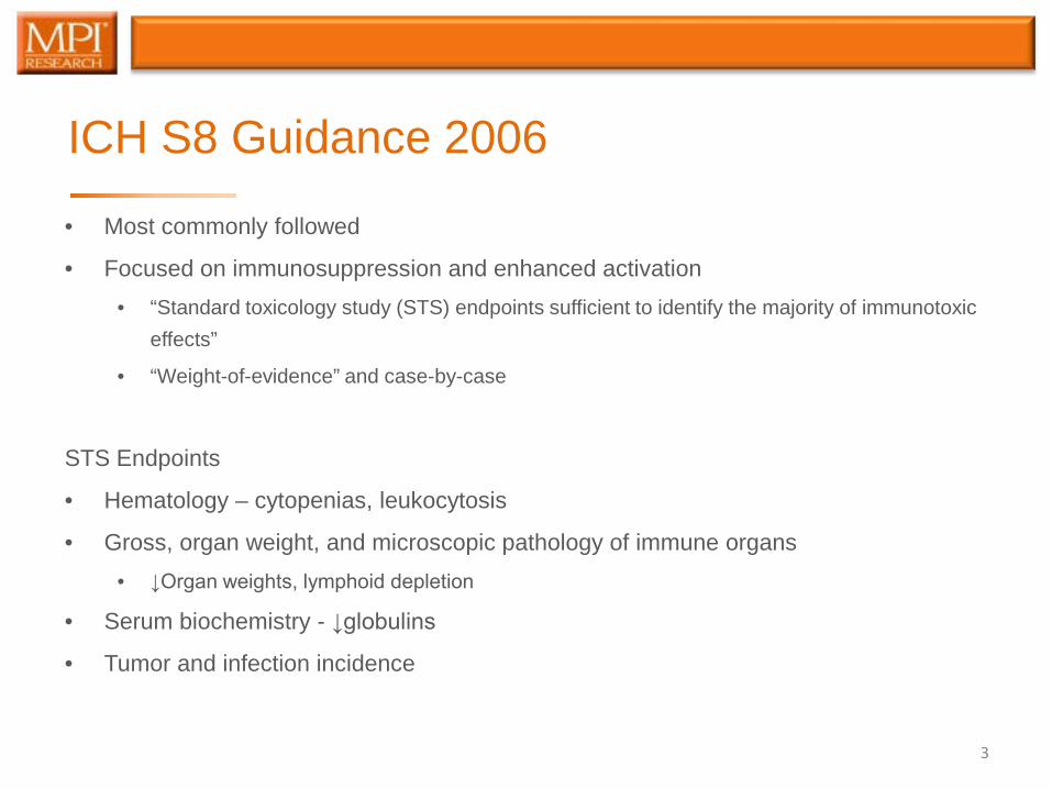
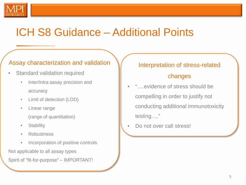
• Regulatory Guidance Overview – ICH S8 (2006), FDA (2002)

• Utilizing parameters for Standard Toxicity Studies (STS)

• Hematology, pathology, etc.

• Lymphocyte Subset Analysis (Immunophenotyping)

• T-cell Dependent Antibody Response Testing (TDAR)

• Biologic validation of ELISA methods

• Translating into humans

2

ICH S8 Guidance 2006

• Most commonly followed

• Focused on immunosuppression and enhanced activation • “Standard toxicology study (STS) endpoints sufficient to identify the majority of immunotoxic

effects”

• “Weight-of-evidence” and case-by-case

STS Endpoints

• Hematology – cytopenias, leukocytosis

• Gross, organ weight, and microscopic pathology of immune organs • ↓Organ weights, lymphoid depletion

• Serum biochemistry - ↓globulins

• Tumor and infection incidence

3

ICH S8 Guidance

• Should include • Statistical analysis • Dose/exposure relationship • Safety margin • Changes that occur as secondary effects (e.g.

stress, anorexia) • Possible cellular or molecular

targets/mechanisms • Reversibility

• Is there potential impact on the immune system?

• Immune tissues or cells

• Increased incidence of infections/tumors

YES? 4

ICH S8 Guidance – Additional Points

Assay characterization and validation • Standard validation required

• Inter/intra assay precision and

accuracy

• Limit of detection (LOD)

• Linear range

(range of quantitation)

• Stability

• Robustness

• Incorporation of positive controls

Not applicable to all assay types

Spirit of “fit-for-purpose” – IMPORTANT!

Interpretation of stress-related

changes • “….evidence of stress should be

compelling in order to justify not

conducting additional immunotoxicity

testing….”

• Do not over call stress!

5

FDA Guidance 2002

General Mention • Use STS endpoints to determine if further testing warranted

• Same weight-of-evidence approach

• Examples, details, and references

Specific Mention • PK studies indicate drug concentrates in immune tissues

• Suggests evaluation of developmental immunotox

1. intended for pregnancy

2. immunosuppression

• Inhalation and dermal studies

• Sensitizing potential

• Adverse immunotoxicity vs. intended pharmacology

6

FDA Guidance 2002

5 adverse event categories • Immunosuppression

• Leukopenia, ↓organ weights, cell depletion, ↓globulins, infections

• TDAR

• Supports separate study of satellite animals

• Immunogenicity

• Hypersensitivity/allergic reactions • Specific examples of Type I, II, III, and IV

• Extensive

• Autoimmunity • Examples, no standard methods

• Immunostimulation • STS and cytokines

7

ICH S8 and FDA Guidance

Additional testing – contingent upon results of STS parameters

• Functional and Non-functional • TDAR (T-cell Dependent antibody response)

• FDA - separate study or satellite animals • ICH S8 – include in STS

• Immunophenotyping of lymphocyte populations

• Natural Killer (NK) Cell Activity Assays – In vitro

• Host resistance assays (pathogens or tumor cells)

• Neutrophil/macrophage functional Assays

• Cell-mediated immunity

• Hypersensitivity/DTH

1st line

8

Standard Toxicology Study (STS) Endpoints

Immunosuppression

• Cytopenias - (granulocytes and lymphocytes)

• Immune organ weight decreases

• Lymph nodes, spleen, thymus

• Immune organ lymphoid depletion

• Often correlates with circulating lymphocytes

• Bacterial sepsis, abscesses, pneumonia

Enhanced immune activation

• Leukocytosis, neutrophilia, left shift

• No microscopic correlates

• Acute phase response (fibrinogen, CRP, etc.)

• Microscopic inflammation not associated with organ toxicity

• E.g. catheter sites, injection sites

9

When to do Immunotoxicity Testing?

Other • Anaphylaxis/hypersensitivity reactions

• Suspect autoimmune

• Hemolysis - ↓red cell mass, ↑TBIL, splenic EMH, ↑hemosiderin pigment

• Thrombocytopenia (suspicious)

• Vasculitis

1. Impact on immune tissues/cells

2. Increased infections

3. Mechanism of action

4. When they tell you to! (regulators)

10

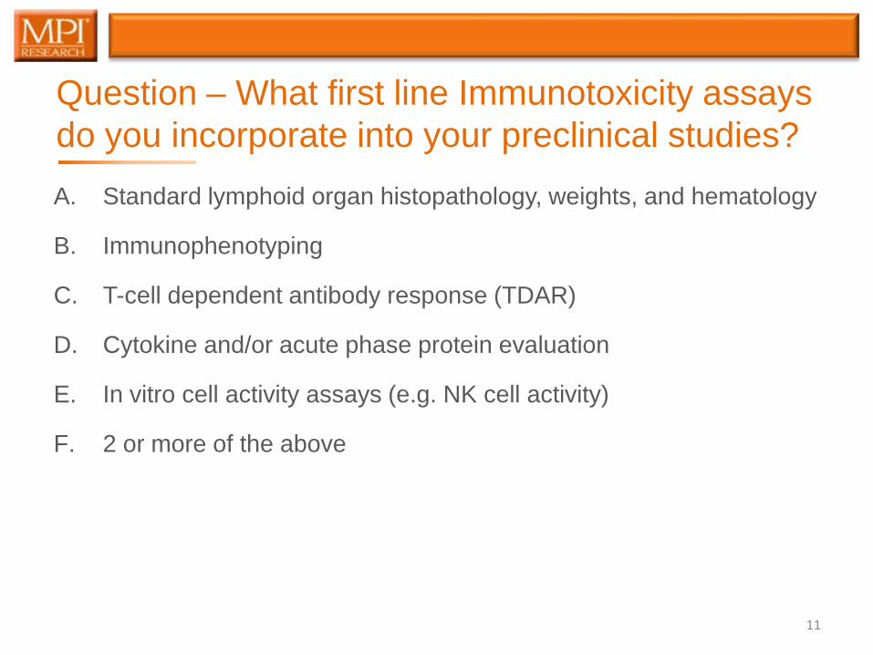
Question – What first line Immunotoxicity assays do you incorporate into your preclinical studies? A. Standard lymphoid organ histopathology, weights, and hematology

B. Immunophenotyping

C. T-cell dependent antibody response (TDAR)

D. Cytokine and/or acute phase protein evaluation

E. In vitro cell activity assays (e.g. NK cell activity)

F. 2 or more of the above

11

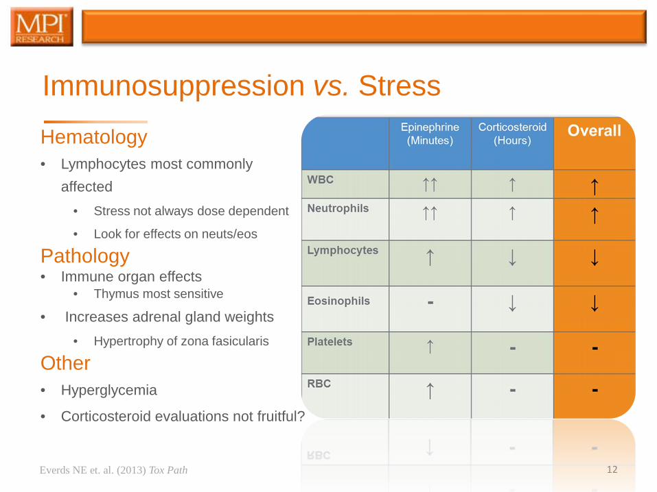
Immunosuppression vs. Stress Hematology • Lymphocytes most commonly

affected • Stress not always dose dependent

• Look for effects on neuts/eos

Pathology • Immune organ effects

• Thymus most sensitive

• Increases adrenal gland weights • Hypertrophy of zona fasicularis

Other • Hyperglycemia

• Corticosteroid evaluations not fruitful?

Everds NE et. al. (2013) Tox Path 12

Stress vs. Immunosuppression – Other Factors

Stress

• Often associated with overt toxicity

• ↓ Food consumption/body weight/clinical observations

• “Tends” to be less consistent/dose dependent

• Thymus most sensitive to stress

Immunosuppression

• Lymphoid effects reaching lower than other toxicity signals

• Likely to be direct effect if no thymic changes

Sometimes have both…..

• Immunotoxicity stress ↓ food consumption ↓marrow and lymphoid cellularity

Guidance specifically addresses (ICH and FDA)

13

Markers of Enhanced Immune Activation

Acute Phase Proteins • Non-specific markers of inflammatory cascade/process

• Most produced by liver in response to cytokine activation (IL-1, IL-6, etc.)

• Hours to days

• Must use appropriate species specific markers

• Fibrinogen (most)

• C-reactive protein (NHP and canines)

• Α-2 macroglobulin (A2M), A-1 acid glycoprotein (AGP) (rats)

• Haptoglobin and serum amyloid A (mice and swine)

Globulins • Total and IgG, IgM, and IgE

• Anaphylaxis

• Validated methods! 14

Markers of Enhanced Immune Activation

Cytokines • Involved in cell-cell messaging

• Many cells secrete – lymphocytes, macrophages, dendritic/APCs

• Minutes to hours – compound specific

• What good are they? • Elucidate mechanisms (pro and anti-inflammatory markers) • Cause or effect of inflammation? • Predictive - early signs

• Luminex/multiplex panels • Methods not standardized – assays generally not as tight as APPs • Validated methods!

• In vivo VS in vitro

• In vitro - most common, recommended for mechanistic studies • In vivo – may not be representative – TGN 1412

15

APPs vs. Cytokines (rats)

Honjo T et. al. (2010) Lab Animals 16

NHP Lymphocyte Immunophenotyping Panel

Immunophenotype Antigen Markers

Lymphocytes CD45

T-cells CD45, CD3

Thelper Cells CD45, CD3, CD4

Tcytotoxic Cells CD45, CD3, CD8

B-cells CD45, CD20

NK Cells CD45, CD159a

Regulatory T Cells CD4, CD25, Foxp3

Couple with hematology

17

TDAR Testing

T-cell Dependent Antibody Response • Immune function assessment

• Immunosuppression

• Ability to mount antibody response to standardized antigen challenge

• Keyhole Limpet Hemocyanin (KLH)

• Sheep Red Blood Cells (SRBC)

• Tetanus Toxoid

• Coordinated activity of macrophages, T-helper cells, and B-cells

• Antigen-specific IgM followed by IgG responses

• Supplements hematology and lymphoid organ assessment

• Further studies required regarding mechanisms of dysfunction

• FDA vs EPA requirements

18

Classic TDAR Response

• IgM precedes IgG

• Isotype switching

• Peak Response

• IgM – 7-14d

• IgG – 14-21d

• Use to time sampling

• IgM will wane

• IgG may persist

Immunization 19

Classic TDAR IgM Response

Immunize 20

TDAR Testing in NHP Overview

Retrospective review of 30 studies in NHP • No gender differences

• No country of origin differences - NHP

• Most used KLH (87%), TT (34%), SRBC (12%)

• Substantial inter/intra-animal variability

• ≤ 4 animals/group only identifies large differences • Combine sexes for more power

• Some differences in magnitudes and timing of responses based on source (rat) Lebrec et. al. (2011) J Immunotoxicol

Lebrec et. al. (2013) J Immunotoxicol 21

TDAR Testing Guidelines

General Considerations • All animals can be immunized

• Separate cohorts not typical

• Immunization does not significantly impact other endpoints (generally)

• Wide individual variation • Individual immune response

• Analytical methods

• Minimum 4-6 animals/sex/group recommended – combine sexes for statistics

• Immunization protocol and analysis should be consistent • Antigen source

• Injection site – SQ, IV, IM, footpad

• Analytical methods – lab to lab comparisons difficult

• Prior viral exposure – false positive reported

22

TDAR Testing

When to immunize? • Compound dependent

• Sufficient time to impact test system – not only exposure • NOT Day1

• 28 Day Studies – Day 7 or 14

• 13 Week Studies – Day 21 or 28

When to draw samples for antibody levels? • 2–4 times following immunization

• 7–14 days following immunization at 7 day intervals

Do I need a positive control group? • Not required

23

TDAR Testing

Recovery groups and secondary responses? • Compound dependent

• Must have knowledge of and account for multiple variables • Half life/exposure – days to months

• 30-45+ days for antibody response to subside

• Test system resolution • Lymphoid repopulation etc.

• Then re-immunize (secondary response) • Faster, more robust, longer

• Altered dynamics (IgG>IgM)

• 13 Week + studies usually required 24

Biological Validation of ELISA Methods

• Cynomolgus monkeys • Control and positive control groups

• 6/group/sex N=36

• Challenged KLH Day 21 and 71 • Primary and secondary responses

• 100 days

• Positive controls group (represents test compound) • Cyclophosphamide beginning Day 1

• Correlated with • Hematology

• Immunophenotyping - lymphocytes

• Histopathology – lymphoid organs

25

Lymphocyte Counts – Pooled Sexes

Day CYP KLH KLH 26

Immunophenotyping Results

27

Pathology – Organ Weights

Test Article-related Organ Weight Changes - Terminal Male and Female (Percent change relative to control)

Group: KLH/CYP KLH Sex M F M F Number Examined 6 6 6 6

Spleen (g) ↓21.67a ↓10.52 ↓2.58 ↓3.13

Thymus (g) ↓62.51a ↓61.25a ↑7.30a ↓12.21

a Significantly different from Antigen 1 Vehicle; (p<0.05) ↑ - Increased ↓ - Decreased M – Male F – Female

28

Microscopic Pathology

Test Article-related Microscopic Observations – Terminal Pooled Lymph Nodes

(iliac, popliteal, inguinal, and mandibular)

Group: Control KLH/CYP KLH

Sex M F M F M F

Number Examined 47 47 46 43 47 48

Lymph nodes (pooled)

Depletion, lymphoid, generalized (minimal to mild) 0 0 7 6 0 3

Depletion, lymphoid, germinal center

-minimal 0 0 0 0 1 0

Hyperplasia, lymphoid, germinal center

-minimal 0 0 0 0 5 2

M – Male F – Female

29

Anti-KLH IgM and IgG Responses

Aulbach et. al. (2013) ACT poster 30

Effects of Reduced Leukocytes

When do reductions actually adversely impact immune function?

• Humans (>40% ↓ lymphocytes; >75% ↓ in granulocytes)

Adversity subjective

Rely on clinical evidence – infections etc.

No consistent guidance for animal studies

• Neutrophils <1000 cells/µL

Hannet I et. al. (1992) Immunol Today 31

Effects of Reduced Lymphocytes on TDAR % Change in Cyclophosphamide Treated Relative Controls

7 days post Immunization

Day 28 Day 78 Lymphocytes -74%a -70%a

T Cells -77%a -78%a CD4+ -70%a -73%a CD8+ -87%a -84%a

B Cells -62% -27% NK Cells -92%a -87%a KLH IgM -72%a -46%a KLH IgG -84%b -60%a

a significant at (p<0.01) b significant at (p<0.05)

32

NHP Conclusions KLH

• Primary (D21) and secondary (D71) immunizations resulted in statistically significant increases in Anti-KLH IgM and IgG within 7-14 days post immunization

• Intermittent cyclophosphamide (CYP) dosing resulted in significant reductions in total lymphocytes and most lymphocyte subtypes as detected by flow cytometry

• Animals dosed with CYP had significant decreases in Anti-KLH IgM and IgG relative to immunized control animals indicating

• Detection of a compound-related reduction in immune function by these methods

33

Translating into Man

• Basic structure of immune systems similar • Lymphoid tissues, leukocytes, innate, acquired, humoral

• Species-specific variants

• Antibody responses

• Antigenic markers

• NHP often the only relevant species based on antibody cross reactivity with human target proteins

• Share significant genetic homology

• Immunoassay cross reactivity

• ICH S6 acknowledges antibody induction in animals not predictive of antibody formation in man 34

Translating into Man - Examples

Similarities • Innate immunity – dendritic cell subsets in rhesus monkeys

• myeloid (CD11c+/CD123neg) and plasmacytoid (CD11c-/CD123+) • cytokine responses similar • DC TLR expression same as human; different from mice

Differences • TGN 1412

• CD28 superagonist – expressed on human but not NHP T-cells

• Led to “cytokine storm” in 6 human volunteers – near fatal

• Recommend in vitro human studies in cases with mechanistic relations

Messaoudi I et. al. (2011) Antioxid Redox Signal Stebbings R et. al. (2007) J Immunol 35

Conclusions • Guidance supports weight-of-evidence case-by-case strategy for inclusion of

immunotoxicity testing • Considerations for species, stress, related mechanisms, pharmacology dictate a

case-by-case approach • STS endpoints drive

• Lymphoid organ effects • Leukocyte effects • Inflammatory biomarkers • Infection incidence

• First Tier • TDAR (T-cell Dependent Antibody Response) • Lymphocyte Immunophenotyping • Acute phase protein and cytokines

• Validated and well-characterized methods • Immunization protocols • Ligand-binding assays

36

References • Everds NE, Snyder PW, Bailey BB, Creasy DM, Foley GL, Rosol TJ, Seller T. Interpreting stress

responses during routine toxicity studies: a review of the biology, impact, and assessment. Tox Path 41(4):560-614, 2013.

• United States Food and Drug Administration, Center for Drug Evaluation and Research (CDER) Guidance for Industry. Immunotoxicology Evaluation of Investigational New Drugs, 2002.

• Gore ER, Gower J, Kurali E, Sui JL, Bynum J, Ennulat D, Herzyk DJ. Primary antibody response to keyhole limpet hemocyanin in rat as a model for immunotoxicity evaluation. Toxicology 197:23-35, 2004.

• ICH Harmonised Tripartite Guideline: Immunotoxicity studies for human pharmaceuticals S8, 2006.

• Lebrec H, Hock MB, Sundsmo JS, Mytych DT, Carlock LL, Joubert MK, Reindel J, Zhou L, Bussiere JL. T-cell-dependent antibody responses in the rat: Forms and sources of keyhole limpet hemocyanin matter. J Immunotoxicol Early online 1-9, 2013.

• Lebrec H, Cowan L, Lagrou M, Krejsa C, Neradilek MB, Polissar NL, Black L, Bussiere J. An inter-laboratory retrospective analysis of immunotoxicological endpoints in non-human primates:T-cell-dependent antibody responses. J immunotoxicol 8(3):238-250, 2011.

• Messaoudi I, Estep R, Robinson B, Wong S. Nonhuman primate models in human immunology. Antioxid Redox Signal 14(2):261-273, 2011.

• Piccotti J, Alvey JD, Reindel JF, Guzman RE. T-cell-dependent antibody response: Assay development in cynomolgus monkeys. J Immunotoxicol 2:191-196, 2005.

37


Recommended