21
Fungal Disease in Snakes Julia Russell Mycology November 2013 http://www.democraticunderground.com/discuss/duboard.php? az=view_all&address=389x3184386
| Date post: | 15-Dec-2015 |
| Category: |
Documents |
| Upload: | carlee-legates |
| View: | 219 times |
| Download: | 3 times |
Fungal Disease in SnakesJulia Russell
MycologyNovember 2013
http://www.democraticunderground.com/discuss/duboard.php?az=view_all&address=389x3184386

Outline
• Introduction to Reptile Fungal Diseases• Snake Fungal Disease (SFD)• Fungi that Could Potentially Cause SFD• SFD Infection Symptoms• Effects of the Spread of SFD• Conclusions
http://community.naturebreak.org/photo/eastern-massasauga-rattlesnake-431?context=user
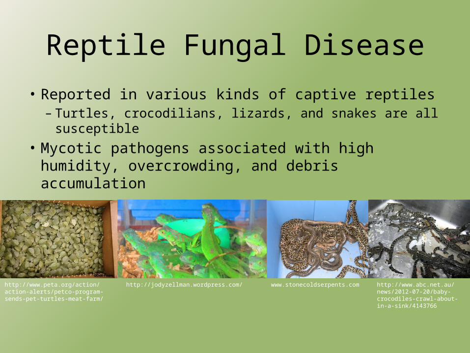
Reptile Fungal Disease
• Reported in various kinds of captive reptiles– Turtles, crocodilians, lizards, and snakes are all susceptible
• Mycotic pathogens associated with high humidity, overcrowding, and debris accumulation
http://www.peta.org/action/action-alerts/petco-program-sends-pet-turtles-meat-farm/
http://jodyzellman.wordpress.com/ www.stonecoldserpents.com http://www.abc.net.au/news/2012-07-20/baby-crocodiles-crawl-about-in-a-sink/4143766
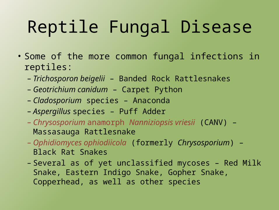
Reptile Fungal Disease
• Some of the more common fungal infections in reptiles:– Trichosporon beigelii – Banded Rock Rattlesnakes– Geotrichium canidum – Carpet Python– Cladosporium species – Anaconda– Aspergillus species – Puff Adder– Chrysosporium anamorph Nanniziopsis vriesii (CANV) –
Massasauga Rattlesnake– Ophidiomyces ophiodiicola (formerly Chrysosporium) – Black Rat
Snakes– Several as of yet unclassified mycoses – Red Milk Snake, Eastern
Indigo Snake, Gopher Snake, Copperhead, as well as other species
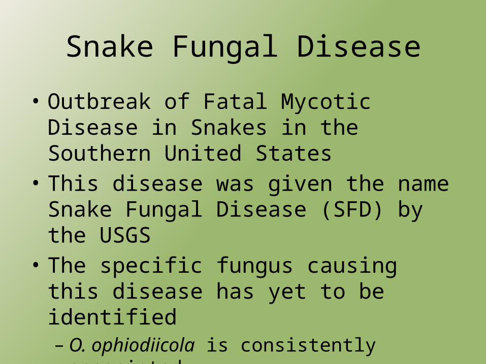
Snake Fungal Disease
• Outbreak of Fatal Mycotic Disease in Snakes in the Southern United States
• This disease was given the name Snake Fungal Disease (SFD) by the USGS
• The specific fungus causing this disease has yet to be identified– O. ophiodiicola is consistently associated– CANV may also be associated

Snake Fungal Disease• SFD noted in species common to the Eastern
United States including:– Northern Watersnake– Massasauga Rattlesnake– Black Rat Snake– Timber Rattlesnake– Pygmy Rattlesnake– Eastern Racer– Milk Snake
http://www.biokids.umich.edu/critters/1127/Sistrurus_catenatus/pictures/
http://commons.wikimedia.org/wiki/File:Timber_Rattlesnake_(dark_coloration).JPGhttp://www.fws.gov/northeast/chinco/
reptilesamphibians.html
http://www.ces.ncsu.edu/gaston/Pests/reptiles/pages/nwater.htm
http://www2.ca.uky.edu/forestryextension/kysnakes/browse/non-venomous/
http://srelherp.uga.edu/snakes/sismil.htm
http://www.virginiaherpetologicalsociety.com/reptiles/snakes/eastern-milk-snake/eastern_milksnake.htm
Fungi That Could Potentially Cause SFD
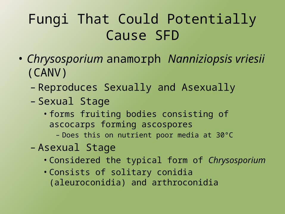
• Chrysosporium anamorph Nanniziopsis vriesii (CANV)– Reproduces Sexually and Asexually– Sexual Stage
• forms fruiting bodies consisting of ascocarps forming ascospores– Does this on nutrient poor media at 30°C
– Asexual Stage• Considered the typical form of Chrysosporium• Consists of solitary conidia (aleuroconidia) and
arthroconidia

Chrysosporium anamorph Nanniziopsis vriesii
• Arthroconidia– 3.5-13µm long and 2-3.5µm wide– Formed in chains by the fragmentation of hyphae– Occasionally are separated by empty cells
• Known as alternate arthroconidia
• Aleuroconidia– 4-6µm long and 2-3µm wide– Produced on the sides of fertile hyphae or at the ends of
short stalks• Arthroconidia predominated under certain cultural
conditions

Chrysosporium anamorph Nanniziopsis vriesii
All figures from (5)

Fungi That Could Potentially Cause SFD
• Ophidiomyces ophiodiicola (formerly Chrysosporium)– Very similar to CANV– Main Characteristics:
• Presence of numerous narrow to slightly clavate conidia• Strong pungent odor in colonies
– Differences from CANV:• Absence of asperulate fertile hyphae• Globose to pyriform conidia are sometimes grouped in
clusters• Presence of an odor in the colonies
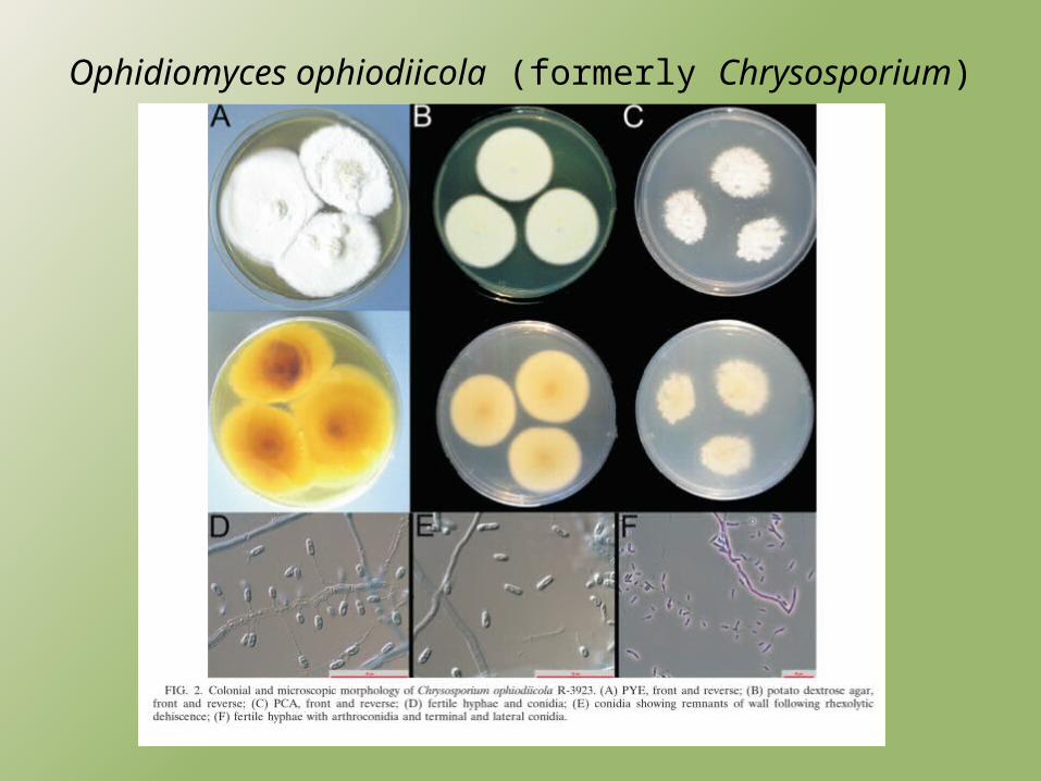
Ophidiomyces ophiodiicola (formerly Chrysosporium)
SFD Infection Symptoms
• CANV and O. ophiodiicola not normally found on healthy animals
• Fungal diseases in reptiles usually secondary– Not the case with CANV and O. ophiodiicola
• Potentially caused by disruptions of the normal defense mechanisms of the skin– Wetter than usual conditions– Stressful environment– Change in pH

SFD Infection Symptoms
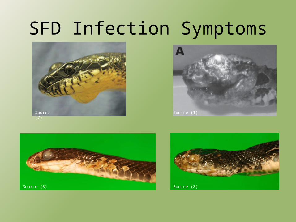
• SFD typically limited to head and ventral regions
• Head Region– Subcutaneous swelling– Lesions– Cutaneous ulcers with granulomas– Thick adherent crusts• Contain numerous right angle branching hyphae with
terminal structures consisting of spores
SFD Infection Symptoms
Source (7) Source (1)
Source (8) Source (8)

SFD Infection Symptoms
• Ventral Region– Edema of the ventral scales– Formation of cutaneous vesicles
• Filled with clear to cloudy serous fluid
– Ruptured vesicles replaced by brown, caseous plaques
– Underlying epidermis is dry and necrotic– Lesions begin where ventral scales overlap and
continue to spread to over 50% of the snakes ventral surface

SFD Infection Symptoms
Source (2)
Source (8)

SFD Infection Symptoms
• Fungal hyphae often extend deep into the epidermis, but usually do not cross into deeper tissue layers
• Fungal infection is usually not the immediate cause of death– Secondary bacterial infection– Osmotic imbalance
http://fineartamerica.com/featured/water-snake-karol-livote.html
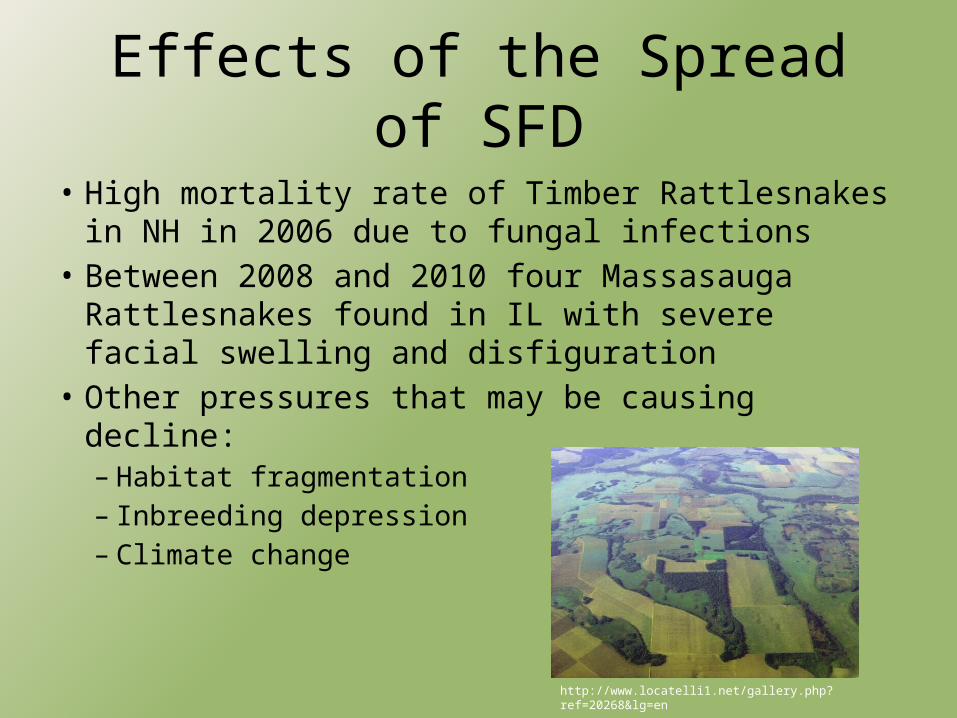
Effects of the Spread of SFD
• High mortality rate of Timber Rattlesnakes in NH in 2006 due to fungal infections
• Between 2008 and 2010 four Massasauga Rattlesnakes found in IL with severe facial swelling and disfiguration
• Other pressures that may be causing decline:– Habitat fragmentation– Inbreeding depression– Climate change
http://www.locatelli1.net/gallery.php?ref=20268&lg=en
Conclusions
• SFD is an emerging disease in Eastern and Midwestern U.S.
• Exact fungal origin is uncertain• Disease may be more prevalent than currently
documented• Will affect small isolated populations the
greatest• Population impacts difficult to assess due to the
cryptic and solitary nature of snakes

Sources(1) Allender M.C., Dreslik M., Wylie S., Phillips C., Wylie D.B., Maddox C., Delaney M.A., & Kinsel M.J. (2011, December).
Chrysosporium sp. Infection in Eastern Massasauga Rattlesnakes. Emerging Infectious Diseases, 17(12), 2383-2384.(2) Bertelsen M.F., Crawshaw G.J., Sigler L., & Smith D.A. (2005, March). Fatal Cutaneous Mycosis in Tentacled Snakes
(Erpeton tentaculum) Caused by Chrysosporium Anamoph of Nannizziopsis vriesii. Journal of Zoo and Wildlife Medicine, 36(1), 82-87.
(3) Clark R.W., Marchand M.N., Clifford B.J., Stechert R., & Stephens S. (2011) Decline of an isolated timber rattlesnake (Cortalus horridus) population: Interaction between climate change, disease, and loss of genetic diversity. Biological Conservation, 144(2011), 886-891.
(4) Jacobson E.R., Cheatwood J.L., & Maxwell L.K. (2000, April). Mycotic Diseases of Reptiles. Seminars in Avian and Exotic Pet Medicine, 9(2), 94-101.
(5) Nichols D.K., Weyant R.S., Lamirande E.W., Sigler L., & Mason R.T. (1999). Fatal Mycotic Dermatitis in Captive Brown Tree Snakes (Boiga irregularis). Journal of Zoo and Wildlife Medicine, 30(1), 111-118.
(6) Pare J.A., Sigler L., Rypien K.L., Gibas C.C. (2003). Survey for the Chrysosporium Anamorph of Nannizziopsis vriesii on the Skin of Healthy Captive Squamate Reptiles and Notes on their Cutaneous Fungal Mycobiota. Journal of Herpetological Medicine and Surgery, 13(4), 10-15.
(7) Rajeev S., Sutton D.A., Wickes B.L., Miller D.L., Giri D., Van Meter M., Thompson E.H., Rinaldi M.G., Romanelli A.M., Cano J.F., & Guarro J. (2009, April). Isolation and Cahracterization of a New Fungal Species Chrysosporium ophiodiicola, from a Mycotic Granuloma of a Black Rat Snake (Elaphe obsoleta obsoleta). Journal of Clinical Microbiology, 2009, 1264-1268.
(8) Sleeman J. (2013, May 2). Snake Fungal Disease in the United States. USGS National Wildlife Health Bulletin 2013-02. Retrieved from: http://www.nwhc.usgs.gov/disease_information/other_diseases/snake_fungal_disease.jsp
(9) Vissiennon T.H. Schuppel K.F., Ullrich E., & Kuijpers A.F.A. (1999). Case Report. A disseminated infection due to Chrysosporium queenslandicum in a garter snake (Thamnophis). Mycosis, 42, 107-110.
Questions?


