Fusion Techniques in Degenerative Disc Disease Guillem Saló Bru, MD, Phd AOSpine Principles Symposium- Cervical Spine Orthopaedic Depatment. Spine Unit. Hospital del Mar. Barcelona. Associated Professor UAB Barcelona, February 2014
Hospital del Mar. Barcelona.Associated Professor UAB Barcelona, February 2014
Disclosure information
I have no financial relationships with commercial entities that produce health-care related products.
Cervical Disc Disease. Introduction.
� Degenerative process.� Natural part of aging process.� Spectrum of clinical sd. associated with:
� Neck pain.� Neurologic dysfunction.
� Incidence unknown.� Radiographic degeneration:
� 90% in patients older than 65 years1.� 76% in patients older than 56 years2.
� Symptomatic degeneration:� 9.5% of men and 12.5% of women
complained with chronic pain3.� 24% overall frequency of neck pain4.
1. Laurence JC. Disc degeneration. Its frequency in relationship to symptoms. Ann Rheum Dis 1969;28:121-37.2. Hortwitz T. Degenerative lesions in the cervical portion of the spine. Ann Intern Med 1940: 55;1178.3. Makela M et al. Prevalence, determinants and consequences of chronic neck pain in Finland. J Epidemiol 1991;134:1356-67.4. Bovim et al. Neck pain in general population. Spine 1994; 19: 1307-9.
1. Spondylotic degeneration with neck pain.Desiccation of nucleus pulpous
Loss of mechanical conditions
Increased strain on the annulus
Tears and protrusion
Excess of motion in zygapophyseal joints
Increased strain in the supporting ligaments
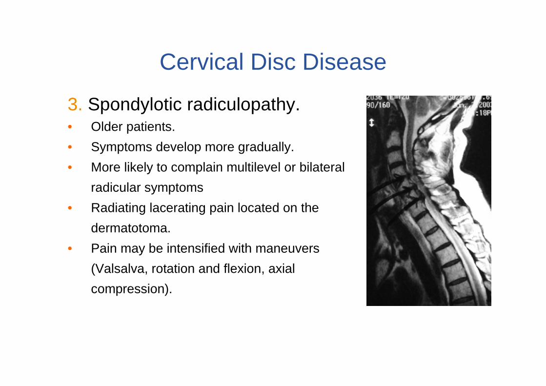
Cervical Disc Disease
1. Spondylotic degeneration with neck pain.• C5-C6 most commonly involved.• Primary Pain generator: Intervertebral disc.• Facet joints can become painful.• Axial neck pain.• Loss of motion.• Interscapular and upper brachial sclerotomal
pain radiation.• Pain is mechanical in nature, worst in flexion and
extension.
Cervical Disc Disease
2. Cervical disc displacement / prolapse.
Disc material prolapsed through tears in annulus
Root impingement / cord impingement.
Nerve dysfunction(directly and through vascular compromise)
• Complete symptomatic reversal after treatment is rare.
• Five categories according neurologic findings:
1. Transverse lesion sd. (corticospinal and spino-thalamic)
2. Motor sd. (corticospinal or anterior horn cells).
3. Central cord sd. (central grey mater of cord).
4. Brown-Séquard sd. (unilateral cord lesion).
5. Brachialgia cord sd. (myeloradiculopathy)
Options of surgical treatement.
Options for this 4 types of patients:•The type of surgical procedure advocated for cervical disc disease is dictated by the location and extent of the pathology.•There are also situations in which similar pathology can be addressed in several ways, with roughly similar results.
• The gold standard for the surgical treatment of cervical radiculopathy. Indications.
– Radiculopathy responds well to the surgical treatment (success rates of surgical treatment greater than 90%).
– Myelopathy is generally a clear indication for surgical intervention, specially if the patient develop signs or symptoms of neurological damage. 90% good to excellent results
– Axial neck pain from degenerative disk disease is a rare indication (success rates of surgical fusion only 60%).
• Most patients will have immediate relief of arm symptoms
• Recovery time: 4-6 weeks for office work, 8-12 weeks for heavy physical work
•1989: Improve of surgical supplies & trapezoidal plate.
Caspar5
•1986-90: Unicortical anchoring plates. Morscher6
•2000: Anterior dynamic plates (load-sharing)7
• 2000-2014: New plates design and materials. Development of
cages8
Smith Robinson Technique2
Cloward Technique3
1. Bailey R, Badgley C. Stabilization of the cervical spine by anterior fusion. J Bone Joint Surg Am 1960; 42: 565-94.2. Smith G, Robinson R The treatment of certain cervical-spine disorders by anterior removal of the intervertebral
disc and interbody fusion. J Bone Joint Surg Am 1958; 40:607-24.3. Cloward R Treatment of acute fractures and fracture-dislocations of the cervical spine by vertebral body fusion. J
Neurosurg 1961;18:201-94. Orozco R, Llobet J Osteosintesis en las fracturas del raquis cervical. Rev Ortop Traumatol 1970;14:285-85. Caspar W, Barbier D, Klara P. Anterior cervical fusion andC aspar plate stabilization for cervical trauma.
Neurosurgery 1989;25:491-502.6. Morscher E, Sutter F, Jenny H, Olerud S Anterior plating of the cervical spine with the hollow screw-plate system
of titanium. Chirurg 1986;57(11):702-7.7. Epstein NE. Anterior dynamic plates in complex cervical reconstructive surgeries. J Spinal Disord Tech 2002; 15:
221-8.8. Garcia CM. Stabilization and replacement devices currently used in arthrodesis and arthroplasties of the cervical
spine. Rev Chil Radiol 2008; 14:181-99.
Anterior fusion: Preoperative evaluation.
• Complaint and Physical Exam– Neck Pain– Arm Pain/Numbness/Tingling– (radiculopathy)– Myelopathy (cord abnormalities)
• Balance. • Unusual sensations.• Slow wide based gait
Anterior fusion. Preoperative evaluation
XRAYS• Bone structure and quality
• Alignment
• Lordosis• Instability
• Other diagnosis of neck pain (Cancer/Infection/Fracture)
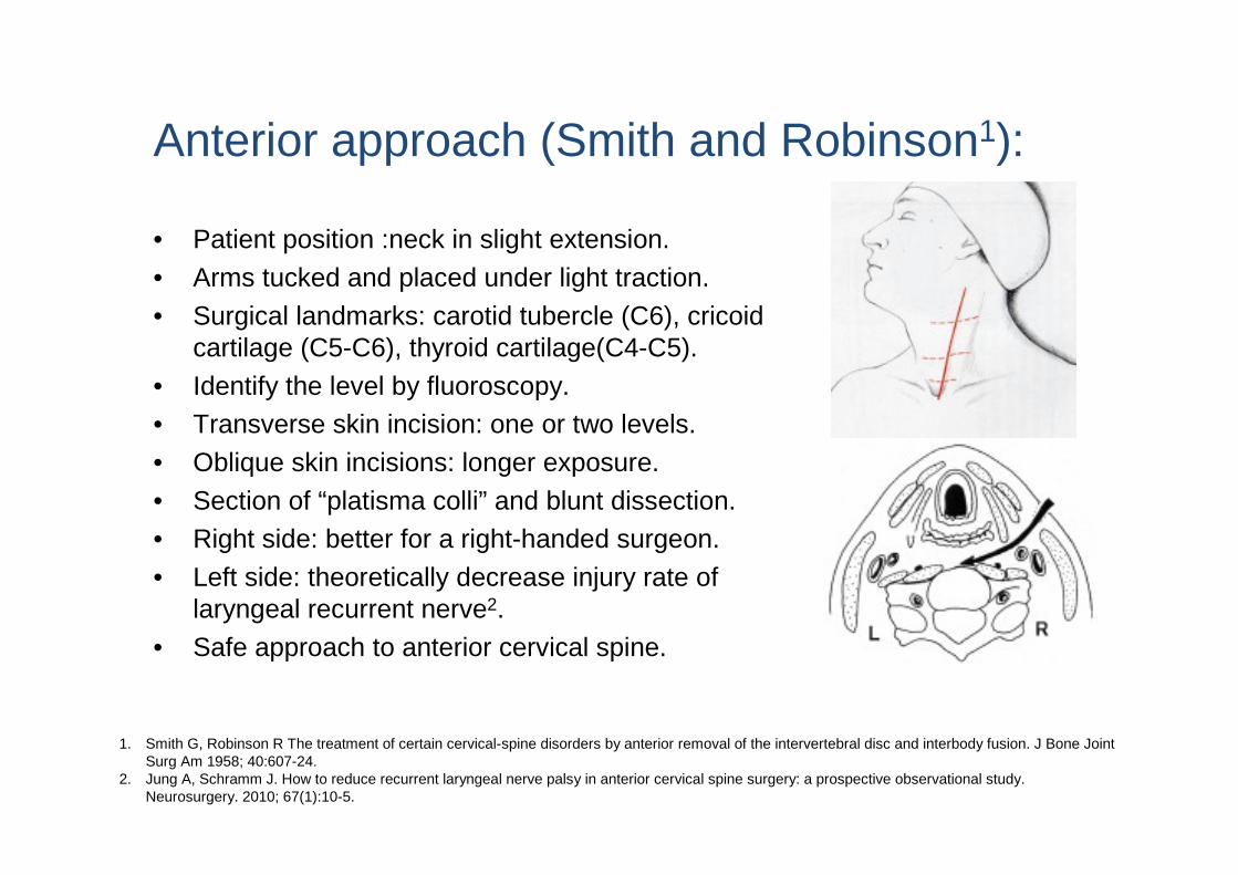
• Identify the level by fluoroscopy.• Transverse skin incision: one or two levels.
• Oblique skin incisions: longer exposure.
• Section of “platisma colli” and blunt dissection.• Right side: better for a right-handed surgeon.
• Left side: theoretically decrease injury rate of laryngeal recurrent nerve2.
• Safe approach to anterior cervical spine.
1. Smith G, Robinson R The treatment of certain cervical-spine disorders by anterior removal of the intervertebral disc and interbody fusion. J Bone Joint Surg Am 1958; 40:607-24.
2. Jung A, Schramm J. How to reduce recurrent laryngeal nerve palsy in anterior cervical spine surgery: a prospective observational study. Neurosurgery. 2010; 67(1):10-5.
Anterior approach (Smith and Robinson1):
Cervical Anterior fusion tecnique
Cervical Anterior fusion tecnique
• Complete removal of disc.• Curettage of vertebral endplates• Posterior osteophytes must be taken down:
• More immediate and complete relief of neural symptoms.
• Increases the foramina decompression.
• Better identification of extruded disk fragments.
• The posterior ligament can be removed or not1.
• It’s important to restore the disc height: The graft or cage should be 2 mm oversized2.
1. Schulte K, Clark CR, Goel VK. Kinematics of the cervical spine following discectomy and stabilization. Spine 1989; 14: 1116-21.
2. An HS, Evanich CJ, Nowicki BH, Haughton VM. Ideal thickness of Smith-Robinson graft for anterior cervical fusion. A cadaver study with computed tomographic correlation. Spine 1993; 18:2043-7.
Scholz M, Reyes PM, Schleicher P, Sawa AG, Baek S, Kandziora F, Marciano FF, Crawford NR. A new stand-alone cervical interbody fusion device: biomechanical comparison with established anterior cervical fixation devices. Spine. 2009; 34(2): 156-60.
Anterior fusion. Role of anterior platting.
• Widely used after 70‘s
• Increases chance of fusion.
• Recommended specially for more than one level of fusion1
• The plate protects the graft against an excessive axial load2.
• In a one-level fusion, the anterior plate is a mechanic device for distribution of loads. This increases the vertebral fusion rate2.
• Not demonstrated an increased risk of adjacent disc degeneration
• Good clinical and radiological results
1. Daffner SD, Wang JC. Anterior cervical fusion: the role of anterior plating Instr Course Lect. 2009;58:689-98.2. Rapoff AJ, O’Brien TJ, Ghanayem AJ, Heisey DM, Zdeblick TA. Anterior cervical graft and plate load sharing. J Spinal Disord. 1999;12(1):45-9.
Anterior fusion. Role of anterior platting.
97 patients. Discectomy plus Anterior fusion only with tricortical graft:– 11% non-fusion rate in one level– 28% non-fusion rate in two levels
The authors recommended to add a plate.
78 patients. Discectomy and fusion– 38 only with a interbody cage (79% of fusion, 32,3% of subsidence,
10,5% of revision surgery)– 40 with a interbody cage and plate.(97,5% of fusion, 9,7% of
subsidence, 0% of revision surgery)
Similar clinical results.
Wright IP, Eisenstein SM. Anterior cervical discectomy and fusion without Instrumentation .Spine. 2007;32(7):772-4.
Song KJ, Taghavi CE, Lee KB, Song JH, Eun JP. The efficacy of plate construct augmentation versus cage alone in anterior cervical fusion. Spine. 2009;34(26):2886-92.
• 28 cage alone / 26 cage + plate
• No differences in fusion (96%) or segmental kyphosis.
• Differences in subsidence: 35.71% vs 11.54% (P<0.05). • Clinical outcomes were similar in the 2 treatment groups.
• Conclusions: The use of cage and plate construct in 2-level ACDF results in a shorter fusion duration and a lower subsidence rate than that of cage alone; however, there is no significant difference in the postoperative global and segmental alignment and clinical outcomes between groups
Anterior fusion. Role of anterior platting.
J Spinal Disord Tech Volume 26, Number 8, December 2013
Anterior fusion. Role of anterior platting.
• 2 level ACDF.• ACDF/Static Plate/autograft: 87.8% per level• ACDF/Dynamic Plate/allograft: 89.8% per level• All pseudarthrosis patients were asymptomatic (10-13 months)
Goldberg, G., Albert, T., et al Short Term Comparison of Cervical Fusion with Static and Dynamic Plating Using Computerized Motion Analysis., SPINE 32:E371-375, 2007
Dynamic Plates
DuBois CM, Bolt PM, Todd AG, Gupta P, Wetzel FT, Phillips FM. Static versus dynamic plating for multilevel anterior cervical discectomy and fusion. Spine J. 2007; 7(2):188-93
• Retrospective. 52 patients.• No clinical differences (84% good/excellent results).• Non-union rate 16 % with dynamic / 5 % with static plate• Conclusion: dynamic plate not offers advantages compared with static plates
• The dynamic plates improved the loads distribution and provides a good resistance against movement.
• It can lead to a segmental kyphosis due the movement between plate and screws.
• It could have a less fusion rate due excessive movement between plate and screws
Anterior fusion. Results.
• Moderate success in reducing neck pain by >50%• Excellent success in reducing arm symptoms by >80%.• Global results good-excellent between 85-95%.• Fusion rate 95%• Complications rate less than 5%.• Gold standard.
1. DePalma AF, Rothman R, Lewinnek G, Canale ST. Anterior interbody fusion for severe cervical disc degeneration. Surg Ginecol Obstet. 1972;134:755-61.
2. Radhakrishnan K, Litchy WJ, O’Fallon WM, Kurland LT. Epidemiology of cervical radiculopaty. A population-based study from Rochester, Minnesota, 1976 through 1990. Brain. 1994;117:325-35.
Lied B, Roenning PA, Sundseth J, Helseth E. Anterior cervical discectomy with fusion in patients with cervical disc degeneration: a prospective outcome study of 258 patients. BMC Surg 2010; 21:10.
Anterior fusion. Results. Cage vs graftLow-quality evidence was found that iliac crest autograft results in better fusion than a cage (RR: 1.87; 95% CI: 1.10–3.17); but more complications (RR: 0.33; 95% CI:0.12–0.92). When fusion of the motion segment is considered to be the working mechanism for pain relief and functional improvement, iliac crest autograft appears to be the golden standard. When ignoring fusion rates and looking at complication rates, a cage as a golden standard has a weak evidence base over iliac crest autograft, but not over discectomy.
Fusion rates were 91% for allograft and autograft and 97% for cage. Adverse events were uncommon in all groups. ACDF with allograft, ACDF with autograft, ACDF with cage, and cervical disc arthroplasty show similar improvements in pain, function, and quality of life with correspondingly low adverse event rates. All ACDF procedures result in high fusion rates.
Anterior fusion. Tantalum cage.
• Prospective. Randomized. 28 tantalum cage vs 33 iliac crest + plate in one level. • At 24 months, equal results for both groups.•The data reported suggest that using porous tantalum as a stand-alone device is less costly and more effective than autograft and plate in ACDF procedures.
Fernandez-Fairen M, Sala P, Dufoo M Jr, Ballester J, Murcia A, Merzthal L. Anterior cervical fusion with tantalum implant: a prospective randomized controlled study. Spine. 2008;33:465-72.
Anterior fusion: Early Complications.1. Complications related to approach
• Cervical Haematoma 5.6%, (surgical intervention only 2.4%).
• Airway occlusion
Lied B, Sundseth J, Helseth E. Immediate (0-6 h), early (6-72 h) and late (>72 h) complications after anterior cervical discectomy with fusion for cervical disc degeneration; discharge six hours after operation is feasible.. Acta Neurichir (Wien). 2008; 150(2):111-8.
2. Donor site complications– Pain.
– Infection
– Bleeding.
– Iatrogenic fracture.3. Other complications
– Wound infection 0.1%– Skin problems (rare)
– Postoperative dysphagia 9.5%
Mortality rate 0.1% Overall morbidity rate was 19.3%
Anterior fusion: Early Complications.
1. Hardware complications (1%).2. Pseudoartroses.3. Subsidence of the graft / cage.
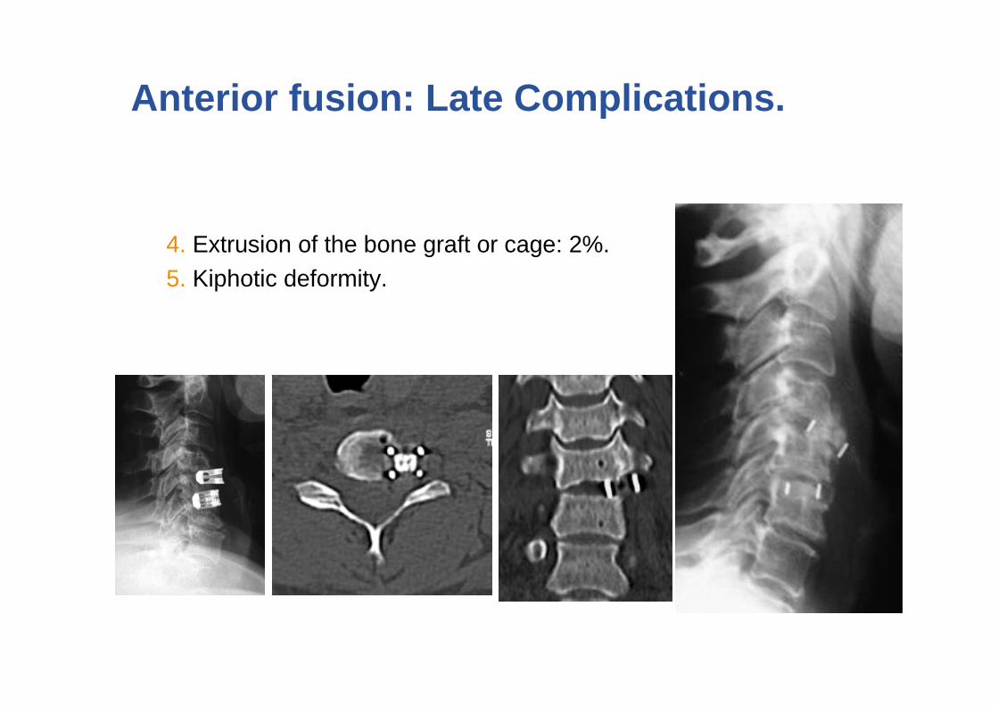
Anterior fusion: Late Complications.
4. Extrusion of the bone graft or cage: 2%.5. Kiphotic deformity.
Anterior fusion: Late Complications.
From a meta-analysis of prospective studies, there is no difference in the rate of ASD for ACDF versus TDA. We also report an overall lower rate of follow-up for patients with ACDF than for those with TDR. Future prospective studies should continue to focus on excellent patient follow-up and accurate assessment of patient symptoms that are attributable to an adjacent level as this has been an under-reported finding in prospective studies..
6. Adjacent disk degeneration.
Anterior fusion: Late Complications.
Conclusions CDA may result in better mid- to long-termfunctional recovery and a lower rate of subsequent surgicalprocedures than ACDF would. A review of the literatureshowed that only an insufficient number of studies hadinvestigated adjacent segment disease; therefore, it ismandatory that adequate future research should focus inthis direction.
For treating symptomatic cervical disc disease, cervical disc arthroplasty appears to provide better function, a lower incidence of reoperationrelated to index surgery at 1 to 5 years, and lower major complication rates compared with fusion. However, cervical disc arthroplasty did not reduce the reoperation rate attributable to adjacent segment degeneration than fusion. Further, it is unclear whether these differences in subsequent surgery including arthroplasty revisions will persist beyond 5 years
Posterior Fusion
Cervical posterior fusion tecnique
Posterior approach
Cervical posterior fusion tecnique
Posterior Fusion (C3-C7 Lateral Masses)
Efficacy: High fusion success Mostly plates, few rod-screw 0-10% Increased kyphosis, Safety: Rare hardware failure, Root at small risk (1.2%), very low risk for vertebral artery
JournalJournalJournalJournal YearYearYearYear
Posterior fusion.
• High efficacy >97% healing
•Poor study design
•Maintenance of stability good >95%
of cases.
•Complications: – Vertebral artery injury rare
– Root injury 0-5%
– Hardware failure rare except screw back
out in plates
Liu H, Ploumis A, Schwender JD, Garvey TA. Posterior cervical lateral mass screw fixation and fusion to treat pseudarthrosis of
anterior cervical fusion. Journal of spinal disorders & techniques 2012;25:138-41.
Conclusions. Take at Home messages.
• Despite of the interest in cervical motion preservation, fusion still is the gold standad of treatement for disc degeneration disease.
• Fusion rate of bone allograft and plate is over 95%.• There is no diferences in fusion rate between cages and graft.• In more than one-level fusion is recommended to add a plate.• Posterior fusion has similar results than anterior fusion in terms of
safety and Efficacy.• The long term results of new implants and matherials (bone cages,