20


1
G: Technical Specifications
GROUP 1 – ESTABLISHMENT OF NILGID
Item No.
NAME OF GOODS, TECHNICAL DESCRIPTION, SPECIFICATIONS, AND STANDARDS
Qty.
1 PER OPERATIVE ULTRASOUND System architecture. Compact and mobile design. Broadband 64 channel (transmitting) digital beam former. Three active transducer ports. 2-13 MHz transducers (Linear and Convex array) support. 15” flicker free VGA monitor. Detachable keyboard for ease of use. Operating modes features. Single 2D, double 2D, 2D + M mode, M mode, PW Doppler mode, CW Doppler mode. Color Doppler mode, Power Doppler mode. Contrast and Tissue harmonic imaging. Integrated Volume 3D. Adjustment of Focal zone, Gain, TGC, Dynamic range, Edge enhancement, Gamma correction. Real time Pan Zoom facility. Image annotation (alphanumeric and body marks). Multiple user defined presets for variety of clinical applications. 2D Cine memory, M Mode scroll memory. Palm control remote control operation.
Archiving and Storage.
Storage of 2D images, 3D volume studies to scanner hard drive, CD or USB device.
Storage of 2D images to DICOM network. Measurement and Calculations package. 4 distance measurements, 2 angle measurements, calculation of diameter, area, and circumference, calculation of volume by height/width/length, planimetry, automatic planimetry and empirical method. Calculation packages including reports, Urology, Cardiac (M and B mode), Obstetrics, Vascular. Transducers. Laparoscopic transducer with 4-way steer able scanning head. Multi frequency (6-10MHz) B/W and colour follow imaging capability. Integrated puncture and biopsy system. Support for radio frequency ablation. Fully immerse able sterilization capability. Intra-operative Linear transducer. Multi frequency (5 – 9 MHz).
1 No.

2
Item No.
NAME OF GOODS, TECHNICAL DESCRIPTION, SPECIFICATIONS, AND STANDARDS
Qty.
B/W and colour follow imaging capability. Live biplane 2D imaging. Integrated puncture and biopsy system. Fully immerse able sterilization capability. Intra-operative bi-plane imaging convex transducer. Multi frequency (5 – 10 MHz). B/W and colour follow imaging capability. Integrated puncture and biopsy system. Fully immerse able sterilization capability. Electronic convex array transducer. Multi frequency (2.5 – 6 MHz). B/W and colour follow imaging capability. Fully immerseable sterilization capability. Colour video printer. B/W thermal video printer. All equipment should be compatible to 220 VAC 50Hz.
2 VENOUS BLOOD PUMP
1 No.
3 CELL SAVER Auto transfusion Cell separator with auto start processing on reaching pre defined pre programmed trigger volumes With integrated/stand alone vacuum source and also capable of working on central vacuum Vacuum Range 0-300 mmHg Graphic Color LCD Auto transfusion reservoirs x 6 of various sizes available Wash Sets with various size bowls Replacement Vacuum Filters x 20 Clamped Empty Line detection Centrifuge Bowl Leak detection Overfilled Waste Bag detection Buffy Coat Sensor On-Board Help and Diagnostics Customizable User Programming and storage
1 No.
4 RAPID INFUSER Pressure infuser designed to function with standard 500 cc and 1000 cc fluid bags having wide opening for rapid easy insertion Infusion with Minimum variation in pressure Maintainable Preset pressure Precise pressure setting Can Accommodate 2x 500, 2x 1000 bags or 1x 3000 cm bag IV pole Mount Pressure limited to 300 mm/Hg Mains or battery operated
4 Nos.

3
Item No.
NAME OF GOODS, TECHNICAL DESCRIPTION, SPECIFICATIONS, AND STANDARDS
Qty.
Mains and Battery Operated Capacity of battery back up ~ 90 minutes
5 HYPER / HYPO THERMIA SYSTEM A whole body warming system with Fluid Temperature Selectable Range of 4°C to 42°C (39.2°F to 107.6°F) Patient Automatic Temp Range of 30°C to 43.5°C (86°F to 110°F) Refrigerant Non-CFC (R-134A) Heater Power 800 Watts Mobile Unit Water Flow Indicator Internal By-Pass with Pre-Heating and/or Pre-Cooling. System complete with adult, paed and infant blankets.
2 Nos.
6 TEG GRAFT Tissue Engineered Graft for Liver Transplant Management
20 Nos.
7 CO2 MONITOR For use with any ventilator or simply to monitor unassisted. Back-lit display, Battery and mains operated. Capnograph / Oximeter w/battery & charger Filters (10) Sample Lines - 4 ft (10) Airway Adapter straight without filter (10) Airway Adapter sidesteam without filter (10) Displays, Indicators & Keys:
o Display - LCD, with back light o Indicators - High / medium / low priority alarm, o Display Trends - Graphical display for ETCO2, respiration rate,
%SpO2, or pulse rate; user selectable scale o Displayed Waveforms - CO2 waveform, oximetry plethysmogram,
Capnograph: o Measurement - Non-dispersive IR absorption o Calibration - Manual 2 point o Measurement Range - 0-10% CO2 STPD (standard temperature and
pressure dry) o Display Range Approximate Accuracy – 0 -100 mmHg; 0-13.3kPa; 0-
10% CO2 +2mmHg or 4% of reading, whichever is greater o System Response Time 2.46s to 90% with 8 ft. patient line o N2O Compensation - Selectable 40% o Averaging - 4 breath average o Flow Rate - 120 + 20ml/min
Respiration Rate: o Range - 0-150 breaths/min o Accuracy - +1 bpm o Averaging - 4 breath average
SpO2: o Range - 0-100% SpO2 o Accuracy - +2% at 70-100% SpO2, +3% at 50-69% SpO2
4 Nos.

4
Item No.
NAME OF GOODS, TECHNICAL DESCRIPTION, SPECIFICATIONS, AND STANDARDS
Qty.
o Averaging - 8 beats Peripheral Pulse Rate:
o Range - 30-254 bpm o Accuracy - +2% bpm or 2%, whichever is greater, at 30 to 254 bpm o Averaging - 8 seconds o Pulse Tone-Pitch corresponds to SpO2 value, Volume adjustable or
OFF Alarms:
o Physiological - User-selectable high and low limits, adjustable volume, screen indicators
o System - Adjustable volume o Silence - Temporary 2 min or indefinite LED indicator
Power: o AC Power - 230V~, 50/60Hz o Battery Charger built-in
8 HEMOSTASIS ANALYSER Eight simultaneous samples via 4 analyzers, each with 2 channels Two independent channels per instrument Maximum sensitivity with software vibration damping Advanced QC and maintenance handling Electromagnetic detection system with torsion wire Full network support with remote viewing Preferably Windows-based software 110V/60Hz or 220V/50Hz Power Analog output via DB9 cable to computer interface box DB9 RS232 serial cable to available com port on PC One-year factory warranty against manufacturer's defects Sample Capacity 8 samples Tests/Assays: Clotting time Clot kinetics, Clot strength, Hemostasis
profile, Clot stability
1 No.
9 LIMON (LIVER MONITOR) Monitor for measuring elimination of ICG-PULSION in all critically ill patients, especially those with sepsis, acute liver or multi-organ failure, after multiple trauma, in patients with chronically reduced hepatic function (hepatitis, liver cirrhosis), for evaluation of liver function in organ donors and recipients, for perioperative monitoring of liver function during liver surgery (resection, porto-caval shunt) for diagnosis and monitoring of congenital liver failure in children and neonates Parameters: Non-invasive sensor and any venous access Quick bed-side results Up to 20 measurements per 24 hours by Plasma Disappearance Rate of ICG-PULSION (PDR), Retention Rate of ICG-PULSION extrapolated to 15 minutes (R15), Blood Clearance of ICG-PULSION (CB), Circulating Blood Volume (BV), and continuous measuring of Arterial Oxygen Saturation (SpO2), Heart Rate (HR)
4 Nos.
5
Item No.
NAME OF GOODS, TECHNICAL DESCRIPTION, SPECIFICATIONS, AND STANDARDS
Qty.
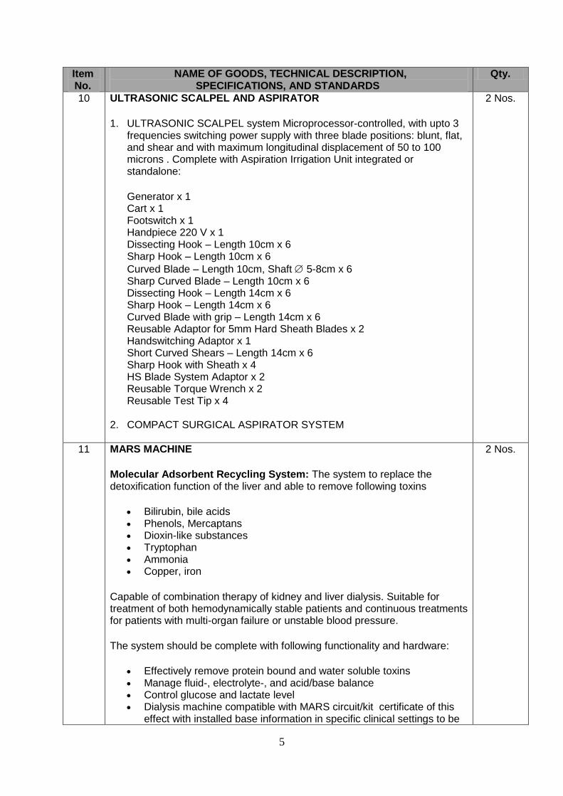
10 ULTRASONIC SCALPEL AND ASPIRATOR
1. ULTRASONIC SCALPEL system Microprocessor-controlled, with upto 3 frequencies switching power supply with three blade positions: blunt, flat, and shear and with maximum longitudinal displacement of 50 to 100 microns . Complete with Aspiration Irrigation Unit integrated or standalone:
Generator x 1 Cart x 1 Footswitch x 1 Handpiece 220 V x 1 Dissecting Hook – Length 10cm x 6 Sharp Hook – Length 10cm x 6
Curved Blade – Length 10cm, Shaft 5-8cm x 6 Sharp Curved Blade – Length 10cm x 6 Dissecting Hook – Length 14cm x 6 Sharp Hook – Length 14cm x 6 Curved Blade with grip – Length 14cm x 6 Reusable Adaptor for 5mm Hard Sheath Blades x 2 Handswitching Adaptor x 1 Short Curved Shears – Length 14cm x 6 Sharp Hook with Sheath x 4 HS Blade System Adaptor x 2 Reusable Torque Wrench x 2 Reusable Test Tip x 4
2. COMPACT SURGICAL ASPIRATOR SYSTEM
2 Nos.
11 MARS MACHINE
Molecular Adsorbent Recycling System: The system to replace the detoxification function of the liver and able to remove following toxins
Bilirubin, bile acids Phenols, Mercaptans Dioxin-like substances Tryptophan Ammonia Copper, iron
Capable of combination therapy of kidney and liver dialysis. Suitable for treatment of both hemodynamically stable patients and continuous treatments for patients with multi-organ failure or unstable blood pressure.
The system should be complete with following functionality and hardware:
Effectively remove protein bound and water soluble toxins Manage fluid-, electrolyte-, and acid/base balance Control glucose and lactate level Dialysis machine compatible with MARS circuit/kit certificate of this
effect with installed base information in specific clinical settings to be
2 Nos.

6
Item No.
NAME OF GOODS, TECHNICAL DESCRIPTION, SPECIFICATIONS, AND STANDARDS
Qty.
provided with the offer. MARS Monitor with Albumin flow rate: 50-250 ml/min, Pressure range
of 100 to 500mmHg and albumin circuit volume of 600ml 20% albumin solution and compatible with quoted dialysis machine
Complete MARS Kit x 100
12 EEG MACHINE Clinical Digital 36 channel EEG with amplifier attached to module with fiberoptic interface, shielded against electrical, radio and magnetic interference. The system shall be network capable on LAN for integration of PC based data acquisition, review, and analysis, with Patient administration software, Workflow management, Frequency and amplitude measurement tools, Storage of measurements to the patient exam and transfer, capability of comparing sections of EEG data, Storable user preferences for both acquisition and review. Complete with PC, trolley, hard cop laser printer, cables available softwares, photo stimulator and accessories
1 No.
13 INTRACRANIAL PRESSURE MONITOR Intra cranial pressure monitor with source-subdural, parenchymal and ventricular monitoring through fiber optic technology measuring intracranial pressure, intracranial temperature and cerebral perfusion pressure
color display with ICP waveform, digital CPP, ICP and ICT readouts.
Continuous recording and display of ICP and CPP values over the most recent 12 or 24 hour period.
Bedrail or pole mounted
Networking capability with hospital bedside monitoring systems.
Built-in rechargeable battery for transport.
High ICP and Low CPP Alarms.
RS232 Serial and ICP Analog Output connections
Complete with Reusable cable and transducers.
3 Nos.
14 SPECIAL BED WITH PRESSURE MATTRESS Surgical ICU Bed with side-rail and footboard positioning controls and capable of following Features:
Cardiac chair position
Emergency Drop Backrest
Trend. angle display
High/low,
Trend./reverse Trend.
Radiolucent from neck through pelvic region
Scale-isolated drainage bag hooks/head-end IV poles
Integrated 8" (20 cm) bed extender
Four-wheel, steel-ring brake system with centrally-located activation pedals
90° backrest for upright chest imaging
Dual pedestal design
4 Nos.

7
Item No.
NAME OF GOODS, TECHNICAL DESCRIPTION, SPECIFICATIONS, AND STANDARDS
Qty.
CPR release for backrest and knee
6" (15 cm) casters
Locking-caster steering
Four traction sockets
Four IV receptacles (scale isolated at head end)
Electric function lock-out controls
In-rail backrest/knee gatch controls
Removable CPR board
Overall Length 90" approx.
Overall Width 40" approx
Weight Capacity 500 lbs
Standard Height Range High 32" Low 18"
Backrest 0° - 90° 0° - 90°
Knee Gatch 0° - 30° 0° - 30°
Trend./reverse Trend. ±12° +10/-12°
Roller bumpers
Manual back up for head and knee control
Integrated pump holder
Nurse controls on footboard and siderails
Fixed patient controls on siderails
Degree indicator for head elevation
Patient restraint locations
Electronic function lockout controls
One-handed dampened siderail release
Auto contour Low air-loss, pressure relieving, non-integrated, ulcer prevention and treatment Mattress having integrated adjusting anatomical zones for prevention of bed sores by means of air pockets and with Fluid and bacterial resistant cover
15 SCD FOR DVT PROPHYLAXIS
Sequential Compression System Non-invasive method of prophylaxis to reduce the incidence of deep vein thrombosis (DVT). For use on patients with high risk conditions such as obesity, advanced age, malignancy, surgery/trauma or prior thromboembolism.
With Pneumatic controller providing intermittent pulses of compressed air that sequentially inflates the multiple chambers of the sleeve beginning at the ankle and moving up the leg resulting s in a wave-like milking action resulting in greatly increased peak blood flow velocity.
With Optional Compression System that can detect each patient's individual vascular refill rate to deliver the customized compression therapy.
With Disposable sterile sleeves and reusable sleeves in various knee and thigh lengths. knee length regular and large, and thigh length small, medium, and large etc.
3 Nos.

8
GROUP 2 – STRENGTHENING OF RADIOLOGY, CARDIOLOGY AND CARDIAC SURGERY DEPARTMENTS
Item No.
NAME OF GOODS, TECHNICAL DESCRIPTION, SPECIFICATIONS, AND STANDARDS
Qty.
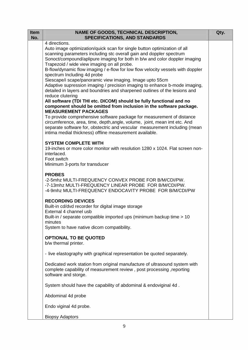
16 COLOUR DOPPLER ULTRASOUND Fully digital beam former having 17000 processing channels color doppler ultrasound system with 12 bit a/d conversion for abdomin,ob/gyne, msk and peripheral vessels studies with integrated data management system having 500gb or more hard disk drive for still and cine clips storage with matrix probe capability 2d/m-mode Doppler Pw, hprf doppler Color flow imaging System capability 2.0 – 18 mhz or more M-mode Tissue harmonic imaging Diffirential tissue hamonic imaging Viewing depth: 30 cms minimum Frame rate 500 or more Built-in cine loop 4000 frames or more Real time freeze & image magnification ability> 4x Pre & post processing Sweep speed: slow, medium, fast Independent linear syeering : 10 – 15 degree for color and b/w System dynamic range : 190 or more DOPPLER MODE SPECIFICATIONS Pulse repetition frequency range . Pwd; 0.5 to 10 khz . Cwd(optional): 1.6 to 50 khz . Lowest detectable velocity range: 0.03cm . Maximum detectable velocity range; pwd= 18 m/sec or more, cwd(optional)= 20 m/sec or more Doppler beam steering Sample gate size : 1-20mm in steps On line 3-ports for transthorasic probes or more for imaging (all ports should have capability) for pw, and color doppler Doppler beam steering and bi-directional stereo audio. Both cw & pw doppler must be continuously Stearable in color blood flow imaging mode in real time Colorized spectrum display Tissue harmonic imaging with 3 harmonic frequencies and with complete functionality . 2D image with color flow Power doppler Live triplex imaging with color flow and pulse wave and also on color flow and cw on all probe. Quad signal color doppler processing for simultaneously reception in
17 Nos.
9
Item No.
NAME OF GOODS, TECHNICAL DESCRIPTION, SPECIFICATIONS, AND STANDARDS
Qty.
4 directions. Auto image optimization/quick scan for single button optimization of all scanning parameters including stc overall gain and doppler spectrum Sonoct/compound/aplipure imaging for both in b/w and color doppler imaging Trapezoid / wide view imaging on all probe. B-flow/dynamic flow imaging / e-flow for low flow velocity vessels with doppler spectrum Including 4d probe Siescape/i scape/panoramic view imaging. Image upto 55cm Adaptive supression imaging / precision imaging to enhance b-mode imaging, detailed in layers and boundries and sharpened outlines of the lesions and reduce clutering All software (TDI THI etc. DICOM) should be fully functional and no component should be omitted from inclusion in the software package. MEASUREMENT PACKAGES To provide comprehensive software package for measurement of distance circumference, area, time, depth,angle, volume, joint, mean imt etc. And separate software for, obstectric and vescular measurement including (mean intima medial thickness) offline measurement available. SYSTEM COMPLETE WITH 19-inches or more color monitor with resolution 1280 x 1024. Flat screen non-interlaced. Foot switch Minimum 3-ports for transducer PROBES -2-5mhz MULTI-FREQUENCY CONVEX PROBE FOR B/M/CDI/PW. -7-13mhz MULTI-FREQUENCY LINEAR PROBE FOR B/M/CDI/PW. -4-9mhz MULTI-FREQUENCY ENDOCAVITY PROBE FOR B/M/CDI/PW RECORDING DEVICES Built-in cd/dvd recorder for digital image storage External 4 channel usb Built-in / separate compatible imported ups (minimum backup time > 10 minutes System to have native dicom compatibility. OPTIONAL TO BE QUOTED b/w thermal printer. - live elastography with graphical representation be quoted separately. Dedicated work station from original manufacture of ultrasound system with complete capability of measurement review , post processing ,reporting software and storge. System should have the capability of abdominal & endoviginal 4d . Abdominal 4d probe Endo viginal 4d probe. Biopsy Adaptors

10
Item No.
NAME OF GOODS, TECHNICAL DESCRIPTION, SPECIFICATIONS, AND STANDARDS
Qty.
17 ACT MACHINE For measurement of clotting to assess the neutralization to heparin. Activated timer clotting. Complete with 100- cartridges. Dual Chamber
1 No.
18 ANGIOGRAPHY MACHINE A fully digital flat panel single plane Cardiac Angiography / Cardiac catheterization system, dedicated for diagnostic & interventional cardiac procedures. The Angiography system should comprise of the following configurations. POSITIONING ARM: Frontal Arm: STAND. Floor/Ceiling mounted stand with motorized movements. Real time display of rotation angulations. Geometry: C-arm / G-arm RAO / LAO +/- 105 ° or more. Cranial / Caudal: Min. +/-45° or more, (Capable of achieving Spider position.) Rotation Speed: 20°/sec or more in LAO / RAO. Isocentric Height: Variable / Fixed. Auto Positioning: Programmable auto positioning of selected angulations. (30 or more programmable positions.) The control panel can be mounted at any side of the patient table. All the rotational / angles should be digital displayed next to the ceiling mounted monitor. Motorized / manual parking / rotation of the positioning arm.
DIGITAL FLAT PANEL. Single plane C-Arm / G-Arm Image matrix of 1024 x 1024 x 12 bits or more. Standard cardiology size with three formats. Built in temperature stabilizer. Digital flat panel detector. Integrated collision protection feature All other standard accessories according to this digital flat panel.
PATIENT SUPPORT/ TABLE. Catherization Table: Floor mounted with up down / vertical, longitudinal and transverse rotational movements. Longitudinal Stroke.: 1000-1500 mm. Lateral stroke: 100-300 mm CPR: In any table position. Table side movement control with one additional control to control the movements from any side of the table. Table top should be of such construction in material and durability to accept patient’s weight of not less than 150 kg plus 100 kg for resuscitation. Table dimensions should be able to accommodate patients of all ages. Table top should have large metal free over hang for un-obstructed imaging coverage.
1 No.

11
Item No.
NAME OF GOODS, TECHNICAL DESCRIPTION, SPECIFICATIONS, AND STANDARDS
Qty.
Complete accessories should be provided including arm holder, hand grip, arm support and arm rest and positioning aids. Left / right table pivoting: + / - 90 degree. X-RAY GENERATOR. Microprocessor based high frequency using fiber optic for data communication between each imaging system. Dedicated X-Ray generator of 100 kW. Radiographic rating minimum 1000 mA. Serial filming exposures with shortest exposure of 1ms, with automatic kV and mA control for optimum image quality. The system should have capability of digital radiography and fluoroscopy. Should have capability of doing digital pulsed fluoroscopy at 12.5/15 & 25 / 30 /Sec. Automatic kV,mA & pulse width regulation facility. DIGITAL IMAGING AND ACQUISITION / FLUOROSCOPY. DIGITAL SYSTEM. Acquisition, storage and display in 1024 x 1024 x 10 bits or more at 12.5 / 15 and 25/30 FPS. Parallel processing capability / multitasking facility. Real time filtering and road map function. Magnetic Disk Capacity for Storage of 25,000 images in 1024 x 1024 x10 bit or more on the magnetic disk of main console. Minimum scene length to be 10 seconds in 1024 matrix. Digital pulsed fluoroscopy with 12.5 / 15 and 25 / 30 FPS in 1024 x 1024 x 10 bits or more. Images to be stored on and retrieved from archival disks for possible manipulation and quantification using available software packages. X-RAY TUBE. Dual focal with at least 1.9 MHU or better anode heat storage capacity to enable continuous heat dissipation during serial exposure. Dual focus, rotating anode. Focus 0.6 and 1.2 mm or better. Dose management with auto adjustment fluoro filters. MONITORING SYSTEM. Flat Screen 18” TFT of 1024 x 1024 matrix. Monitors should be ceiling mounted in the operation room.
Brightness of the imaging monitors: 600 cd/m or better. Two monitors for live images and road mapping in the operating room 18” TFT. Two monitors for live images and road mapping in the control room 18” TFT. One 18” TFT monitor for live images and road mapping in the conference room remote from the Cath Lab. One integrated/Seprate monitor of 17 Inches for images of IVUS One additional monitor 18” TFT to display hemodynamic data in the operating room. All the monitors will be of Medial Grade, complied with international standards for medical monitors. CONTROLS: All controls of digital imaging system, incl. Post Processing & Quantification (LVA,QCA) analysis shall be in the control room while replay / display of

12
Item No.
NAME OF GOODS, TECHNICAL DESCRIPTION, SPECIFICATIONS, AND STANDARDS
Qty.
reference image should be available in the examination room. RECORDING / ARCHIVING & COMMUNICATION SYSTEM: Recording / archiving system should be DICOM-3 compatible. The digital images should be stored as backup on CD/DVD. DICOM (send/storage, commitment, retrieve/query) Ethernet connection to connect with other terminals. Integrated Intercom system.
REVIEW STATIONS (01). DICOM-3 Compatible. Edge enhancement, adjustable view speeds & post processing. High resolution 18” TFT Monitor. CD/DVD writer and CD/DVD ROM drive. Image storage capacity 3 x 80 GB with at least 10000 RPM Speed at each review station and SCSI/ Equivalent Controller at each review station. The bidders should quote their own licensed software. SOFTWARE / HARDWARE PACKAGES. Complete analysis package for the following cardiac applications. - Dynamic pre and post PTCA / valvotomy comparison with one image live and other reference. - Automatic loop replay after acquisition or fluoroscopy. - Dynamic real time pan / zoom. - Dynamic real time digital image processing like edge enhancement or gamma correction, noise reduction (spatial filtration.) - Simultaneous display of fluoroscopy and reference images, not only as static images but as dynamic loop. Standard Quantification packages for QCA & LVA analysis. Online image density (gray scale) correction. Facility to review previous studies in the examination room from the patients old CD. All controls of digital imaging system incl. Post-processing & Quantification (LVA & QCA) shall be in the examination as well as control room.
SURGICAL SHADOW LESS LIGHT. Ceiling suspended. For Angiographic and related surgical procedures.
RADIATION PROTECTION. Ceiling suspended tilt able lead glass for radiation protection of operators head & neck regions & lower body parts region. Collision tolerant. Lower body radiation protection flaps. Lead lining of room where necessary. QUALITY AND SAFETY STANDARDS. FDA 510 K approval CE (MDD) compliance. OTHER ALLIED ACCESSORIES: One postscript level Laser Printer or Video printer for taking image printouts on paper. This printer is to be connected with the Main Digital Imaging System. Paper for 500 prints should be delivered with the printer.
13
Item No.
NAME OF GOODS, TECHNICAL DESCRIPTION, SPECIFICATIONS, AND STANDARDS
Qty.
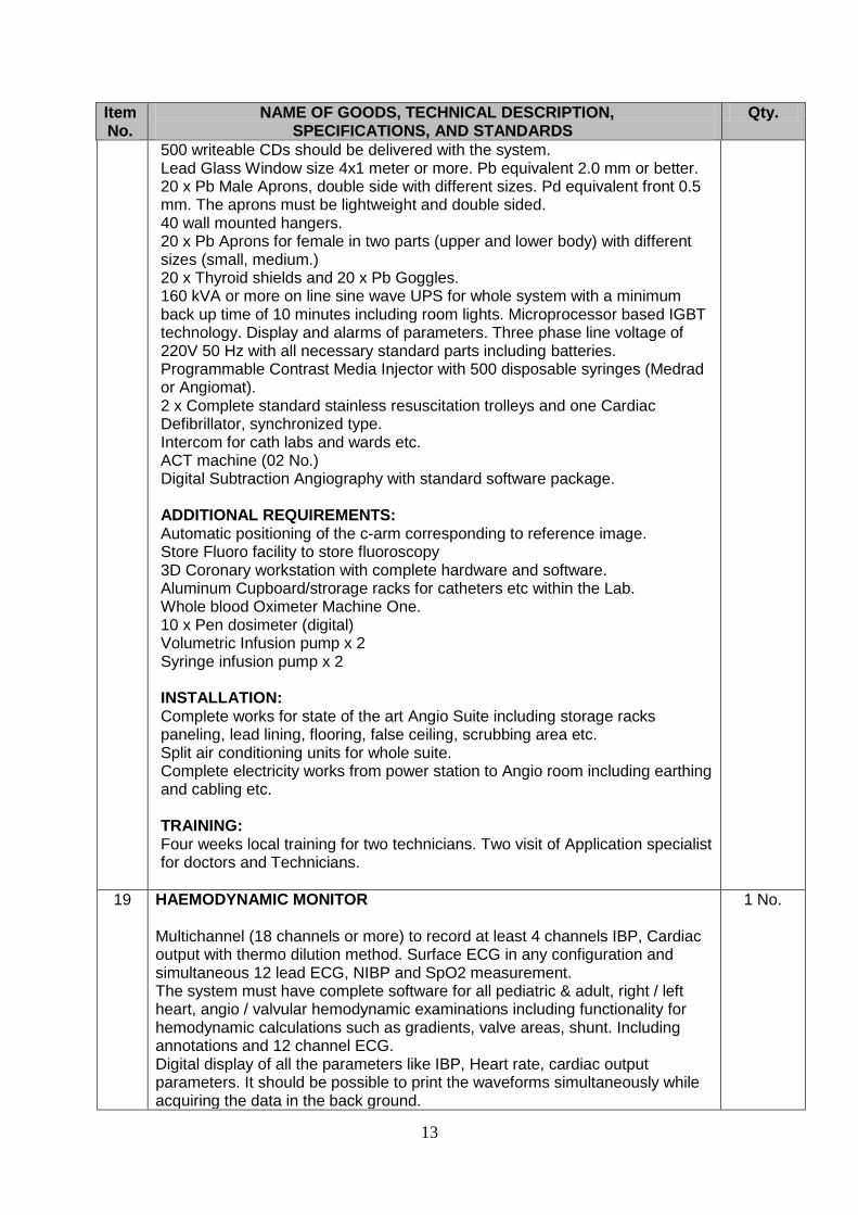
500 writeable CDs should be delivered with the system. Lead Glass Window size 4x1 meter or more. Pb equivalent 2.0 mm or better. 20 x Pb Male Aprons, double side with different sizes. Pd equivalent front 0.5 mm. The aprons must be lightweight and double sided. 40 wall mounted hangers. 20 x Pb Aprons for female in two parts (upper and lower body) with different sizes (small, medium.) 20 x Thyroid shields and 20 x Pb Goggles. 160 kVA or more on line sine wave UPS for whole system with a minimum back up time of 10 minutes including room lights. Microprocessor based IGBT technology. Display and alarms of parameters. Three phase line voltage of 220V 50 Hz with all necessary standard parts including batteries. Programmable Contrast Media Injector with 500 disposable syringes (Medrad or Angiomat). 2 x Complete standard stainless resuscitation trolleys and one Cardiac Defibrillator, synchronized type. Intercom for cath labs and wards etc. ACT machine (02 No.) Digital Subtraction Angiography with standard software package. ADDITIONAL REQUIREMENTS: Automatic positioning of the c-arm corresponding to reference image. Store Fluoro facility to store fluoroscopy 3D Coronary workstation with complete hardware and software. Aluminum Cupboard/strorage racks for catheters etc within the Lab. Whole blood Oximeter Machine One. 10 x Pen dosimeter (digital) Volumetric Infusion pump x 2 Syringe infusion pump x 2 INSTALLATION: Complete works for state of the art Angio Suite including storage racks paneling, lead lining, flooring, false ceiling, scrubbing area etc. Split air conditioning units for whole suite. Complete electricity works from power station to Angio room including earthing and cabling etc. TRAINING: Four weeks local training for two technicians. Two visit of Application specialist for doctors and Technicians.
19 HAEMODYNAMIC MONITOR Multichannel (18 channels or more) to record at least 4 channels IBP, Cardiac output with thermo dilution method. Surface ECG in any configuration and simultaneous 12 lead ECG, NIBP and SpO2 measurement. The system must have complete software for all pediatric & adult, right / left heart, angio / valvular hemodynamic examinations including functionality for hemodynamic calculations such as gradients, valve areas, shunt. Including annotations and 12 channel ECG. Digital display of all the parameters like IBP, Heart rate, cardiac output parameters. It should be possible to print the waveforms simultaneously while acquiring the data in the back ground.
1 No.

14
Item No.
NAME OF GOODS, TECHNICAL DESCRIPTION, SPECIFICATIONS, AND STANDARDS
Qty.
It should be possible to store the waveforms on the hard disk / MOD of the physiological recording system. The system should include following: 20 x Invasive blood pressure transducer. Reusable interface/cable for 12 lead ECG (05 Nos.), NIBP (05 Nos.) and Cardiac Output. Holder for mounting the IBP transducer alongside the patient table. The system should have 10 GB or more hard disk for permanent storage. Two color monitors 17 inch Active matrix TFT inside the control room. One monitor in the examination room that should be installed on the ceiling suspension with two monitors of imaging system. DVD/CD writer for archiving of study data and waveforms stored on hard disk. Facility of freezing the homodynamic data and simultaneous recovery of recent data / compare stored data with current waveform. Upgradeable to Electrophysiology
20 PORTABLE ECHO WITH TEE A complete dedicated digital Echocardiography unit for wide range of premium performance application of cardiovascular imaging in pediatrics and adult. Dedicated Portable Echo with built in workstation / data management system for digital acquisition, storage and review of complete ultrasound studies including static and dynamic clips in DICOM format, read/write zoom. Studies can be reviewed and output to CD / DVD/MOD. The machine must have sharp and high quality image reproduction with heavy duty performance. It should have minimum following specification : DISPLAY: High resolution 1280x 1240 non-interlaced, flicker free. Display size Min. 15” Flat/TFT, tilt able and swive able type. OPERATING MODES: B, 2D Imaging, M-Mode, Power Doppler, HPRF, Spectral Doppler, Color Doppler, Velocity Mode, Pw Doppler, Duplex And Triplex Doppler, CW Doppler Steerable and ECG Gating. CONTROL PANEL: Alphanumeric keyboard with built-in trackball. Direct access to system functions through dedicated keys. Indicator lights identify activated keys. Audio volume control with bidirectional / stereo speakers and foot switch User selectable image magnification control. Adjustable transmit focusing control. Total and Lateral Gran Compensation controls (6 or more). CALIPER / MEASUREMENTS : 6 to 8 calipers for measurement per screen trace length measurements for: Distance, angle, distance depth from skin line, area, circumferences, compound / volume, slope, time, heart rate and acceleration. APPLICATION: Cardiac, Peripheral, pediatric, adult cephalic and transesophageal with all required software for measurements and according with transducer
1 No.

15
Item No.
NAME OF GOODS, TECHNICAL DESCRIPTION, SPECIFICATIONS, AND STANDARDS
Qty.
OPERATING MODES: 2D tissue, 2D angio flow, color M-Mode, tissue velocity M-mode, tissue strain imaging, disynchrony imaging, continuous wave Doppler, tissue m-mode, pulse wave Doppler, tissue velocity imaging, tissue synchronization, blood flow imaging, blood flow angio flow imaging. DISPLAY MODES: Live and stored display format: full size and split screen. Review image format: for still and cine, simultaneous capability B+PW, B+ CFM/TVI+PW, CW, B+ or triplex mode, B+ color split screen display. Tissue Imaging, 2D mode, multi dimensional mode, M-mode, color Doppler imaging, color flow imaging, color Doppler imaging, color angio, color m-mode, blood flow imaging, blood flow angio imaging, tissue velocity imaging, tissue velocity imaging mode, tissue tracking mode, tissue synchronization imaging mode, PW / HPRF Doppler, CW Doppler, contrast imaging, LVO Contrast, Vascular / abdominal contrast, vascular calculations, cardiac measurements, FRAME RATE Min. 200fps in B-Mode and 100fps in Doppler mode. CINE MEMORY Min. Cine Memory for 500 frames or 250MB min. IMAGE VIEWING DEPTH: 20 – 280 mm or more for cardiac application IMAGING MODES / TECHNIQUES: Tissue harmonic Imaging, Tissue Doppler Imaging, Color Angio, Tissue Velocity Imaging Tissue Imaging (Display real time Doppler shift information from moving tissue to better visualize and quantity myocardial function). Capability to display time difference in myocardial motion in color for CRT (Cardiac resynchronization therapy). Quantitative strain rate imaging; An advanced quantitative technique of Tissue Doppler Velocity. Strain rate is a measure of the contractile motion of myocardium. Auto-Tracking contrast quantification: quantitative technique for on-line assessment of contrast agent images. Contrast plus sequencing technology: a real time, low mechanical index, non-linear imaging technique for contrast agent examinations. The software should have the capability to show contrast agent only, tissue only or contrast and tissue displays. Contrast Harmonic Imaging capability. Vascular imaging software for carotids with IMT measurement. STRESS ECHO Integrated multi stage stress echo system for advance and flexible stress echo Acquisition and measurement for LV B-Mode imaging. quantitative analysis for contrast during stress. Examinations Used with TDI protocols. EXTERNAL STORAGE DEVICE Built-in MOD/CD / DVD Drive WITH 10 DISKETTES.

16
Item No.
NAME OF GOODS, TECHNICAL DESCRIPTION, SPECIFICATIONS, AND STANDARDS
Qty.
SYSTEM DYNAMIC RANGE Dynamic range minimum 160 dB or more COMMUNICATION SOFTWARE System should conform to DICOM 3 communication software for: Image Storage, print, Query / Retrieve, Network Communication. STANDARD TRANSDUCERS: Linear Probe multi frequency to cover frequency of 6.0-8.0 MHz. Multi frequency Phased array sector probe to cover 2.0/2.5–4.0 MHz. Multi frequency Phased array sector probe to cover 5.0 – 8.0MHz. CW Pencil Probe Steerable TEE Probe Probes: Should be light weight, capable of multiple centre frequencies on transmit for both 2D, color Doppler PW/CW (Steerable) Imaging and to perform Harmonics. PORTS: Active Switch able transducer transthoracic port excluding pencil port Video Output USB / RS 232 Networking ACCESSORIES: Digital Color Thermal Printer with 10 Packs of 100.. Online UPS for 10 min. backup time for complete unit including Printer. Digital B/W Thermal Printer with 50 rolls of papers. Jelly 20 L in bottles. FUTURE UPGRADES: The unit should have the capability for up gradation of future software (shall be provided free of cost within the warranty period). POWER REQUIREMENT 220-240VAC 50 Hz
21 OPHTHALMIC MICROSURGICAL SYSTEM FOR ANTERIOR & POSTERIOR SEGMENT PROCEDURES Equipped with dual pump system (peristaltic and venturi), dual linear programmable foot control, irrigation, irrigation/aspiration, anterior vitrectomy, high speed posterior vitrectomy – 4000 cuts/min. or better, air fluid exchange, viscous fluid injection, phacoemulsification, phacofragmentation, dual fiber xenon light source, touch-screen controls, trolley with motorized IV pole along with Reusable I/A handpiece for coaxial technique - 1, Reusable curved I/A tip with silicon sleeve, 20GA - 1, Single use cassette with I/A tubing and drainage bag – 20, Reusable bipolar cable – 1, Reusable bipolar forceps, Reusable bipolar eraser, 23GA single use endo diathermy probe – 5, 25GA single use endo diathermy probe – 5, 23GA single use multiport endo illumination probe – 5, 25GA single use multiport endo illumination probe – 5, 23GA single use high speed vitrectomy probe – 5, 25GA single use high speed vitrectomy probe – 5, Single use air injection tubing set with filter – 5, Single use viscous fluid removal kit – 10, Single use silicon oil injection tube – 5,
1 No.
17
Item No.
NAME OF GOODS, TECHNICAL DESCRIPTION, SPECIFICATIONS, AND STANDARDS
Qty.
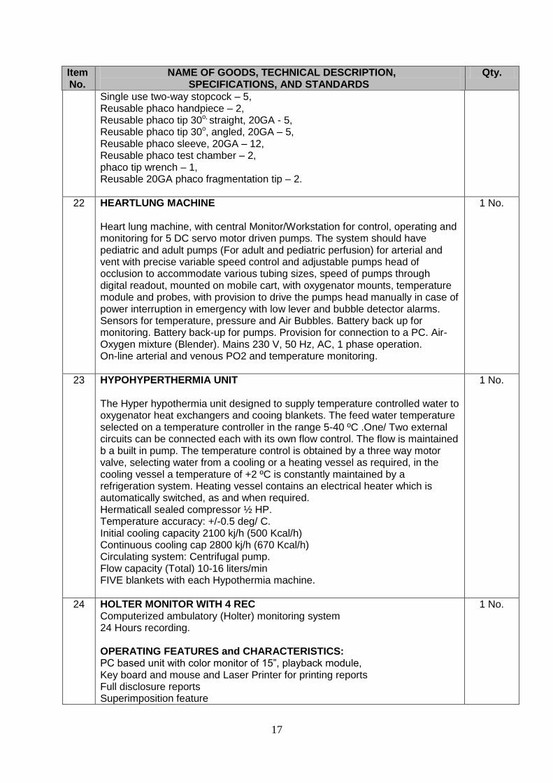
Single use two-way stopcock – 5, Reusable phaco handpiece – 2, Reusable phaco tip 30o, straight, 20GA - 5, Reusable phaco tip 30o, angled, 20GA – 5, Reusable phaco sleeve, 20GA – 12, Reusable phaco test chamber – 2, phaco tip wrench – 1, Reusable 20GA phaco fragmentation tip – 2.
22 HEARTLUNG MACHINE Heart lung machine, with central Monitor/Workstation for control, operating and monitoring for 5 DC servo motor driven pumps. The system should have pediatric and adult pumps (For adult and pediatric perfusion) for arterial and vent with precise variable speed control and adjustable pumps head of occlusion to accommodate various tubing sizes, speed of pumps through digital readout, mounted on mobile cart, with oxygenator mounts, temperature module and probes, with provision to drive the pumps head manually in case of power interruption in emergency with low lever and bubble detector alarms. Sensors for temperature, pressure and Air Bubbles. Battery back up for monitoring. Battery back-up for pumps. Provision for connection to a PC. Air-Oxygen mixture (Blender). Mains 230 V, 50 Hz, AC, 1 phase operation. On-line arterial and venous PO2 and temperature monitoring.
1 No.
23 HYPOHYPERTHERMIA UNIT The Hyper hypothermia unit designed to supply temperature controlled water to oxygenator heat exchangers and cooing blankets. The feed water temperature selected on a temperature controller in the range 5-40 ºC .One/ Two external circuits can be connected each with its own flow control. The flow is maintained b a built in pump. The temperature control is obtained by a three way motor valve, selecting water from a cooling or a heating vessel as required, in the cooling vessel a temperature of +2 ºC is constantly maintained by a refrigeration system. Heating vessel contains an electrical heater which is automatically switched, as and when required. Hermaticall sealed compressor ½ HP. Temperature accuracy: +/-0.5 deg/ C. Initial cooling capacity 2100 kj/h (500 Kcal/h) Continuous cooling cap 2800 kj/h (670 Kcal/h) Circulating system: Centrifugal pump. Flow capacity (Total) 10-16 liters/min FIVE blankets with each Hypothermia machine.
1 No.
24 HOLTER MONITOR WITH 4 REC Computerized ambulatory (Holter) monitoring system 24 Hours recording. OPERATING FEATURES and CHARACTERISTICS: PC based unit with color monitor of 15”, playback module, Key board and mouse and Laser Printer for printing reports Full disclosure reports Superimposition feature
1 No.

18
Item No.
NAME OF GOODS, TECHNICAL DESCRIPTION, SPECIFICATIONS, AND STANDARDS
Qty.
Analysis of ST slope, heart rate, R-R variability & arrhythmia Reports: in tabular and summary form., graphical and analytical, arrhythmia data and patient information: Heart rates (min., max, & avg.), R-R variability calculations, test duration, ventricular and supraventricular ectopics ST segment Reports: adjustable ST- segment criteria, time, duration, max deviation & slope, deviation severity index & number 2 channel simultaneous high resolution holter (solid state)recorder with event marker, time clock Number of Solid state Recorders: required quantity to be indicated below PARAMETERS: 2 channel simultaneous ECG acquisition OPERATING REQUIREMENTS: Main unit : AC 220V / 5OHz; Recorders: Standard long life dry batteries UPS for main unit for at least 30-min
25 BALLOON PUMP Self contained mobile console with ECG amplifier with possible selection 5 leads arterial blood pressure amplifier. Discriminative triggering circuit to command balloon actions on patient’s ECG arterial blood pressure curve or internal simulator 80 BPM. Colour graphic displays at least of 10” for display of arterial and pressure heart rate balloon volume used and alarm conditions with trouble shooting procedures. Wave form displays for ECG, arterial pressure and balloon pressure on three channel memory type oscilloscope. Fall safe system. V Pacing switch. Progressive viewing sequence. Integrated battery power supply to take patient to catheterization labs, operating theatre or other hospital: 60 minute autonomy. CO2 / helium tank wrench. 5 lead ECG cable, male connector pressure, and transducer adopter, chart recorder. Cardiac output computer 220 V, 50 Hz, Ac. One spare set of patient cable. System should be complete to display all the parameters. Disposable balloon catheters of varying sizes for use with the pump 50 nos. (An offer on placement basis on purchase of Balloons shall be given and will be given preference)
2 Nos.
26 FIBRILATOR Mode of operation continuous Rated out put Fully variable 10 Volt,50Hz ,0.10 mA Fuses T 160 ma Permissible operating temperature range +10°c tc+40°c ELECTRODES Fibrillator electrodes with double side electrode Fibrillator with bulldog clip and single plate electrodes Fibrillator Electrodes with two bulldog clips. Complete with Alligator Clamp.
1 No.

19
Item No.
NAME OF GOODS, TECHNICAL DESCRIPTION, SPECIFICATIONS, AND STANDARDS
Qty.
27 RAD ROOM RETRO FIT Double detector 17”x17” on existing three radiology suits of DOW Radiology Detectors are preferably of GOS technology but not mandatory Complete with workstations and software on placement basis against Rate contract of films for films. The firms will also be required to upgrade the existing 4 x CR Systems and network the DRs and CRs with existing printers. The total load of films is approximately 2500 packs per month with expected increase @ 7 -10% per annum. The forms will be requied to replace / upgrade the systems on 5-7 year basis.
3 Nos.
28 POWER INJECTOR Total 5 (3 x CT / Flouro and 2 x MRI compatible) Power Injector for radiology department on placement against rate contracts of syringes.
5 Nos.
SPECIAL NOTE:
The Purchaser will evaluate and compare the bids on itemized basis OR on the basis of a group OR a combination of groups OR as total of groups.
Equipment must be quoted with all the standard accessories.
UPS/Power protection for the equipment shall be incorporated in the systems, otherwise prices must be quoted separately.
All the civil works will be carried-out by the Dow University of Health Sciences, Karachi with the consultation of the responsive bidder.
Responsive bidder will provide the consultancy regarding the installation of the Equipment according to the GMP / WHO Standards (if applicable).
All site specific work to be required in the system viz. Lead Glass/special antistatic flooring, environment control/radiation protection must be quoted separately.
The bidder shall separately quote the price of service contract inclusive of parts as well as excluding the parts for 5 years (minimum) in term of %age for total contract value.


