AUTISM SCIENCE DIGEST: THE JOURNAL OF AUTISMONE ISSUE 04 www.ausmone.org NANCY MULLAN, MD, is an author, lecturer, and sought-after clinician best known for her natural approach to treatment and recovery from ASD. She was educated at the University of Pennsylvania, Tufts University, and the University of Chicago. Dr. Mullan has been practicing for 30 years and is excited to be on the cutting edge of the newest innovations in non- pharmaceutical ASD therapies. Currently, Dr. Mullan is practicing nutritional medicine and psychiatry in Burbank, California, treating children on the autism spectrum and adults with hormonal, gastroenterologic, neurologic, and/or metabolic dysfunction. 66 AUTISM SCIENCE DIGEST: THE JOURNAL OF AUTISMONE ISSUE 04 www.ausmone.org
Transcript
AUTISM SCIENCE DIGEST: THE JOURNAL OF AUTISMONE ISSUE 04 www.autismone.org
NaNcy MullaN, MD, is an author, lecturer, and sought-after clinician best known for her natural approach to treatment and recovery from ASD. She was educated at the University of Pennsylvania, Tufts University, and the University of Chicago. Dr. Mullan has been practicing for 30 years and is excited to be on the cutting edge of the newest innovations in non-pharmaceutical ASD therapies. Currently, Dr. Mullan is practicing nutritional medicine and psychiatry in Burbank, California, treating children on the autism spectrum and adults with hormonal, gastroenterologic, neurologic, and/or metabolic dysfunction.
66 AUTISM SCIENCE DIGEST: THE JOURNAL OF AUTISMONE ISSUE 04 www.autismone.org
www.autismone.org AUTISM SCIENCE DIGEST: THE JOURNAL OF AUTISMONE ISSUE 04 67
The adult human intestine is home to an almost inconceivable number of microorganisms. The size of the population—up to 100 trillion—far exceeds that of all other microbial communities associated with the body’s surfaces and is ~ 10 times greater than the total number of our somatic and germ cells. Thus, it seems appropriate to view ourselves as a composite of many species and our genetic landscape as an amalgam of genes embedded in our Homo sapiens genome and in the genomes of our affiliated microbial partners (the microbiome).
Our gut microbiota can be pictured as a microbial organ placed within a host organ. It is composed of different cell lineages with a capacity to communicate with one another and the host; it mediates physiologically important chemical transformations; and it can manage and repair itself through self-replication. The gut microbiome, which may contain > 100 times the number of genes in our genome, endows us with functional features that we have not had to evolve ourselves. (p.1915)
Colonization of the newborn’s GI tract starts immediately after birth. The earliest bacteria colonized can modulate the expression of genes in the host epithelial cells, thus creating a favorable environment for
Gastrointestinal Balance and
neurotransmitter Formation
By AMy yASko, PhD, AMD, FAAIM, AnD nAnCy MUllAn, MD
The complexiTy of The gasTroinTesTinal TracTover the past few decades, research has significantly changed the way that scientists understand gastrointestinal (GI) function and dysfunction. We have come to recognize that the GI tract is, in fact, far more subtle and complex than initially thought. Studies have illuminated how the complex interaction of the immune system, the neuroendocrine system, and the microbial environment within the gut affects not only GI function but also the function of other organs and systems in the body, most notably the brain and nervous system. We now understand that body systems and organs function within a web of physiology and biochemistry, and we no longer consider each system as discrete and isolated from the others.
During this same time period, autism spectrum disorders (ASDs)—consisting of autism, pervasive developmental delay (PDD), speech delay, attention-deficit disorder (ADD) or attention-deficit/hyperactivity disorder (ADhD)—have been acknowledged as multifactorial conditions. A number of causal factors must come together to create the constellation of symptoms that result in a diagnosis of one or several of these conditions. Prominent among these factors is GI dysfunction.1-3 The investigation of how the gastrointestinal environment and its function influence the central nervous system and other neurologic functions is, therefore, particularly pertinent.
Bäckhed and colleagues4 describe the human gut microbiome (the microbial community that resides in the intestine) as follows:
Studies have illuminated how the complex interaction of the immune system, the neuroendocrine system, and the microbial environment within the gut affects not
only GI function but also the function of other organs and systems in the body, most notably the brain and nervous system.
AUTISM SCIENCE DIGEST: THE JOURNAL OF AUTISMONE ISSUE 04 www.autismone.org
aMy yasko PhD, aMD, FaaIM, holds a doctorate in microbiology, immunology, and infectious diseases with an award for outstanding academic excellence from Albany Medical College.
She completed two research fellowships at Strong Memorial hospital in Rochester ny; one as a member of the Dept. of Pediatrics and Infectious Diseases, the other as a member of the Wilmont Cancer Center. Dr. yasko was also a fellow in the Department of hematology at yale Medical Center prior to joining a biotechnology company in Connecticut. She later co founded a successful biotechnology company, where she was recognized as an expert in the field of DnA/RnA based diagnostics and therapeutics. Prior to shifting her focus to integrative healthcare she was consultant to the medical, pharmaceutical, and research communities for almost 20 years with an expertise in biochemistry, molecular biology, and biotechnology. Dr. yasko continued her education in the area of integrative healthcare, receiving two additional degrees, a Doctor of naturopathy and a Doctor of natural health.
AUTISM SCIENCE DIGEST: THE JOURNAL OF AUTISMONE ISSUE 04 www.autismone.org68
www.autismone.org AUTISM SCIENCE DIGEST: THE JOURNAL OF AUTISMONE ISSUE 04 69
themselves. This can prevent the growth of other bacteria introduced at a later time. Therefore, this initial colonization is very relevant to the final composition of the adult individual’s flora.5 The predominant intestinal mucosa-associated bacterial community is host-specific and significantly different from the fecal bacterial community.6
Gastrointestinal mucus serves as a matrix for the flora contained in the GI tract.7 The integrity of the mucosal barrier is maintained by a single layer of tightly fitted epithelial cells called enterocytes that comprise a surface area greater than 400 square meters, which is 200 times greater than the surface of the skin. Approximately 70 percent of the body’s immune system is present in specialized lymphatic compartments within the GI mucosa and in the intercellular spaces along its epithelium. Experimental evidence demonstrates that the gastrointestinal mucosal immune system functions separately from the bloodborne (e.g., white blood cells and immunoglobulins) immune system.8 It is significant to note that the gastrointestinal tract houses a separate immune system – the enteric immune system.
In the remainder of this article, we discuss factors contributing to chronic microbial imbalance in the gastrointestinal tract and review some of the microorganisms (such as clostridia, streptococci, and Helicobacter pylori) that can have far-reaching effects when out of balance. We focus, in particular, on the influence of pathogenic organisms on neurotransmitter formation because of the essential role of neurotransmitters as the body’s chemical messengers.
WhaT causes chronic microbial imbalanceGut mucosal integrity can be compromised by a number of factors, including chronic microbial imbalance (dysbiosis), inflammation, and immune system dysregulation. Dysbiosis can produce mucosal damage, and the accompanying inflammatory processes may lead to a disruption in microvilli function and gut permeability. These, in turn, can manifest as gluten, casein, and/or lactose intolerance; food allergies or intolerance; abdominal pain and discomfort; or abnormal bowel function. non-GI-related symptoms can also appear, such as headaches, skin irritations, chronic joint pain, anxiety, or depression.
Imbalances in the flora of the GI tract may begin as early as birth. Maternal streptococci, for example, can be transmitted from the mother to the neonate during delivery. Although researchers originally believed that transmission occurred solely via vaginal delivery,9 more recent data suggest that streptococcal infection can also occur in infants who have been delivered via cesarean section.10 The rate of mother-to-infant transmission of streptococci during vaginal delivery is between 20 and 30 percent.
Increased gut acidity also can predispose to microbial imbalance. Some of the factors that contribute to increased gut acidity and gut flora imbalances include vitamin B12 deficiency, decreased pancreatic or liver function, genetics/blood type, and antibiotic use.
VItaMIN B12 DeFIcIeNcy: Intrinsic factor, a substrate produced by the gastric lining, is necessary for the uptake of vitamin B12 by the small intestine. Sufficient stomach acid is necessary for intrinsic factor activation. The secretion of stomach acid and the secretion of intrinsic factor parallel one another,11 and loss of gastric secreting
cells decreases both intrinsic factor and stomach acid.12 Proton pump inhibitors, a group of drugs widely used for peptic ulcer disease and other hyperacidic conditions, antagonize stomach acid levels and lead to decreases in intrinsic factor and decreased uptake of B12.13
A significant lack of B12 has been found in autism, irrespective of age.14 Efforts by the body to increase B12 levels lead to increased intrinsic factor and would be expected to increase stomach acid.13,15
While the growth of many bacteria is inhibited in an acidic (low ph) environment, streptococci are among those bacteria that can survive and flourish at a lower ph.16 Increased stomach acid secondary to B12 deficiency, therefore, leads to a gut environment that predisposes to the growth of streptococci and other pathogenic bacteria that can survive in an acid milieu, such as Escherichia coli.16,17
DecreaseD PaNcreatIc or lIVer FuNctIoN: one of the roles of bile is to neutralize stomach acid. lack of bile due to decreased pancreatic or liver function contributes to an acidic gut environment. Impairments in liver or pancreatic function due to toxin overload or infectious diseases such as rubella (which is known to infect the pancreas), therefore, decrease the body’s ability to neutralize excess acid.
GeNetIcs aND BlooD tyPe: Genetics and blood type can predispose to colonization with streptococci. For example, single nucleotide polymorphisms (SnPs) that decrease the level of B12 in the body can contribute to an environment that is conducive to the growth of streptococci. Blood type also appears to play a role, which may be related in part to the increased acidity seen in those with blood type o.18 Effects of different carbohydrate groupings on the surface of blood cells of varying blood types may also have an impact on the ability of streptococci to aggregate in the system.19
aNtIBIotIcs: normal flora help to protect the gut from the growth of pathogenic organisms. Antibiotic use is well known to cause imbalances in normal gut flora. The use of antibiotics without concurrent addition of probiotics, therefore, can predispose to the growth of streptococci as well as other pathogenic organisms such as clostridia. Fecal flora studies of children with ASD have found that the number of clostridial species are greater and the clostridial counts higher in ASD children when compared with controls.1,3
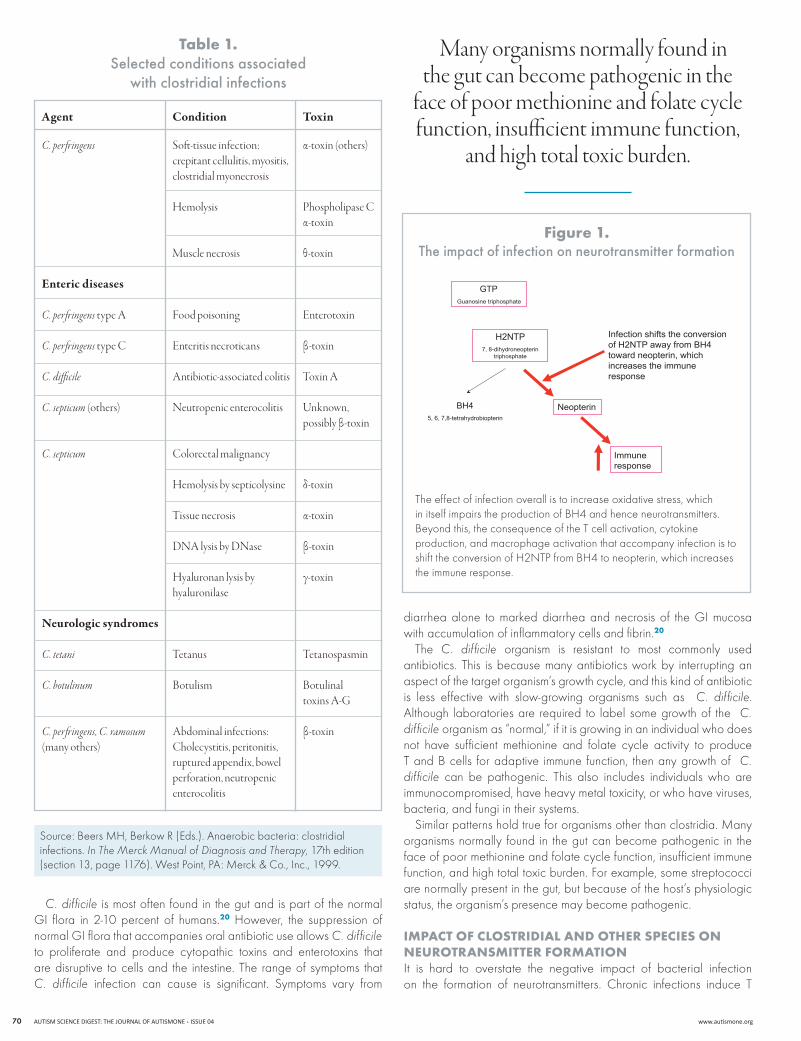
closTridial and oTher species Clostridia are spore-forming, Gram-positive anaerobes. The growth of anaerobes is inhibited or significantly slowed in the presence of oxygen. The dearth of oxygen in the lumen of the small and large intestines predisposes this environment to the growth of anaerobic organisms. There are more than 50 species of clostridia, a number of which cause significant illness. To name only a few examples, Clostridium botulinum causes botulism, C. perfringens causes gas gangrene and food poisoning, C. tetani causes tetanus, and C. difficile causes pseudomembranous colitis. Table 1 provides a more comprehensive list of the many conditions associated with clostridial infection.
Gut mucosal integrity can be compromised by a number of factors, including chronic microbial imbalance (dysbiosis), inflammation, and immune system dysregulation.
AUTISM SCIENCE DIGEST: THE JOURNAL OF AUTISMONE ISSUE 04 www.autismone.org70
C. difficile is most often found in the gut and is part of the normal GI flora in 2-10 percent of humans.20 however, the suppression of normal GI flora that accompanies oral antibiotic use allows C. difficile to proliferate and produce cytopathic toxins and enterotoxins that are disruptive to cells and the intestine. The range of symptoms that C. difficile infection can cause is significant. Symptoms vary from
diarrhea alone to marked diarrhea and necrosis of the GI mucosa with accumulation of inflammatory cells and fibrin.20
The C. difficile organism is resistant to most commonly used antibiotics. This is because many antibiotics work by interrupting an aspect of the target organism’s growth cycle, and this kind of antibiotic is less effective with slow-growing organisms such as C. difficile. Although laboratories are required to label some growth of the C. difficile organism as “normal,” if it is growing in an individual who does not have sufficient methionine and folate cycle activity to produce T and B cells for adaptive immune function, then any growth of C. difficile can be pathogenic. This also includes individuals who are immunocompromised, have heavy metal toxicity, or who have viruses, bacteria, and fungi in their systems.
Similar patterns hold true for organisms other than clostridia. Many organisms normally found in the gut can become pathogenic in the face of poor methionine and folate cycle function, insufficient immune function, and high total toxic burden. For example, some streptococci are normally present in the gut, but because of the host’s physiologic status, the organism’s presence may become pathogenic.
impacT of closTridial and oTher species on neuroTransmiTTer formaTionIt is hard to overstate the negative impact of bacterial infection on the formation of neurotransmitters. Chronic infections induce T
C. perfringens type C Enteritis necroticans β-toxin
C. difficile Antibiotic-associated colitis Toxin A
C. septicum (others) Neutropenic enterocolitis Unknown, possibly β-toxin
C. septicum Colorectal malignancy
Hemolysis by septicolysine δ-toxin
Tissue necrosis α-toxin
DNA lysis by DNase β-toxin
Hyaluronan lysis by γ-toxin hyaluronilase
Neurologic syndromes
C. tetani Tetanus Tetanospasmin
C. botulinum Botulism Botulinal toxins A-G
C. perfringens, C. ramosum Abdominal infections: β-toxin (many others) Cholecystitis, peritonitis, ruptured appendix, bowel perforation, neutropenic enterocolitis
Source: Beers Mh, Berkow R (Eds.). Anaerobic bacteria: clostridial infections. In The Merck Manual of Diagnosis and Therapy, 17th edition (section 13, page 1176). West Point, PA: Merck & Co., Inc., 1999.
Many organisms normally found in the gut can become pathogenic in the
face of poor methionine and folate cycle function, insufficient immune function,
and high total toxic burden.
7, 8-dihydroneopterintriphosphate
Infection shifts the conversionof H2NTP away from BH4toward neopterin, whichincreases the immuneresponse
GTPGuanosine triphosphate
H2NTP
Neopterin
Immuneresponse
BH45, 6, 7,8-tetrahydrobiopterin
The effect of infection overall is to increase oxidative stress, which in itself impairs the production of Bh4 and hence neurotransmitters. Beyond this, the consequence of the T cell activation, cytokine production, and macrophage activation that accompany infection is to shift the conversion of h2nTP from Bh4 to neopterin, which increases the immune response.
figure 1. the impact of infection on neurotransmitter formation
www.autismone.org AUTISM SCIENCE DIGEST: THE JOURNAL OF AUTISMONE ISSUE 04 71
cell activation, cytokine formation, and macrophage activation, increasing oxidative stress, which impairs neurotransmitter formation (Figure 1).
Dihydroxyphenylpropionic acid (DhPPA) is a marker for bacterial infection with clostridial species,21 Pseudomonas species,22 and E. coli.23 The production of DhPPA that results from infection with these bacteria may reduce formation of the neurotransmitter dopamine through its depletion of the enzyme tyrosinase.24 There are two pathways for dopamine synthesis in the body. In the primary pathway, phenylalanine is hydroxylated to tyrosine in a reaction catalyzed by phenylalanine hydroxylase. This reaction is dependent on tetrahydrobiopterin (Bh4) (see Figure 2). The resulting tyrosine is hydroxylated to l-DoPA (3,4-dihydroxyphenylalanine) by the enzyme tyrosine hydroxylase in a second Bh4-dependent reaction. l-DoPA is then decarboxylated in a reaction catalyzed by aromatic acid decarboxylase to synthesize dopamine. Dopamine is the substrate for the subsequent synthesis of the catecholamines norepinephrine and epinephrine.
This primary pathway for dopamine synthesis can be compromised by a number of factors. For example, microbes can increase the serum phenylalanine-tyrosine ratio, which inhibits the first step in the pathway. Besides inhibiting this pathway, high levels of phenylalanine may also cause a drop in serotonin and/or GABA (gamma-aminobutyric acid), which may result in obsessive-compulsive disorder (oCD) behaviors.25 The exigencies of tyrosine hydroxylase activity also have an impact on this dopamine synthesis pathway.26-28 If levels of norepinephrine and epinephrine increase as the result of stressors (including infections), feedback inhibition on tyrosine hydroxylase will result. Increased glutamate also decreases tyrosine hydroxylase
activity. Glutamate (via nMDA receptors) and dopamine (via D2 receptors) decrease tyrosine hydroxlase phosphorylation by decreasing cAMP, a messenger derived from adenosine triphosphate (ATP) that is important in many biological processes. Because tyrosine hydroxylase activity is stimulated by phosphorylation, a situation of decreased tyrosine hydroxylase phosphorylation leads to decreased enzyme activity and lower levels of dopamine.29
The levels of Bh4 in the body are critical to both dopamine and serotonin synthesis. however, Bh4 is a vulnerable molecule and can be deficient for a number of reasons, some of which involve gut bacteria. As a substrate for various reactions, Bh4 is especially subject to depletion. Biopterin deficiency will reduce available Bh4, as will oxidative stress. In addition, the presence of aluminum or lead inhibits the activity of dihydrobiopterin reductase, which catalyzes Bh4 synthesis from dihydrobiopterin (Bh2). Bh4 levels also are profoundly decreased by infection because the body’s immune response produces neopterin, which reduces the production of Bh4 (see Figure 1). Finally, individuals with MThFR A1298C mutations may have a reduced ability to synthesize Bh4.30 The product of the MThFR enzyme reaction, 5-methyltetrahydrofolate, has been shown to be directly related to Bh4 levels31-33 and to be reversible, with Bh4 production being the outcome of the reverse reaction.34-36
Although the primary sequence for dopamine production is through the pathway shown in Figure 2, this pathway may not always function. Alternatively, the enzyme tyrosinase can act on tyrosine to produce l-DoPA in a one-step process,37 following which aromatic acid decarboxylase can act on l-DoPA to produce dopamine. This alternative pathway is able to circumvent the blockages that can result from problems with phenylalanine levels, tyrosine hydroxylase activity,
figure 2. the primary pathway for dopamine synthesis
AUTISM SCIENCE DIGEST: THE JOURNAL OF AUTISMONE ISSUE 04 www.autismone.org72
lack of Bh4, or oxidative stress. With the depletion of tyrosinase that results from bacterial infection-induced production of DhPPA, however, both the primary and the secondary pathways for the production of dopamine can be compromised. In addition, the production of norepinephrine and epinephrine are likely to be decreased because dopamine subsequently converts to norephinephrine and ephinephrine.
biofilms and sTrepTococciA biofilm is a community of one or several diverse species of organisms that firmly fix to a surface and grow within a self-produced polymer matrix. Ultimately, the group of organisms begins to function as a unit. The community aspect of biofilm formations offers a number of advantages, including protection from hostile environmental conditions and the opportunity to sequester nutritional resources. In addition, a microbial community offers the possibility for a variety of different organisms (bacteria, fungi, viruses, and single-celled parasites) to live together, exchange genetic information, communicate through chemical signaling, act cooperatively, and, in so doing, enhance their own survival and the survival of the collective. It has come to be recognized that the formation of biofilms is the preferred form of growth for organisms, whereas growth of planktonic (single-celled or free-floating) organisms is an artifact of in vitro culture.38 Certain organisms have evolved genetically to be viable only in a biofilm.39 The take-home message here is that organisms don’t really live on their own in nature – they live in communities.
Biofilms can form quickly. In a human body that is experiencing nutrient depletion or high oxidative stress, E. coli bacteria can activate genes that form biofilms in a variety of environments within
24 hours.40-42 In ASD patients, in particular, the GI tract is a natural place for biofilms to form; the lack of cell-mediated immunity in these patients predisposes them to biofilm growth. Moreover, the range of GI symptoms caused by biofilms is clearly identifiable in the ASD population.2
Among many other organisms, streptococci have been identified in biofilms. This form of organization gives streptococci an adaptational advantage and the capacity to thrive in a wide range of ph conditions and environments in which they otherwise would not survive.43 Generally, bacteria elicit a B-cell-mediated immune response and viruses a T-cell-mediated immune response. Streptococci, however, prompt the elaboration of a large number of extracellular toxins, all of which have the ability to nonspecifically stimulate T cells. once an immune response is mounted against streptococcal species, therefore, the response involves both B cells and T cells, resulting in a major inflammatory reaction. In addition, streptococcal infection increases the production of hydrogen peroxide (h2o2), thereby increasing oxidative stress in the body, and depletes the peroxidase enzyme necessary for the production of thyroid hormone, possibly reducing levels of thyroxine (T4) and triiodothyronine (T3).44 This and other consequences of streptococcal infection are summarized in Figure 3.
Several species of streptococci are well characterized as causing neuropsychiatric symptoms45 and disorders such as autism and anorexia nervosa.46 Autoantibodies to group A streptococcal sugar moieties are implicated in oCD, Tourette syndrome, chronic tic disorders, ADhD, and Sydenham chorea, a neuropsychiatric complication of rheumatic fever.47,48 Sydenham chorea is one of the best examples of postinfectious autoimmunity produced by molecular
figure 3. consequences of streptococcal infection
In ASD patients, in particular, the GI tract is a natural place for biofilms to form; the lack of cell-mediated immunity in these patients predisposes them to biofilm growth.
www.autismone.org AUTISM SCIENCE DIGEST: THE JOURNAL OF AUTISMONE ISSUE 04 73
The association between chronic bacterial infection and neurologic and psychiatric disorders can additionally be explained by the fact that streptococcal and other bacterial infections cause the breakdown of tryptophan, a direct precursor of serotonin. States of persistent immune activation diminish the availability of free serum tryptophan and compromise serotonin production. As immune activation accelerates tryptophan degradation, tryptophan depletion, in turn, may downregulate the immune response. In addition, a breakdown product of tryptophan degradation, quinolinic acid, begins to accumulate. Quinolinic acid is another excitotoxin and may contribute to the development of the neuropsychiatric disorders seen in the presence of chronic infection and serotonin depletion.50
guT imbalances and heavy meTalsBacterial infection promotes accumulation and retention of heavy metals in the body,51-56 which contributes to oxidative stress and impairs neurotransmitter formation. Metals may be retained in the body through several mechanisms.51-56 We have documented the sequestration of aluminum and lead in the organisms of the microbiome and the impacts of retention of these metals at length elsewhere.57,58 The elimination of abnormal gastrointestinal flora and excretion of the metals they retain, therefore, may be essential for proper function of the biochemical pathways in the body, along with maintenance of proper balance among the organisms that should be present in the gastrointestinal tract.58
Streptoccal infections act in a number of ways to retain heavy metals in the body, including sequestering metals within the cell walls of the bacteria. Another mechanism related to metal retention involves sulfhydryl groups, which ordinarily bind and eliminate toxic and heavy metals. Streptococcus produces an extracellular enzyme called sulfhydryl protease that is capable of cleaving these sulfhydryl groups, leading to a deficiency of sulfur-containing moieties in the body. Streptococci proliferate in the presence of iron59 and reduce the capacity of the body to excrete this heavy metal. Iron, in turn, increases the virulence of many bacteria, including streptococci. In addition, iron is necessary for biofilm formation.60-63 Excess iron can increase microbial imbalances in the body, including but not limited to streptococcal infections.
Aluminum is a well-documented and undisputed neurotoxin that is associated with cognitive, psychological, and motor abnormalities. Both clinical observation and animal experiments have documented neurotoxicity from excess brain exposure to aluminum,64 which has been found in elevated levels in the brains of patients with Parkinson-ism,65-67 amyotrophic lateral sclerosis (AlS),66,67 and Alzheimer-type dementia.68-72 Aluminum induces encephalopathy67,73,74 and causes neuroanatomical and neurochemical changes in the brain, including neurofilament disturbances65,69,75-77 followed by nerve cell loss.73,78 Primate studies have provided evidence of aluminum’s ability to induce seizures.79-82
While staphylococci are especially prone to retaining aluminum,54 it is likely that other bacteria also can do so. Moreover, aluminum may increase the propensity for bacteria to form a biofilm, in part because of its pro-oxidant activity.83 It has been characterized as having direct effects on biofilm activity in other systems.84
mimicry (when molecules are similar and the body confuses an invader with the body’s own tissue) between host and antigen. Following a pharyngeal infection with group A streptococcus, chorea antibodies produced in response to a group A streptococcal carbohydrate target the surface of human neuronal cells, producing abrupt, spontaneous movements of the face and extremities, oCD behaviors, hyperactivity, emotional lability, and other psychiatric symptoms.47 The binding of cross-reacting antibodies to human basal ganglia results in changes to neuronal signaling transduction and neurotransmitter synthesis and release.46
Infection with group A beta-hemolytic Streptococcus (GABhS) can result in a pediatric syndrome labeled as pediatric autoimmune neuropsychiatric disorders associated with streptococcal infections (PAnDAS). Individuals with PAnDAS develop tics, oCD symptoms, and, in some cases, psychosis, without the comorbid chorea component. When sera from GABhS-immunized mice were tested for immunoreactivity to mouse brain, a subset was found to be immunoreactive to several brain regions, including the deep cerebellar nuclei (DCn), the globus pallidus (one of three nuclei that make up the basal ganglia), and the thalamus (involved in sensory perception and regulation of motor functions).48 GABhS mice showing serum immunoreactivity to DCn also had increased IgG deposits in the DCn and exhibited abnormal behavior. The motoric and behavioral disturbances that result from an immune response to GABhS suggest that anti-GABhS antibodies cross-reacting with brain components are part of the pathophysiology of these disturbances.48
Streptococcal infection leads to elevated levels of the inflammatory cytokines TnF-alpha and nF-kB. high levels of TnF-alpha have been implicated in Tourette syndrome, facial tics, oCD behavior, and schizophrenia. The increased TnF-alpha levels that occur with streptococcal infection cause increased levels of glutamate, an excitatory neurotransmitter. When the normal processes that regulate glutamate levels malfunction and toxic levels build up in the synaptic junctions, glutamate toxicity can produce neuron loss. Moreover, the neuroinflammatory response in and of itself also contributes to glutamate excitotoxicity and neuronal loss. The two pathogenic mechanisms, glutamate excitotoxicity and neuroinflammation, seem to be linked.49
To remove excess glutamate, the brain requires sufficient levels of oxygen and energy. however, glutamate release leads to the release of insulin, which results in decreased glucose levels. Because the amount of glucose in the brain regulates the removal of excess glutamate from the synapses, a drop in blood glucose disrupts the removal process and allows a buildup of toxic glutamate. (hypoglycemia or low calorie/starvation conditions can induce a similar cycle involving glutamate release, insulin release, decreased glucose, and inability to remove excess glutamate from the brain.) Excess glutamate is problematic because it depletes glutathione. Glutathione is one of the body’s most powerful antioxidants and helps to protect neurons from damage. Glutathione depletion consequently leads to the death of additional neurons. If glutamate itself were not already reducing glutathione levels, the high TnF-alpha levels resulting from streptococcal infection also would result in decreased glutathione because TnF-alpha levels are inversely correlated with glutathione levels.
Bacterial infection promotes accumulation and retention of heavy metals in the body, which contributes to oxidative stress and impairs neurotransmitter formation.
AUTISM SCIENCE DIGEST: THE JOURNAL OF AUTISMONE ISSUE 04 www.autismone.org74
figure 4. Helicobacter pylori
Helicobacter pylori in auTism specTrum disordersHelicobacter pylori (H. pylori) is a Gram-negative, spiral-shaped bacterium that lives in the stomach and duodenum. This ulcer-causing gastric pathogen is able to colonize the harsh acidic environment of the human stomach. Although the stomach is protected from its own gastric juice by a thick layer of mucus that covers the stomach lining, H. pylori takes advantage of this protection by living in the mucus lining itself. It does so using long, whip-like flagella that facilitate locomotion through the mucus layer (see Figure 4). Although the organism is best known for its etiologic role in ulcers and impaired digestion, it also can induce vasovagal symptoms after eating, including weakness, skin pallor, profuse sweating, and sensations of loss of consciousness that resolve after eradication of the infection.85
In the mucus lining, H. pylori survives the stomach’s acidic conditions by producing urease, an enzyme that catalyzes hydrolysis of urea into ammonia and bicarbonate. As strong bases, ammonia and bicarbonate produce a cloud of alkalinity around the bacterium, making it impossible for the body’s normal defenses (such as T cells, natural killer cells, and other white blood cells) to get to it in the gastric mucus layer. Polymorphs, white blood cells containing a segmented nucleus and that are first responders to infection sites, release superoxide radicals on stomach lining cells in an increasing inflammatory response. other factors that contribute to H. pylori colonization of the gastric mucosa include adhesins, molecules that make the bacteria adhere to the mucosa, and genes encoding proteins with chemotaxis into the mucus.86,87 Incidentally—but importantly— H. pylori infection increases TnF-alpha levels. The organism binds to the mucin and uses it to burrow into the cells lining the mucosal layer to create an infection.88 It very deeply infects those cells and is distributed evenly throughout (see Figure 5).
Source: Schreiber S, konradt M, Groll C, Scheid P, hanauer G, Werling ho, et al. The spatial orientation of Helicobacter pylori in the gastric mucus. PNAS. 2004;101(14):5024–9.
H. pylori infection increases TNF-alpha levels.
Distribution of H. pylori and H. felis in the mucus layer of mice and Mongolian gerbils. (A) The tissue surface of the H. pylori-infected gerbil depicted from the luminal side of the antrum. Several focus planes have been digitally combined, the H. pylori in the mucus layer subsequently highlighted in red. (B) The gastric mucosa and mucus of the H. felis-infected mouse and the H. pylori-infected gerbil are shown as schematic cross sections. The first 25 μm of the mucus layer on the tissue side (“juxtamucosal” mucus) are subdivided into 5 μm of sections. The numbers represent the percentage of bacteria present within each section. The first 10 μm from the luminal surface is referred to as “luminal mucus,” the rest of the mucus layer as “central mucus.” H. felis was found located between 5 and 25 μm from the tissue surface. H. pylori, however, colonizes the whole section 0-25 μm from the tissue surface. Some H. pylori were attached to cells.
figure 5. H. pylori invasion
www.autismone.org AUTISM SCIENCE DIGEST: THE JOURNAL OF AUTISMONE ISSUE 04 75
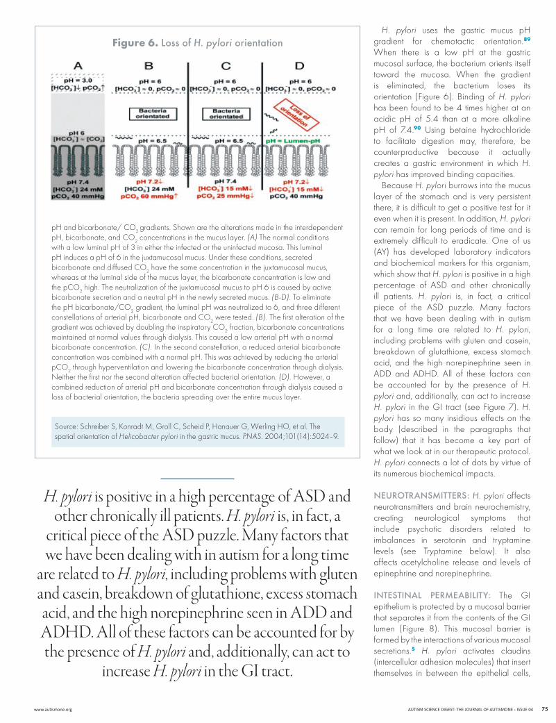
H. pylori uses the gastric mucus ph gradient for chemotactic orientation.89 When there is a low ph at the gastric mucosal surface, the bacterium orients itself toward the mucosa. When the gradient is eliminated, the bacterium loses its orientation (Figure 6). Binding of H. pylori has been found to be 4 times higher at an acidic ph of 5.4 than at a more alkaline ph of 7.4.90 Using betaine hydrochloride to facilitate digestion may, therefore, be counterproductive because it actually creates a gastric environment in which H. pylori has improved binding capacities.
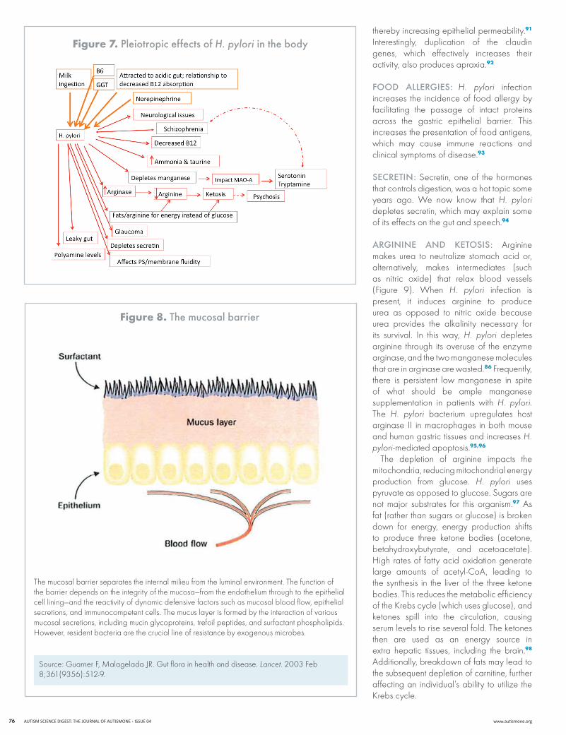
Because H. pylori burrows into the mucus layer of the stomach and is very persistent there, it is difficult to get a positive test for it even when it is present. In addition, H. pylori can remain for long periods of time and is extremely difficult to eradicate. one of us (Ay) has developed laboratory indicators and biochemical markers for this organism, which show that H. pylori is positive in a high percentage of ASD and other chronically ill patients. H. pylori is, in fact, a critical piece of the ASD puzzle. Many factors that we have been dealing with in autism for a long time are related to H. pylori, including problems with gluten and casein, breakdown of glutathione, excess stomach acid, and the high norepinephrine seen in ADD and ADhD. All of these factors can be accounted for by the presence of H. pylori and, additionally, can act to increase H. pylori in the GI tract (see Figure 7). H. pylori has so many insidious effects on the body (described in the paragraphs that follow) that it has become a key part of what we look at in our therapeutic protocol. H. pylori connects a lot of dots by virtue of its numerous biochemical impacts.
NeurotraNsMItters: H. pylori affects neurotransmitters and brain neurochemistry, creating neurological symptoms that include psychotic disorders related to imbalances in serotonin and tryptamine levels (see Tryptamine below). It also affects acetylcholine release and levels of epinephrine and norepinephrine.
INtestINal PerMeaBIlIty: The GI epithelium is protected by a mucosal barrier that separates it from the contents of the GI lumen (Figure 8). This mucosal barrier is formed by the interactions of various mucosal secretions.5 H. pylori activates claudins (intercellular adhesion molecules) that insert themselves in between the epithelial cells,
figure 6. loss of H. pylori orientation
H. pylori is positive in a high percentage of ASD and other chronically ill patients. H. pylori is, in fact, a
critical piece of the ASD puzzle. Many factors that we have been dealing with in autism for a long time
are related to H. pylori, including problems with gluten and casein, breakdown of glutathione, excess stomach acid, and the high norepinephrine seen in ADD and ADHD. All of these factors can be accounted for by the presence of H. pylori and, additionally, can act to
increase H. pylori in the GI tract.
Source: Schreiber S, konradt M, Groll C, Scheid P, hanauer G, Werling ho, et al. The spatial orientation of Helicobacter pylori in the gastric mucus. PNAS. 2004;101(14):5024–9.
ph and bicarbonate/ Co2 gradients. Shown are the alterations made in the interdependent ph, bicarbonate, and Co2 concentrations in the mucus layer. (A) The normal conditions with a low luminal ph of 3 in either the infected or the uninfected mucosa. This luminal ph induces a ph of 6 in the juxtamucosal mucus. Under these conditions, secreted bicarbonate and diffused Co2 have the same concentration in the juxtamucosal mucus, whereas at the luminal side of the mucus layer, the bicarbonate concentration is low and the pCo2 high. The neutralization of the juxtamucosal mucus to ph 6 is caused by active bicarbonate secretion and a neutral ph in the newly secreted mucus. (B-D). To eliminate the ph bicarbonate/Co2 gradient, the luminal ph was neutralized to 6, and three different constellations of arterial ph, bicarbonate and Co2 were tested. (B). The first alteration of the gradient was achieved by doubling the inspiratory Co2 fraction, bicarbonate concentrations maintained at normal values through dialysis. This caused a low arterial ph with a normal bicarbonate concentration. (C). In the second constellation, a reduced arterial bicarbonate concentration was combined with a normal ph. This was achieved by reducing the arterial pCo2 through hyperventilation and lowering the bicarbonate concentration through dialysis. neither the first nor the second alteration affected bacterial orientation. (D). however, a combined reduction of arterial ph and bicarbonate concentration through dialysis caused a loss of bacterial orientation, the bacteria spreading over the entire mucus layer.
AUTISM SCIENCE DIGEST: THE JOURNAL OF AUTISMONE ISSUE 04 www.autismone.org76
figure 7. Pleiotropic effects of H. pylori in the body
figure 8. the mucosal barrier
Source: Guarner F, Malagelada JR. Gut flora in health and disease. Lancet. 2003 Feb 8;361(9356):512-9.
The mucosal barrier separates the internal milieu from the luminal environment. The function of the barrier depends on the integrity of the mucosa—from the endothelium through to the epithelial cell lining—and the reactivity of dynamic defensive factors such as mucosal blood flow, epithelial secretions, and immunocompetent cells. The mucus layer is formed by the interaction of various mucosal secretions, including mucin glycoproteins, trefoil peptides, and surfactant phospholipids. however, resident bacteria are the crucial line of resistance by exogenous microbes.
thereby increasing epithelial permeability.91 Interestingly, duplication of the claudin genes, which effectively increases their activity, also produces apraxia.92
FooD allerGIes: H. pylori infection increases the incidence of food allergy by facilitating the passage of intact proteins across the gastric epithelial barrier. This increases the presentation of food antigens, which may cause immune reactions and clinical symptoms of disease.93
secretIN: Secretin, one of the hormones that controls digestion, was a hot topic some years ago. We now know that H. pylori depletes secretin, which may explain some of its effects on the gut and speech.94
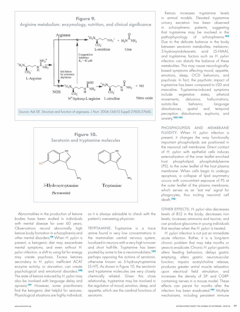
arGININe aND ketosIs: Arginine makes urea to neutralize stomach acid or, alternatively, makes intermediates (such as nitric oxide) that relax blood vessels (Figure 9). When H. pylori infection is present, it induces arginine to produce urea as opposed to nitric oxide because urea provides the alkalinity necessary for its survival. In this way, H. pylori depletes arginine through its overuse of the enzyme arginase, and the two manganese molecules that are in arginase are wasted.86 Frequently, there is persistent low manganese in spite of what should be ample manganese supplementation in patients with H. pylori. The H. pylori bacterium upregulates host arginase II in macrophages in both mouse and human gastric tissues and increases H. pylori-mediated apoptosis.95,96
The depletion of arginine impacts the mitochondria, reducing mitochondrial energy production from glucose. H. pylori uses pyruvate as opposed to glucose. Sugars are not major substrates for this organism.97 As fat (rather than sugars or glucose) is broken down for energy, energy production shifts to produce three ketone bodies (acetone, betahydroxybutyrate, and acetoacetate). high rates of fatty acid oxidation generate large amounts of acetyl-CoA, leading to the synthesis in the liver of the three ketone bodies. This reduces the metabolic efficiency of the krebs cycle (which uses glucose), and ketones spill into the circulation, causing serum levels to rise several fold. The ketones then are used as an energy source in extra hepatic tissues, including the brain.98 Additionally, breakdown of fats may lead to the subsequent depletion of carnitine, further affecting an individual’s ability to utilize the krebs cycle.
www.autismone.org AUTISM SCIENCE DIGEST: THE JOURNAL OF AUTISMONE ISSUE 04 77
figure 10. seratonin and tryptamine molecules
figure 9. arginine metabolism: enzymology, nutrition, and clinical significance
Source: Ash DE. Structure and function of arginases. J Nutr. 2004;134(10 Suppl):2760S-2764S.
Abnormalities in the production of ketone bodies have been studied in individuals with mental disease for over 60 years. observations record abnormally high ketone body formation in schizophrenia and other mental disorders.99 When H. pylori is present, a ketogenic diet may exacerbate mental symptoms, and even without H. pylori infection, a shift to using fat for energy may create psychosis. Excess ketones secondary to H. pylori, inefficient ACAT enzyme activity, or starvation, can create psychological and emotional disorders.100 The state of ketosis induced by H. pylori may also be involved with language delay and apraxia.101 however, some practitioners find the ketogenic diet helpful for seizures. Physiological situations are highly individual,
so it is always advisable to check with the patient’s overseeing physician.
tryPtaMINe: Tryptamine is a trace amine found in very low concentrations in the mammalian central nervous system, localized in neurons with a very high turnover and short half-life. Tryptamine has been posited by some to be a neuromodulator,102 perhaps opposing the actions of serotonin, otherwise known as 5-hydroxytryptamine (5-hT). As shown in Figure 10, the serotonin and tryptamine molecules are very closely chemically related. Given this close relationship, tryptamine may be involved in the regulation of mood, emotion, sleep, and appetite, which are the cardinal functions of serotonin.
ketosis increases tryptamine levels in animal models. Elevated tryptamine urinary excretion has been observed in schizophrenic patients, suggesting that tryptamine may be involved in the pathophysiology of schizophrenia.103 Due to the delicate balance in the body between serotonin metabolites, melatonin, 5-hydroxyindoleacetic acid (5-hIAA), and tryptamine, factors such as H. pylori infection can disturb the balance of these metabolites. This may cause neurologically-based symptoms affecting mood, appetite, emotions, sleep, oCD behaviors, and psychosis. In fact, the psychotic impact of tryptamine has been compared to lSD and mescaline. Tryptamine-induced symptoms include vegetative states, athetoid movements, delusions, hallucinations, autistic-like behavior, language disturbances, spatial and temporal perception disturbances, euphoria, and anxiety.103-105
PhosPholIPIDs aND MeMBraNe FluIDIty: When H. pylori infection is present, it changes the way functionally important phospholipids are positioned in the neuronal cell membrane. Direct contact of H. pylori with epithelial cells induces externalization of the inner leaflet enriched host phospholipid, phosphatidylserine (PS), to the outer leaflet of the host plasma membrane. When cells begin to undergo apoptosis, a collapse of lipid asymmetry occurs with concomitant exposure of PS at the outer leaflet of the plasma membrane, which serves as an “eat me” signal for phagocytes, thus inciting neuronal cell death.106
other eFFects: H. pylori also decreases levels of B12 in the body; decreases iron levels; increases ammonia and taurine; and can produce glaucoma in young individuals that resolves when the H. pylori is treated.
H. pylori infection is not just an immediate acute infection. Rather, it is a long-term chronic problem that may take months or years to eradicate. Chronic H. pylori gastritis alters feeding behaviors, delays gastric emptying, alters gastric neuromuscular function, impairs acetylcholine release, produces greater antral muscle relaxation upon electrical field stimulation, and increases the density of SP- and CGRP-containing nerves in a mouse model. These effects can persist for months after the infection has been eradicated.107 Multiple mechanisms, including persistent immune
AUTISM SCIENCE DIGEST: THE JOURNAL OF AUTISMONE ISSUE 04 www.autismone.org78
RefeRences
gut activation and altered brain neurochemistry, may be responsible for the maintenance of abnormal feeding behaviors, providing an explanation for the lack of apparent symptom resolution following the eradication of H. pylori.108 An extended period may be necessary before gastric physiology can return to normal after successful H. pylori treatment.109
TreaTmenT consideraTionsMany organisms normally found in the gut can become pathogenic in the face of poor methionine and folate cycle function, insufficient immune function, and a high toxic burden. Therefore, it is essential to treat imbalanced gut flora whenever these conditions are present. Although organisms such as E. coli and streptococci often are reported on stool analyses as normal flora, any such organisms appearing on the stool culture of an immunocompromised individual should be considered possible pathogens. These organisms’ potential pathogenic impacts in significantly immunocompromised individuals are amply documented, even if mainstream opinion continues to consider them nonpathogenic.
our treatment approach emphasizes specialty nutritional products developed for the purpose of rebalancing. The use of prescription antibiotics often leads to further depletion of normal flora and additional imbalances, whereas herbal agents and specialty nutritional products can be utilized more safely.
Where iron is concerned, we do not give iron for documented or imagined iron deficiency, such as a low red blood cell count. We regard laboratory findings of iron deficiency or iron excretion from the body as an indication for lactoferrin and/or other specialty nutritional products designed to place iron appropriately in the body.
Vitamin D plays a critical role in mucosal barrier homeostasis by preserving the integrity of tight junction complexes and the healing capacity of the colonic epithelium. Vitamin D deficiency, therefore, may compromise the mucosal barrier.110 Vitamin D also seems to decrease levels of nF-kB, a cytokine that when elevated is linked to symptomatic behavior as well as inflammation associated with GI infections and PAnDAS.111 We, therefore, consider supplementation with vitamin D3 when indicated by laboratory testing.
As we hope to have demonstrated, addressing GI balance plays a vital role in enhancing neurotransmitter balance and overall function. Appropriate support with probiotic flora can lay the groundwork for an internal GI environment more conducive to normal bacterial growth. Additional steps to optimize the GI environment include focusing on ph levels, reducing hyperacidity, using digestive enzymes, reaching ideal B12 levels, and implementing other GI supports appropriate to the individual. All of these measures can help to promote the growth of beneficial flora rather than favor dysbiosis. For example, achieving sufficient levels of B12 will preclude the increase in acid formation that occurs when the body stimulates production of intrinsic factor in an attempt to raise B12 stores.
It is important to directly address dysbiotic and imbalanced flora in immunocompromised individuals. Treatment of streptococci and H. pylori, along with clostridia, Pseudomonas species, E. coli, and Klebsiella, among others, should be strongly emphasized no matter whether they are reported as normal or imbalanced. Getting the gut environment in balance, populating it with appropriate normal flora, and eradicating symptom-producing dysbiotic and imbalanced flora are key steps to achieving gastrointestinal health, gut epithelial wall integrity, neurotransmitter balance, and symptom resolution.
Many organisms normally found in the gut can become pathogenic in the face of poor methionine and folate cycle function, insufficient immune function, and a high toxic burden. Therefore, it is essential to treat imbalanced gut flora whenever
these conditions are present.
1. Parracho hM, Bingham Mo, Gibson GR, McCartney Al. Differences between the gut microflora of children with autistic spectrum disorders and that of healthy children. J Med Microbiol. 2005;54(10):987-91.
2. hicks J. Biofilm: a cause of chronic gastrointestinal issues in ASD. Autism Science Digest. 2011, Issue 2, pp. 47-53.
3. Finegold SM, Molitoris D, Song y, liu C, Vaisanen Ml, Bolte E, et al. Gastrointestinal microflora studies in late-onset autism. Clin Infect Dis. 2002;35(Suppl 1):S6-S16.
4. Bäckhed F, ley RE, Sonnenburg Jl, Peterson DA, Gordon JI. host-bacterial mutualism in the human intestine. Science. 2005;307(5717):1915-20.
5. Guarner F, Malagelada JR. Gut flora in health and disease. Lancet. 2003; 361(9356):512-9.
6. Zoetendal EG, von Wright A, Vilpponen-Salmela T, Ben-Amor k, Akkermans AD, de Vos WM. Mucosa-associated bacteria in the human gastrointestinal tract are uniformly distributed along the colon and differ from the community recovered from feces. Appl Environ Microbiol. 2002;68(7):3401-7.
7. Bengmark S. Immunonutrition: role of biosurfactants, fiber, and probiotic bacteria. Nutrition. 1998;14(7-8):585-94.
8. Takahashi I, kiyono h. Gut as the largest immunologic tissue. J Parenter Enteral Nutr. 1999;23(5 Suppl):S7-S12.
9. kowalska B, niemiec kT, Drejewicz h, Polak k, kubik P, Elmidaoui A, et al. [Prevalence of group B streptococcal colonization in pregnant women and their newborns based on the results of examination of patients in the obstetric and gynecology department of the national Research Institute of Mother and Child—a pilot study.] Ginekol Pol. 2003;74(10):1223-7.
10. krasnianin E, Skret -Magierlo J, Witalis J, Barnas E, kluz T, koziel A, et al. The incidence of streptococcus group B in 100 parturient women and the transmission of pathogens to the newborn. Ginekol Pol. 2009;80(4):285-9.
11. Ardeman S, Chanarin I. Intrinsic factor secretion in gastric atrophy. Gut. 1966;7(1):99-101.
www.autismone.org AUTISM SCIENCE DIGEST: THE JOURNAL OF AUTISMONE ISSUE 04 79
12. Giorgini Gl Jr, Iannuccilli E, leduc lP, Thayer WR Jr. Control of intrinsic factor secretion in rats. Relation of vitamin B12 deficiency and gastric hormonal secretion. Am J Dig Dis. 1973;18(4):332-6.
13. kapadia C. Cobalamin (vitamin B12) deficiency: is it a problem for our aging population and is the problem compounded by drugs that inhibit gastric acid secretion? J Clin Gastroenterol. 2000;30(1):4-6.
14. Zimmer Mh, hart lC, Manning-Courtney P, Murray DS, Bing nM, Summer S. Food variety as a predictor of nutritional status among children with autism. J Autism Dev Disord. 2011 May 10.
15. Smith CD, herkes SB, Behrns kE, Fairbanks VF, kelly kA, Sarr MG. Gastric acid secretion and vitamin B12 absorption after vertical Roux-en-y gastric bypass for morbid obesity. Ann Surg. 1993;218(1):91-6.
16. Cotter PD, hill C. Surviving the acid test: responses of gram-positive bacteria to low ph. Microbiol Mol Biol Rev. 2003;67(3):429-53.
17. Berry ED, Cutter Cn. Effects of acid adaptation of Escherichia coli o157:h7 on efficacy of acetic acid spray washes to decontaminate beef carcass tissue. Appl Environ Microbiol. 2000;66(4):1493-8.
18. Roberts JA. Blood groups and susceptibility to disease: a review. Brit J Prev Soc Med. 1957;11(3):107-25.
19. ligtenberg AJ, Veerman EC, de Graaff J, nieuw Amerongen AV. Saliva-induced aggregation of oral streptococci and the influence of blood group reactive substances. Arch Oral Biol. 1990;35 Suppl:141S-143S.
20. Brooks G, Carroll kC, Butel J, Morse S, Mietzner T. Jawetz, Melnick & Adelberg’s Medical Microbiology, twenty-fifth edition. new york, ny: McGraw-hill Medical, 2010.
21. Bralley JA, lord RS. Laboratory Evaluations in Molecular Medicine: Nutrients, Toxicants, and Cell Regulators. norcross, GA: Institute for Advances in Molecular Medicine, 2001.
22. Shukla oP. Microbial transformation of quinoline by a Pseudomonas sp. Appl Environ Microbiol. 1986;51(6):1332-42.
23. Burlingame R, Chapman PJ. Catabolism of phenylpropionic acid and its 3-hydroxy derivative by Escherichia coli. J Bacteriol. 1983;155(1):113-21.
24. kahn V, Ben-Shalom n, Zakin V. P-hydroxy-phenylpropionic acid (PhPPA) and 3,4-dihydroxy-phenylpropionic acid (3,4-DPPA) as substrates for mushroom tyrosinase. J Food Biochem. 1999;23(1):75-94.
25. Wannemacher RW Jr, klainer AS, Dinterman RE, Beisel WR. The significance and mechanism of an increased serum phenylalanine-tyrosine ratio during infection. Am J Clin Nutr. 1976;29(9):997-1006.
26. Alterio J, Ravassard P, haavik J, le Caer JP, Biguet nF, Waksman G, et al. human tyrosine hydroxylase isoforms: inhibition by excess tetrahydropterin and unusual behavior of isoform 3 after cAMP-dependent protein kinase phosphorylation. J Biol Chem.1998;273(17):10196-201.
27. Dogan MD, Sumners C, Broxson CS, Clark n, Tümer n. Central angiotensin II increases biosynthesis of tyrosine hydroxylase in the rat adrenal medulla. Biochem Biophys Res Commun. 2004;313(3):623-6.
28. Gabor R, Regunathan S, Sourkes Tl. Central regulation of adrenal tyrosine hydroxylase: interaction between dopamine and GABA systems. Neuropharmacology.1989;28(5):521-7.
29. lindgren n, Xu ZD, herrera-Marschitz M, haycock J, hökfelt T, Fisone G. Dopamine D2 receptors regulate tyrosine hydroxylase activity and phosphorylation at Ser40 in rat striatum. Eur J Neurosci. 2001;13(4):773-80.
30. yasko A. Genetic Bypass: Using Nutrition to Bypass Genetic Mutations. new york, ny: Matrix Development Publishing, 2005.
31. leeming RJ, harpey JP, Brown SM, Blair JA. Tetrahydrofolate and hydroxycobolamin in the management of dihydropteridine reductase deficiency. J Ment Defic Res. 1982;26(1):21-5.
32. Griffith TM, Chaytor AT, Bakker lM, Edwards Dh. 5-methyltetrahydrofolate and tetrahydrobiopterin can modulate electrotonically mediated endothelium-dependent vascular relaxation. Proc Natl Acad Sci USA. 2005;102(19):7008-13.
33. hamon CG, Blair JA, Barford PA. The effect of tetrahydrofolate on tetrahydrobiopterin metabolism. J Ment Defic Res. 1986;30(2):179-83.
34. Matthews RG, kaufman S. Characterization of the dihydropterin reductase activity of pig liver methylenetetrahydrofolate reductase. J Biol Chem. 1980;255(13):6014-7.
35. Robien k, Ulrich CM. 5,10 methylenetetrahy-drofolate reductase polymorphisms and leuke-mia risk: a huGE minireview. Am J Epidemiol. 2003;157(7):571-82.
36. Bottiglieri T, hyland k, laundy M, Godfrey P, Carney MW, Toone Bk, et al. Folate deficiency, biopterin and monoamine metabolism in depression. Psychol Med. 1992;22(4):871-6.
37. Rios M, habecker B, Sasaoka T, Eisenhofer G, Tian h, landis S, et al. Catecholamine synthesis is medicated by tryosinase in the absence of tyrosine hydroxylase. J Neurosci. 1999;19(9):3519-26.
38. Jefferson kk. What drives bacteria to produce a biofilm? FEMS Microbiol Lett. 2004;236(2):163-73.
39. li yh, lau PC, lee Jh, Ellen RP, Cvitkovitch DG. natural genetic transformation of Streptococcus mutans growing in biofilms. J Bacteriol. 2001;183(3):897-908.
40. Cho h, Jönsson h, Campbell k, Melke P, Williams JW, Jedynak B, et al. Self-organization in high-density bacterial colonies: efficient crowd control. PLoS Biol. 2007;5(7):e302.
41. Vieira hl, Freire P, Arraiano CM. Effect of Escherichia coli morphogene bolA on biofilms. Appl Environ Microbiol. 2004;70(9):5682-4.
42. Aldea M, hernandez-Chico C, de la Campa AG, kushner SR, Vicente M. Identification, cloning and expression of boIA, an ftsZ-dependent morphogene of Escherichia coli. J Bacteriol. 1988;170(11):5169-76.
43. Cvitkovitch DG, li yh, Ellen RP. Quorum sensing and biofilm formation in streptococcal infections. J Clin Invest. 2003;112(11):1626-32.
44. Doran kS, nizet V. Molecular pathogenesis of neonatal group B streptococcal infection: no longer in its infancy. Mol Microbiol. 2004;54(1):23-31.
45. Mell lk, Davis Rl, owens D. Association between streptococcal infection and obsessive-compulsive disorder, Tourette’s syndrome, and tic disorder. Pediatrics. 2005;116(1);56-60.
46. Fujinami Rn, Sweeten Tl. letting antibodies get to your head. Nat Med. 2003;9(7):823-5.
47. kirvan CA, Swedo SE, heuser JS, Cunningham MW. Mimicry and autoantibody-mediated neuronal cell signaling in Sydenham chorea. Nat Med. 2003;9(7):914-20.
48. hoffman kl, hornig M, yaddanapudi k, Jabado o, lipkin WI. A murine model for neuropsychiatric disorders associated with group A beta-hemolytic streptococcal infection. J Neurosci. 2004;24(7):1780-91.
49. Tolosa l, Caraballo-Miralles V, olmos G, lladó J. TnF-alpha potentiates glutamate-induced spinal cord motoneuron death via nF-kB. Mol Cell Neurosci. 2011;46(1):176-86.
50. Wirleitner B, neurauter G, Schröcksnadel k, Frick B, Fuchs D. Interferon-gamma-induced conversion of tryptophan: immunologic and neuropsychiatric aspects. Curr Med Chem. 2003;10(16):1581-91.
51. Gadd GM. heavy metal accumulation by bacteria and other microorganisms. Cell Mol Life Sci. 1990;46(8):834-40.
52. Perdrial n, liewig n, Delphin JE, Elsass F. TEM evidence for intracellular accumulation of lead by bacteria in subsurface environments. Chem Geol. 2008;253(3-4):196-204.
53. Summers Ao, Silver S. Microbial transformations of metals. Ann Rev Microbiol. 1978;32:637-72.
54. Bradley TJ, Parker MS. Binding of aluminum ions by staphylococcus aurens 893.Experientia.1968;24(11):1175-6.
55. Wood JM, Wang hk. Microbial resistance to heavy metals. Environ Sci Technol. 1983;17(12):582A-592A.
56. Strandberg GW, Shumate SE II, Parrott JR Jr. Microbial cells as biosorbents for heavy metals: accumulation of uranium by Saccharomyces cerevisiae and Pseudomonas aeruginosa. Appl Environ Microbiol. 1981;41(1):237-45.
57. yasko A, Mullan n. how bacterial imbalances may predispose to seizure disorder. The Autism File Global. 2010, Issue 38, pp. 86-90.
58. yasko A. Autism: Pathways to Recovery. Bethel, Maine: neurological Research Institute, 2009.
59. Crichton R. Iron Metabolism: From Molecular Mechanisms to Clinical Consequences, 3rd edition. hoboken, nJ: John Wiley & Sons, 2009.
60. Griffiths E. Iron and bacterial virulence- -a brief overview. Biol Met. 1991;4(1):7-13.
61. Wooldridge kG, Williams Ph. Iron uptake mechanisms of pathogenic bacteria. FEMS Microbiol Rev. 1993;12(4):325-48.
62. Payne SM, Finkelstein RA. The critical role of iron in host-bacterial interactions. J Clin Invest. 1978;61(6):1428-40.
63. Payne SM. Iron acquisition in microbial pathogenesis. Trends Microbiol. 1993;1(2):66-9.
64. Agency for Toxic Substances and Disease Registry. Toxicological profile for aluminum. Atlanta, GA: ATSDR, September 2008.
AUTISM SCIENCE DIGEST: THE JOURNAL OF AUTISMONE ISSUE 04 www.autismone.org80
65. Garruto RM, Fukatsu R, yanagihara R, Gajdusek DC. hook G, Fiori CE. Imaging of calcium and aluminum neurofibrillary tangle-bearing neurons in Parkinsonism-dementia of Guam. Proc Natl Acad Sci USA. 1984;81(6):1875-9. Correction: Proc Natl Acad Sci USA. 1984;81(13):4240.
66. Perl DP, Gajdusek DC, Garruto RM, yanagihara RT, Gibbs CJ. Intraneuronal aluminum accumulation in amyotrophic lateral sclerosis and Parkinsonism-dementia of Guam. Science. 1982;217(4564):1053-5.
67. nayak P, Chatterjee Ak. Effects of aluminum exposure on brain glutamate and GABA systems: an experimental study in rats. Food Chem Toxicol. 2001;39(12):1285-9.
68. Deloncle R, Guillard o. Mechanism of Alzheimer’s disease: arguments for a neurotransmitter-aluminium complex implication. Neurochem Res.1990;15(12):1239-45.
69. Crapper DR, krishnan SS, Quittkat S. Aluminium, neurofibrillary degeneration and Alzheimer’s disease. Brain. 1976;99(1):67-80.
70. Flaten TP. Aluminium as a risk factor in Alzheimer’s disease, with emphasis on drinking water. Brain Res Bull. 2001;55(2):187-96.
71. McDermott JR, Smith AI, Iqbal k, Wisniewski hM. Brain aluminum in aging and Alzheimer’s disease. Neurology. 1979;29(6):809-14.
72. Swegert CV, Dave kR, katyare SS. Effect of aluminium-induced Alzheimer like condition on oxidative energy metabolism in rat liver, brain and heart mitochondria. Mech Ageing Dev. 1999;112(1):27-42.
73. Ghetti B, Musicco M, norton J, Bugiani o. nerve cell loss in the progressive encephalopathy induced by aluminum powder. A morphologic and semiquantitative study of the Purkinje cells. Neuropathol Appl Neurobiol. 1985;11(1):31-53.
74. Gulya k, Rakonczay Z, kasa P. Cholinotoxic effects of aluminum in rat brain. J Neurochem. 1990;54(3):1020-6.
75. Perl DP, Brody AR. Alzheimer’s disease: X-ray spectrometric evidence of aluminum accumulation in neurofibrillary tangle-bearing neurons. Science. 1980;208(4441):297-9.
76. Simpson J, yates CM, Whyler Dk, Wilson h, Dewar AJ, Gordon A. Biochemical studies on rabbits with aluminum induced neurofilament accumulations. Neurochem Res. 1985;10(2):229-38.
77. yates CM, Simpson J, Russell D, Gordon A. Cholinergic enzymes in neurofibrillary degeneration produced by aluminum. Brain Res. 1980;197(1):269-74.
78. Bilkei-Gorzó A. neurotoxic effect of enteral aluminium. Food Chem Toxicol. 1993;31(5):357-61.
79. Faeth Wh, Walker AE, kaplan AD, Warner WA. Threshold studies on production of experimental epilepsy with alumina cream. Proc Soc Exp Biol Med. 1955;88(3):329-31.
80. kopeloff lM, Chusid JG, kopeloff n. Chronic experimental epilepsy in Macaca mulatta. Neurology.1954;4(3):218-27.
81. lockard JS, Wyler AR. The influence of attending on seizure activity in epileptic monkeys. Epilepsia. 1979;20(2):157-68.
82. Mayanagi y. Alumina cream-induced temporal lobe epilepsy in the monkey as an experimental model. Folia Psychiatr Neurol Jpn. 1979;33(3):457-62.
83. Exley C. The pro-oxidant activity of aluminum. Free Radic Biol Med. 2004;36(3):380-7.
84. Sonak S, Bhosle nB. observations on biofilm bacteria isolated from aluminium panels immersed in estuarine waters. Biofouling. 1995;8:243-54.
85. lugon JR, Moreira MD, de Almeida JMR, Silva AS, Esberard ECB, Bousquet-Santos k, et al. Cardiovascular autonomic response to food ingestion in patients with gastritis: a comparison between Helicobacter pylori-positive and -negative patients. Helicobacter. 2006;11:173-80.
86. De Reuse h, Skouloubris S. nitrogen metabolism. In hlT Mobley, Gl Mendz, Sl hazell (Eds.), Helicobacter pylori: Physiology and Genetics (Chapter 11). Washington, DC: ASM Press, 2001.
87. Celli JP, Turner BS, Afdhal nh, keates S, Chiran I, kelly C, et al. Helicobacter pylori moves through mucus by reducing mucin viscoelasticity. PNAS. 2009;106(34):14321-6.
88. yea SS, yang yI, Jang Wh, lee yJ, Bae hS, Paik kh. Association between TnF-alpha promoter polymorphism and Helicobacter pylori cagA subtype infection. J Clin Pathol. 2001;54(9):703-6.
89. Schreiber S, konradt M, Groll C, Scheid P, hanauer G, Werling ho, et al. The spatial orientation of Helicobacter pylori in the gastric mucus. PNAS. 2004;101(14):5024–9.
90. Corthésy-Theulaz I, Porta n, Pringault E, Racine l, Bogdanova A, kraehenbuhl JP, et al. Adhesion of Helicobacter pylori to polarized T84 human intestinal cell monolayers is ph dependent. Infect Immun. 1996;64(9):3827-32.
91. Fedwick JP, lapointe Tk, Meddings JB, Sherman PM, Buret AG. Helicobacter pylori activates myosin light-chain kinase to disrupt claudin-4 and claudin-5 and increase epithelial permeability. Infect Immun. 2005;73(12):7844-52.
92. Sommerville MJ, Mervis CB, young EJ, Seo EJ, del Campo M, Bamforth S, et al. Severe expressive-language delay related to duplication of the Williams-Beuren locus. N Engl J Med. 2005;353(16):1694-701.
93. Matysiak-Budnik T, heyman MJ. Food allergy and Helicobacter pylori. J Pediatr Gastroenterol Nutr. 2002;34(1):5-12.
94. love JW. Peptic ulceration may be a hormonal deficiency disease. Med Hypotheses. 2008;70(6):1103-7.
95. Ash DE. Structure and function of arginases. J Nutr. 2004;134(10 Suppl):2760S-2764S.
96. Das P, lahiri A, lahiri A, Chakravortty D. Modulation of the arginase pathway in the context of microbial pathogenesis: a metabolic enzyme moonlighting as an immune modulator. PLoS Pathog. 2010;6(6):e1000899.
97. Chalk PA, Roberts AD, Blows WM. Metabolism of pyruvate and glucose by intact cells of helicobacter pylori studied by 13C nMR spectroscopy. Microbiology. 1994;140(Pt 8):2085-92.
98. hartman Al, Gasior M, Vining EP, Rogawski MA. The neuropharmacology of the ketogenic diet. Pediatr Neurol. 2007;36(5):281-92.
99. kitays JI, Altschule MD. Blood ketone concentration in patients with mental and emotional disorders. AMA Arch Neurol Psychiatry. 1952;68(4):506-9.
100. Tyni T, Palotie A, Viinikka l, Valanne l, Salo Mk, von Döbein U, et al. long-chain 3-hydroxyacyl-coenzyme A dehydrogenase deficiency with the G1528C mutation: clinical presentation of thirteen patients. J Pediatr. 1997;130(1):67-76.
101. Watanabe C, oishi T, yamamoto T, Sasaki k, Tosaka M, Sata T, et al. Chorea and Broca aphasia induced by diabetic ketoacidosis in a type 1 diabetic patient diagnosed as Moyamoya disease. Diabetes Res Clin Pract. 2005;67(2):180-5.
102. yu AM, Granvil CP, haining Rl, krausz kW, Corchero J, küpfer A, et al. The relative contribution of monoamine oxidase and cytochrome P450 isozymes to the metabolic deamination of the trace amine tryptamine. J Pharmacol Exp Ther. 2003;304(2):539-46.
103. Szara S. The comparison of the psychotic effect of tryptamine derivatives with the effects of mescaline and lSD-25 in self-experiments. In S Garattini, V Ghetti (Eds.), Psychotropic Drugs (pp. 460-7). new york: Elsevier, 1957.
104. yilmaz y, Gul CB, Arabul M, Eren MA. Helicobacter pylori: a role in schizophrenia? Med Sci Monit. 2008;14(7):hy13-16.
105. De hert M, hautekeete M, De Wilde D, Peuskens J. high prevalence of Helicobacter pylori in institutionalized schizophrenia patients. Schizophr Res. 1997;26(2-3):243-4.
106. Murata-kamiya n, kikuchi k, hayashi T, higashi h, hatakeyama M. Helicobacter pylori exploits host membrane phosphatidylserine for delivery, localization, and pathophysiological action of the CagA oncoprotein. Cell Host Microbe. 2010;7(5):399-411.
107. Bercik P, Verdú EF, Foster JA, lu J, Scharringa A, kean I, et al. Role of gut-brain axis in persistent abnormal feeding behavior in mice following eradication of Helicobacter pylori infection. Am J Physiol Regul Integr Comp Physiol. 2009;296(3):R587-R594.
108. Zheng h, Patterson lM, Phifer CB, Berthoud hR. Brain stem melanocortinergic modulation of meal size and identification of hypothalamic PoMC projections. Am J Physiol Regul Integr Comp Physiol. 2005;289(1):R247–R258.
109. ladas SD, katsogridakis J, Malamou h, Giannopoulou h, kesse-Elia M, Raptis SA. Helicobacter pylori may induce bile reflux: link between H pylori and bile induced injury to gastric epithelium. Gut. 1996;38(1):15-8.
110. kong J, Zhang Z, Musch MW, ning G, Sun J, hart J, et al. novel role of the vitamin D receptor in maintaining the integrity of the intestinal mucosal barrier. Am J Physiol Gastrointest Liver Physiol. 2008;294(1):G208-G216.
111. Cohen-lahav M, Shany S, Tobvin D, Chaimovitz C, Douvdevani A. Vitamin D decreases nFkB activity by increasing IkBα levels. Nephrol Dial Transplant. 2006;21:889-97.