25
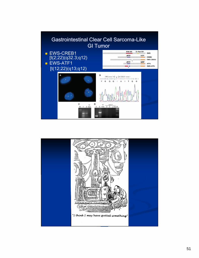
51 Gastrointestinal Clear Cell Sarcoma-Like GI Tumor EWS-CREB1 [t(2;22)(q32.3;q12) EWS-ATF1 [t(12;22)(q13;q12)
| Date post: | 13-May-2019 |
| Category: |
Documents |
| Upload: | phamnguyet |
| View: | 216 times |
| Download: | 0 times |
51
Gastrointestinal Clear Cell Sarcoma-Like GI Tumor
EWS-CREB1 [t(2;22)(q32.3;q12)
EWS-ATF1 [t(12;22)(q13;q12)

52
Gastrointestinal Stromal Tumor Arises from Interstitial Cells of Cajal – Peristalsis Control
Resemble Smooth Muscle & Schwann Cells
95% C-KIT, 98% DOG1, 70% CD34 Positive; C-Kit Mutation
Carney’s Triad (gastric GIST, paraganglioma, pulmonary chondroma), Neurofibromatosis Type 1, Carney-Stratakis (paraganglioma, GIST), Familial GIST (germline mutation KIT/PDGFRA)
STI-571: PDGFRA & c-Kit Mutated Tumors
BRAF (13%), IGF1R (Most), HIF-1A Targets (Carney-Stratakis) - Also EGFR, MET, NY-ESO
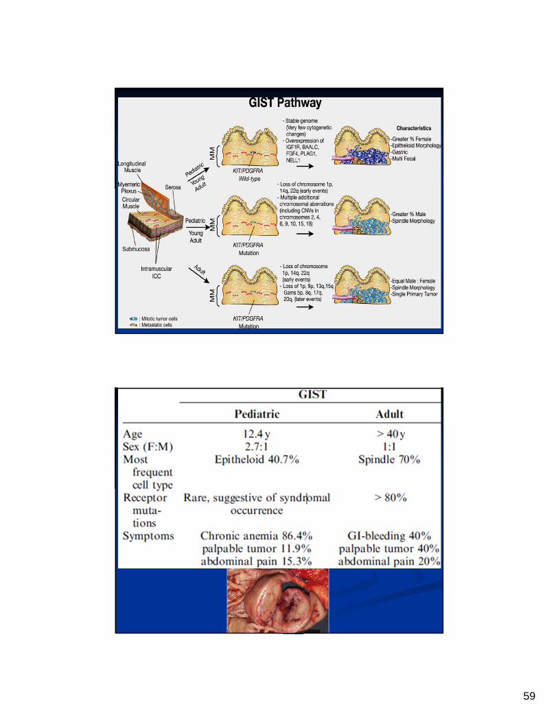
Gastrointestinal Stromal Tumors in Pediatrics
Represent About 1-2% of GISTs Age:
>10 yrs 60% <1 yr 20% 1-5yr 12% 6-10yr 8%
F:M Gender Ratio 1.5:1.0 Stomach (antrum) 52% Small Intestine 20% Colon/Rectum 20% Size
<5cm 20% >5-10 cm 28% >10 cm 24%
Symptoms GI Bleeding, Abdominal Palpable Mass,
Abdominal Distention, Intestinal Obstruction

53

54

55
56
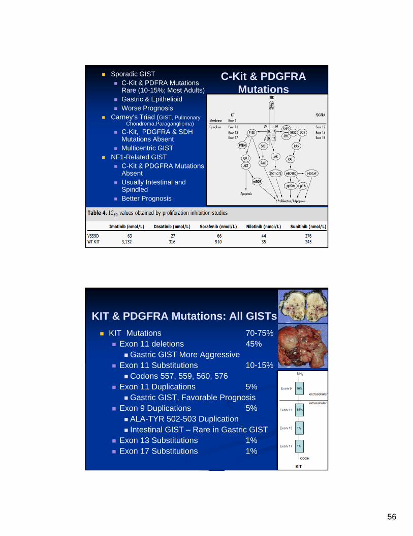
C-Kit & PDGFRA Mutations
Sporadic GIST C-Kit & PDFRA Mutations
Rare (10-15%; Most Adults) Gastric & Epithelioid Worse Prognosis
Carney’s Triad (GIST, Pulmonary Chondroma,Paraganglioma)
C-Kit, PDGFRA & SDH Mutations Absent
Multicentric GIST NF1-Related GIST
C-Kit & PDGFRA Mutations Absent
Usually Intestinal and Spindled
Better Prognosis
KIT & PDGFRA Mutations: All GISTs
KIT Mutations 70-75% Exon 11 deletions 45%
Gastric GIST More Aggressive Exon 11 Substitutions 10-15%
Codons 557, 559, 560, 576 Exon 11 Duplications 5%
Gastric GIST, Favorable Prognosis Exon 9 Duplications 5%
ALA-TYR 502-503 Duplication Intestinal GIST – Rare in Gastric GIST
Exon 13 Substitutions 1% Exon 17 Substitutions 1%
57
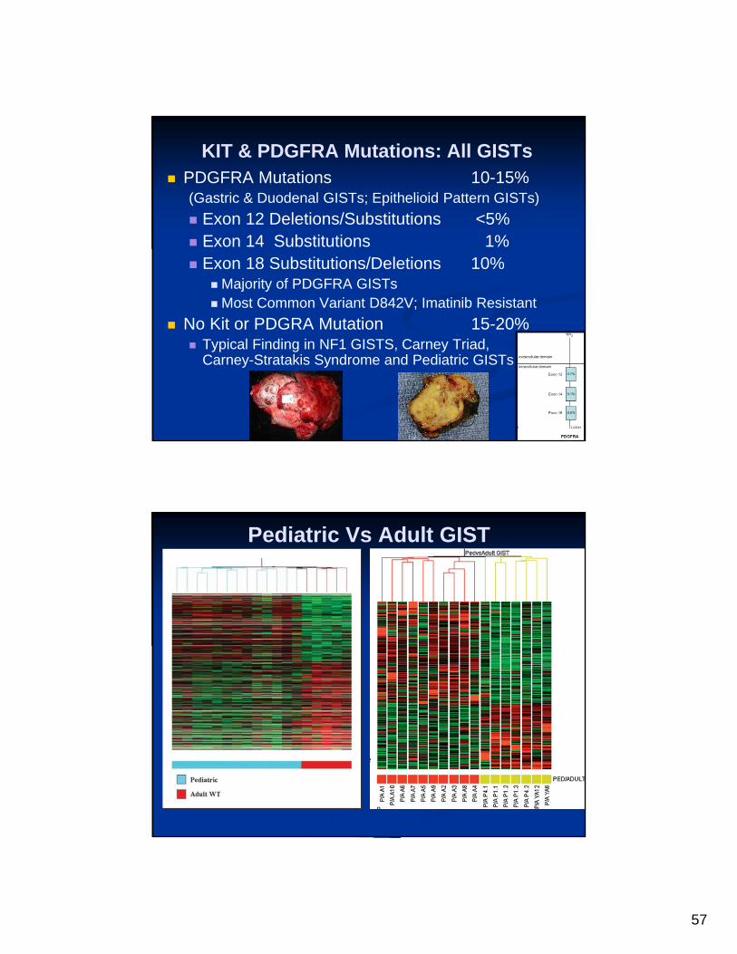
KIT & PDGFRA Mutations: All GISTs PDGFRA Mutations 10-15%
(Gastric & Duodenal GISTs; Epithelioid Pattern GISTs)
Exon 12 Deletions/Substitutions <5% Exon 14 Substitutions 1% Exon 18 Substitutions/Deletions 10%
Majority of PDGFRA GISTs Most Common Variant D842V; Imatinib Resistant
No Kit or PDGRA Mutation 15-20% Typical Finding in NF1 GISTS, Carney Triad,
Carney-Stratakis Syndrome and Pediatric GISTs
Pediatric Vs Adult GIST
58
59

60
Recurrence
Local 20%
Metastatic 4%
Survival
Alive 80%
DOD 4-13%
DOC 4%
Unknown 12%
Case History 6 Month Old Boy
Recent Onset of Vomiting,
Abdominal Pain & Cramping
Intermittent Rectal Bleeding
Diagnostic Imaging: Intussusception

61
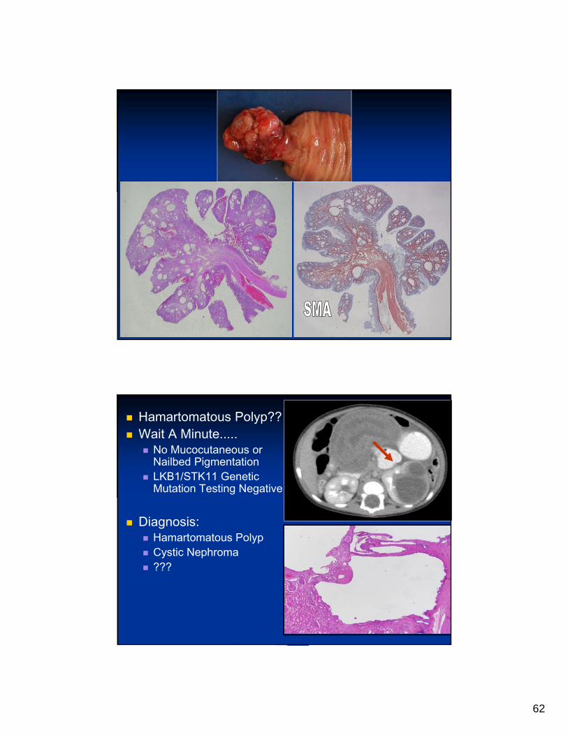
62
Hamartomatous Polyp?? Wait A Minute.....
No Mucocutaneous or Nailbed Pigmentation
LKB1/STK11 Genetic Mutation Testing Negative
Diagnosis: Hamartomatous Polyp Cystic Nephroma ???

63
PPB Family Tumor Susceptibility Syndrome
PPB Family Tumor Susceptibility Syndrome: Multiple Tumors Exist or Develop. Bilateral and/or Multifocal Lung Cysts in 15% of
children Bilateral Type I PPB In Several Cases Cystic nephroma: Most Common Non-
Pulmonary Neoplasm (~10% in PPB Patients or Relatives)
*Small Subset: PPB, Cystic Nephroma and Small Bowel Polyps
Several PPB Patients with Sertoli-Leydig Cell Ovarian Tumors or Nasal Chondromesenchymal Hamartoma
Unique Set of Diseases Different than Other Familial Neoplasia Syndrome
PPB Family Tumor Susceptibility Syndrome
Treatment-Related Second Malignant Neoplasms Not Different from Other Cancer Survivors 3 PPB Children With Apparent Treatment-Related
Malignancies:Glioblastoma Multiforme (radiation PPB brain
metastasis)Thyroid Carcinoma After Chest RadiationAML After Chemotherapy for PPB (Alkylating
Agents & Etoposide)

64
Burkitt Lymphoma Aggressive B-cell NHL with Extremely High
Proliferation Index & Characteristic Tranlocation (8q24- MYC)
Endemic (most cases) Associated with Early EBV Infection and Increased EBV Viral Loads
Promoters: Plasmodium Falciparum, Arbovirus and Plant Tumor Promoters
Sporadic: EBV in 20-30% of Cases Low Socioeconimic Status and Early EBV Infection
Immunodeficiency: EBV in 30-40% More Common in HIV: May Occur with High CD4
T-Cell Counts in HIV
Polyclonal B-Cell Activation in HIV and Malaria
Burkitt Lymphoma Endemic BL
4-10/100,000 Children; 2M:1F
Most <15 Years of Age
Equatorial Africa & New Papua-Guinea
Sporadic BL
40% of All Childhood Lymphomas
0.3/100,000 Children, 3M:1F
Industrialized Nations
Caucasian>Asian or African-American
Immunodeficiency BL: Low Incidence
Decreasing Lymphoma Incidence in HIV Children: Highly Active Anti-Retroviral Therapy

65
Burkitt Lymphoma Endemic BL:
Jaw/Facial Bones (50-60%), Breast, Abdomen, Bone Marrrow (10%)
Typically Lack Leukemic Presentation
Sporadic BL: Abdomenal Mass (Ileocecal Region)
Ovaries, Kidneys, Breasts
Jaws Rarely
Immunodeficiency BL: Nodal and Bone Marrow
Burkitt Lymphoma Often Present with Bulkly Disease
(Stage III/IV)
Symptoms Present for Few Weeks
Immunophenotype: IgM, B-Cell Antigens (CD19, CD20, CD22, CD79a), CD10, CD38, CD45, Bcl-6, Ki-67 >95%
Rarely Weak Bcl-2, MUM1/IRF-4 in Subset, Lack TdT
Recommended IHC Panel: CD10, CD20, Bcl-6, Bcl-2, Ki67, EBER-1
(in situ), EBV-LMP

66
Burkitt Lymphoma Molecular and Cytogenetics
MYC Translocations (8q24) with IgH(80%, 14q32), Kappa light Chain (15%, 2p11), Lambda Light Chain (5%, 22q11), NonIg Partners Rare
10% Lack MYC Translocation by FISH Alone
Other Genetic and Epigenetic Alterations
BAX, P16, p53, p73, p130/Rb2, Bcl-6
Complex Cytogenetics More Common in Adults
Additional Abnormalities Correlate with Poor Prognosis
Burkitt Lymphoma Differential Diagnosis:
Diffuse Large B Cell Lymphoma
Lymphoblastic Leukemia Lymphoma
Unclassified B Cell Lymphoma
Small Round Cell Tumor: (Ewing Tumor, Neuroblastoma, Rhabdomyosarcoma)
Myeloid Sarcoma
Prognosis: Highly Aggressive, But Curable
Better Prognosis for Children than Adults
Treatment: Intensive Chemotherapy and Intrathecal Prophylaxis
Anti-CD20 (Rituximab)
Cure Rate Up To 90% Low Stage &
60-80% in Advanced Stage
Relapses Typically Occurs 1 Year from Diagnosis

67
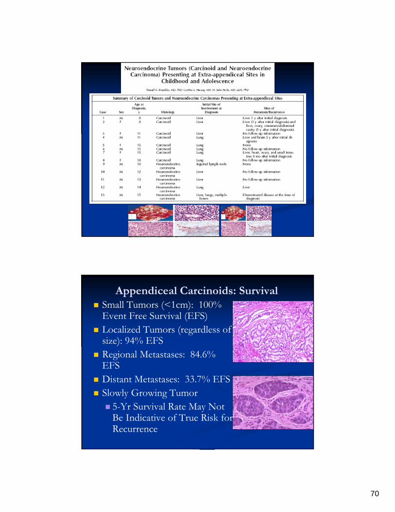
Carcinoid Tumors: What’s The Big Deal?
Incidence: 2 per 100,000 per Year
Pernicious Anemia & Atrophic Gastritis: Carcinoid Tumors (11%)
Zollinger-Ellison Disease & MEN 1 LOH on chromosome 11q13
(MENIN) 26-78% Carcinoid Tumors
Associated with Other Tumors (13%)
Carcinoid Tumors GI carcinoids:
Incidental Finding at Appendectomy (1 in 200-300)
Recurrent Abdominal Pain Melena and Bleeding (Rectal)
Bronchopulmonary carcinoids: Hemoptysis, Pneumonia Cough Only
76% of Carcinoids Found at Autopsy

68
Carcinoid Syndrome Occurs in 2-20%
Flushing Attacks (23-65%) Erythema Upper Body Associated
with Other Symptoms Spontaneous or Triggered by
Stress, Foods, Exercise May last for minutes or hours
Diarrhea (32-75%) Cardiac Manifestations (11-66%)
Fibrosis of Endocardium (Heart Failure)
Other Symptoms (Asthma-Like Attack, Skin Lesions, Arthralgias, Mental Status Changes)
Pediatric Carcinoid Tumors
Most Common Tumor of Appendix Second Most Common GI Tract
Tumor After Lymphoma 1:100,000 in Children Per Year Acute Appendicitis Common
Presenting Symptom Usually Localized at Appendix Tip Small Localized Tumors: 100% EFS Locally Invasive Tumors: Ileocecal
Resection Adequate Most Children, No Long-Term Follow-Up Available

69
Prognostic factors: Carcinoids Tumor Site
Appendix > Small Intestine > Colorectal > Liver/Pancreas
Tumor Size (survival) <1cm : 100%; 1.1-2.0
cm: 82%; >2 cm: 39%
Depth of Invasion Metastases
(Liver Metastases, Unfavorable)
Mitotic Index <10/10 HPF,
Favorable
70
Appendiceal Carcinoids: Survival Small Tumors (<1cm): 100%
Event Free Survival (EFS) Localized Tumors (regardless of
size): 94% EFS Regional Metastases: 84.6%
EFS Distant Metastases: 33.7% EFS Slowly Growing Tumor 5-Yr Survival Rate May Not
Be Indicative of True Risk for Recurrence

71
Carcinoid Treatment Tumor Size < 1 cmAppendectomy Alone
Tumor Size 1 to 2 cmUnclearAggressive Surgery With
Serosal Invasion Tumor Size >2 cm Full Cancer SurgeryRight Hemicolectomy Lymph Nodes
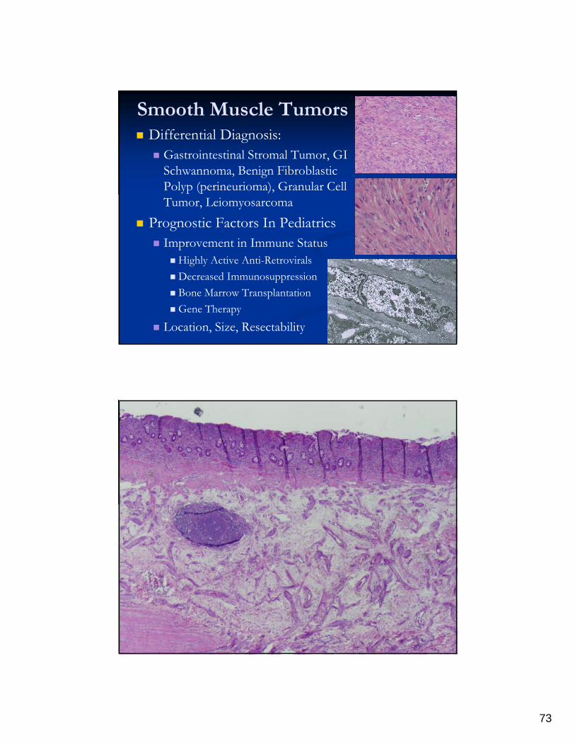
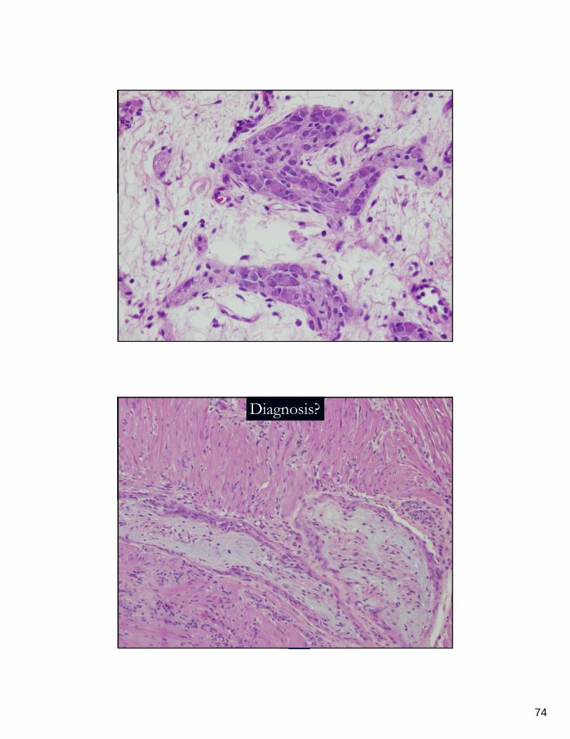
Smooth Muscle Tumors Arise in Association with Muscularis
Mucosae or Propria
Most Common in Esophagus and Colon
Adults: Esophagus with Tiny Seedling Leiomyomas In Inner Muscularis Propria on about 50% of Gastroesophageal Carcinoma Resections
Colonic Leiomyomas Typically Found with Screening for Colorectal Adenomas
Well-Circumscribed, Whorled Cut Surface Similar to Leiomyomas at Other Sites

72
Smooth Muscle Tumors Smooth Muscle Differentiation
Spindle Cells with Eosinophilic Cytoplasm and Blunt Nuclei
“Perpendicularly” Oriented Fascicles (Herring Bone-Like)
Minimal Mitotic Activity
Immunophenotype: SMA, Desmin, h-Caldesmon >70%
Focal: Keratin, EMA, CD34, S100
Negative: CD117
Smooth Muscle Tumors Pediatric Leiomyomas
HIV, Immune Suppression, Immunodeficiency Disorders, Solid Organ Transplantation
EBV-Association: CD21 Receptor on Smooth Muscle Cells
Tumors Tend To Be Multicentric – Not Mets
Involve Parenchyma Organs Rather than Soft Tissue
Bland But with Primitive Round Cell Component
Variable Mitotic Activity & Lymphocytic Infiltrate
May Have Perivascular Myopericytoma Growth Pattern
Better Behavior Than Conventional Type
EBER-1
73
Smooth Muscle Tumors Differential Diagnosis:
Gastrointestinal Stromal Tumor, GI Schwannoma, Benign Fibroblastic Polyp (perineurioma), Granular Cell Tumor, Leiomyosarcoma
Prognostic Factors In Pediatrics Improvement in Immune Status
Highly Active Anti-Retrovirals
Decreased Immunosuppression
Bone Marrow Transplantation
Gene Therapy
Location, Size, Resectability
74
Diagnosis?

75

![IOS Press Evaluationof the expressionof C-kit (CD117) in … · 2019. 7. 31. · mors, Ewing sarcoma and gastrointestinal stromal tu-mors (GISTs) [1,19,21–30]. Oncogenic c-kit muta-tion](https://static.documents.pub/doc/80x56/60f75ccb0c4cf50672185423/ios-press-evaluationof-the-expressionof-c-kit-cd117-in-2019-7-31-mors-ewing.jpg)


