78
GASTROINTESTINAL SYSTEM PROBLEM 4 ALMIRA NABILA VALMAI 405130193
| Date post: | 07-Dec-2015 |
| Category: |
Documents |
| Upload: | almira-valmai |
| View: | 224 times |
| Download: | 0 times |

GASTROINTESTINAL SYSTEM PROBLEM 4
ALMIRA NABILA VALMAI405130193
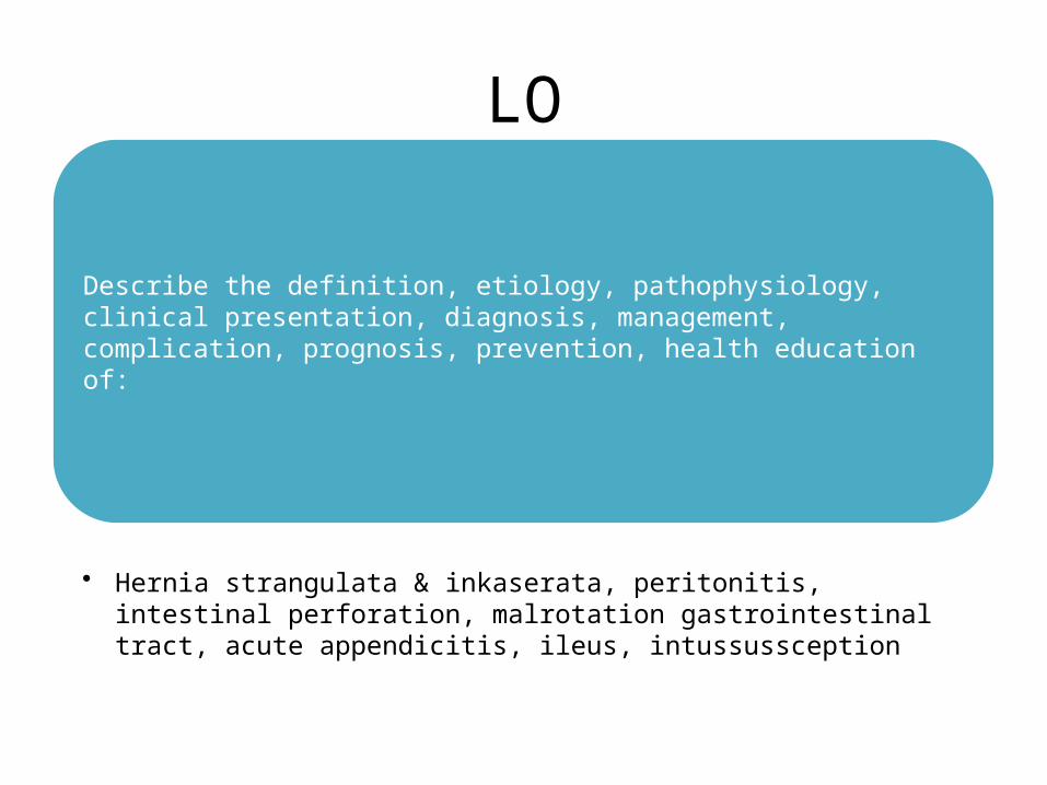
LO
Describe the definition etiology pathophysiology clinical presentation diagnosis management complication prognosis prevention health education of
bull Hernia strangulata amp inkaserata peritonitis intestinal perforation malrotation gastrointestinal tract acute appendicitis ileus intussussception
HERNIA STRANGULATA amp INKASERATALO 1
Hernia EtiologyAny condition that increases the pressure in the intra abdominal cavity may contribute to the formation of a hernia including the followingbull Marked obesitybull Heavy liftingbull Coughingbull Straining with defecation or urinationbull Ascitesbull Peritoneal dialysisbull Ventriculoperitoneal shuntbull Chronic obstructive pulmonary disease (COPD)bull Family history of hernias[16]
PathophysiologyTypes of Hernia ndash Location
bull Indirect herniaAn indirect inguinal hernia follows the tract through the inguinal canal This results from a persistent process vaginalis The inguinal canal begins in the intra-abdominal cavity at the internal inguinal ring located approximately midway between the pubic symphysis and the anterior iliac spine The canal courses down along the inguinal ligament to the external ring located medial to the inferior epigastric arteries subcutaneously and slightly above the pubic tubercle
bull Direct herniaA direct inguinal hernia usually occurs due to a defect or weakness in the transversalis fascia area of the Hesselbach triangle The triangle is defined inferiorly by the inguinal ligament laterally by the inferior epigastric arteries and medially by the conjoined tendon[5]
bull Femoral herniaThe femoral hernia follows the tract below the inguinal ligament through the femoral canal The canal lies medial to the femoral vein and lateral to the lacunar (Gimbernat) ligament
bull Umbilical herniaThe umbilical hernia occurs through the umbilical fibromuscular ring which usually obliterates by 2 years of age They are congenital in origin and are repaired if they persist in children older than age 2-4 years[2 5]
bull Richter herniaThe Richter hernia occurs when only the antimesenteric border of the bowel herniates through the fascial defect The Richter hernia involves only a portion of the circumference of the bowel As such the bowel may not be obstructed even if the hernia is incarcerated or strangulated and the patient may not present with vomiting The Richter hernia can occur with any of the various abdominal hernias and is particularly dangerous as a portion of strangulated bowel may be reduced unknowingly into the abdominal cavity leading to perforation and peritonitis[6]
bull Incisional herniaThis iatrogenic hernia occurs in 2-10 of all abdominal operations secondary to breakdown of the fascial closure of prior surgery Even after repair recurrence rates approach 20-45
bull Spigelian herniaThis rare form of abdominal wall hernia occurs through a defect in the spigelian fascia which is defined by the lateral edge of the rectus muscle at the semilunar line (costal arch to the pubic tubercle) The two subtypes are interstitial and subcutaneous which are best defined using CT and assist with optimizing the surgical approach when indicated[7 8 9]
bull Obturator herniaThis hernia passes through the obturator foramen following the path of the obturator nerves and muscles Obturator hernias occur with a female-to-male ratio of 61 because of a gender-specific larger canal diameter and predominately in the elderly
Types of Hernia - Conditionbull Reducible herniaThis term refers to the ability to return the contents of the hernia into the abdominal cavity either spontaneously or manually
bull Incarcerated herniaAn incarcerated hernia is no longer reducible The vascular supply of the bowel is not compromised however bowel obstruction is common
bull Strangulated hernia A strangulated hernia occurs when the vascular supply of the bowel is compromised secondary to incarceration of hernia contents
Differential Diagnoses
bull Epididymitisbull Hidradenitis Suppurativabull Hydrocelebull Lymphogranuloma Venereumbull Testicular Torsion
Medication
For strangulated hernias start broad-spectrum antibiotics Antibiotics are administered routinely if ischemic bowel is suspected
Multiple regimens that cover for bowel perforation andor ischemic bowel can be used Cover for both aerobic and anaerobic gram-negative bacteria
Complications
bull If strangulation of the hernia is missed bowel perforation and peritonitis can occur
bull Hernias can reappear in the same location even after surgical repair
PERITONITISLO 2
PERITONITIS
bull PRIMARYbull SECONDARY
PRIMARY (SPONTANEOUS) BACTERIAL PERITONITIS
bull complicates ascites but does not cause it (occurs in 10 of cirrhotic ascites) higher risk in patients with GI bleed
bull 13 of patients are asymptomatic thus do not hesitate to do a diagnostic paracentesis in ascites even if no clinical indication of infection
bull fever chills abdominal pain ileus hypotension worsening encephalopathy acute kidney injury
bull Gram-negatives compose 70 of pathogens E coli (most common) Streptococcus Klebsiella
bull time of diagnosis and 1 gkg on day 3) decreases mortality by lowering risk of acute renal failure
PRIMARY (SPONTANEOUS) BACTERIAL PERITONITIS
bull Diagnosisndash absolute neutrophil count in peritoneal fluid gt025x10 cellsL (250
cellsmm)ndash Gram stain positive in only 10-50 of patientsndash culture positive in lt80 of patients (not needed for diagnosis)
bull Prophylaxis consider in patients withndash cirrhosis or GI bleed IV ceftriaxone daily or norfloxacin bid x 7 dndash previous episode of SBP long-term prophylaxis with daily norfloxacin or
TMP-SMX
bull Treatmentndash IV antibiotics (cefotaxime 2 g IV q8h is the treatment of choice for 5 d
modify if responseinadequate or culture shows resistant organisms)ndash IV albumin (15 gkg at
SECONDARY BACTERIAL PERITONITIS
bull develops when bacteria contaminate the peritoneum as a result of spillage from an intraabdom- inal viscus
bull Gram-negative bacilli particularly E coli are common bloodstream isolates but Bacteroides fragilis bacteremia occurs as well
bull The severity of abdominal pain and the clinical course depend on the inciting process
bull Secondary peritonitis can result pri- marily from chemical irritation or bacterial contamination
SECONDARY BACTERIAL PERITONITIS
CLINICAL MANIFESTATIONbull epigastric pain from a ruptured gastric ulcerbull Unusual locations of the appendix (including a retro- cecal position) can
complicate this presentation further bull lie motionless often with knees drawn up to avoid stretching the nerve fibers of
the peritoneal cavity bull Coughing and sneezing which increase pressure within the peritoneal cavity are
associated with sharp pain bull may or may not be pain localized to the infected or diseased organ from which
secondary peri- tonitis has arisen bull abnormal findings on abdominal examination with marked voluntary and
involuntary guarding of the anterior abdominal musculature bull tenderness especially rebound tenderness bull febrile bull leukocytosis and a left shift of the WBCs to earlier granulocyte forms
SECONDARY BACTERIAL PERITONITIS
TREATMENTbull early administration of antibiotics aimed particularly at
aerobic gram-negative bacilli and an- aerobes bull Mild to moderate broad-spectrum pen- icillinβ1113100-lactamase
inhibitor combinations (eg ticarcillinclavulanate 31 g q6h IV) or cefoxitin (2 g q24h IV)
bull hospitalization in intensive care imipenem (500 mg q6h IV) meropenem (1 g q8h IV) or combinations of drugs (ampicillin plus metronidizole plus ciprofloxacin)
bull usually requires both surgical intervention and antibiotic administration
INTESTINAL PERFORATIONLO 3
MALROTATION GASTROINTESTINAL TRACTLO 4
Intestinal malrotation
bull Intestinal malrotation when the intestines donrsquot make the turns as they should resulting in the congenital (present at birth)
bull When a fetus is about five weeks old her intestine exits her abdomen into the amniotic fluid (where therersquos more space) and continues to grow there At around ten weeks the intestine re-enters the abdomen and makes two turns
Source httpwwwchildrenshospitalorgconditions-and-treatmentsconditionsintestinal-malrotation
bull Intestinal malrotation itself isnrsquot much of a concern but it puts your child at higher risk for two serious complications
bull volvulus ndash when the intestine twists in on itself potentially cutting off the blood supplybull intestinal obstruction ndash when a stalk of fibrous tissue
known as Laddrsquos bands creates a blockage that prevents the intestine from functioning
Source httpwwwchildrenshospitalorgconditions-and-treatmentsconditionsintestinal-malrotation
Source httpwwwchildrenshospitalorgconditions-and-treatmentsconditionsintestinal-malrotation
bull Itrsquos fairly common that a baby is born with intestinal malrotation it affects about one in every 500 babies in the United States Some babies may not have symptoms until they become children teens or adults Others may go through their entire life with no symptoms never have a problem and never be diagnosed
bull Of babies who are diagnosed with intestinal malrotationbull 25 to 40 percent are diagnosed in the first week of lifebull 50 to 60 percent are diagnosed within the first month of lifebull 75 to 90 percent are diagnosed by age 1bull Although malrotation occurs equally among boys and girls boys are
more likely to become symptomatic by the first month of life
Source httpwwwchildrenshospitalorgconditions-and-treatmentsconditionsintestinal-malrotation
ACUTE APPENDICITISLO 5
A condition characterized by inflammation of the appendix
most common cause of acute inflammation in the right lower quadrant of the abdominal cavity
prevalent in countries in which people consume a diet low in fiber and high in refined carbohydrates
DEFINITION
bullThe appendix is located in the lower quadrant of the abdomen or more specifically the right iliac fossa
bullIt is a slender worm-shaped pouch averaging 5mdash10cm in length
RACIAL amp DIETARY FACTORS- bull MORE COMMON IN WHITE RACES bull YOUNG MALES ARE AFFECTED MORE OFTEN bull DIET RICH IN MEAT PRECIPITATES APPENDICITIS bull FAMILIAL TENDENCY
SOCIO-ECONOMIC STATUSbull IT IS COMMON IN MIDDLE CLASS amp RICH PEOPLE
OBSTRUCTION OF THE LUMEN bull A) IN THE LUMEN-INTESTINAL WARM eg ROUND
WORMTHREDWORM ETC VEGETABLEFRUIT SEEDFECES MATERIAL BARIUM
bull B) IN THE WALL-STRUCTURE NEOPLASM
ETIOLOGY
Non-modifiable
bullAge all age groups old
bullGender male(male- female =21)
bullHereditary tumor formation in the opening of the appendix
Modifiable
bullDiet People whose diet is low in fiber and rich in refined carbohydrates
bullInfections Gastrointestinal infections such as Amoebiasis Bacterial Gastroenteritis
PATHOPHYSIOLOGY
Decreased O2 supply in the appendix
Appendix starts to be necrotic bacteria invade the appendix
Disruption of cell membrane of appendix
Episodes of constipation
Occlusion of appendix by fecalith
Decreased flowdrainage of mucosal secretions
vasoncongestion
Decreased blood supply in the appendix
Start of Inflammatory Process
Release of Chemical Mediators Activation of the Vomiting center in the medulla
Neutrophils to area
Swelling of Appendix
Risk for infection(if appendix ruptures)
Histamine Prostaglandin Leukotrienes Bradykinin Pus Formation
(phagocytized bacteria and dead cells)
Prostaglandin Bradykinin
Pain in the RLQ of Abdomen
Stimulation of Vagus Nerve
Suppression of Sympathetic GI function
NVAnorexia
Risk for Deficient Fluid Volume
Risk for Imbalanced Nutrition less than body requirements
Acute pain
Interleukin-1
Increased WBC
Inflammation of Appendix
Appendectomy
Tissue TraumaOpen Wound
Disruption of Cell Membrane
Noriceptors of the Dermis
Send Impulse to CNS
Pain on Surgical Site
Start of Inflammatory Process
Release of Prostaglandin Bradykinin Activity Intolerance
bull Only 55 have classical featuresbull Atypical 45bull History 24-36 hoursbull Abdominal pain (diffuse and periumbilical localizing to the RIF)bull Anorexia (almost always)bull Vomiting (75)bull Low grade fever
bull If gt38 suspect perforationbull Tenderness guarding and rebound Be gentlebull Rovsingrsquos psoas obturator signs unreliable and late
bull Tender Appendicular massbull Atypical
bull (loin high RUQ deep pelvic)bull Diarrhea ( not always gastroenteritis)bull Urinary frequency
bull The Extremes of Agebull Children lt 5 rapid progressionbull Pain in the elderly is less intense
CLINICAL PRESENTATION
A Physical Exambull Findings depend on duration of illness prior to exambull Early on patients may not have localized tendernessbull With progression there is tenderness to deep palpation over McBurneyrsquos pointbull McBurneyrsquos Point just below the middle of a line connecting the umbilicus and
the ASISbull Rovsingrsquos pain in RLQ with palpation to LLQbull Rectal exam pain can be most pronounced if the patient has pelvic appendixbull Additional components that may be helpful in diagnosis rebound tenderness
voluntary guarding muscular rigidity tenderness on rectalbull Psoas sign place patient in L lateral decubitus and extend R leg at the hip If
there is pain with this movement then the sign is positivebull Obturator sign passively flex the R hip and knee and internally rotate the hip If
there is increased pain then the sign is positivebull Fever another late findingbull At the onset of pain fever is usually not found bull Temperatures gt39 C are uncommon in first 24 h but not uncommon after
rupture
DIAGNOSIS
Laboratory amp Radiology Result
CBC WBC count reveal moderate leukocytosis (10000 to 16000mm3) with shift to the left
Ultrasound studies amp CT scans May reveal right liver quadrant density or localized distention of the bowel
Abdominal x-ray visualize shadow consistent with fecalith in appendix perforation will reveal free air
B Laboratory amp Radiology
bull M Antibiotics for infection Analgesic agent (morphine) can be given for pain after the surgery
bull E Within 12 hrs of surgery you may get up and move around You can usually return to normal activities in 2-3 weeks after laparoscopic surgery
bull T Pretreatment of foods with lactase preparations (eg lactacid drops) before ingestion can reduce symptoms
Ingestion of lactase enzyme tablets with the first bite of food can reduce symptoms bull H To care wound perform dressing changes and irrigations as prescribe avoid taking laxative
or applying heat to abdomen when abdominal pain of unknown cause is experienced Reinforce need for follow-up appointment with the surgeon Call your physician for increased pain at the incision site
bull O Document bowel sounds and the passing of flatus or bowel movements (these are signs of the return of peristalsis)
Watch for surgical complications such as continuing pain or fever which indicate an abscess or wound dehiscence Stitches removed between fifth and seventh day (usually in physicians office)
bull D Liquid or soft diet until the infection subsides Soft diet is low in fiber and easily breaks down in the gastrointestinal tract
MANAGEMENT
PRE-OPERATIVE MANAGEMENTbull NPO diet in preparation for surgery bull An intravenous drip is used to hydrate the
patient bull Antibiotics given intravenously such as
Cefuroxime and Metronidazole bull If the stomach is empty (no food in the past
six hours) general anesthesia is usually used
bull Otherwise spinal anesthesia may be used
Removal of the appendix Performed as soon as possible to decrease
the risk of perforation
Two ways performed1 Laparotomy2 Laparoscopy
Appendectomy
POST- OPERATIVE MANAGEMENTbull Assist patient to position of comfort such as semi-fowlers with
knees are flexedbull Restrict activity that may aggravate pain such as coughing
and ambulationbull Apply ice bag to abdomen for comfortbull Advise avoidance of enemas or harsh laxatives increased
fluids and stool softeners may be used for postoperative constipation
bull Give narcotic analgesic as ordered and administer oral fluids when tolerated
bull Monitor frequently for signs and symptoms of worsening condition indicating perforation abscess or peritonitis (increasing severity of pain tenderness rigidity distention absent bowel sounds fever malaise and tachycardia)
bull If a drain is left in place at the area of the incision monitor carefully for signs of intestinal obstruction secondary hemorrhage or secondary abscesses (eg fever tachycardia and increased leukocyte count)
Complication InterventionsPERITONITIS bullObserve for abdominal tenderness fever
vomiting abdominal rigidity and tachycardiabullEmploy constant nasogastric suctionbullCorrect dehydrationbullAdmin Antibiotics as prescribed
PELVIC ABSCESS bullEvaluate for anorexia chills fever and diaphoresisbullObserve for diarrhea which may indicate pelvic abscessbullPrepare pt for rectal exambullPrepare pt for surgical drainage procedure
SUPHRENIC ABSCESS bullAssess pt for chills fever diaphoresisbullPrepare for x-ray exam and surgical drainage of abscess
PARALYTIC ILEUS bullAssess bowel soundsbullReplace fluids and electrolytes by IV routebullEmploy nasogastric intubation and suction
COMPLICATIONS
ABA-The Appendix- 4th year Lectures
bull Mortality from 02 to 1bull Complications increase with perforation bull Morbidity
bull Wound abscess bull Wound infection (less with MacBurneyrsquos incision)bull Wound dehiscencebull Intra-abdominal abscess bull Faecal fistula bull Intestinal obstruction bull Adhesive band bull inguinal hernia bull Fertility
PROGNOSIS
Prevents
bull Eat foods high in fiber such as fresh fruits and vegetables
Ascariasis
bull Disease bull Ascariasis ascariasis infection roundworm infection
Pathophysiological mechanism
Adult worms move throughout the GI tract and may become incarcerated leading to
obstructive pathology
The worms may die rarr leading to inflammation necrosis infection and abscess
formation
If they migrate through an existing perforation in the bowel wall rarr secondary to
tuberculosis or typhoid rarr cause a granulomatous peritonitis
Larvae during migration rarr may be deposited in the brain spinal cord kidney or other
organs rarr leading to granuloma formation inflammation or infection
They may become entwined in a bolus and obstruct the small bowel this is most
common in the terminal ileum although other more proximal sites have been rarely
reported
Risk Factors
bull Preschool age or youngerbull Eating unsanitary foodbull Drinking unclean water
Symptomsbull Small burdens of worms in the intestine may cause no symptoms
bull The patient may have symptoms of pneumonitis with cough dyspnea wheezing chest pain and low grade fever during the migration of the larvae through the liver and lungs
bull In heavy worm burdens the adult worms actively migrate in the intestine resulting in intestinal blockage vomiting abdominal pain colic nausea anorexia and intermittent diarrhea
bull A heavy worm burden in children may lead to severe nutritional impairment and retardation in growth
bull Worms exiting through the nose or mouth
Laboratory Studiesbull Early infection (larval migration)
ndash Complete blood count (CBC) may show eosinophiliandash Sputum analysis may reveal larvae or Charcot-Leyden crystalsndash Stool examination findings are typically normal in absence of previous
infection (during the first 40 d)ndash Ascaris specific antibodies (not useful in acute infection and not
protective)ndash Increases in IgE and later IgG
bull Established infection (adult phase) ndash Stool examination findings include characteristic eggs Adult females lay
about 200000 eggs per day aiding microscopic identification of characteristic eggs
Imaging Studies
bull Early infection (larval migration) ndash Chest radiography may reveal patchy infiltrates of eosinophilic pneumonia
bull Established infection (adult phase)ndash Abdominal radiography may reveal adult worms (especially with
contrast) ndash Obstructing Ascaris lesions cause cylindrical filling defects on contrast
computed tomography (CT) scansndash Cholangiopancreatography by endoscopy (ERCP) or magnetic
resonance imaging (MRCP or magnetic resonance cholangiopancreatography) may detect adult worms in bile or pancreatic ducts8
ndash Ultrasonography may detect worms in the gallbladder
Treatmentbull Albendazole ndash 400 mg PO once for all ages
bull Mebendazole ndash 100 mg bid PO for 3 days or 500 mg PO once for all
agesbull Piperazine citrate ndash 150 mgkg PO initially followed by 6 doses of 65
mgkg at 12 hr intervals PO which causes neuromuscular paralysis of the parasite (the treatment of choice for intestinal or biliary obstruction)
Treatmentbull Pyrantel pamoate ndash 11 mgkg PO once maximum 1 g
bull Nitazoxanide ndash 100 mg bid PO for 3 days for children 1-3 yrs of age ndash 200 mg bid PO for 3 days for children 4-11 yrndash 500 mg bid PO for 3 days for adolescents and adults
produces cure rates comparable with single-dose albendazole
bull Surgery may be required for cases with severe obstruction
complicationsbull Intestinal obstruction - 63 bull Bile duct obstruction - 23bull Perforation peritonitis or both - 32bull Volvulus - 27bull Hepatitic abscess - 21bull Appendicitis - 21bull Pancreatitis - 1bull Cerebral encephalitis - 1bull Intussusception - 05bull Other sites of pathology (lt05) include Meckel diverticulum the gallbladder ears
eyes nose lungs kidneys vagina urethra heart placenta spleen thoracic cavity and umbilicus
bull In endemic regions ascariasis is a significant part of the differential diagnosis for intestinal obstruction appendicitis biliary tract disease pancreatitis intussusception and volvulus
ILEUSLO 6
Paralytic ileus
Definition Failure of appropriate forward movement of bowel contents
Causes Neurogenikmetabolikobat2aninfeksiiskemi usus
Symtomps Mild stomach painbloatingnauseavomitingkonstipasi
PF Silent abdomen (-) distensi timpanik
PP Laboratorium leukosit darahkadar elektrolytureumglukosa darahamilase Foto polos rontgen dilatasi usus halus dan besar diagfragma elevation
Treatment farmako Metoklopramid u gastroparesis sisaprid u ileus paralitik pasca operasi klonidin u mengatasi ileus paralitik
Treatment non-farmako Gum chewing pasca operasi menunjukan perbaikan ikeus
Prognosis Prognosis ileus paralitik baik bila penyakit primernya dapat diats
Ileus Obstruction (Ascaris)
bull Ascaris lumbricoides (A lumbricoides) is the most common intestinal helminth parasite and it is estimated that the infected population is 08-12 billion worldwide The highest prevalence of ascariasis occurs in tropical and semitropical countries where sanitation is poor
httpwwwncbinlmnihgovpmcarticlesPMC4194592
bull large numbers of adult Ascaris in the small intestine can cause
bull abdominal distension bull abdominal pain bull obstructive ileus bull malnutrition
A single worm of A lumbricoides can enter the ampulla of Vater and result in biliary colic obstructive jaundice ascending cholangitis acalculous cholecystitis or acute pancreatitis
httpwwwncbinlmnihgovpmcarticlesPMC4194592
httpwwwncbinlmnihgovpmcarticlesPMC4194592
httpwwwncbinlmnihgovpmcarticlesPMC4194592
httpwwwncbinlmnihgovpmcarticlesPMC4194592
bull TerminologyA lumbricoides the human roundworm is one of the most common soil transmitted parasites in the world infecting about 12 billion people globally
httpwwwncbinlmnihgovpmcarticlesPMC4194592
Factors that result in Ascaris-related intestinal obstruction
There are 4 major factors that result in Ascaris-related intestinal obstruction
bull Multiple worms can form a large bolus resulting in mechanical obstruction of the
bowel lumen This is the most frequent cause of Ascaris- related bowel
obstruction
bull The worm bolus may serve as a lead point in intussusception or a pivot in small-
bowel volvulus
bull Ascaris worms may inhabit the ileocecal valve where roundworm secretion of
neurotoxins prompts small-bowel contraction This action coupled with high
worm burden in the ileocecal valve can obstruct the intestine
bull A host inflammatory reaction to worm-derived hemolysins endocrinolysins and
anaphylatoxins can be severe enough to obstruct the gut lumen
Patient presentation and symptomatology
bull Surendran and Paulose conducted a 5-year study of intestinal obstruction by A lumbricoides and categorized small-bowel obstruction resulting from Ascarisas either acute or subacute
bull Patients with subacute obstruction in this study experienced diffuse and colicky abdominal pain fever vomiting that was more pronounced at the onset of symptoms and diarrhea without blood and mucus
bull Acute obstruction had far more ominous presenting signs and symptoms Patients were often ill for several days before presentation These persons showed signs of severe dehydration and had a toxic appearance Vomiting abdominal pain and distention (Figure 1) fever leukocytosis and obstipation were frequently noted
Distended abdomen is characteristic of a child with severe ascariasis (Reprinted from Despommier D et al Parasitic Diseases 2001[24])
Treatment
bull The anthelmintics of choice are mebendazole and albendazole bull These medications are used for intestinal ascariasis as well as HPA bull Mebendazole100 mg is given twice daily for 3 days and provides
protection against Trichuris trichiura (whipworm) as well as Ancylostoma duodenale and Necator americanus (hookworm)
bull Alternatively albendazole may be used the advantage of this drug is that it is given in a single 400-mg dose
bull pyrantel pamoate 11 mgkg (up to a maximum of 1 g) orally daily for 3 days
httpwwwcdcgovparasiteshookwormhealth_professionalsindexhtmls_cid=cs_074httpemedicinemedscapecomarticle212510-treatment
httpwwwmedscapecomviewarticle451597_3
Recommended criteria for surgical exploration include the following
bull Passage of blood per rectumbull Multiple air fluid levels on abdominal radiographsbull An ill child with abdominal distension and
rebound tendernessbull Unsatisfactory response to conservative therapybull Appendicitis and primary peritonitisbull Hepatobiliary diseasebull Pancreatic pseudocyst
httpemedicinemedscapecomarticle212510-treatmentd9
bull PrognosisThe prognosis for ascariasis is excellent However in higher worm burden infections serious complications such as obstruction are more common
bull Patient EducationRecommend good personal hygiene and food handling techniques discriminate defecation hand-washing cleaning fruits and vegetables and avoiding soil consumption Educational programs should also address the use of human feces as fertilizer a practice that persists in many communities internationally
httpemedicinemedscapecomarticle788398-followupe6
INTUSSUSSCEPTIONLO 7
Intussusception
bull DefinitionThe invagination or telescoping of a proximal segment of bowel (intussusceptum) into lumen of a distal segment (intussuscipiens)
Etiology bull Idiopatic (most common in children)bull Neoplasmndash Benign polyp leiomyoma lipoma lympoma
adenoma of appendix appendiceal stump granulomandash Malignant
bull Primary (more common in colon)bull (more common in small bowel)
bull Postoperativebull Meckelrsquos diverticulumbull Colitisbull Many cases thought to be related to viral
gastroenteritis in children
Pathophysiology bull The invaginated segment is carried distally by
peristalsisbull Mesentery and vessel become involved with
the intraluminal loop and are squeezed within the engulfing segment causing venous congestion
bull Types enteroenteric enterocolic and colocolic
Childrenbull Well nourished infantbull Cramping abdominal
painbull Vomittingbull Diarrhea (often-jelly
stools)bull A palpable tender
sausage shaped mass in the abdomen
Adults bull Intermitten painbull Nausea and vomittingbull Often red blood per
rectumbull Often nonspecific
complains
Examination
bull Abdominal studiesbull Barium studiesbull Ultrasoundbull CT
Treatment
bull Air reductionbull Contrast reduction
bull Surgical exploration and resection of the intussuscepted bowel loops
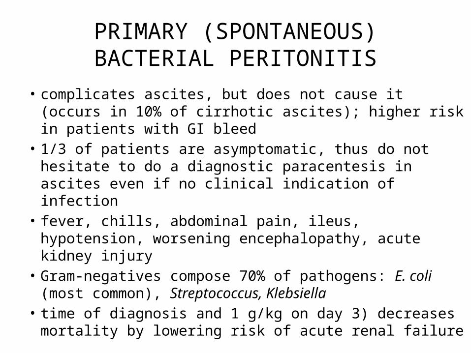
PRIMARY (SPONTANEOUS) BACTERIAL PERITONITIS
bull Diagnosisndash absolute neutrophil count in peritoneal fluid gt025x10 cellsL (250
cellsmm)ndash Gram stain positive in only 10-50 of patientsndash culture positive in lt80 of patients (not needed for diagnosis)
bull Prophylaxis consider in patients withndash cirrhosis or GI bleed IV ceftriaxone daily or norfloxacin bid x 7 dndash previous episode of SBP long-term prophylaxis with daily norfloxacin or
TMP-SMX
bull Treatmentndash IV antibiotics (cefotaxime 2 g IV q8h is the treatment of choice for 5 d
modify if responseinadequate or culture shows resistant organisms)ndash IV albumin (15 gkg at
SECONDARY BACTERIAL PERITONITIS
bull develops when bacteria contaminate the peritoneum as a result of spillage from an intraabdom- inal viscus
bull Gram-negative bacilli particularly E coli are common bloodstream isolates but Bacteroides fragilis bacteremia occurs as well
bull The severity of abdominal pain and the clinical course depend on the inciting process
bull Secondary peritonitis can result pri- marily from chemical irritation or bacterial contamination
SECONDARY BACTERIAL PERITONITIS
CLINICAL MANIFESTATIONbull epigastric pain from a ruptured gastric ulcerbull Unusual locations of the appendix (including a retro- cecal position) can
complicate this presentation further bull lie motionless often with knees drawn up to avoid stretching the nerve fibers of
the peritoneal cavity bull Coughing and sneezing which increase pressure within the peritoneal cavity are
associated with sharp pain bull may or may not be pain localized to the infected or diseased organ from which
secondary peri- tonitis has arisen bull abnormal findings on abdominal examination with marked voluntary and
involuntary guarding of the anterior abdominal musculature bull tenderness especially rebound tenderness bull febrile bull leukocytosis and a left shift of the WBCs to earlier granulocyte forms
SECONDARY BACTERIAL PERITONITIS
TREATMENTbull early administration of antibiotics aimed particularly at
aerobic gram-negative bacilli and an- aerobes bull Mild to moderate broad-spectrum pen- icillinβ1113100-lactamase
inhibitor combinations (eg ticarcillinclavulanate 31 g q6h IV) or cefoxitin (2 g q24h IV)
bull hospitalization in intensive care imipenem (500 mg q6h IV) meropenem (1 g q8h IV) or combinations of drugs (ampicillin plus metronidizole plus ciprofloxacin)
bull usually requires both surgical intervention and antibiotic administration
INTESTINAL PERFORATIONLO 3
MALROTATION GASTROINTESTINAL TRACTLO 4
Intestinal malrotation
bull Intestinal malrotation when the intestines donrsquot make the turns as they should resulting in the congenital (present at birth)
bull When a fetus is about five weeks old her intestine exits her abdomen into the amniotic fluid (where therersquos more space) and continues to grow there At around ten weeks the intestine re-enters the abdomen and makes two turns
Source httpwwwchildrenshospitalorgconditions-and-treatmentsconditionsintestinal-malrotation
bull Intestinal malrotation itself isnrsquot much of a concern but it puts your child at higher risk for two serious complications
bull volvulus ndash when the intestine twists in on itself potentially cutting off the blood supplybull intestinal obstruction ndash when a stalk of fibrous tissue
known as Laddrsquos bands creates a blockage that prevents the intestine from functioning
Source httpwwwchildrenshospitalorgconditions-and-treatmentsconditionsintestinal-malrotation
Source httpwwwchildrenshospitalorgconditions-and-treatmentsconditionsintestinal-malrotation
bull Itrsquos fairly common that a baby is born with intestinal malrotation it affects about one in every 500 babies in the United States Some babies may not have symptoms until they become children teens or adults Others may go through their entire life with no symptoms never have a problem and never be diagnosed
bull Of babies who are diagnosed with intestinal malrotationbull 25 to 40 percent are diagnosed in the first week of lifebull 50 to 60 percent are diagnosed within the first month of lifebull 75 to 90 percent are diagnosed by age 1bull Although malrotation occurs equally among boys and girls boys are
more likely to become symptomatic by the first month of life
Source httpwwwchildrenshospitalorgconditions-and-treatmentsconditionsintestinal-malrotation
ACUTE APPENDICITISLO 5
A condition characterized by inflammation of the appendix
most common cause of acute inflammation in the right lower quadrant of the abdominal cavity
prevalent in countries in which people consume a diet low in fiber and high in refined carbohydrates
DEFINITION
bullThe appendix is located in the lower quadrant of the abdomen or more specifically the right iliac fossa
bullIt is a slender worm-shaped pouch averaging 5mdash10cm in length
RACIAL amp DIETARY FACTORS- bull MORE COMMON IN WHITE RACES bull YOUNG MALES ARE AFFECTED MORE OFTEN bull DIET RICH IN MEAT PRECIPITATES APPENDICITIS bull FAMILIAL TENDENCY
SOCIO-ECONOMIC STATUSbull IT IS COMMON IN MIDDLE CLASS amp RICH PEOPLE
OBSTRUCTION OF THE LUMEN bull A) IN THE LUMEN-INTESTINAL WARM eg ROUND
WORMTHREDWORM ETC VEGETABLEFRUIT SEEDFECES MATERIAL BARIUM
bull B) IN THE WALL-STRUCTURE NEOPLASM
ETIOLOGY
Non-modifiable
bullAge all age groups old
bullGender male(male- female =21)
bullHereditary tumor formation in the opening of the appendix
Modifiable
bullDiet People whose diet is low in fiber and rich in refined carbohydrates
bullInfections Gastrointestinal infections such as Amoebiasis Bacterial Gastroenteritis
PATHOPHYSIOLOGY
Decreased O2 supply in the appendix
Appendix starts to be necrotic bacteria invade the appendix
Disruption of cell membrane of appendix
Episodes of constipation
Occlusion of appendix by fecalith
Decreased flowdrainage of mucosal secretions
vasoncongestion
Decreased blood supply in the appendix
Start of Inflammatory Process
Release of Chemical Mediators Activation of the Vomiting center in the medulla
Neutrophils to area
Swelling of Appendix
Risk for infection(if appendix ruptures)
Histamine Prostaglandin Leukotrienes Bradykinin Pus Formation
(phagocytized bacteria and dead cells)
Prostaglandin Bradykinin
Pain in the RLQ of Abdomen
Stimulation of Vagus Nerve
Suppression of Sympathetic GI function
NVAnorexia
Risk for Deficient Fluid Volume
Risk for Imbalanced Nutrition less than body requirements
Acute pain
Interleukin-1
Increased WBC
Inflammation of Appendix
Appendectomy
Tissue TraumaOpen Wound
Disruption of Cell Membrane
Noriceptors of the Dermis
Send Impulse to CNS
Pain on Surgical Site
Start of Inflammatory Process
Release of Prostaglandin Bradykinin Activity Intolerance
bull Only 55 have classical featuresbull Atypical 45bull History 24-36 hoursbull Abdominal pain (diffuse and periumbilical localizing to the RIF)bull Anorexia (almost always)bull Vomiting (75)bull Low grade fever
bull If gt38 suspect perforationbull Tenderness guarding and rebound Be gentlebull Rovsingrsquos psoas obturator signs unreliable and late
bull Tender Appendicular massbull Atypical
bull (loin high RUQ deep pelvic)bull Diarrhea ( not always gastroenteritis)bull Urinary frequency
bull The Extremes of Agebull Children lt 5 rapid progressionbull Pain in the elderly is less intense
CLINICAL PRESENTATION
A Physical Exambull Findings depend on duration of illness prior to exambull Early on patients may not have localized tendernessbull With progression there is tenderness to deep palpation over McBurneyrsquos pointbull McBurneyrsquos Point just below the middle of a line connecting the umbilicus and
the ASISbull Rovsingrsquos pain in RLQ with palpation to LLQbull Rectal exam pain can be most pronounced if the patient has pelvic appendixbull Additional components that may be helpful in diagnosis rebound tenderness
voluntary guarding muscular rigidity tenderness on rectalbull Psoas sign place patient in L lateral decubitus and extend R leg at the hip If
there is pain with this movement then the sign is positivebull Obturator sign passively flex the R hip and knee and internally rotate the hip If
there is increased pain then the sign is positivebull Fever another late findingbull At the onset of pain fever is usually not found bull Temperatures gt39 C are uncommon in first 24 h but not uncommon after
rupture
DIAGNOSIS
Laboratory amp Radiology Result
CBC WBC count reveal moderate leukocytosis (10000 to 16000mm3) with shift to the left
Ultrasound studies amp CT scans May reveal right liver quadrant density or localized distention of the bowel
Abdominal x-ray visualize shadow consistent with fecalith in appendix perforation will reveal free air
B Laboratory amp Radiology
bull M Antibiotics for infection Analgesic agent (morphine) can be given for pain after the surgery
bull E Within 12 hrs of surgery you may get up and move around You can usually return to normal activities in 2-3 weeks after laparoscopic surgery
bull T Pretreatment of foods with lactase preparations (eg lactacid drops) before ingestion can reduce symptoms
Ingestion of lactase enzyme tablets with the first bite of food can reduce symptoms bull H To care wound perform dressing changes and irrigations as prescribe avoid taking laxative
or applying heat to abdomen when abdominal pain of unknown cause is experienced Reinforce need for follow-up appointment with the surgeon Call your physician for increased pain at the incision site
bull O Document bowel sounds and the passing of flatus or bowel movements (these are signs of the return of peristalsis)
Watch for surgical complications such as continuing pain or fever which indicate an abscess or wound dehiscence Stitches removed between fifth and seventh day (usually in physicians office)
bull D Liquid or soft diet until the infection subsides Soft diet is low in fiber and easily breaks down in the gastrointestinal tract
MANAGEMENT
PRE-OPERATIVE MANAGEMENTbull NPO diet in preparation for surgery bull An intravenous drip is used to hydrate the
patient bull Antibiotics given intravenously such as
Cefuroxime and Metronidazole bull If the stomach is empty (no food in the past
six hours) general anesthesia is usually used
bull Otherwise spinal anesthesia may be used
Removal of the appendix Performed as soon as possible to decrease
the risk of perforation
Two ways performed1 Laparotomy2 Laparoscopy
Appendectomy
POST- OPERATIVE MANAGEMENTbull Assist patient to position of comfort such as semi-fowlers with
knees are flexedbull Restrict activity that may aggravate pain such as coughing
and ambulationbull Apply ice bag to abdomen for comfortbull Advise avoidance of enemas or harsh laxatives increased
fluids and stool softeners may be used for postoperative constipation
bull Give narcotic analgesic as ordered and administer oral fluids when tolerated
bull Monitor frequently for signs and symptoms of worsening condition indicating perforation abscess or peritonitis (increasing severity of pain tenderness rigidity distention absent bowel sounds fever malaise and tachycardia)
bull If a drain is left in place at the area of the incision monitor carefully for signs of intestinal obstruction secondary hemorrhage or secondary abscesses (eg fever tachycardia and increased leukocyte count)
Complication InterventionsPERITONITIS bullObserve for abdominal tenderness fever
vomiting abdominal rigidity and tachycardiabullEmploy constant nasogastric suctionbullCorrect dehydrationbullAdmin Antibiotics as prescribed
PELVIC ABSCESS bullEvaluate for anorexia chills fever and diaphoresisbullObserve for diarrhea which may indicate pelvic abscessbullPrepare pt for rectal exambullPrepare pt for surgical drainage procedure
SUPHRENIC ABSCESS bullAssess pt for chills fever diaphoresisbullPrepare for x-ray exam and surgical drainage of abscess
PARALYTIC ILEUS bullAssess bowel soundsbullReplace fluids and electrolytes by IV routebullEmploy nasogastric intubation and suction
COMPLICATIONS
ABA-The Appendix- 4th year Lectures
bull Mortality from 02 to 1bull Complications increase with perforation bull Morbidity
bull Wound abscess bull Wound infection (less with MacBurneyrsquos incision)bull Wound dehiscencebull Intra-abdominal abscess bull Faecal fistula bull Intestinal obstruction bull Adhesive band bull inguinal hernia bull Fertility
PROGNOSIS
Prevents
bull Eat foods high in fiber such as fresh fruits and vegetables
Ascariasis
bull Disease bull Ascariasis ascariasis infection roundworm infection
Pathophysiological mechanism
Adult worms move throughout the GI tract and may become incarcerated leading to
obstructive pathology
The worms may die rarr leading to inflammation necrosis infection and abscess
formation
If they migrate through an existing perforation in the bowel wall rarr secondary to
tuberculosis or typhoid rarr cause a granulomatous peritonitis
Larvae during migration rarr may be deposited in the brain spinal cord kidney or other
organs rarr leading to granuloma formation inflammation or infection
They may become entwined in a bolus and obstruct the small bowel this is most
common in the terminal ileum although other more proximal sites have been rarely
reported
Risk Factors
bull Preschool age or youngerbull Eating unsanitary foodbull Drinking unclean water
Symptomsbull Small burdens of worms in the intestine may cause no symptoms
bull The patient may have symptoms of pneumonitis with cough dyspnea wheezing chest pain and low grade fever during the migration of the larvae through the liver and lungs
bull In heavy worm burdens the adult worms actively migrate in the intestine resulting in intestinal blockage vomiting abdominal pain colic nausea anorexia and intermittent diarrhea
bull A heavy worm burden in children may lead to severe nutritional impairment and retardation in growth
bull Worms exiting through the nose or mouth
Laboratory Studiesbull Early infection (larval migration)
ndash Complete blood count (CBC) may show eosinophiliandash Sputum analysis may reveal larvae or Charcot-Leyden crystalsndash Stool examination findings are typically normal in absence of previous
infection (during the first 40 d)ndash Ascaris specific antibodies (not useful in acute infection and not
protective)ndash Increases in IgE and later IgG
bull Established infection (adult phase) ndash Stool examination findings include characteristic eggs Adult females lay
about 200000 eggs per day aiding microscopic identification of characteristic eggs
Imaging Studies
bull Early infection (larval migration) ndash Chest radiography may reveal patchy infiltrates of eosinophilic pneumonia
bull Established infection (adult phase)ndash Abdominal radiography may reveal adult worms (especially with
contrast) ndash Obstructing Ascaris lesions cause cylindrical filling defects on contrast
computed tomography (CT) scansndash Cholangiopancreatography by endoscopy (ERCP) or magnetic
resonance imaging (MRCP or magnetic resonance cholangiopancreatography) may detect adult worms in bile or pancreatic ducts8
ndash Ultrasonography may detect worms in the gallbladder
Treatmentbull Albendazole ndash 400 mg PO once for all ages
bull Mebendazole ndash 100 mg bid PO for 3 days or 500 mg PO once for all
agesbull Piperazine citrate ndash 150 mgkg PO initially followed by 6 doses of 65
mgkg at 12 hr intervals PO which causes neuromuscular paralysis of the parasite (the treatment of choice for intestinal or biliary obstruction)
Treatmentbull Pyrantel pamoate ndash 11 mgkg PO once maximum 1 g
bull Nitazoxanide ndash 100 mg bid PO for 3 days for children 1-3 yrs of age ndash 200 mg bid PO for 3 days for children 4-11 yrndash 500 mg bid PO for 3 days for adolescents and adults
produces cure rates comparable with single-dose albendazole
bull Surgery may be required for cases with severe obstruction
complicationsbull Intestinal obstruction - 63 bull Bile duct obstruction - 23bull Perforation peritonitis or both - 32bull Volvulus - 27bull Hepatitic abscess - 21bull Appendicitis - 21bull Pancreatitis - 1bull Cerebral encephalitis - 1bull Intussusception - 05bull Other sites of pathology (lt05) include Meckel diverticulum the gallbladder ears
eyes nose lungs kidneys vagina urethra heart placenta spleen thoracic cavity and umbilicus
bull In endemic regions ascariasis is a significant part of the differential diagnosis for intestinal obstruction appendicitis biliary tract disease pancreatitis intussusception and volvulus
ILEUSLO 6
Paralytic ileus
Definition Failure of appropriate forward movement of bowel contents
Causes Neurogenikmetabolikobat2aninfeksiiskemi usus
Symtomps Mild stomach painbloatingnauseavomitingkonstipasi
PF Silent abdomen (-) distensi timpanik
PP Laboratorium leukosit darahkadar elektrolytureumglukosa darahamilase Foto polos rontgen dilatasi usus halus dan besar diagfragma elevation
Treatment farmako Metoklopramid u gastroparesis sisaprid u ileus paralitik pasca operasi klonidin u mengatasi ileus paralitik
Treatment non-farmako Gum chewing pasca operasi menunjukan perbaikan ikeus
Prognosis Prognosis ileus paralitik baik bila penyakit primernya dapat diats
Ileus Obstruction (Ascaris)
bull Ascaris lumbricoides (A lumbricoides) is the most common intestinal helminth parasite and it is estimated that the infected population is 08-12 billion worldwide The highest prevalence of ascariasis occurs in tropical and semitropical countries where sanitation is poor
httpwwwncbinlmnihgovpmcarticlesPMC4194592
bull large numbers of adult Ascaris in the small intestine can cause
bull abdominal distension bull abdominal pain bull obstructive ileus bull malnutrition
A single worm of A lumbricoides can enter the ampulla of Vater and result in biliary colic obstructive jaundice ascending cholangitis acalculous cholecystitis or acute pancreatitis
httpwwwncbinlmnihgovpmcarticlesPMC4194592
httpwwwncbinlmnihgovpmcarticlesPMC4194592
httpwwwncbinlmnihgovpmcarticlesPMC4194592
httpwwwncbinlmnihgovpmcarticlesPMC4194592
bull TerminologyA lumbricoides the human roundworm is one of the most common soil transmitted parasites in the world infecting about 12 billion people globally
httpwwwncbinlmnihgovpmcarticlesPMC4194592
Factors that result in Ascaris-related intestinal obstruction
There are 4 major factors that result in Ascaris-related intestinal obstruction
bull Multiple worms can form a large bolus resulting in mechanical obstruction of the
bowel lumen This is the most frequent cause of Ascaris- related bowel
obstruction
bull The worm bolus may serve as a lead point in intussusception or a pivot in small-
bowel volvulus
bull Ascaris worms may inhabit the ileocecal valve where roundworm secretion of
neurotoxins prompts small-bowel contraction This action coupled with high
worm burden in the ileocecal valve can obstruct the intestine
bull A host inflammatory reaction to worm-derived hemolysins endocrinolysins and
anaphylatoxins can be severe enough to obstruct the gut lumen
Patient presentation and symptomatology
bull Surendran and Paulose conducted a 5-year study of intestinal obstruction by A lumbricoides and categorized small-bowel obstruction resulting from Ascarisas either acute or subacute
bull Patients with subacute obstruction in this study experienced diffuse and colicky abdominal pain fever vomiting that was more pronounced at the onset of symptoms and diarrhea without blood and mucus
bull Acute obstruction had far more ominous presenting signs and symptoms Patients were often ill for several days before presentation These persons showed signs of severe dehydration and had a toxic appearance Vomiting abdominal pain and distention (Figure 1) fever leukocytosis and obstipation were frequently noted
Distended abdomen is characteristic of a child with severe ascariasis (Reprinted from Despommier D et al Parasitic Diseases 2001[24])
Treatment
bull The anthelmintics of choice are mebendazole and albendazole bull These medications are used for intestinal ascariasis as well as HPA bull Mebendazole100 mg is given twice daily for 3 days and provides
protection against Trichuris trichiura (whipworm) as well as Ancylostoma duodenale and Necator americanus (hookworm)
bull Alternatively albendazole may be used the advantage of this drug is that it is given in a single 400-mg dose
bull pyrantel pamoate 11 mgkg (up to a maximum of 1 g) orally daily for 3 days
httpwwwcdcgovparasiteshookwormhealth_professionalsindexhtmls_cid=cs_074httpemedicinemedscapecomarticle212510-treatment
httpwwwmedscapecomviewarticle451597_3
Recommended criteria for surgical exploration include the following
bull Passage of blood per rectumbull Multiple air fluid levels on abdominal radiographsbull An ill child with abdominal distension and
rebound tendernessbull Unsatisfactory response to conservative therapybull Appendicitis and primary peritonitisbull Hepatobiliary diseasebull Pancreatic pseudocyst
httpemedicinemedscapecomarticle212510-treatmentd9
bull PrognosisThe prognosis for ascariasis is excellent However in higher worm burden infections serious complications such as obstruction are more common
bull Patient EducationRecommend good personal hygiene and food handling techniques discriminate defecation hand-washing cleaning fruits and vegetables and avoiding soil consumption Educational programs should also address the use of human feces as fertilizer a practice that persists in many communities internationally
httpemedicinemedscapecomarticle788398-followupe6
INTUSSUSSCEPTIONLO 7
Intussusception
bull DefinitionThe invagination or telescoping of a proximal segment of bowel (intussusceptum) into lumen of a distal segment (intussuscipiens)
Etiology bull Idiopatic (most common in children)bull Neoplasmndash Benign polyp leiomyoma lipoma lympoma
adenoma of appendix appendiceal stump granulomandash Malignant
bull Primary (more common in colon)bull (more common in small bowel)
bull Postoperativebull Meckelrsquos diverticulumbull Colitisbull Many cases thought to be related to viral
gastroenteritis in children
Pathophysiology bull The invaginated segment is carried distally by
peristalsisbull Mesentery and vessel become involved with
the intraluminal loop and are squeezed within the engulfing segment causing venous congestion
bull Types enteroenteric enterocolic and colocolic
Childrenbull Well nourished infantbull Cramping abdominal
painbull Vomittingbull Diarrhea (often-jelly
stools)bull A palpable tender
sausage shaped mass in the abdomen
Adults bull Intermitten painbull Nausea and vomittingbull Often red blood per
rectumbull Often nonspecific
complains
Examination
bull Abdominal studiesbull Barium studiesbull Ultrasoundbull CT
Treatment
bull Air reductionbull Contrast reduction
bull Surgical exploration and resection of the intussuscepted bowel loops

SECONDARY BACTERIAL PERITONITIS
bull develops when bacteria contaminate the peritoneum as a result of spillage from an intraabdom- inal viscus
bull Gram-negative bacilli particularly E coli are common bloodstream isolates but Bacteroides fragilis bacteremia occurs as well
bull The severity of abdominal pain and the clinical course depend on the inciting process
bull Secondary peritonitis can result pri- marily from chemical irritation or bacterial contamination
SECONDARY BACTERIAL PERITONITIS
CLINICAL MANIFESTATIONbull epigastric pain from a ruptured gastric ulcerbull Unusual locations of the appendix (including a retro- cecal position) can
complicate this presentation further bull lie motionless often with knees drawn up to avoid stretching the nerve fibers of
the peritoneal cavity bull Coughing and sneezing which increase pressure within the peritoneal cavity are
associated with sharp pain bull may or may not be pain localized to the infected or diseased organ from which
secondary peri- tonitis has arisen bull abnormal findings on abdominal examination with marked voluntary and
involuntary guarding of the anterior abdominal musculature bull tenderness especially rebound tenderness bull febrile bull leukocytosis and a left shift of the WBCs to earlier granulocyte forms
SECONDARY BACTERIAL PERITONITIS
TREATMENTbull early administration of antibiotics aimed particularly at
aerobic gram-negative bacilli and an- aerobes bull Mild to moderate broad-spectrum pen- icillinβ1113100-lactamase
inhibitor combinations (eg ticarcillinclavulanate 31 g q6h IV) or cefoxitin (2 g q24h IV)
bull hospitalization in intensive care imipenem (500 mg q6h IV) meropenem (1 g q8h IV) or combinations of drugs (ampicillin plus metronidizole plus ciprofloxacin)
bull usually requires both surgical intervention and antibiotic administration
INTESTINAL PERFORATIONLO 3
MALROTATION GASTROINTESTINAL TRACTLO 4
Intestinal malrotation
bull Intestinal malrotation when the intestines donrsquot make the turns as they should resulting in the congenital (present at birth)
bull When a fetus is about five weeks old her intestine exits her abdomen into the amniotic fluid (where therersquos more space) and continues to grow there At around ten weeks the intestine re-enters the abdomen and makes two turns
Source httpwwwchildrenshospitalorgconditions-and-treatmentsconditionsintestinal-malrotation
bull Intestinal malrotation itself isnrsquot much of a concern but it puts your child at higher risk for two serious complications
bull volvulus ndash when the intestine twists in on itself potentially cutting off the blood supplybull intestinal obstruction ndash when a stalk of fibrous tissue
known as Laddrsquos bands creates a blockage that prevents the intestine from functioning
Source httpwwwchildrenshospitalorgconditions-and-treatmentsconditionsintestinal-malrotation
Source httpwwwchildrenshospitalorgconditions-and-treatmentsconditionsintestinal-malrotation
bull Itrsquos fairly common that a baby is born with intestinal malrotation it affects about one in every 500 babies in the United States Some babies may not have symptoms until they become children teens or adults Others may go through their entire life with no symptoms never have a problem and never be diagnosed
bull Of babies who are diagnosed with intestinal malrotationbull 25 to 40 percent are diagnosed in the first week of lifebull 50 to 60 percent are diagnosed within the first month of lifebull 75 to 90 percent are diagnosed by age 1bull Although malrotation occurs equally among boys and girls boys are
more likely to become symptomatic by the first month of life
Source httpwwwchildrenshospitalorgconditions-and-treatmentsconditionsintestinal-malrotation
ACUTE APPENDICITISLO 5
A condition characterized by inflammation of the appendix
most common cause of acute inflammation in the right lower quadrant of the abdominal cavity
prevalent in countries in which people consume a diet low in fiber and high in refined carbohydrates
DEFINITION
bullThe appendix is located in the lower quadrant of the abdomen or more specifically the right iliac fossa
bullIt is a slender worm-shaped pouch averaging 5mdash10cm in length
RACIAL amp DIETARY FACTORS- bull MORE COMMON IN WHITE RACES bull YOUNG MALES ARE AFFECTED MORE OFTEN bull DIET RICH IN MEAT PRECIPITATES APPENDICITIS bull FAMILIAL TENDENCY
SOCIO-ECONOMIC STATUSbull IT IS COMMON IN MIDDLE CLASS amp RICH PEOPLE
OBSTRUCTION OF THE LUMEN bull A) IN THE LUMEN-INTESTINAL WARM eg ROUND
WORMTHREDWORM ETC VEGETABLEFRUIT SEEDFECES MATERIAL BARIUM
bull B) IN THE WALL-STRUCTURE NEOPLASM
ETIOLOGY
Non-modifiable
bullAge all age groups old
bullGender male(male- female =21)
bullHereditary tumor formation in the opening of the appendix
Modifiable
bullDiet People whose diet is low in fiber and rich in refined carbohydrates
bullInfections Gastrointestinal infections such as Amoebiasis Bacterial Gastroenteritis
PATHOPHYSIOLOGY
Decreased O2 supply in the appendix
Appendix starts to be necrotic bacteria invade the appendix
Disruption of cell membrane of appendix
Episodes of constipation
Occlusion of appendix by fecalith
Decreased flowdrainage of mucosal secretions
vasoncongestion
Decreased blood supply in the appendix
Start of Inflammatory Process
Release of Chemical Mediators Activation of the Vomiting center in the medulla
Neutrophils to area
Swelling of Appendix
Risk for infection(if appendix ruptures)
Histamine Prostaglandin Leukotrienes Bradykinin Pus Formation
(phagocytized bacteria and dead cells)
Prostaglandin Bradykinin
Pain in the RLQ of Abdomen
Stimulation of Vagus Nerve
Suppression of Sympathetic GI function
NVAnorexia
Risk for Deficient Fluid Volume
Risk for Imbalanced Nutrition less than body requirements
Acute pain
Interleukin-1
Increased WBC
Inflammation of Appendix
Appendectomy
Tissue TraumaOpen Wound
Disruption of Cell Membrane
Noriceptors of the Dermis
Send Impulse to CNS
Pain on Surgical Site
Start of Inflammatory Process
Release of Prostaglandin Bradykinin Activity Intolerance
bull Only 55 have classical featuresbull Atypical 45bull History 24-36 hoursbull Abdominal pain (diffuse and periumbilical localizing to the RIF)bull Anorexia (almost always)bull Vomiting (75)bull Low grade fever
bull If gt38 suspect perforationbull Tenderness guarding and rebound Be gentlebull Rovsingrsquos psoas obturator signs unreliable and late
bull Tender Appendicular massbull Atypical
bull (loin high RUQ deep pelvic)bull Diarrhea ( not always gastroenteritis)bull Urinary frequency
bull The Extremes of Agebull Children lt 5 rapid progressionbull Pain in the elderly is less intense
CLINICAL PRESENTATION
A Physical Exambull Findings depend on duration of illness prior to exambull Early on patients may not have localized tendernessbull With progression there is tenderness to deep palpation over McBurneyrsquos pointbull McBurneyrsquos Point just below the middle of a line connecting the umbilicus and
the ASISbull Rovsingrsquos pain in RLQ with palpation to LLQbull Rectal exam pain can be most pronounced if the patient has pelvic appendixbull Additional components that may be helpful in diagnosis rebound tenderness
voluntary guarding muscular rigidity tenderness on rectalbull Psoas sign place patient in L lateral decubitus and extend R leg at the hip If
there is pain with this movement then the sign is positivebull Obturator sign passively flex the R hip and knee and internally rotate the hip If
there is increased pain then the sign is positivebull Fever another late findingbull At the onset of pain fever is usually not found bull Temperatures gt39 C are uncommon in first 24 h but not uncommon after
rupture
DIAGNOSIS
Laboratory amp Radiology Result
CBC WBC count reveal moderate leukocytosis (10000 to 16000mm3) with shift to the left
Ultrasound studies amp CT scans May reveal right liver quadrant density or localized distention of the bowel
Abdominal x-ray visualize shadow consistent with fecalith in appendix perforation will reveal free air
B Laboratory amp Radiology
bull M Antibiotics for infection Analgesic agent (morphine) can be given for pain after the surgery
bull E Within 12 hrs of surgery you may get up and move around You can usually return to normal activities in 2-3 weeks after laparoscopic surgery
bull T Pretreatment of foods with lactase preparations (eg lactacid drops) before ingestion can reduce symptoms
Ingestion of lactase enzyme tablets with the first bite of food can reduce symptoms bull H To care wound perform dressing changes and irrigations as prescribe avoid taking laxative
or applying heat to abdomen when abdominal pain of unknown cause is experienced Reinforce need for follow-up appointment with the surgeon Call your physician for increased pain at the incision site
bull O Document bowel sounds and the passing of flatus or bowel movements (these are signs of the return of peristalsis)
Watch for surgical complications such as continuing pain or fever which indicate an abscess or wound dehiscence Stitches removed between fifth and seventh day (usually in physicians office)
bull D Liquid or soft diet until the infection subsides Soft diet is low in fiber and easily breaks down in the gastrointestinal tract
MANAGEMENT
PRE-OPERATIVE MANAGEMENTbull NPO diet in preparation for surgery bull An intravenous drip is used to hydrate the
patient bull Antibiotics given intravenously such as
Cefuroxime and Metronidazole bull If the stomach is empty (no food in the past
six hours) general anesthesia is usually used
bull Otherwise spinal anesthesia may be used
Removal of the appendix Performed as soon as possible to decrease
the risk of perforation
Two ways performed1 Laparotomy2 Laparoscopy
Appendectomy
POST- OPERATIVE MANAGEMENTbull Assist patient to position of comfort such as semi-fowlers with
knees are flexedbull Restrict activity that may aggravate pain such as coughing
and ambulationbull Apply ice bag to abdomen for comfortbull Advise avoidance of enemas or harsh laxatives increased
fluids and stool softeners may be used for postoperative constipation
bull Give narcotic analgesic as ordered and administer oral fluids when tolerated
bull Monitor frequently for signs and symptoms of worsening condition indicating perforation abscess or peritonitis (increasing severity of pain tenderness rigidity distention absent bowel sounds fever malaise and tachycardia)
bull If a drain is left in place at the area of the incision monitor carefully for signs of intestinal obstruction secondary hemorrhage or secondary abscesses (eg fever tachycardia and increased leukocyte count)
Complication InterventionsPERITONITIS bullObserve for abdominal tenderness fever
vomiting abdominal rigidity and tachycardiabullEmploy constant nasogastric suctionbullCorrect dehydrationbullAdmin Antibiotics as prescribed
PELVIC ABSCESS bullEvaluate for anorexia chills fever and diaphoresisbullObserve for diarrhea which may indicate pelvic abscessbullPrepare pt for rectal exambullPrepare pt for surgical drainage procedure
SUPHRENIC ABSCESS bullAssess pt for chills fever diaphoresisbullPrepare for x-ray exam and surgical drainage of abscess
PARALYTIC ILEUS bullAssess bowel soundsbullReplace fluids and electrolytes by IV routebullEmploy nasogastric intubation and suction
COMPLICATIONS
ABA-The Appendix- 4th year Lectures
bull Mortality from 02 to 1bull Complications increase with perforation bull Morbidity
bull Wound abscess bull Wound infection (less with MacBurneyrsquos incision)bull Wound dehiscencebull Intra-abdominal abscess bull Faecal fistula bull Intestinal obstruction bull Adhesive band bull inguinal hernia bull Fertility
PROGNOSIS
Prevents
bull Eat foods high in fiber such as fresh fruits and vegetables
Ascariasis
bull Disease bull Ascariasis ascariasis infection roundworm infection
Pathophysiological mechanism
Adult worms move throughout the GI tract and may become incarcerated leading to
obstructive pathology
The worms may die rarr leading to inflammation necrosis infection and abscess
formation
If they migrate through an existing perforation in the bowel wall rarr secondary to
tuberculosis or typhoid rarr cause a granulomatous peritonitis
Larvae during migration rarr may be deposited in the brain spinal cord kidney or other
organs rarr leading to granuloma formation inflammation or infection
They may become entwined in a bolus and obstruct the small bowel this is most
common in the terminal ileum although other more proximal sites have been rarely
reported
Risk Factors
bull Preschool age or youngerbull Eating unsanitary foodbull Drinking unclean water
Symptomsbull Small burdens of worms in the intestine may cause no symptoms
bull The patient may have symptoms of pneumonitis with cough dyspnea wheezing chest pain and low grade fever during the migration of the larvae through the liver and lungs
bull In heavy worm burdens the adult worms actively migrate in the intestine resulting in intestinal blockage vomiting abdominal pain colic nausea anorexia and intermittent diarrhea
bull A heavy worm burden in children may lead to severe nutritional impairment and retardation in growth
bull Worms exiting through the nose or mouth
Laboratory Studiesbull Early infection (larval migration)
ndash Complete blood count (CBC) may show eosinophiliandash Sputum analysis may reveal larvae or Charcot-Leyden crystalsndash Stool examination findings are typically normal in absence of previous
infection (during the first 40 d)ndash Ascaris specific antibodies (not useful in acute infection and not
protective)ndash Increases in IgE and later IgG
bull Established infection (adult phase) ndash Stool examination findings include characteristic eggs Adult females lay
about 200000 eggs per day aiding microscopic identification of characteristic eggs
Imaging Studies
bull Early infection (larval migration) ndash Chest radiography may reveal patchy infiltrates of eosinophilic pneumonia
bull Established infection (adult phase)ndash Abdominal radiography may reveal adult worms (especially with
contrast) ndash Obstructing Ascaris lesions cause cylindrical filling defects on contrast
computed tomography (CT) scansndash Cholangiopancreatography by endoscopy (ERCP) or magnetic
resonance imaging (MRCP or magnetic resonance cholangiopancreatography) may detect adult worms in bile or pancreatic ducts8
ndash Ultrasonography may detect worms in the gallbladder
Treatmentbull Albendazole ndash 400 mg PO once for all ages
bull Mebendazole ndash 100 mg bid PO for 3 days or 500 mg PO once for all
agesbull Piperazine citrate ndash 150 mgkg PO initially followed by 6 doses of 65
mgkg at 12 hr intervals PO which causes neuromuscular paralysis of the parasite (the treatment of choice for intestinal or biliary obstruction)
Treatmentbull Pyrantel pamoate ndash 11 mgkg PO once maximum 1 g
bull Nitazoxanide ndash 100 mg bid PO for 3 days for children 1-3 yrs of age ndash 200 mg bid PO for 3 days for children 4-11 yrndash 500 mg bid PO for 3 days for adolescents and adults
produces cure rates comparable with single-dose albendazole
bull Surgery may be required for cases with severe obstruction
complicationsbull Intestinal obstruction - 63 bull Bile duct obstruction - 23bull Perforation peritonitis or both - 32bull Volvulus - 27bull Hepatitic abscess - 21bull Appendicitis - 21bull Pancreatitis - 1bull Cerebral encephalitis - 1bull Intussusception - 05bull Other sites of pathology (lt05) include Meckel diverticulum the gallbladder ears
eyes nose lungs kidneys vagina urethra heart placenta spleen thoracic cavity and umbilicus
bull In endemic regions ascariasis is a significant part of the differential diagnosis for intestinal obstruction appendicitis biliary tract disease pancreatitis intussusception and volvulus
ILEUSLO 6
Paralytic ileus
Definition Failure of appropriate forward movement of bowel contents
Causes Neurogenikmetabolikobat2aninfeksiiskemi usus
Symtomps Mild stomach painbloatingnauseavomitingkonstipasi
PF Silent abdomen (-) distensi timpanik
PP Laboratorium leukosit darahkadar elektrolytureumglukosa darahamilase Foto polos rontgen dilatasi usus halus dan besar diagfragma elevation
Treatment farmako Metoklopramid u gastroparesis sisaprid u ileus paralitik pasca operasi klonidin u mengatasi ileus paralitik
Treatment non-farmako Gum chewing pasca operasi menunjukan perbaikan ikeus
Prognosis Prognosis ileus paralitik baik bila penyakit primernya dapat diats
Ileus Obstruction (Ascaris)
bull Ascaris lumbricoides (A lumbricoides) is the most common intestinal helminth parasite and it is estimated that the infected population is 08-12 billion worldwide The highest prevalence of ascariasis occurs in tropical and semitropical countries where sanitation is poor
httpwwwncbinlmnihgovpmcarticlesPMC4194592
bull large numbers of adult Ascaris in the small intestine can cause
bull abdominal distension bull abdominal pain bull obstructive ileus bull malnutrition
A single worm of A lumbricoides can enter the ampulla of Vater and result in biliary colic obstructive jaundice ascending cholangitis acalculous cholecystitis or acute pancreatitis
httpwwwncbinlmnihgovpmcarticlesPMC4194592
httpwwwncbinlmnihgovpmcarticlesPMC4194592
httpwwwncbinlmnihgovpmcarticlesPMC4194592
httpwwwncbinlmnihgovpmcarticlesPMC4194592
bull TerminologyA lumbricoides the human roundworm is one of the most common soil transmitted parasites in the world infecting about 12 billion people globally
httpwwwncbinlmnihgovpmcarticlesPMC4194592
Factors that result in Ascaris-related intestinal obstruction
There are 4 major factors that result in Ascaris-related intestinal obstruction
bull Multiple worms can form a large bolus resulting in mechanical obstruction of the
bowel lumen This is the most frequent cause of Ascaris- related bowel
obstruction
bull The worm bolus may serve as a lead point in intussusception or a pivot in small-
bowel volvulus
bull Ascaris worms may inhabit the ileocecal valve where roundworm secretion of
neurotoxins prompts small-bowel contraction This action coupled with high
worm burden in the ileocecal valve can obstruct the intestine
bull A host inflammatory reaction to worm-derived hemolysins endocrinolysins and
anaphylatoxins can be severe enough to obstruct the gut lumen
Patient presentation and symptomatology
bull Surendran and Paulose conducted a 5-year study of intestinal obstruction by A lumbricoides and categorized small-bowel obstruction resulting from Ascarisas either acute or subacute
bull Patients with subacute obstruction in this study experienced diffuse and colicky abdominal pain fever vomiting that was more pronounced at the onset of symptoms and diarrhea without blood and mucus
bull Acute obstruction had far more ominous presenting signs and symptoms Patients were often ill for several days before presentation These persons showed signs of severe dehydration and had a toxic appearance Vomiting abdominal pain and distention (Figure 1) fever leukocytosis and obstipation were frequently noted
Distended abdomen is characteristic of a child with severe ascariasis (Reprinted from Despommier D et al Parasitic Diseases 2001[24])
Treatment
bull The anthelmintics of choice are mebendazole and albendazole bull These medications are used for intestinal ascariasis as well as HPA bull Mebendazole100 mg is given twice daily for 3 days and provides
protection against Trichuris trichiura (whipworm) as well as Ancylostoma duodenale and Necator americanus (hookworm)
bull Alternatively albendazole may be used the advantage of this drug is that it is given in a single 400-mg dose
bull pyrantel pamoate 11 mgkg (up to a maximum of 1 g) orally daily for 3 days
httpwwwcdcgovparasiteshookwormhealth_professionalsindexhtmls_cid=cs_074httpemedicinemedscapecomarticle212510-treatment
httpwwwmedscapecomviewarticle451597_3
Recommended criteria for surgical exploration include the following
bull Passage of blood per rectumbull Multiple air fluid levels on abdominal radiographsbull An ill child with abdominal distension and
rebound tendernessbull Unsatisfactory response to conservative therapybull Appendicitis and primary peritonitisbull Hepatobiliary diseasebull Pancreatic pseudocyst
httpemedicinemedscapecomarticle212510-treatmentd9
bull PrognosisThe prognosis for ascariasis is excellent However in higher worm burden infections serious complications such as obstruction are more common
bull Patient EducationRecommend good personal hygiene and food handling techniques discriminate defecation hand-washing cleaning fruits and vegetables and avoiding soil consumption Educational programs should also address the use of human feces as fertilizer a practice that persists in many communities internationally
httpemedicinemedscapecomarticle788398-followupe6
INTUSSUSSCEPTIONLO 7
Intussusception
bull DefinitionThe invagination or telescoping of a proximal segment of bowel (intussusceptum) into lumen of a distal segment (intussuscipiens)
Etiology bull Idiopatic (most common in children)bull Neoplasmndash Benign polyp leiomyoma lipoma lympoma
adenoma of appendix appendiceal stump granulomandash Malignant
bull Primary (more common in colon)bull (more common in small bowel)
bull Postoperativebull Meckelrsquos diverticulumbull Colitisbull Many cases thought to be related to viral
gastroenteritis in children
Pathophysiology bull The invaginated segment is carried distally by
peristalsisbull Mesentery and vessel become involved with
the intraluminal loop and are squeezed within the engulfing segment causing venous congestion
bull Types enteroenteric enterocolic and colocolic
Childrenbull Well nourished infantbull Cramping abdominal
painbull Vomittingbull Diarrhea (often-jelly
stools)bull A palpable tender
sausage shaped mass in the abdomen
Adults bull Intermitten painbull Nausea and vomittingbull Often red blood per
rectumbull Often nonspecific
complains
Examination
bull Abdominal studiesbull Barium studiesbull Ultrasoundbull CT
Treatment
bull Air reductionbull Contrast reduction
bull Surgical exploration and resection of the intussuscepted bowel loops
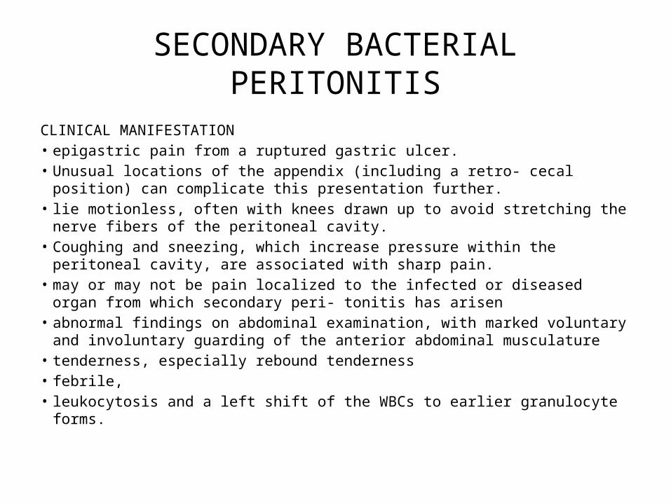
SECONDARY BACTERIAL PERITONITIS
TREATMENTbull early administration of antibiotics aimed particularly at
aerobic gram-negative bacilli and an- aerobes bull Mild to moderate broad-spectrum pen- icillinβ1113100-lactamase
inhibitor combinations (eg ticarcillinclavulanate 31 g q6h IV) or cefoxitin (2 g q24h IV)
bull hospitalization in intensive care imipenem (500 mg q6h IV) meropenem (1 g q8h IV) or combinations of drugs (ampicillin plus metronidizole plus ciprofloxacin)
bull usually requires both surgical intervention and antibiotic administration
INTESTINAL PERFORATIONLO 3
MALROTATION GASTROINTESTINAL TRACTLO 4
Intestinal malrotation
bull Intestinal malrotation when the intestines donrsquot make the turns as they should resulting in the congenital (present at birth)
bull When a fetus is about five weeks old her intestine exits her abdomen into the amniotic fluid (where therersquos more space) and continues to grow there At around ten weeks the intestine re-enters the abdomen and makes two turns
Source httpwwwchildrenshospitalorgconditions-and-treatmentsconditionsintestinal-malrotation
bull Intestinal malrotation itself isnrsquot much of a concern but it puts your child at higher risk for two serious complications
bull volvulus ndash when the intestine twists in on itself potentially cutting off the blood supplybull intestinal obstruction ndash when a stalk of fibrous tissue
known as Laddrsquos bands creates a blockage that prevents the intestine from functioning
Source httpwwwchildrenshospitalorgconditions-and-treatmentsconditionsintestinal-malrotation
Source httpwwwchildrenshospitalorgconditions-and-treatmentsconditionsintestinal-malrotation
bull Itrsquos fairly common that a baby is born with intestinal malrotation it affects about one in every 500 babies in the United States Some babies may not have symptoms until they become children teens or adults Others may go through their entire life with no symptoms never have a problem and never be diagnosed
bull Of babies who are diagnosed with intestinal malrotationbull 25 to 40 percent are diagnosed in the first week of lifebull 50 to 60 percent are diagnosed within the first month of lifebull 75 to 90 percent are diagnosed by age 1bull Although malrotation occurs equally among boys and girls boys are
more likely to become symptomatic by the first month of life
Source httpwwwchildrenshospitalorgconditions-and-treatmentsconditionsintestinal-malrotation
ACUTE APPENDICITISLO 5
A condition characterized by inflammation of the appendix
most common cause of acute inflammation in the right lower quadrant of the abdominal cavity
prevalent in countries in which people consume a diet low in fiber and high in refined carbohydrates
DEFINITION
bullThe appendix is located in the lower quadrant of the abdomen or more specifically the right iliac fossa
bullIt is a slender worm-shaped pouch averaging 5mdash10cm in length
RACIAL amp DIETARY FACTORS- bull MORE COMMON IN WHITE RACES bull YOUNG MALES ARE AFFECTED MORE OFTEN bull DIET RICH IN MEAT PRECIPITATES APPENDICITIS bull FAMILIAL TENDENCY
SOCIO-ECONOMIC STATUSbull IT IS COMMON IN MIDDLE CLASS amp RICH PEOPLE
OBSTRUCTION OF THE LUMEN bull A) IN THE LUMEN-INTESTINAL WARM eg ROUND
WORMTHREDWORM ETC VEGETABLEFRUIT SEEDFECES MATERIAL BARIUM
bull B) IN THE WALL-STRUCTURE NEOPLASM
ETIOLOGY
Non-modifiable
bullAge all age groups old
bullGender male(male- female =21)
bullHereditary tumor formation in the opening of the appendix
Modifiable
bullDiet People whose diet is low in fiber and rich in refined carbohydrates
bullInfections Gastrointestinal infections such as Amoebiasis Bacterial Gastroenteritis
PATHOPHYSIOLOGY
Decreased O2 supply in the appendix
Appendix starts to be necrotic bacteria invade the appendix
Disruption of cell membrane of appendix
Episodes of constipation
Occlusion of appendix by fecalith
Decreased flowdrainage of mucosal secretions
vasoncongestion
Decreased blood supply in the appendix
Start of Inflammatory Process
Release of Chemical Mediators Activation of the Vomiting center in the medulla
Neutrophils to area
Swelling of Appendix
Risk for infection(if appendix ruptures)
Histamine Prostaglandin Leukotrienes Bradykinin Pus Formation
(phagocytized bacteria and dead cells)
Prostaglandin Bradykinin
Pain in the RLQ of Abdomen
Stimulation of Vagus Nerve
Suppression of Sympathetic GI function
NVAnorexia
Risk for Deficient Fluid Volume
Risk for Imbalanced Nutrition less than body requirements
Acute pain
Interleukin-1
Increased WBC
Inflammation of Appendix
Appendectomy
Tissue TraumaOpen Wound
Disruption of Cell Membrane
Noriceptors of the Dermis
Send Impulse to CNS
Pain on Surgical Site
Start of Inflammatory Process
Release of Prostaglandin Bradykinin Activity Intolerance
bull Only 55 have classical featuresbull Atypical 45bull History 24-36 hoursbull Abdominal pain (diffuse and periumbilical localizing to the RIF)bull Anorexia (almost always)bull Vomiting (75)bull Low grade fever
bull If gt38 suspect perforationbull Tenderness guarding and rebound Be gentlebull Rovsingrsquos psoas obturator signs unreliable and late
bull Tender Appendicular massbull Atypical
bull (loin high RUQ deep pelvic)bull Diarrhea ( not always gastroenteritis)bull Urinary frequency
bull The Extremes of Agebull Children lt 5 rapid progressionbull Pain in the elderly is less intense
CLINICAL PRESENTATION
A Physical Exambull Findings depend on duration of illness prior to exambull Early on patients may not have localized tendernessbull With progression there is tenderness to deep palpation over McBurneyrsquos pointbull McBurneyrsquos Point just below the middle of a line connecting the umbilicus and
the ASISbull Rovsingrsquos pain in RLQ with palpation to LLQbull Rectal exam pain can be most pronounced if the patient has pelvic appendixbull Additional components that may be helpful in diagnosis rebound tenderness
voluntary guarding muscular rigidity tenderness on rectalbull Psoas sign place patient in L lateral decubitus and extend R leg at the hip If
there is pain with this movement then the sign is positivebull Obturator sign passively flex the R hip and knee and internally rotate the hip If
there is increased pain then the sign is positivebull Fever another late findingbull At the onset of pain fever is usually not found bull Temperatures gt39 C are uncommon in first 24 h but not uncommon after
rupture
DIAGNOSIS
Laboratory amp Radiology Result
CBC WBC count reveal moderate leukocytosis (10000 to 16000mm3) with shift to the left
Ultrasound studies amp CT scans May reveal right liver quadrant density or localized distention of the bowel
Abdominal x-ray visualize shadow consistent with fecalith in appendix perforation will reveal free air
B Laboratory amp Radiology
bull M Antibiotics for infection Analgesic agent (morphine) can be given for pain after the surgery
bull E Within 12 hrs of surgery you may get up and move around You can usually return to normal activities in 2-3 weeks after laparoscopic surgery
bull T Pretreatment of foods with lactase preparations (eg lactacid drops) before ingestion can reduce symptoms
Ingestion of lactase enzyme tablets with the first bite of food can reduce symptoms bull H To care wound perform dressing changes and irrigations as prescribe avoid taking laxative
or applying heat to abdomen when abdominal pain of unknown cause is experienced Reinforce need for follow-up appointment with the surgeon Call your physician for increased pain at the incision site
bull O Document bowel sounds and the passing of flatus or bowel movements (these are signs of the return of peristalsis)
Watch for surgical complications such as continuing pain or fever which indicate an abscess or wound dehiscence Stitches removed between fifth and seventh day (usually in physicians office)
bull D Liquid or soft diet until the infection subsides Soft diet is low in fiber and easily breaks down in the gastrointestinal tract
MANAGEMENT
PRE-OPERATIVE MANAGEMENTbull NPO diet in preparation for surgery bull An intravenous drip is used to hydrate the
patient bull Antibiotics given intravenously such as
Cefuroxime and Metronidazole bull If the stomach is empty (no food in the past
six hours) general anesthesia is usually used
bull Otherwise spinal anesthesia may be used
Removal of the appendix Performed as soon as possible to decrease
the risk of perforation
Two ways performed1 Laparotomy2 Laparoscopy
Appendectomy
POST- OPERATIVE MANAGEMENTbull Assist patient to position of comfort such as semi-fowlers with
knees are flexedbull Restrict activity that may aggravate pain such as coughing
and ambulationbull Apply ice bag to abdomen for comfortbull Advise avoidance of enemas or harsh laxatives increased
fluids and stool softeners may be used for postoperative constipation
bull Give narcotic analgesic as ordered and administer oral fluids when tolerated
bull Monitor frequently for signs and symptoms of worsening condition indicating perforation abscess or peritonitis (increasing severity of pain tenderness rigidity distention absent bowel sounds fever malaise and tachycardia)
bull If a drain is left in place at the area of the incision monitor carefully for signs of intestinal obstruction secondary hemorrhage or secondary abscesses (eg fever tachycardia and increased leukocyte count)
Complication InterventionsPERITONITIS bullObserve for abdominal tenderness fever
vomiting abdominal rigidity and tachycardiabullEmploy constant nasogastric suctionbullCorrect dehydrationbullAdmin Antibiotics as prescribed
PELVIC ABSCESS bullEvaluate for anorexia chills fever and diaphoresisbullObserve for diarrhea which may indicate pelvic abscessbullPrepare pt for rectal exambullPrepare pt for surgical drainage procedure
SUPHRENIC ABSCESS bullAssess pt for chills fever diaphoresisbullPrepare for x-ray exam and surgical drainage of abscess
PARALYTIC ILEUS bullAssess bowel soundsbullReplace fluids and electrolytes by IV routebullEmploy nasogastric intubation and suction
COMPLICATIONS
ABA-The Appendix- 4th year Lectures
bull Mortality from 02 to 1bull Complications increase with perforation bull Morbidity
bull Wound abscess bull Wound infection (less with MacBurneyrsquos incision)bull Wound dehiscencebull Intra-abdominal abscess bull Faecal fistula bull Intestinal obstruction bull Adhesive band bull inguinal hernia bull Fertility
PROGNOSIS
Prevents
bull Eat foods high in fiber such as fresh fruits and vegetables
Ascariasis
bull Disease bull Ascariasis ascariasis infection roundworm infection
Pathophysiological mechanism
Adult worms move throughout the GI tract and may become incarcerated leading to
obstructive pathology
The worms may die rarr leading to inflammation necrosis infection and abscess
formation
If they migrate through an existing perforation in the bowel wall rarr secondary to
tuberculosis or typhoid rarr cause a granulomatous peritonitis
Larvae during migration rarr may be deposited in the brain spinal cord kidney or other
organs rarr leading to granuloma formation inflammation or infection
They may become entwined in a bolus and obstruct the small bowel this is most
common in the terminal ileum although other more proximal sites have been rarely
reported
Risk Factors
bull Preschool age or youngerbull Eating unsanitary foodbull Drinking unclean water
Symptomsbull Small burdens of worms in the intestine may cause no symptoms
bull The patient may have symptoms of pneumonitis with cough dyspnea wheezing chest pain and low grade fever during the migration of the larvae through the liver and lungs
bull In heavy worm burdens the adult worms actively migrate in the intestine resulting in intestinal blockage vomiting abdominal pain colic nausea anorexia and intermittent diarrhea
bull A heavy worm burden in children may lead to severe nutritional impairment and retardation in growth
bull Worms exiting through the nose or mouth
Laboratory Studiesbull Early infection (larval migration)
ndash Complete blood count (CBC) may show eosinophiliandash Sputum analysis may reveal larvae or Charcot-Leyden crystalsndash Stool examination findings are typically normal in absence of previous
infection (during the first 40 d)ndash Ascaris specific antibodies (not useful in acute infection and not
protective)ndash Increases in IgE and later IgG
bull Established infection (adult phase) ndash Stool examination findings include characteristic eggs Adult females lay
about 200000 eggs per day aiding microscopic identification of characteristic eggs
Imaging Studies
bull Early infection (larval migration) ndash Chest radiography may reveal patchy infiltrates of eosinophilic pneumonia
bull Established infection (adult phase)ndash Abdominal radiography may reveal adult worms (especially with
contrast) ndash Obstructing Ascaris lesions cause cylindrical filling defects on contrast
computed tomography (CT) scansndash Cholangiopancreatography by endoscopy (ERCP) or magnetic
resonance imaging (MRCP or magnetic resonance cholangiopancreatography) may detect adult worms in bile or pancreatic ducts8
ndash Ultrasonography may detect worms in the gallbladder
Treatmentbull Albendazole ndash 400 mg PO once for all ages
bull Mebendazole ndash 100 mg bid PO for 3 days or 500 mg PO once for all
agesbull Piperazine citrate ndash 150 mgkg PO initially followed by 6 doses of 65
mgkg at 12 hr intervals PO which causes neuromuscular paralysis of the parasite (the treatment of choice for intestinal or biliary obstruction)
Treatmentbull Pyrantel pamoate ndash 11 mgkg PO once maximum 1 g
bull Nitazoxanide ndash 100 mg bid PO for 3 days for children 1-3 yrs of age ndash 200 mg bid PO for 3 days for children 4-11 yrndash 500 mg bid PO for 3 days for adolescents and adults
produces cure rates comparable with single-dose albendazole
bull Surgery may be required for cases with severe obstruction
complicationsbull Intestinal obstruction - 63 bull Bile duct obstruction - 23bull Perforation peritonitis or both - 32bull Volvulus - 27bull Hepatitic abscess - 21bull Appendicitis - 21bull Pancreatitis - 1bull Cerebral encephalitis - 1bull Intussusception - 05bull Other sites of pathology (lt05) include Meckel diverticulum the gallbladder ears
eyes nose lungs kidneys vagina urethra heart placenta spleen thoracic cavity and umbilicus
bull In endemic regions ascariasis is a significant part of the differential diagnosis for intestinal obstruction appendicitis biliary tract disease pancreatitis intussusception and volvulus
ILEUSLO 6
Paralytic ileus
Definition Failure of appropriate forward movement of bowel contents
Causes Neurogenikmetabolikobat2aninfeksiiskemi usus
Symtomps Mild stomach painbloatingnauseavomitingkonstipasi
PF Silent abdomen (-) distensi timpanik
PP Laboratorium leukosit darahkadar elektrolytureumglukosa darahamilase Foto polos rontgen dilatasi usus halus dan besar diagfragma elevation
Treatment farmako Metoklopramid u gastroparesis sisaprid u ileus paralitik pasca operasi klonidin u mengatasi ileus paralitik
Treatment non-farmako Gum chewing pasca operasi menunjukan perbaikan ikeus
Prognosis Prognosis ileus paralitik baik bila penyakit primernya dapat diats
Ileus Obstruction (Ascaris)
bull Ascaris lumbricoides (A lumbricoides) is the most common intestinal helminth parasite and it is estimated that the infected population is 08-12 billion worldwide The highest prevalence of ascariasis occurs in tropical and semitropical countries where sanitation is poor
httpwwwncbinlmnihgovpmcarticlesPMC4194592
bull large numbers of adult Ascaris in the small intestine can cause
bull abdominal distension bull abdominal pain bull obstructive ileus bull malnutrition
A single worm of A lumbricoides can enter the ampulla of Vater and result in biliary colic obstructive jaundice ascending cholangitis acalculous cholecystitis or acute pancreatitis
httpwwwncbinlmnihgovpmcarticlesPMC4194592
httpwwwncbinlmnihgovpmcarticlesPMC4194592
httpwwwncbinlmnihgovpmcarticlesPMC4194592
httpwwwncbinlmnihgovpmcarticlesPMC4194592
bull TerminologyA lumbricoides the human roundworm is one of the most common soil transmitted parasites in the world infecting about 12 billion people globally
httpwwwncbinlmnihgovpmcarticlesPMC4194592
Factors that result in Ascaris-related intestinal obstruction
There are 4 major factors that result in Ascaris-related intestinal obstruction
bull Multiple worms can form a large bolus resulting in mechanical obstruction of the
bowel lumen This is the most frequent cause of Ascaris- related bowel
obstruction
bull The worm bolus may serve as a lead point in intussusception or a pivot in small-
bowel volvulus
bull Ascaris worms may inhabit the ileocecal valve where roundworm secretion of
neurotoxins prompts small-bowel contraction This action coupled with high
worm burden in the ileocecal valve can obstruct the intestine
bull A host inflammatory reaction to worm-derived hemolysins endocrinolysins and
anaphylatoxins can be severe enough to obstruct the gut lumen
Patient presentation and symptomatology
bull Surendran and Paulose conducted a 5-year study of intestinal obstruction by A lumbricoides and categorized small-bowel obstruction resulting from Ascarisas either acute or subacute
bull Patients with subacute obstruction in this study experienced diffuse and colicky abdominal pain fever vomiting that was more pronounced at the onset of symptoms and diarrhea without blood and mucus
bull Acute obstruction had far more ominous presenting signs and symptoms Patients were often ill for several days before presentation These persons showed signs of severe dehydration and had a toxic appearance Vomiting abdominal pain and distention (Figure 1) fever leukocytosis and obstipation were frequently noted
Distended abdomen is characteristic of a child with severe ascariasis (Reprinted from Despommier D et al Parasitic Diseases 2001[24])
Treatment
bull The anthelmintics of choice are mebendazole and albendazole bull These medications are used for intestinal ascariasis as well as HPA bull Mebendazole100 mg is given twice daily for 3 days and provides
protection against Trichuris trichiura (whipworm) as well as Ancylostoma duodenale and Necator americanus (hookworm)
bull Alternatively albendazole may be used the advantage of this drug is that it is given in a single 400-mg dose
bull pyrantel pamoate 11 mgkg (up to a maximum of 1 g) orally daily for 3 days
httpwwwcdcgovparasiteshookwormhealth_professionalsindexhtmls_cid=cs_074httpemedicinemedscapecomarticle212510-treatment
httpwwwmedscapecomviewarticle451597_3
Recommended criteria for surgical exploration include the following
bull Passage of blood per rectumbull Multiple air fluid levels on abdominal radiographsbull An ill child with abdominal distension and
rebound tendernessbull Unsatisfactory response to conservative therapybull Appendicitis and primary peritonitisbull Hepatobiliary diseasebull Pancreatic pseudocyst
httpemedicinemedscapecomarticle212510-treatmentd9
bull PrognosisThe prognosis for ascariasis is excellent However in higher worm burden infections serious complications such as obstruction are more common
bull Patient EducationRecommend good personal hygiene and food handling techniques discriminate defecation hand-washing cleaning fruits and vegetables and avoiding soil consumption Educational programs should also address the use of human feces as fertilizer a practice that persists in many communities internationally
httpemedicinemedscapecomarticle788398-followupe6
INTUSSUSSCEPTIONLO 7
Intussusception
bull DefinitionThe invagination or telescoping of a proximal segment of bowel (intussusceptum) into lumen of a distal segment (intussuscipiens)
Etiology bull Idiopatic (most common in children)bull Neoplasmndash Benign polyp leiomyoma lipoma lympoma
adenoma of appendix appendiceal stump granulomandash Malignant
bull Primary (more common in colon)bull (more common in small bowel)
bull Postoperativebull Meckelrsquos diverticulumbull Colitisbull Many cases thought to be related to viral
gastroenteritis in children
Pathophysiology bull The invaginated segment is carried distally by
peristalsisbull Mesentery and vessel become involved with
the intraluminal loop and are squeezed within the engulfing segment causing venous congestion
bull Types enteroenteric enterocolic and colocolic
Childrenbull Well nourished infantbull Cramping abdominal
painbull Vomittingbull Diarrhea (often-jelly
stools)bull A palpable tender
sausage shaped mass in the abdomen
Adults bull Intermitten painbull Nausea and vomittingbull Often red blood per
rectumbull Often nonspecific
complains
Examination
bull Abdominal studiesbull Barium studiesbull Ultrasoundbull CT
Treatment
bull Air reductionbull Contrast reduction
bull Surgical exploration and resection of the intussuscepted bowel loops

Source httpwwwchildrenshospitalorgconditions-and-treatmentsconditionsintestinal-malrotation
bull Itrsquos fairly common that a baby is born with intestinal malrotation it affects about one in every 500 babies in the United States Some babies may not have symptoms until they become children teens or adults Others may go through their entire life with no symptoms never have a problem and never be diagnosed
bull Of babies who are diagnosed with intestinal malrotationbull 25 to 40 percent are diagnosed in the first week of lifebull 50 to 60 percent are diagnosed within the first month of lifebull 75 to 90 percent are diagnosed by age 1bull Although malrotation occurs equally among boys and girls boys are
more likely to become symptomatic by the first month of life
Source httpwwwchildrenshospitalorgconditions-and-treatmentsconditionsintestinal-malrotation
ACUTE APPENDICITISLO 5
A condition characterized by inflammation of the appendix
most common cause of acute inflammation in the right lower quadrant of the abdominal cavity
prevalent in countries in which people consume a diet low in fiber and high in refined carbohydrates
DEFINITION
bullThe appendix is located in the lower quadrant of the abdomen or more specifically the right iliac fossa
bullIt is a slender worm-shaped pouch averaging 5mdash10cm in length
RACIAL amp DIETARY FACTORS- bull MORE COMMON IN WHITE RACES bull YOUNG MALES ARE AFFECTED MORE OFTEN bull DIET RICH IN MEAT PRECIPITATES APPENDICITIS bull FAMILIAL TENDENCY
SOCIO-ECONOMIC STATUSbull IT IS COMMON IN MIDDLE CLASS amp RICH PEOPLE
OBSTRUCTION OF THE LUMEN bull A) IN THE LUMEN-INTESTINAL WARM eg ROUND
WORMTHREDWORM ETC VEGETABLEFRUIT SEEDFECES MATERIAL BARIUM
bull B) IN THE WALL-STRUCTURE NEOPLASM
ETIOLOGY
Non-modifiable
bullAge all age groups old
bullGender male(male- female =21)
bullHereditary tumor formation in the opening of the appendix
Modifiable
bullDiet People whose diet is low in fiber and rich in refined carbohydrates
bullInfections Gastrointestinal infections such as Amoebiasis Bacterial Gastroenteritis
PATHOPHYSIOLOGY
Decreased O2 supply in the appendix
Appendix starts to be necrotic bacteria invade the appendix
Disruption of cell membrane of appendix
Episodes of constipation
Occlusion of appendix by fecalith
Decreased flowdrainage of mucosal secretions
vasoncongestion
Decreased blood supply in the appendix
Start of Inflammatory Process
Release of Chemical Mediators Activation of the Vomiting center in the medulla
Neutrophils to area
Swelling of Appendix
Risk for infection(if appendix ruptures)
Histamine Prostaglandin Leukotrienes Bradykinin Pus Formation
(phagocytized bacteria and dead cells)
Prostaglandin Bradykinin
Pain in the RLQ of Abdomen
Stimulation of Vagus Nerve
Suppression of Sympathetic GI function
NVAnorexia
Risk for Deficient Fluid Volume
Risk for Imbalanced Nutrition less than body requirements
Acute pain
Interleukin-1
Increased WBC
Inflammation of Appendix
Appendectomy
Tissue TraumaOpen Wound
Disruption of Cell Membrane
Noriceptors of the Dermis
Send Impulse to CNS
Pain on Surgical Site
Start of Inflammatory Process
Release of Prostaglandin Bradykinin Activity Intolerance
bull Only 55 have classical featuresbull Atypical 45bull History 24-36 hoursbull Abdominal pain (diffuse and periumbilical localizing to the RIF)bull Anorexia (almost always)bull Vomiting (75)bull Low grade fever
bull If gt38 suspect perforationbull Tenderness guarding and rebound Be gentlebull Rovsingrsquos psoas obturator signs unreliable and late
bull Tender Appendicular massbull Atypical
bull (loin high RUQ deep pelvic)bull Diarrhea ( not always gastroenteritis)bull Urinary frequency
bull The Extremes of Agebull Children lt 5 rapid progressionbull Pain in the elderly is less intense
CLINICAL PRESENTATION
A Physical Exambull Findings depend on duration of illness prior to exambull Early on patients may not have localized tendernessbull With progression there is tenderness to deep palpation over McBurneyrsquos pointbull McBurneyrsquos Point just below the middle of a line connecting the umbilicus and
the ASISbull Rovsingrsquos pain in RLQ with palpation to LLQbull Rectal exam pain can be most pronounced if the patient has pelvic appendixbull Additional components that may be helpful in diagnosis rebound tenderness
voluntary guarding muscular rigidity tenderness on rectalbull Psoas sign place patient in L lateral decubitus and extend R leg at the hip If
there is pain with this movement then the sign is positivebull Obturator sign passively flex the R hip and knee and internally rotate the hip If
there is increased pain then the sign is positivebull Fever another late findingbull At the onset of pain fever is usually not found bull Temperatures gt39 C are uncommon in first 24 h but not uncommon after
rupture
DIAGNOSIS
Laboratory amp Radiology Result
CBC WBC count reveal moderate leukocytosis (10000 to 16000mm3) with shift to the left
Ultrasound studies amp CT scans May reveal right liver quadrant density or localized distention of the bowel
Abdominal x-ray visualize shadow consistent with fecalith in appendix perforation will reveal free air
B Laboratory amp Radiology
bull M Antibiotics for infection Analgesic agent (morphine) can be given for pain after the surgery
bull E Within 12 hrs of surgery you may get up and move around You can usually return to normal activities in 2-3 weeks after laparoscopic surgery
bull T Pretreatment of foods with lactase preparations (eg lactacid drops) before ingestion can reduce symptoms
Ingestion of lactase enzyme tablets with the first bite of food can reduce symptoms bull H To care wound perform dressing changes and irrigations as prescribe avoid taking laxative
or applying heat to abdomen when abdominal pain of unknown cause is experienced Reinforce need for follow-up appointment with the surgeon Call your physician for increased pain at the incision site
bull O Document bowel sounds and the passing of flatus or bowel movements (these are signs of the return of peristalsis)
Watch for surgical complications such as continuing pain or fever which indicate an abscess or wound dehiscence Stitches removed between fifth and seventh day (usually in physicians office)
bull D Liquid or soft diet until the infection subsides Soft diet is low in fiber and easily breaks down in the gastrointestinal tract
MANAGEMENT
PRE-OPERATIVE MANAGEMENTbull NPO diet in preparation for surgery bull An intravenous drip is used to hydrate the
patient bull Antibiotics given intravenously such as
Cefuroxime and Metronidazole bull If the stomach is empty (no food in the past
six hours) general anesthesia is usually used
bull Otherwise spinal anesthesia may be used
Removal of the appendix Performed as soon as possible to decrease
the risk of perforation
Two ways performed1 Laparotomy2 Laparoscopy
Appendectomy
POST- OPERATIVE MANAGEMENTbull Assist patient to position of comfort such as semi-fowlers with
knees are flexedbull Restrict activity that may aggravate pain such as coughing
and ambulationbull Apply ice bag to abdomen for comfortbull Advise avoidance of enemas or harsh laxatives increased
fluids and stool softeners may be used for postoperative constipation
bull Give narcotic analgesic as ordered and administer oral fluids when tolerated
bull Monitor frequently for signs and symptoms of worsening condition indicating perforation abscess or peritonitis (increasing severity of pain tenderness rigidity distention absent bowel sounds fever malaise and tachycardia)
bull If a drain is left in place at the area of the incision monitor carefully for signs of intestinal obstruction secondary hemorrhage or secondary abscesses (eg fever tachycardia and increased leukocyte count)
Complication InterventionsPERITONITIS bullObserve for abdominal tenderness fever
vomiting abdominal rigidity and tachycardiabullEmploy constant nasogastric suctionbullCorrect dehydrationbullAdmin Antibiotics as prescribed
PELVIC ABSCESS bullEvaluate for anorexia chills fever and diaphoresisbullObserve for diarrhea which may indicate pelvic abscessbullPrepare pt for rectal exambullPrepare pt for surgical drainage procedure
SUPHRENIC ABSCESS bullAssess pt for chills fever diaphoresisbullPrepare for x-ray exam and surgical drainage of abscess
PARALYTIC ILEUS bullAssess bowel soundsbullReplace fluids and electrolytes by IV routebullEmploy nasogastric intubation and suction
COMPLICATIONS
ABA-The Appendix- 4th year Lectures
bull Mortality from 02 to 1bull Complications increase with perforation bull Morbidity
bull Wound abscess bull Wound infection (less with MacBurneyrsquos incision)bull Wound dehiscencebull Intra-abdominal abscess bull Faecal fistula bull Intestinal obstruction bull Adhesive band bull inguinal hernia bull Fertility
PROGNOSIS
Prevents
bull Eat foods high in fiber such as fresh fruits and vegetables
Ascariasis
bull Disease bull Ascariasis ascariasis infection roundworm infection
Pathophysiological mechanism
Adult worms move throughout the GI tract and may become incarcerated leading to
obstructive pathology
The worms may die rarr leading to inflammation necrosis infection and abscess
formation
If they migrate through an existing perforation in the bowel wall rarr secondary to
tuberculosis or typhoid rarr cause a granulomatous peritonitis
Larvae during migration rarr may be deposited in the brain spinal cord kidney or other
organs rarr leading to granuloma formation inflammation or infection
They may become entwined in a bolus and obstruct the small bowel this is most
common in the terminal ileum although other more proximal sites have been rarely
reported
Risk Factors
bull Preschool age or youngerbull Eating unsanitary foodbull Drinking unclean water
Symptomsbull Small burdens of worms in the intestine may cause no symptoms
bull The patient may have symptoms of pneumonitis with cough dyspnea wheezing chest pain and low grade fever during the migration of the larvae through the liver and lungs
bull In heavy worm burdens the adult worms actively migrate in the intestine resulting in intestinal blockage vomiting abdominal pain colic nausea anorexia and intermittent diarrhea
bull A heavy worm burden in children may lead to severe nutritional impairment and retardation in growth
bull Worms exiting through the nose or mouth
Laboratory Studiesbull Early infection (larval migration)
ndash Complete blood count (CBC) may show eosinophiliandash Sputum analysis may reveal larvae or Charcot-Leyden crystalsndash Stool examination findings are typically normal in absence of previous
infection (during the first 40 d)ndash Ascaris specific antibodies (not useful in acute infection and not
protective)ndash Increases in IgE and later IgG
bull Established infection (adult phase) ndash Stool examination findings include characteristic eggs Adult females lay
about 200000 eggs per day aiding microscopic identification of characteristic eggs
Imaging Studies
bull Early infection (larval migration) ndash Chest radiography may reveal patchy infiltrates of eosinophilic pneumonia
bull Established infection (adult phase)ndash Abdominal radiography may reveal adult worms (especially with
contrast) ndash Obstructing Ascaris lesions cause cylindrical filling defects on contrast
computed tomography (CT) scansndash Cholangiopancreatography by endoscopy (ERCP) or magnetic
resonance imaging (MRCP or magnetic resonance cholangiopancreatography) may detect adult worms in bile or pancreatic ducts8
ndash Ultrasonography may detect worms in the gallbladder
Treatmentbull Albendazole ndash 400 mg PO once for all ages
bull Mebendazole ndash 100 mg bid PO for 3 days or 500 mg PO once for all
agesbull Piperazine citrate ndash 150 mgkg PO initially followed by 6 doses of 65
mgkg at 12 hr intervals PO which causes neuromuscular paralysis of the parasite (the treatment of choice for intestinal or biliary obstruction)
Treatmentbull Pyrantel pamoate ndash 11 mgkg PO once maximum 1 g
bull Nitazoxanide ndash 100 mg bid PO for 3 days for children 1-3 yrs of age ndash 200 mg bid PO for 3 days for children 4-11 yrndash 500 mg bid PO for 3 days for adolescents and adults
produces cure rates comparable with single-dose albendazole
bull Surgery may be required for cases with severe obstruction
complicationsbull Intestinal obstruction - 63 bull Bile duct obstruction - 23bull Perforation peritonitis or both - 32bull Volvulus - 27bull Hepatitic abscess - 21bull Appendicitis - 21bull Pancreatitis - 1bull Cerebral encephalitis - 1bull Intussusception - 05bull Other sites of pathology (lt05) include Meckel diverticulum the gallbladder ears
eyes nose lungs kidneys vagina urethra heart placenta spleen thoracic cavity and umbilicus
bull In endemic regions ascariasis is a significant part of the differential diagnosis for intestinal obstruction appendicitis biliary tract disease pancreatitis intussusception and volvulus
ILEUSLO 6
Paralytic ileus
Definition Failure of appropriate forward movement of bowel contents
Causes Neurogenikmetabolikobat2aninfeksiiskemi usus
Symtomps Mild stomach painbloatingnauseavomitingkonstipasi
PF Silent abdomen (-) distensi timpanik
PP Laboratorium leukosit darahkadar elektrolytureumglukosa darahamilase Foto polos rontgen dilatasi usus halus dan besar diagfragma elevation
Treatment farmako Metoklopramid u gastroparesis sisaprid u ileus paralitik pasca operasi klonidin u mengatasi ileus paralitik
Treatment non-farmako Gum chewing pasca operasi menunjukan perbaikan ikeus
Prognosis Prognosis ileus paralitik baik bila penyakit primernya dapat diats
Ileus Obstruction (Ascaris)
bull Ascaris lumbricoides (A lumbricoides) is the most common intestinal helminth parasite and it is estimated that the infected population is 08-12 billion worldwide The highest prevalence of ascariasis occurs in tropical and semitropical countries where sanitation is poor
httpwwwncbinlmnihgovpmcarticlesPMC4194592
bull large numbers of adult Ascaris in the small intestine can cause
bull abdominal distension bull abdominal pain bull obstructive ileus bull malnutrition
A single worm of A lumbricoides can enter the ampulla of Vater and result in biliary colic obstructive jaundice ascending cholangitis acalculous cholecystitis or acute pancreatitis
httpwwwncbinlmnihgovpmcarticlesPMC4194592
httpwwwncbinlmnihgovpmcarticlesPMC4194592
httpwwwncbinlmnihgovpmcarticlesPMC4194592
httpwwwncbinlmnihgovpmcarticlesPMC4194592
bull TerminologyA lumbricoides the human roundworm is one of the most common soil transmitted parasites in the world infecting about 12 billion people globally
httpwwwncbinlmnihgovpmcarticlesPMC4194592
Factors that result in Ascaris-related intestinal obstruction
There are 4 major factors that result in Ascaris-related intestinal obstruction
bull Multiple worms can form a large bolus resulting in mechanical obstruction of the
bowel lumen This is the most frequent cause of Ascaris- related bowel
obstruction
bull The worm bolus may serve as a lead point in intussusception or a pivot in small-
bowel volvulus
bull Ascaris worms may inhabit the ileocecal valve where roundworm secretion of
neurotoxins prompts small-bowel contraction This action coupled with high
worm burden in the ileocecal valve can obstruct the intestine
bull A host inflammatory reaction to worm-derived hemolysins endocrinolysins and
anaphylatoxins can be severe enough to obstruct the gut lumen
Patient presentation and symptomatology
bull Surendran and Paulose conducted a 5-year study of intestinal obstruction by A lumbricoides and categorized small-bowel obstruction resulting from Ascarisas either acute or subacute
bull Patients with subacute obstruction in this study experienced diffuse and colicky abdominal pain fever vomiting that was more pronounced at the onset of symptoms and diarrhea without blood and mucus
bull Acute obstruction had far more ominous presenting signs and symptoms Patients were often ill for several days before presentation These persons showed signs of severe dehydration and had a toxic appearance Vomiting abdominal pain and distention (Figure 1) fever leukocytosis and obstipation were frequently noted
Distended abdomen is characteristic of a child with severe ascariasis (Reprinted from Despommier D et al Parasitic Diseases 2001[24])
Treatment
bull The anthelmintics of choice are mebendazole and albendazole bull These medications are used for intestinal ascariasis as well as HPA bull Mebendazole100 mg is given twice daily for 3 days and provides
protection against Trichuris trichiura (whipworm) as well as Ancylostoma duodenale and Necator americanus (hookworm)
bull Alternatively albendazole may be used the advantage of this drug is that it is given in a single 400-mg dose
bull pyrantel pamoate 11 mgkg (up to a maximum of 1 g) orally daily for 3 days
httpwwwcdcgovparasiteshookwormhealth_professionalsindexhtmls_cid=cs_074httpemedicinemedscapecomarticle212510-treatment
httpwwwmedscapecomviewarticle451597_3
Recommended criteria for surgical exploration include the following
bull Passage of blood per rectumbull Multiple air fluid levels on abdominal radiographsbull An ill child with abdominal distension and
rebound tendernessbull Unsatisfactory response to conservative therapybull Appendicitis and primary peritonitisbull Hepatobiliary diseasebull Pancreatic pseudocyst
httpemedicinemedscapecomarticle212510-treatmentd9
bull PrognosisThe prognosis for ascariasis is excellent However in higher worm burden infections serious complications such as obstruction are more common
bull Patient EducationRecommend good personal hygiene and food handling techniques discriminate defecation hand-washing cleaning fruits and vegetables and avoiding soil consumption Educational programs should also address the use of human feces as fertilizer a practice that persists in many communities internationally
httpemedicinemedscapecomarticle788398-followupe6
INTUSSUSSCEPTIONLO 7
Intussusception
bull DefinitionThe invagination or telescoping of a proximal segment of bowel (intussusceptum) into lumen of a distal segment (intussuscipiens)
Etiology bull Idiopatic (most common in children)bull Neoplasmndash Benign polyp leiomyoma lipoma lympoma
adenoma of appendix appendiceal stump granulomandash Malignant
bull Primary (more common in colon)bull (more common in small bowel)
bull Postoperativebull Meckelrsquos diverticulumbull Colitisbull Many cases thought to be related to viral
gastroenteritis in children
Pathophysiology bull The invaginated segment is carried distally by
peristalsisbull Mesentery and vessel become involved with
the intraluminal loop and are squeezed within the engulfing segment causing venous congestion
bull Types enteroenteric enterocolic and colocolic
Childrenbull Well nourished infantbull Cramping abdominal
painbull Vomittingbull Diarrhea (often-jelly
stools)bull A palpable tender
sausage shaped mass in the abdomen
Adults bull Intermitten painbull Nausea and vomittingbull Often red blood per
rectumbull Often nonspecific
complains
Examination
bull Abdominal studiesbull Barium studiesbull Ultrasoundbull CT
Treatment
bull Air reductionbull Contrast reduction
bull Surgical exploration and resection of the intussuscepted bowel loops

Start of Inflammatory Process
Release of Chemical Mediators Activation of the Vomiting center in the medulla
Neutrophils to area
Swelling of Appendix
Risk for infection(if appendix ruptures)
Histamine Prostaglandin Leukotrienes Bradykinin Pus Formation
(phagocytized bacteria and dead cells)
Prostaglandin Bradykinin
Pain in the RLQ of Abdomen
Stimulation of Vagus Nerve
Suppression of Sympathetic GI function
NVAnorexia
Risk for Deficient Fluid Volume
Risk for Imbalanced Nutrition less than body requirements
Acute pain
Interleukin-1
Increased WBC
Inflammation of Appendix
Appendectomy
Tissue TraumaOpen Wound
Disruption of Cell Membrane
Noriceptors of the Dermis
Send Impulse to CNS
Pain on Surgical Site
Start of Inflammatory Process
Release of Prostaglandin Bradykinin Activity Intolerance
bull Only 55 have classical featuresbull Atypical 45bull History 24-36 hoursbull Abdominal pain (diffuse and periumbilical localizing to the RIF)bull Anorexia (almost always)bull Vomiting (75)bull Low grade fever
bull If gt38 suspect perforationbull Tenderness guarding and rebound Be gentlebull Rovsingrsquos psoas obturator signs unreliable and late
bull Tender Appendicular massbull Atypical
bull (loin high RUQ deep pelvic)bull Diarrhea ( not always gastroenteritis)bull Urinary frequency
bull The Extremes of Agebull Children lt 5 rapid progressionbull Pain in the elderly is less intense
CLINICAL PRESENTATION
A Physical Exambull Findings depend on duration of illness prior to exambull Early on patients may not have localized tendernessbull With progression there is tenderness to deep palpation over McBurneyrsquos pointbull McBurneyrsquos Point just below the middle of a line connecting the umbilicus and
the ASISbull Rovsingrsquos pain in RLQ with palpation to LLQbull Rectal exam pain can be most pronounced if the patient has pelvic appendixbull Additional components that may be helpful in diagnosis rebound tenderness
voluntary guarding muscular rigidity tenderness on rectalbull Psoas sign place patient in L lateral decubitus and extend R leg at the hip If
there is pain with this movement then the sign is positivebull Obturator sign passively flex the R hip and knee and internally rotate the hip If
there is increased pain then the sign is positivebull Fever another late findingbull At the onset of pain fever is usually not found bull Temperatures gt39 C are uncommon in first 24 h but not uncommon after
rupture
DIAGNOSIS
Laboratory amp Radiology Result
CBC WBC count reveal moderate leukocytosis (10000 to 16000mm3) with shift to the left
Ultrasound studies amp CT scans May reveal right liver quadrant density or localized distention of the bowel
Abdominal x-ray visualize shadow consistent with fecalith in appendix perforation will reveal free air
B Laboratory amp Radiology
bull M Antibiotics for infection Analgesic agent (morphine) can be given for pain after the surgery
bull E Within 12 hrs of surgery you may get up and move around You can usually return to normal activities in 2-3 weeks after laparoscopic surgery
bull T Pretreatment of foods with lactase preparations (eg lactacid drops) before ingestion can reduce symptoms
Ingestion of lactase enzyme tablets with the first bite of food can reduce symptoms bull H To care wound perform dressing changes and irrigations as prescribe avoid taking laxative
or applying heat to abdomen when abdominal pain of unknown cause is experienced Reinforce need for follow-up appointment with the surgeon Call your physician for increased pain at the incision site
bull O Document bowel sounds and the passing of flatus or bowel movements (these are signs of the return of peristalsis)
Watch for surgical complications such as continuing pain or fever which indicate an abscess or wound dehiscence Stitches removed between fifth and seventh day (usually in physicians office)
bull D Liquid or soft diet until the infection subsides Soft diet is low in fiber and easily breaks down in the gastrointestinal tract
MANAGEMENT
PRE-OPERATIVE MANAGEMENTbull NPO diet in preparation for surgery bull An intravenous drip is used to hydrate the
patient bull Antibiotics given intravenously such as
Cefuroxime and Metronidazole bull If the stomach is empty (no food in the past
six hours) general anesthesia is usually used
bull Otherwise spinal anesthesia may be used
Removal of the appendix Performed as soon as possible to decrease
the risk of perforation
Two ways performed1 Laparotomy2 Laparoscopy
Appendectomy
POST- OPERATIVE MANAGEMENTbull Assist patient to position of comfort such as semi-fowlers with
knees are flexedbull Restrict activity that may aggravate pain such as coughing
and ambulationbull Apply ice bag to abdomen for comfortbull Advise avoidance of enemas or harsh laxatives increased
fluids and stool softeners may be used for postoperative constipation
bull Give narcotic analgesic as ordered and administer oral fluids when tolerated
bull Monitor frequently for signs and symptoms of worsening condition indicating perforation abscess or peritonitis (increasing severity of pain tenderness rigidity distention absent bowel sounds fever malaise and tachycardia)
bull If a drain is left in place at the area of the incision monitor carefully for signs of intestinal obstruction secondary hemorrhage or secondary abscesses (eg fever tachycardia and increased leukocyte count)
Complication InterventionsPERITONITIS bullObserve for abdominal tenderness fever
vomiting abdominal rigidity and tachycardiabullEmploy constant nasogastric suctionbullCorrect dehydrationbullAdmin Antibiotics as prescribed
PELVIC ABSCESS bullEvaluate for anorexia chills fever and diaphoresisbullObserve for diarrhea which may indicate pelvic abscessbullPrepare pt for rectal exambullPrepare pt for surgical drainage procedure
SUPHRENIC ABSCESS bullAssess pt for chills fever diaphoresisbullPrepare for x-ray exam and surgical drainage of abscess
PARALYTIC ILEUS bullAssess bowel soundsbullReplace fluids and electrolytes by IV routebullEmploy nasogastric intubation and suction
COMPLICATIONS
ABA-The Appendix- 4th year Lectures
bull Mortality from 02 to 1bull Complications increase with perforation bull Morbidity
bull Wound abscess bull Wound infection (less with MacBurneyrsquos incision)bull Wound dehiscencebull Intra-abdominal abscess bull Faecal fistula bull Intestinal obstruction bull Adhesive band bull inguinal hernia bull Fertility
PROGNOSIS
Prevents
bull Eat foods high in fiber such as fresh fruits and vegetables
Ascariasis
bull Disease bull Ascariasis ascariasis infection roundworm infection
Pathophysiological mechanism
Adult worms move throughout the GI tract and may become incarcerated leading to
obstructive pathology
The worms may die rarr leading to inflammation necrosis infection and abscess
formation
If they migrate through an existing perforation in the bowel wall rarr secondary to
tuberculosis or typhoid rarr cause a granulomatous peritonitis
Larvae during migration rarr may be deposited in the brain spinal cord kidney or other
organs rarr leading to granuloma formation inflammation or infection
They may become entwined in a bolus and obstruct the small bowel this is most
common in the terminal ileum although other more proximal sites have been rarely
reported
Risk Factors
bull Preschool age or youngerbull Eating unsanitary foodbull Drinking unclean water
Symptomsbull Small burdens of worms in the intestine may cause no symptoms
bull The patient may have symptoms of pneumonitis with cough dyspnea wheezing chest pain and low grade fever during the migration of the larvae through the liver and lungs
bull In heavy worm burdens the adult worms actively migrate in the intestine resulting in intestinal blockage vomiting abdominal pain colic nausea anorexia and intermittent diarrhea
bull A heavy worm burden in children may lead to severe nutritional impairment and retardation in growth
bull Worms exiting through the nose or mouth
Laboratory Studiesbull Early infection (larval migration)
ndash Complete blood count (CBC) may show eosinophiliandash Sputum analysis may reveal larvae or Charcot-Leyden crystalsndash Stool examination findings are typically normal in absence of previous
infection (during the first 40 d)ndash Ascaris specific antibodies (not useful in acute infection and not
protective)ndash Increases in IgE and later IgG
bull Established infection (adult phase) ndash Stool examination findings include characteristic eggs Adult females lay
about 200000 eggs per day aiding microscopic identification of characteristic eggs
Imaging Studies
bull Early infection (larval migration) ndash Chest radiography may reveal patchy infiltrates of eosinophilic pneumonia
bull Established infection (adult phase)ndash Abdominal radiography may reveal adult worms (especially with
contrast) ndash Obstructing Ascaris lesions cause cylindrical filling defects on contrast
computed tomography (CT) scansndash Cholangiopancreatography by endoscopy (ERCP) or magnetic
resonance imaging (MRCP or magnetic resonance cholangiopancreatography) may detect adult worms in bile or pancreatic ducts8
ndash Ultrasonography may detect worms in the gallbladder
Treatmentbull Albendazole ndash 400 mg PO once for all ages
bull Mebendazole ndash 100 mg bid PO for 3 days or 500 mg PO once for all
agesbull Piperazine citrate ndash 150 mgkg PO initially followed by 6 doses of 65
mgkg at 12 hr intervals PO which causes neuromuscular paralysis of the parasite (the treatment of choice for intestinal or biliary obstruction)
Treatmentbull Pyrantel pamoate ndash 11 mgkg PO once maximum 1 g
bull Nitazoxanide ndash 100 mg bid PO for 3 days for children 1-3 yrs of age ndash 200 mg bid PO for 3 days for children 4-11 yrndash 500 mg bid PO for 3 days for adolescents and adults
produces cure rates comparable with single-dose albendazole
bull Surgery may be required for cases with severe obstruction
complicationsbull Intestinal obstruction - 63 bull Bile duct obstruction - 23bull Perforation peritonitis or both - 32bull Volvulus - 27bull Hepatitic abscess - 21bull Appendicitis - 21bull Pancreatitis - 1bull Cerebral encephalitis - 1bull Intussusception - 05bull Other sites of pathology (lt05) include Meckel diverticulum the gallbladder ears
eyes nose lungs kidneys vagina urethra heart placenta spleen thoracic cavity and umbilicus
bull In endemic regions ascariasis is a significant part of the differential diagnosis for intestinal obstruction appendicitis biliary tract disease pancreatitis intussusception and volvulus
ILEUSLO 6
Paralytic ileus
Definition Failure of appropriate forward movement of bowel contents
Causes Neurogenikmetabolikobat2aninfeksiiskemi usus
Symtomps Mild stomach painbloatingnauseavomitingkonstipasi
PF Silent abdomen (-) distensi timpanik
PP Laboratorium leukosit darahkadar elektrolytureumglukosa darahamilase Foto polos rontgen dilatasi usus halus dan besar diagfragma elevation
Treatment farmako Metoklopramid u gastroparesis sisaprid u ileus paralitik pasca operasi klonidin u mengatasi ileus paralitik
Treatment non-farmako Gum chewing pasca operasi menunjukan perbaikan ikeus
Prognosis Prognosis ileus paralitik baik bila penyakit primernya dapat diats
Ileus Obstruction (Ascaris)
bull Ascaris lumbricoides (A lumbricoides) is the most common intestinal helminth parasite and it is estimated that the infected population is 08-12 billion worldwide The highest prevalence of ascariasis occurs in tropical and semitropical countries where sanitation is poor
httpwwwncbinlmnihgovpmcarticlesPMC4194592
bull large numbers of adult Ascaris in the small intestine can cause
bull abdominal distension bull abdominal pain bull obstructive ileus bull malnutrition
A single worm of A lumbricoides can enter the ampulla of Vater and result in biliary colic obstructive jaundice ascending cholangitis acalculous cholecystitis or acute pancreatitis
httpwwwncbinlmnihgovpmcarticlesPMC4194592
httpwwwncbinlmnihgovpmcarticlesPMC4194592
httpwwwncbinlmnihgovpmcarticlesPMC4194592
httpwwwncbinlmnihgovpmcarticlesPMC4194592
bull TerminologyA lumbricoides the human roundworm is one of the most common soil transmitted parasites in the world infecting about 12 billion people globally
httpwwwncbinlmnihgovpmcarticlesPMC4194592
Factors that result in Ascaris-related intestinal obstruction
There are 4 major factors that result in Ascaris-related intestinal obstruction
bull Multiple worms can form a large bolus resulting in mechanical obstruction of the
bowel lumen This is the most frequent cause of Ascaris- related bowel
obstruction
bull The worm bolus may serve as a lead point in intussusception or a pivot in small-
bowel volvulus
bull Ascaris worms may inhabit the ileocecal valve where roundworm secretion of
neurotoxins prompts small-bowel contraction This action coupled with high
worm burden in the ileocecal valve can obstruct the intestine
bull A host inflammatory reaction to worm-derived hemolysins endocrinolysins and
anaphylatoxins can be severe enough to obstruct the gut lumen
Patient presentation and symptomatology
bull Surendran and Paulose conducted a 5-year study of intestinal obstruction by A lumbricoides and categorized small-bowel obstruction resulting from Ascarisas either acute or subacute
bull Patients with subacute obstruction in this study experienced diffuse and colicky abdominal pain fever vomiting that was more pronounced at the onset of symptoms and diarrhea without blood and mucus
bull Acute obstruction had far more ominous presenting signs and symptoms Patients were often ill for several days before presentation These persons showed signs of severe dehydration and had a toxic appearance Vomiting abdominal pain and distention (Figure 1) fever leukocytosis and obstipation were frequently noted
Distended abdomen is characteristic of a child with severe ascariasis (Reprinted from Despommier D et al Parasitic Diseases 2001[24])
Treatment
bull The anthelmintics of choice are mebendazole and albendazole bull These medications are used for intestinal ascariasis as well as HPA bull Mebendazole100 mg is given twice daily for 3 days and provides
protection against Trichuris trichiura (whipworm) as well as Ancylostoma duodenale and Necator americanus (hookworm)
bull Alternatively albendazole may be used the advantage of this drug is that it is given in a single 400-mg dose
bull pyrantel pamoate 11 mgkg (up to a maximum of 1 g) orally daily for 3 days
httpwwwcdcgovparasiteshookwormhealth_professionalsindexhtmls_cid=cs_074httpemedicinemedscapecomarticle212510-treatment
httpwwwmedscapecomviewarticle451597_3
Recommended criteria for surgical exploration include the following
bull Passage of blood per rectumbull Multiple air fluid levels on abdominal radiographsbull An ill child with abdominal distension and
rebound tendernessbull Unsatisfactory response to conservative therapybull Appendicitis and primary peritonitisbull Hepatobiliary diseasebull Pancreatic pseudocyst
httpemedicinemedscapecomarticle212510-treatmentd9
bull PrognosisThe prognosis for ascariasis is excellent However in higher worm burden infections serious complications such as obstruction are more common
bull Patient EducationRecommend good personal hygiene and food handling techniques discriminate defecation hand-washing cleaning fruits and vegetables and avoiding soil consumption Educational programs should also address the use of human feces as fertilizer a practice that persists in many communities internationally
httpemedicinemedscapecomarticle788398-followupe6
INTUSSUSSCEPTIONLO 7
Intussusception
bull DefinitionThe invagination or telescoping of a proximal segment of bowel (intussusceptum) into lumen of a distal segment (intussuscipiens)
Etiology bull Idiopatic (most common in children)bull Neoplasmndash Benign polyp leiomyoma lipoma lympoma
adenoma of appendix appendiceal stump granulomandash Malignant
bull Primary (more common in colon)bull (more common in small bowel)
bull Postoperativebull Meckelrsquos diverticulumbull Colitisbull Many cases thought to be related to viral
gastroenteritis in children
Pathophysiology bull The invaginated segment is carried distally by
peristalsisbull Mesentery and vessel become involved with
the intraluminal loop and are squeezed within the engulfing segment causing venous congestion
bull Types enteroenteric enterocolic and colocolic
Childrenbull Well nourished infantbull Cramping abdominal
painbull Vomittingbull Diarrhea (often-jelly
stools)bull A palpable tender
sausage shaped mass in the abdomen
Adults bull Intermitten painbull Nausea and vomittingbull Often red blood per
rectumbull Often nonspecific
complains
Examination
bull Abdominal studiesbull Barium studiesbull Ultrasoundbull CT
Treatment
bull Air reductionbull Contrast reduction
bull Surgical exploration and resection of the intussuscepted bowel loops

httpwwwncbinlmnihgovpmcarticlesPMC4194592
httpwwwncbinlmnihgovpmcarticlesPMC4194592
httpwwwncbinlmnihgovpmcarticlesPMC4194592
bull TerminologyA lumbricoides the human roundworm is one of the most common soil transmitted parasites in the world infecting about 12 billion people globally
httpwwwncbinlmnihgovpmcarticlesPMC4194592
Factors that result in Ascaris-related intestinal obstruction
There are 4 major factors that result in Ascaris-related intestinal obstruction
bull Multiple worms can form a large bolus resulting in mechanical obstruction of the
bowel lumen This is the most frequent cause of Ascaris- related bowel
obstruction
bull The worm bolus may serve as a lead point in intussusception or a pivot in small-
bowel volvulus
bull Ascaris worms may inhabit the ileocecal valve where roundworm secretion of
neurotoxins prompts small-bowel contraction This action coupled with high
worm burden in the ileocecal valve can obstruct the intestine
bull A host inflammatory reaction to worm-derived hemolysins endocrinolysins and
anaphylatoxins can be severe enough to obstruct the gut lumen
Patient presentation and symptomatology
bull Surendran and Paulose conducted a 5-year study of intestinal obstruction by A lumbricoides and categorized small-bowel obstruction resulting from Ascarisas either acute or subacute
bull Patients with subacute obstruction in this study experienced diffuse and colicky abdominal pain fever vomiting that was more pronounced at the onset of symptoms and diarrhea without blood and mucus
bull Acute obstruction had far more ominous presenting signs and symptoms Patients were often ill for several days before presentation These persons showed signs of severe dehydration and had a toxic appearance Vomiting abdominal pain and distention (Figure 1) fever leukocytosis and obstipation were frequently noted
Distended abdomen is characteristic of a child with severe ascariasis (Reprinted from Despommier D et al Parasitic Diseases 2001[24])
Treatment
bull The anthelmintics of choice are mebendazole and albendazole bull These medications are used for intestinal ascariasis as well as HPA bull Mebendazole100 mg is given twice daily for 3 days and provides
protection against Trichuris trichiura (whipworm) as well as Ancylostoma duodenale and Necator americanus (hookworm)
bull Alternatively albendazole may be used the advantage of this drug is that it is given in a single 400-mg dose
bull pyrantel pamoate 11 mgkg (up to a maximum of 1 g) orally daily for 3 days
httpwwwcdcgovparasiteshookwormhealth_professionalsindexhtmls_cid=cs_074httpemedicinemedscapecomarticle212510-treatment
httpwwwmedscapecomviewarticle451597_3
Recommended criteria for surgical exploration include the following
bull Passage of blood per rectumbull Multiple air fluid levels on abdominal radiographsbull An ill child with abdominal distension and
rebound tendernessbull Unsatisfactory response to conservative therapybull Appendicitis and primary peritonitisbull Hepatobiliary diseasebull Pancreatic pseudocyst
httpemedicinemedscapecomarticle212510-treatmentd9
bull PrognosisThe prognosis for ascariasis is excellent However in higher worm burden infections serious complications such as obstruction are more common
bull Patient EducationRecommend good personal hygiene and food handling techniques discriminate defecation hand-washing cleaning fruits and vegetables and avoiding soil consumption Educational programs should also address the use of human feces as fertilizer a practice that persists in many communities internationally
httpemedicinemedscapecomarticle788398-followupe6
INTUSSUSSCEPTIONLO 7
Intussusception
bull DefinitionThe invagination or telescoping of a proximal segment of bowel (intussusceptum) into lumen of a distal segment (intussuscipiens)
Etiology bull Idiopatic (most common in children)bull Neoplasmndash Benign polyp leiomyoma lipoma lympoma
adenoma of appendix appendiceal stump granulomandash Malignant
bull Primary (more common in colon)bull (more common in small bowel)
bull Postoperativebull Meckelrsquos diverticulumbull Colitisbull Many cases thought to be related to viral
gastroenteritis in children
Pathophysiology bull The invaginated segment is carried distally by
peristalsisbull Mesentery and vessel become involved with
the intraluminal loop and are squeezed within the engulfing segment causing venous congestion
bull Types enteroenteric enterocolic and colocolic
Childrenbull Well nourished infantbull Cramping abdominal
painbull Vomittingbull Diarrhea (often-jelly
stools)bull A palpable tender
sausage shaped mass in the abdomen
Adults bull Intermitten painbull Nausea and vomittingbull Often red blood per
rectumbull Often nonspecific
complains
Examination
bull Abdominal studiesbull Barium studiesbull Ultrasoundbull CT
Treatment
bull Air reductionbull Contrast reduction
bull Surgical exploration and resection of the intussuscepted bowel loops

httpwwwncbinlmnihgovpmcarticlesPMC4194592
httpwwwncbinlmnihgovpmcarticlesPMC4194592
bull TerminologyA lumbricoides the human roundworm is one of the most common soil transmitted parasites in the world infecting about 12 billion people globally
httpwwwncbinlmnihgovpmcarticlesPMC4194592
Factors that result in Ascaris-related intestinal obstruction
There are 4 major factors that result in Ascaris-related intestinal obstruction
bull Multiple worms can form a large bolus resulting in mechanical obstruction of the
bowel lumen This is the most frequent cause of Ascaris- related bowel
obstruction
bull The worm bolus may serve as a lead point in intussusception or a pivot in small-
bowel volvulus
bull Ascaris worms may inhabit the ileocecal valve where roundworm secretion of
neurotoxins prompts small-bowel contraction This action coupled with high
worm burden in the ileocecal valve can obstruct the intestine
bull A host inflammatory reaction to worm-derived hemolysins endocrinolysins and
anaphylatoxins can be severe enough to obstruct the gut lumen
Patient presentation and symptomatology
bull Surendran and Paulose conducted a 5-year study of intestinal obstruction by A lumbricoides and categorized small-bowel obstruction resulting from Ascarisas either acute or subacute
bull Patients with subacute obstruction in this study experienced diffuse and colicky abdominal pain fever vomiting that was more pronounced at the onset of symptoms and diarrhea without blood and mucus
bull Acute obstruction had far more ominous presenting signs and symptoms Patients were often ill for several days before presentation These persons showed signs of severe dehydration and had a toxic appearance Vomiting abdominal pain and distention (Figure 1) fever leukocytosis and obstipation were frequently noted
Distended abdomen is characteristic of a child with severe ascariasis (Reprinted from Despommier D et al Parasitic Diseases 2001[24])
Treatment
bull The anthelmintics of choice are mebendazole and albendazole bull These medications are used for intestinal ascariasis as well as HPA bull Mebendazole100 mg is given twice daily for 3 days and provides
protection against Trichuris trichiura (whipworm) as well as Ancylostoma duodenale and Necator americanus (hookworm)
bull Alternatively albendazole may be used the advantage of this drug is that it is given in a single 400-mg dose
bull pyrantel pamoate 11 mgkg (up to a maximum of 1 g) orally daily for 3 days
httpwwwcdcgovparasiteshookwormhealth_professionalsindexhtmls_cid=cs_074httpemedicinemedscapecomarticle212510-treatment
httpwwwmedscapecomviewarticle451597_3
Recommended criteria for surgical exploration include the following
bull Passage of blood per rectumbull Multiple air fluid levels on abdominal radiographsbull An ill child with abdominal distension and
rebound tendernessbull Unsatisfactory response to conservative therapybull Appendicitis and primary peritonitisbull Hepatobiliary diseasebull Pancreatic pseudocyst
httpemedicinemedscapecomarticle212510-treatmentd9
bull PrognosisThe prognosis for ascariasis is excellent However in higher worm burden infections serious complications such as obstruction are more common
bull Patient EducationRecommend good personal hygiene and food handling techniques discriminate defecation hand-washing cleaning fruits and vegetables and avoiding soil consumption Educational programs should also address the use of human feces as fertilizer a practice that persists in many communities internationally
httpemedicinemedscapecomarticle788398-followupe6
INTUSSUSSCEPTIONLO 7
Intussusception
bull DefinitionThe invagination or telescoping of a proximal segment of bowel (intussusceptum) into lumen of a distal segment (intussuscipiens)
Etiology bull Idiopatic (most common in children)bull Neoplasmndash Benign polyp leiomyoma lipoma lympoma
adenoma of appendix appendiceal stump granulomandash Malignant
bull Primary (more common in colon)bull (more common in small bowel)
bull Postoperativebull Meckelrsquos diverticulumbull Colitisbull Many cases thought to be related to viral
gastroenteritis in children
Pathophysiology bull The invaginated segment is carried distally by
peristalsisbull Mesentery and vessel become involved with
the intraluminal loop and are squeezed within the engulfing segment causing venous congestion
bull Types enteroenteric enterocolic and colocolic
Childrenbull Well nourished infantbull Cramping abdominal
painbull Vomittingbull Diarrhea (often-jelly
stools)bull A palpable tender
sausage shaped mass in the abdomen
Adults bull Intermitten painbull Nausea and vomittingbull Often red blood per
rectumbull Often nonspecific
complains
Examination
bull Abdominal studiesbull Barium studiesbull Ultrasoundbull CT
Treatment
bull Air reductionbull Contrast reduction
bull Surgical exploration and resection of the intussuscepted bowel loops

httpwwwncbinlmnihgovpmcarticlesPMC4194592
bull TerminologyA lumbricoides the human roundworm is one of the most common soil transmitted parasites in the world infecting about 12 billion people globally
httpwwwncbinlmnihgovpmcarticlesPMC4194592
Factors that result in Ascaris-related intestinal obstruction
There are 4 major factors that result in Ascaris-related intestinal obstruction
bull Multiple worms can form a large bolus resulting in mechanical obstruction of the
bowel lumen This is the most frequent cause of Ascaris- related bowel
obstruction
bull The worm bolus may serve as a lead point in intussusception or a pivot in small-
bowel volvulus
bull Ascaris worms may inhabit the ileocecal valve where roundworm secretion of
neurotoxins prompts small-bowel contraction This action coupled with high
worm burden in the ileocecal valve can obstruct the intestine
bull A host inflammatory reaction to worm-derived hemolysins endocrinolysins and
anaphylatoxins can be severe enough to obstruct the gut lumen
Patient presentation and symptomatology
bull Surendran and Paulose conducted a 5-year study of intestinal obstruction by A lumbricoides and categorized small-bowel obstruction resulting from Ascarisas either acute or subacute
bull Patients with subacute obstruction in this study experienced diffuse and colicky abdominal pain fever vomiting that was more pronounced at the onset of symptoms and diarrhea without blood and mucus
bull Acute obstruction had far more ominous presenting signs and symptoms Patients were often ill for several days before presentation These persons showed signs of severe dehydration and had a toxic appearance Vomiting abdominal pain and distention (Figure 1) fever leukocytosis and obstipation were frequently noted
Distended abdomen is characteristic of a child with severe ascariasis (Reprinted from Despommier D et al Parasitic Diseases 2001[24])
Treatment
bull The anthelmintics of choice are mebendazole and albendazole bull These medications are used for intestinal ascariasis as well as HPA bull Mebendazole100 mg is given twice daily for 3 days and provides
protection against Trichuris trichiura (whipworm) as well as Ancylostoma duodenale and Necator americanus (hookworm)
bull Alternatively albendazole may be used the advantage of this drug is that it is given in a single 400-mg dose
bull pyrantel pamoate 11 mgkg (up to a maximum of 1 g) orally daily for 3 days
httpwwwcdcgovparasiteshookwormhealth_professionalsindexhtmls_cid=cs_074httpemedicinemedscapecomarticle212510-treatment
httpwwwmedscapecomviewarticle451597_3
Recommended criteria for surgical exploration include the following
bull Passage of blood per rectumbull Multiple air fluid levels on abdominal radiographsbull An ill child with abdominal distension and
rebound tendernessbull Unsatisfactory response to conservative therapybull Appendicitis and primary peritonitisbull Hepatobiliary diseasebull Pancreatic pseudocyst
httpemedicinemedscapecomarticle212510-treatmentd9
bull PrognosisThe prognosis for ascariasis is excellent However in higher worm burden infections serious complications such as obstruction are more common
bull Patient EducationRecommend good personal hygiene and food handling techniques discriminate defecation hand-washing cleaning fruits and vegetables and avoiding soil consumption Educational programs should also address the use of human feces as fertilizer a practice that persists in many communities internationally
httpemedicinemedscapecomarticle788398-followupe6
INTUSSUSSCEPTIONLO 7
Intussusception
bull DefinitionThe invagination or telescoping of a proximal segment of bowel (intussusceptum) into lumen of a distal segment (intussuscipiens)
Etiology bull Idiopatic (most common in children)bull Neoplasmndash Benign polyp leiomyoma lipoma lympoma
adenoma of appendix appendiceal stump granulomandash Malignant
bull Primary (more common in colon)bull (more common in small bowel)
bull Postoperativebull Meckelrsquos diverticulumbull Colitisbull Many cases thought to be related to viral
gastroenteritis in children
Pathophysiology bull The invaginated segment is carried distally by
peristalsisbull Mesentery and vessel become involved with
the intraluminal loop and are squeezed within the engulfing segment causing venous congestion
bull Types enteroenteric enterocolic and colocolic
Childrenbull Well nourished infantbull Cramping abdominal
painbull Vomittingbull Diarrhea (often-jelly
stools)bull A palpable tender
sausage shaped mass in the abdomen
Adults bull Intermitten painbull Nausea and vomittingbull Often red blood per
rectumbull Often nonspecific
complains
Examination
bull Abdominal studiesbull Barium studiesbull Ultrasoundbull CT
Treatment
bull Air reductionbull Contrast reduction
bull Surgical exploration and resection of the intussuscepted bowel loops

Treatment
bull The anthelmintics of choice are mebendazole and albendazole bull These medications are used for intestinal ascariasis as well as HPA bull Mebendazole100 mg is given twice daily for 3 days and provides
protection against Trichuris trichiura (whipworm) as well as Ancylostoma duodenale and Necator americanus (hookworm)
bull Alternatively albendazole may be used the advantage of this drug is that it is given in a single 400-mg dose
bull pyrantel pamoate 11 mgkg (up to a maximum of 1 g) orally daily for 3 days
httpwwwcdcgovparasiteshookwormhealth_professionalsindexhtmls_cid=cs_074httpemedicinemedscapecomarticle212510-treatment
httpwwwmedscapecomviewarticle451597_3
Recommended criteria for surgical exploration include the following
bull Passage of blood per rectumbull Multiple air fluid levels on abdominal radiographsbull An ill child with abdominal distension and
rebound tendernessbull Unsatisfactory response to conservative therapybull Appendicitis and primary peritonitisbull Hepatobiliary diseasebull Pancreatic pseudocyst
httpemedicinemedscapecomarticle212510-treatmentd9
bull PrognosisThe prognosis for ascariasis is excellent However in higher worm burden infections serious complications such as obstruction are more common
bull Patient EducationRecommend good personal hygiene and food handling techniques discriminate defecation hand-washing cleaning fruits and vegetables and avoiding soil consumption Educational programs should also address the use of human feces as fertilizer a practice that persists in many communities internationally
httpemedicinemedscapecomarticle788398-followupe6
INTUSSUSSCEPTIONLO 7
Intussusception
bull DefinitionThe invagination or telescoping of a proximal segment of bowel (intussusceptum) into lumen of a distal segment (intussuscipiens)
Etiology bull Idiopatic (most common in children)bull Neoplasmndash Benign polyp leiomyoma lipoma lympoma
adenoma of appendix appendiceal stump granulomandash Malignant
bull Primary (more common in colon)bull (more common in small bowel)
bull Postoperativebull Meckelrsquos diverticulumbull Colitisbull Many cases thought to be related to viral
gastroenteritis in children
Pathophysiology bull The invaginated segment is carried distally by
peristalsisbull Mesentery and vessel become involved with
the intraluminal loop and are squeezed within the engulfing segment causing venous congestion
bull Types enteroenteric enterocolic and colocolic
Childrenbull Well nourished infantbull Cramping abdominal
painbull Vomittingbull Diarrhea (often-jelly
stools)bull A palpable tender
sausage shaped mass in the abdomen
Adults bull Intermitten painbull Nausea and vomittingbull Often red blood per
rectumbull Often nonspecific
complains
Examination
bull Abdominal studiesbull Barium studiesbull Ultrasoundbull CT
Treatment
bull Air reductionbull Contrast reduction
bull Surgical exploration and resection of the intussuscepted bowel loops

Examination
bull Abdominal studiesbull Barium studiesbull Ultrasoundbull CT
Treatment
bull Air reductionbull Contrast reduction
bull Surgical exploration and resection of the intussuscepted bowel loops

Treatment
bull Air reductionbull Contrast reduction
bull Surgical exploration and resection of the intussuscepted bowel loops


![BGD Lecture - Gastrointestinal System Development · [Expand] Historic drawing of the developing gastrointestinal tract (Kollman) BGD Lecture - Gastrointestinal System Development](https://static.documents.pub/doc/80x56/5d60c3df88c9937e068b5994/bgd-lecture-gastrointestinal-system-development-expand-historic-drawing.jpg)


