THE NATURE OF THE GECKO VISUAL PIGMENT* BY FREDERICK CRESCITELLI (From tke Department of Zoology, University of California, Los Angeles) (Received for publication, April 11, 1956) The scotopic visual pigments of terrestrial animals are, like the corre- sponding pigment of the human retina, generally regarded as members of the rhodopsin system; i.e., a group of chromoproteins possessing a characteristic spectrum with an absorption maximum at about 500 m/~ and containing re- tinenex as a chromophore. It is expected, therefore, that geckos would possess the typical terrestrial type of visual pigment. Recently the spectral sensi- tivity function of one of these lizards was determined by Denton (1). He reported this curve to be displaced, by about 20 m#, toward the red end from the position of the human aphakic sensitivity curve. This result means, either that the gecko visual pigment is a rhodopsin and that the preretinal media selectively modify the transmitted light or, alternatively, that the visual pigment is not a rhodopsin (or not a rhodopsin alone). This investigation was initiated as an attempt to ascertain the reason for the unusual sensitiv- ity function of this lizard. It has been commonly assumed in most of the literature dealing with the gecko visual system that rhodopsin is the photopigment of the retinal rods. Detwiler (2) implied this in his paper on the retina of Gekko swinhonis Guen. filer. Walls (3, 4) employed the word rhodopsin in writing about the pigment of the gecko rods but it is abundantly clear that he did not view this protein as a definite chemical entity but rather visualized a variety of rhodopsin pig- ments slightly different in different vertebrates. Underwood (5) considered the presence of rhodopsin in the rods as a feature of gecko ophthalmology. In spite of all this, there is nothing in the recent literature on the biochem- ical nature of this chromoprotein for the case of the geckos. Two older papers cited by A. C. Krause (6) which this writer has not seen, are apparently con- cerned with the gecko visual pigment. The first is by W. Krause, published in 1893 and the second is by K6ttgen and Abelsdorff, published in 1895. A. C. Krause (6) listed 500 m/z as the spectral maximum for the gecko pig- ment examined by K6ttgen and Abelsdorff but in view of the methods that were available in the year 1895 such a figure requires confirmation. * Aided by a grant from the Division of Research Grants and Fellowships, National Institutes of Health, United States Public Health Service, and by a grant from the University Board of Research. 217 .]'. GEN. PHYSIOL., 1956, Vol. 40, No. 2 The Journal of General Physiology on November 17, 2018 jgp.rupress.org Downloaded from http://doi.org/10.1085/jgp.40.2.217 Published Online: 20 November, 1956 | Supp Info:
Transcript
THE NATURE OF THE GECKO VISUAL PIGMENT*
BY FREDERICK CRESCITELLI
(From tke Department of Zoology, University of California, Los Angeles)
(Received for publication, April 11, 1956)
The scotopic visual pigments of terrestrial animals are, like the corre- sponding pigment of the human retina, generally regarded as members of the rhodopsin system; i.e., a group of chromoproteins possessing a characteristic spectrum with an absorption maximum at about 500 m/~ and containing re- tinenex as a chromophore. I t is expected, therefore, that geckos would possess the typical terrestrial type of visual pigment. Recently the spectral sensi- tivity function of one of these lizards was determined by Denton (1). He reported this curve to be displaced, by about 20 m#, toward the red end from the position of the human aphakic sensitivity curve. This result means, either that the gecko visual pigment is a rhodopsin and that the preretinal media selectively modify the transmitted light or, alternatively, that the visual pigment is not a rhodopsin (or not a rhodopsin alone). This investigation was initiated as an attempt to ascertain the reason for the unusual sensitiv- ity function of this lizard.
I t has been commonly assumed in most of the literature dealing with the gecko visual system that rhodopsin is the photopigment of the retinal rods. Detwiler (2) implied this in his paper on the retina of Gekko swinhonis Guen. filer. Walls (3, 4) employed the word rhodopsin in writing about the pigment of the gecko rods but it is abundantly clear that he did not view this protein as a definite chemical entity but rather visualized a variety of rhodopsin pig- ments slightly different in different vertebrates. Underwood (5) considered the presence of rhodopsin in the rods as a feature of gecko ophthalmology. In spite of all this, there is nothing in the recent literature on the biochem- ical nature of this chromoprotein for the case of the geckos. Two older papers cited by A. C. Krause (6) which this writer has not seen, are apparently con- cerned with the gecko visual pigment. The first is by W. Krause, published in 1893 and the second is by K6ttgen and Abelsdorff, published in 1895. A. C. Krause (6) listed 500 m/z as the spectral maximum for the gecko pig- ment examined by K6ttgen and Abelsdorff but in view of the methods that were available in the year 1895 such a figure requires confirmation.
* Aided by a grant from the Division of Research Grants and Fellowships, National Institutes of Health, United States Public Health Service, and by a grant from the University Board of Research.
217 .]'. GEN. PHYSIOL., 1956, Vol. 40, No. 2
The Journal of General Physiology
on November 17, 2018jgp.rupress.org Downloaded from http://doi.org/10.1085/jgp.40.2.217Published Online: 20 November, 1956 | Supp Info:
The present investigation involved the extraction of the photosensitive pigments from the retinae of several species of geckos, the determination of the absorption spectra of these pigments, and the comparison of the ab- sorption curves with Denton's sensitivity function. For the study, seven species of geckos, all nocturnal, were employed. I As yet it has not been pos- sible to obtain Gekko gekko, the species used by Denton in his work. In this report the specific details will be given for the case of only one of the seven species; i.e., Phyllums milii (White) from the Warumbungle Mountains, New South Wales, Australia. This course was adopted in the interest of economy and is justified by the following considerations: (a) Phyllurus miUi (White) is a member of the same family--the Gekkonidae---as is Gekko gekko; (b) retinal extracts from Phyllurus were purer than the others; and (c) these extracts contained the greatest concentration of photosensitive pigment. These last two characteristics made it possible to obtain the most precise data for the case of PhyUurus. In any case, the results obtained with this gecko are in no way strikingly different or unique. As a tentative generaliza- tion, and within the framework of ideas to be presented, the present investi- gation describes the main scotopic pigment of nocturnal geckos.
Analytical Procedure The apparatus and methods were identical with those previously employed for
the analysis of the lamprey pigment (7). After dark-adapting the animals for at least an hour, the heads were removed and the eyes were excised and placed in 4 per cent potassium alum for 30 to 60 minutes. The retinae were extruded through an opening in the cornea and were then placed in the alum solution for 16 to 20 hours. The hardened tissue was then washed twice with distilled water and once with borate--KC1 buffer (pH = 8.3). The photosensitive pigment was then extracted from the retinae with 2 per cent digitonin made up in the alkaline borate-KC1 buffer. The volume of digitonin solution varied in separate experiments from 0.5 to 1.5 ml. depending on the amount of retinal tissue which was available. The complete ex- traction was accomplished in two successive steps, employing two-thirds of the total volume of digitonin solution in the initial step. All these procedures were carried out in a dark room, using for illumination a deep red photographic safe light.
Since such extracts are likely to be somewhat unstable for a few days after prep- aration, the usual procedure was to store them in a refrigerator at about 10°C. for 1 to 3 weeks before analysis. At convenient intervals 0.5 ml. of the extract was trans- ferred to a microcell and optical density measurements were made from 700 to 340
1 The author is greatly indebted to the following for providing some of the ani- mals: Mr. S. Kellner and Mr. H. G. Cogger, Sydney, Australia; Dr. P. Tardent and Dr. P. Dohrn, Stazione Zoologica, Naples, Italy; Dr. R. B. Cowles, Mr. B. Brattstrom, Mr. J. Cunninglmm, and Mr. D. Belkin all of Los Angeles. The author is especially grateful to Dr. G. L. Walls and to Mr. G. Underwood for a stimulating correspond- ence on the subject of this investigation.
FREDERICK CRESCITELLI 219
mg. For these measurements, made with the extract at 20 q- 0.5°C., a Beckman DU spectrophotometer with a photomultiplier attachment was employed. Data for the absorption spectrum of each unbleached extract were first collected following the schedule given previously (7). Following this, the microcell with the extract was then transferred to a bleaching chamber, also at 20°C., in which the total volume of extract was illuminated with colored light provided by a B and L grating mono- chromator. To eliminate any possibility of contamination by the second order spec- trum, interference filters were also used. These were placed between the exit slit of the monochromator and the entrance slit of the bleaching chamber. Colored light was employed as routine in these analyses in order to determine whether or not each extract was homogeneous with respect to photosensitive components. For a com- plete analysis the typical schedule of bleaching was as follows: (a) an initial exposure to red light (660 mg or longer) followed by a second series of density measurements, (b) a second exposure to light of slightly shorter wave length (640 mtt) again fol- lowed by density measurements, (c) a repetition of the exposure measurement se- quence using light of still shorter wave length (606 rag). The final bleach was of sufficiently long duration to remove all remnants of the photolabile pigment. Varia- tions from the above schedule were occasionally required in order to meet the condi- tions of particular extracts. Applying this procedure, and using the present equip- ment, it has been possible to resolve prepared mixtures of rhodopsin and porphy- ropsin and, in addition, to identify both of these photopigments in extracts of the same retina, in several species (data to be published).
Following the final step in the schedule listed above, the extract was usually ex- posed to white light or to colored light of wave length much shorter than 606 m/~. Selective density losses as the result of such an exposure do not necessarily indicate the presence of an additional photosensitive pigment in the original unbleached extract. Exposure of a bleached visual pigment solution to light containing shorter wave lengths may result in isomerization of the products of the previous bleaching (8) leading to selective density changes. One way to reduce or to eliminate isomeri- zation is to employ a carbonyl-trapping reagent such as hydroxylamine (NH~OH) which of itself does not destroy the visual pigment (9). For this, and for other reasons given later, each analysis of an extract contained at least one experiment with added NH2OH as a component.
RESULTS
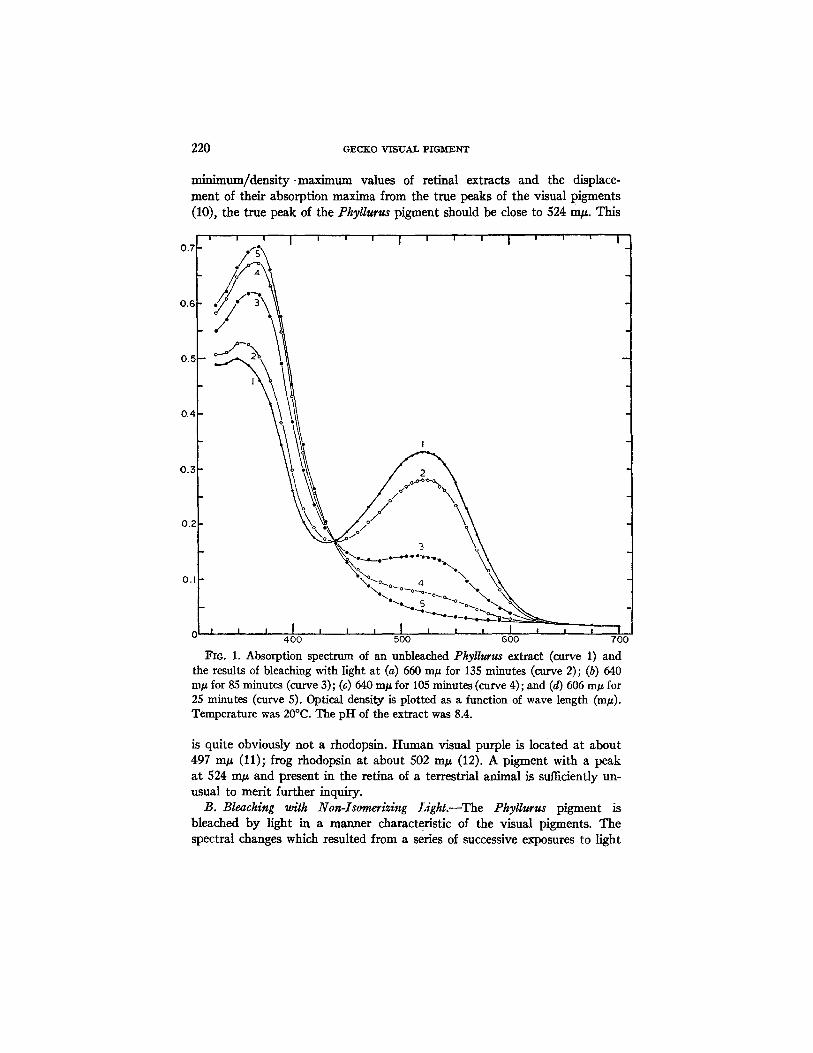
A. Absorption Spectrum of the Unbleached Phyllurus Extract.--A relatively pure retinal extract from this gecko yielded, before bleaching, an absorp- tion spectrum which is typical of solutions of visual pigments (curve 1, Fig. 1). This curve is characterized by an absorption max imum a t 522 m/~, an absorption minimum a t 432 m/~, and a ratio, density min imum/dens i ty max- imum of about 0.50. Assuming, as will later be shown to be true, tha t this extract contained only one photosensitive component, the position of the max imum a t 522 m g is very close to, bu t not identical with, the true peak of the pure pigment. On the basis of the relationship between the density
220 GECKO VISUAL PIGM[ENT
min;mum/density ~maximum values of retinal extracts and the displace- ment of their absorption maxima from the true peaks of the visual pigments (10), the true peak of the Phyllurus pigment should be close to 524 m#. This
0.7
0.6
, , n I n J u I ' ' '
0.~
0.4
0.3
0.2
0. I ~o~o~oI~o e~ o
"".- 5 °"'°~o-
400 500 600 700
Fro. 1. Absorption spectrum of an unbleached Pkyllurus extract (curve 1) and the results of bleaching with light at (a) 650 m# for 135 minutes (curve 2); (b) 640 In# for 85 minutes (curve 3); (c) 640 m/~ for 105 minutes (curve 4); and (d) 605 m/~ for 25 minutes (curve 5). Optical density is plotted as a function of wave length (m/~). Temperature was 20°C. The pH of the extract was 8.4.
is quite obviously not a rhodopsin. Human visual purple is located at about 497 m/z (11); frog rhodopsin at about 502 m/z (12). A pigment with a peak at 524 m/z and present in the retina of a terrestrial animal is sufficiently un- usual to merit further inquiry.
B. Bleaching with Non-Isomerizing Light.--The Phyllurus pigment is bleached by light in a manner characteristic of the visual pigments. The spectral changes which resulted from a series of successive exposures to light
FREDERICK CRESCITELLI 221
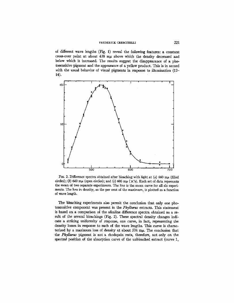
of different wave lengths (Fig. 1) reveal the following features: a constant cross-over point at about 438 In# above which the density decreased and below which it increased. The results suggest the disappearance of a pho- tosensitive pigment and the appearance of a yellow product. This is in accord with the usual behavior of visual pigments in response to illumination (12- 14).
I00
5C • O0
500 600 700
FIO. 2. Difference spectra obtained after bleaching with light at (a) 660 m/~ (filled circles); (b) 640 In# (open circles); and (c) 606 m/~ (×'s). Each set of data represents the mean of two separate experiments. The line is the mean curve for all six experi- ments. The loss in density, as the per cent of the maximum, is plotted as a function of wave length.
The bleaching experiments also permit the conclusion that only one pho- tosensitive component was present in the Pkyllurus extracts. This statement is based on a comparison of the alkaline difference spectra obtained as a re- sult of the several bleachings (Fig. 2). These spectral density changes indi- cate a striking uniformity of response, one curve, in fact, representing the density losses in response to each of the wave lengths. This curve is charac- terized by a maximum loss of density at about 524 In#. The conclusion that the Phyllurus pigment is not a rhodopsin rests, therefore, not only on the spectral position of the absorption curve of the unbleached extract (curve 1,
222 GECKO VISUAL PIGMENT
Fig. 1) but also on the position of the alkaline difference spectrum (Fig. 2). The two methods of analysis yielded identical results.
C. Bleaching with Isomerizing Light.--A 10 minute exposure to white light following the final bleach with non-isomerizing colored light caused a small and selective loss in density, maximal at about 423 m/~ (curve 2, Fig. 3). I t is doubtful, as the results of the hydroxylamine experiment will indicate, whether this change can be interpreted as evidence of a violet-absorbing pigment. Instead this spectral change was probably the result of an isomer- izing action on the products of the preceding bleaches by the short wave length components of the white light.
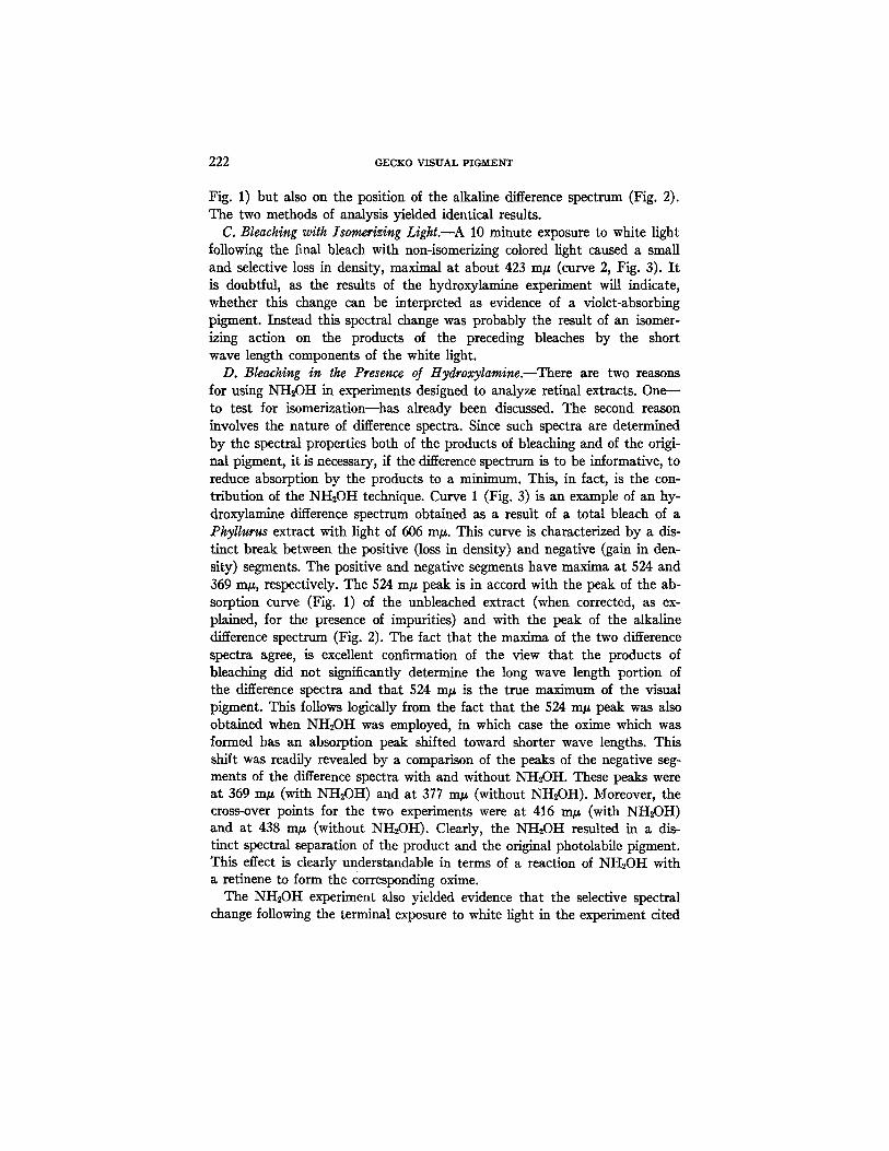
D. Bleaching in the Presence of Hydroxylamine.--There are two reasons for using NH,OH in experiments designed to analyze retinal extracts. One-- to test for isomerization--has already been discussed. The second reason involves the nature of difference spectra. Since such spectra are determined by the spectral properties both of the products of bleaching and of the origi- nal pigment, it is necessary, if the difference spectrum is to be informative, to reduce absorption by the products to a minimum. This, in fact, is the con- tribution of the NH2OH technique. Curve 1 (Fig. 3) is an example of an hy- droxylamine difference spectrum obtained as a result of a total bleach of a PhyUurus extract with light of 606 m#. This curve is characterized by a dis- tinct break between the positive (loss in density) and negative (gain in den- sity) segments. The positive and negative segments have maxima at 524 and 369 m#, respectively. The 524 m/z peak is in accord with the peak of the ab- sorption curve (Fig. 1) of the unbleached extract (when corrected, as ex- plained, for the presence of impurities) and with the peak of the alkaline difference spectrum (Fig. 2). The fact that the maxima of the two difference spectra agree, is excellent confirmation of the view that the products of bleaching did not significantly determine the long wave length portion of the difference spectra and that 524 m/z is the true maximum of the visual pigment. This follows logically from the fact that the 524 m/z peak was also obtained when NH~0H was employed, in which case the oxime which was formed has an absorption peak shifted toward shorter wave lengths. This shift was readily revealed by a comparison of the peaks of the negative seg- ments of the difference spectra with and without NH,OH. These peaks were at 369 m/, (with NH2OH) and at 377 m/~ (without NH,0H). Moreover, the cross-over points for the two experiments were at 416 m/z (with NH,OH) and at 438 m/z (without NH2OH). Clearly, the NH2OH resulted in a dis- tinct spectral separation of the product and the original photolabile pigment. This effect is clearly understandable in terms of a reaction of NH2OH with a retinene to form the Corresponding oxime.
The NHsOH experiment also yielded evidence that the selective spectral change following the terminal exposure to white light in the experiment cited
0.I~
0 .I0
0.05
0.05
0.10
0.15
FREDERICK CRESCITELLI 223
I I I ~ I
O O
, , , I , , , I , , , I , , , f 400 500 600 700
FIG. 3. The NH2OH difference spectrum following a total bleach with light at 606 mlz (curve 1). Curve 2 is the alkaline difference spectrum (without NH2OH) obtained after bleaching with white light following total bleach of the extract with red light. Curve 3 is another experiment similar to that represented by curve 2 ex- cept for the presence of NH2OH in the extract. Ordinate values above the zero indi- cate loss of density; value below zero indicate gain of density. The filled triangles are the spectral densities for a digitonin solution (pH = 8.3) of crystalline aU-trans retinenel to which was added NH2OH to yield the oxime. These points were scaled for plotting with the difference spectrum.
previously (curve 2, Fig. 3) was probably the result of isomerization. The presence of NH2OH in the extract almost entirely abolished the white light effect (curve 3, Fig. 3). In view of this no serious defense can be made of the idea tha t a violet-absorbing pigment was present in the Phyllurus extract.
To summarize the preceding sections, it appears tha t only one photosen- sitive pigment was present as a significant component of the Phyllurus ex-
224 GECKO VISUAL PIGMENT
tract. The absorption spectrum of the relatively pure extract as well as the difference spectra with and without NH~OH agree in showing that this pig- ment is characterized by an absorption maximum at 524 m~ and is, there- fore, not a rhodopsin.
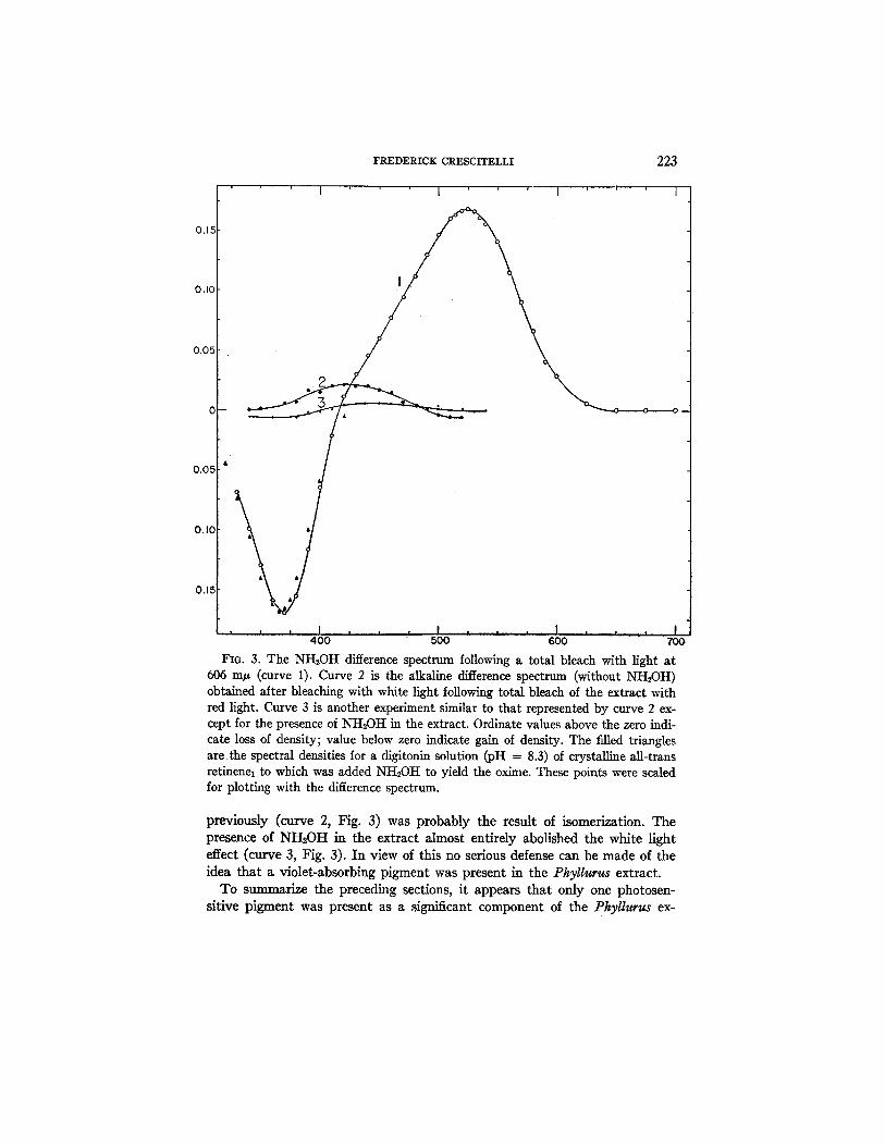
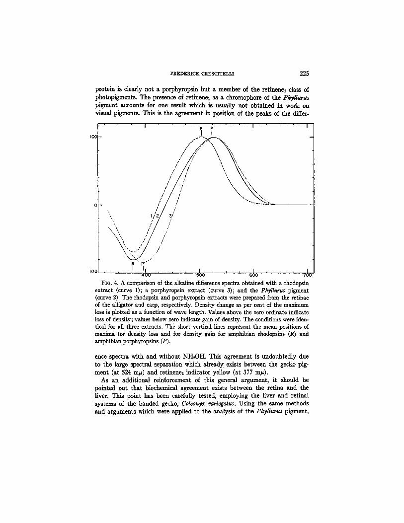
R. Tke Nature of tke Ckromopkore.--The evidence of the preceding sec- tions leads to the conclusion that the PhyUurus chromoprotein is a typical visual pigment with retinene as a prosthetic group. The position at 524 m# raises the obvious question whether this pigment may be a porphyropsin and therefore, a remarkable exception to the generalization that porphyropsin is a retinal constituent of animals with a fresh water habitat (15). The original definition of porphyropsin (16) requires that retinene2 be a part of the chro- moprotein. It is doubtful whether the data on the PkyUurus pigment would conform with this definition. A good indication as to the nature of the chro- mophore is often obtained from the position of the product peaks (indicator yellow and retinene oxime) under conditions of constant pH. The product peaks as determined from the negative difference spectrum occur approxi- mately at 377 m~ without NHsOH (curve 2, Fig. 4 ) a n d at 369 m~ with NH2OH (curve 1, Fig. 3). These figures are to be compared with the corresponding average product peaks--377 and 369 m~---of the rhodopsin system of a number of Amphibia which have been examined in this labora- tory. In contrast, the product peaks of the porphyropsin system in Amphibia (unpublished data from this laboratory) occur at 397 and 383 m~. These comparisons suggest that the Phyllurus pigment, in spite of its position at 524 m~, contains as a carotenoid moiety, not retinene~ but retinenel. This conclusion is reinforced by two further comparisons. The first of these in- volves a comparison, under similar conditions, of the negative difference spectrum in the NH2OH experiment with the absorption spectrum of a reti- nenel oxime (Fig. 3). The retinenel oxime was prepared by adding to a solu- tion of the crystalline all-trans retinenel in 2 per cent digitonin (pH --- 8.3) a small volume of freshly neutralized NH~OH? The absorption curve of this oxime (filled-in triangles), when corrected to scale with the difference spec- trum, strongly suggests that retinene~ oxime was in fact the product of bleaching of the Phyllurus pigment in the NH~OH experiment. In the second comparison (Fig. 4) the alkaline difference spectra obtained after bleaching of a rhodopsin solution (curve 1), a porphyropsin solution (curve 3), and the Phyllurus extract (curve 2) are shown plotted together on a comparable scale. It is clear that, whereas most of the positive section of the Phyllurus difference spectrum is similar to the corresponding portion of the porphyrop- sin, the negative section, which is related to the products of bleaching, re- sembles the corresponding section of the rhodopsin. The Phyllurus chromo-
2 The crystalline retinene was kindly provided by Dr. Grove Baxter of Distillation Products Industries.
FREDERICK CRESCITELLI 225
protein is clearly not a porphyropsin but a member of the retinenel class of photopigments. The presence of retinenel as a chromophore of the Phyllurgs pigment accounts for one result which is usually not obtained in work on visual pigments. This is the agreement in position of the peaks of the differ-
IOC -
/ "..
/ °~'.° I / "..°
/ "%.. t "°%
,, / - ,,, i/
~ I I / '.. / ?
~ I / ~"
ix, i I ~"
, o o , , ,I , , , I , , , , , , 4 0 500 6 0 7 0
I~G. 4. A comparison of the alkaline difference spectra obtained with a rhodopsin extract (curve 1); a porphyropsin extract (curve 3); and the Phyll~rus pigment (curve 2). The rhodopsin and porphyropsin extracts were prepared from the retinae of the alligator and carp, respectively. Density change as per cent of the maximum loss is plotted as a function of wave length. Values above the zero ordinate indicate loss of density; values below zero indicate gain of density. The conditions were iden- tical for all three extracts. The short vertical lines represent the mean positions of maxim& for density loss and for density gain for amphibian rhodopsins (R) and amphibian porphyropsins (P).
ence spectra with and without NH~OH. This agreement is undoubtedly due to the large spectral separation which already exists between the gecko pig- ment (at 524 rap) and retinenel indicator yellow (at 377 mp).
As an additional reinforcement of this general argument, it should be pointed out that biochemical agreement exists between the retina and the liver. This point has been carduUy tested, employing the liver and retinal systems of the banded gecko, Coleonyx variegatus. Using the same methods and arguments which were applied to the analysis of the Phyllurus pigment,
226 GECKO VISUAL PIGMENT
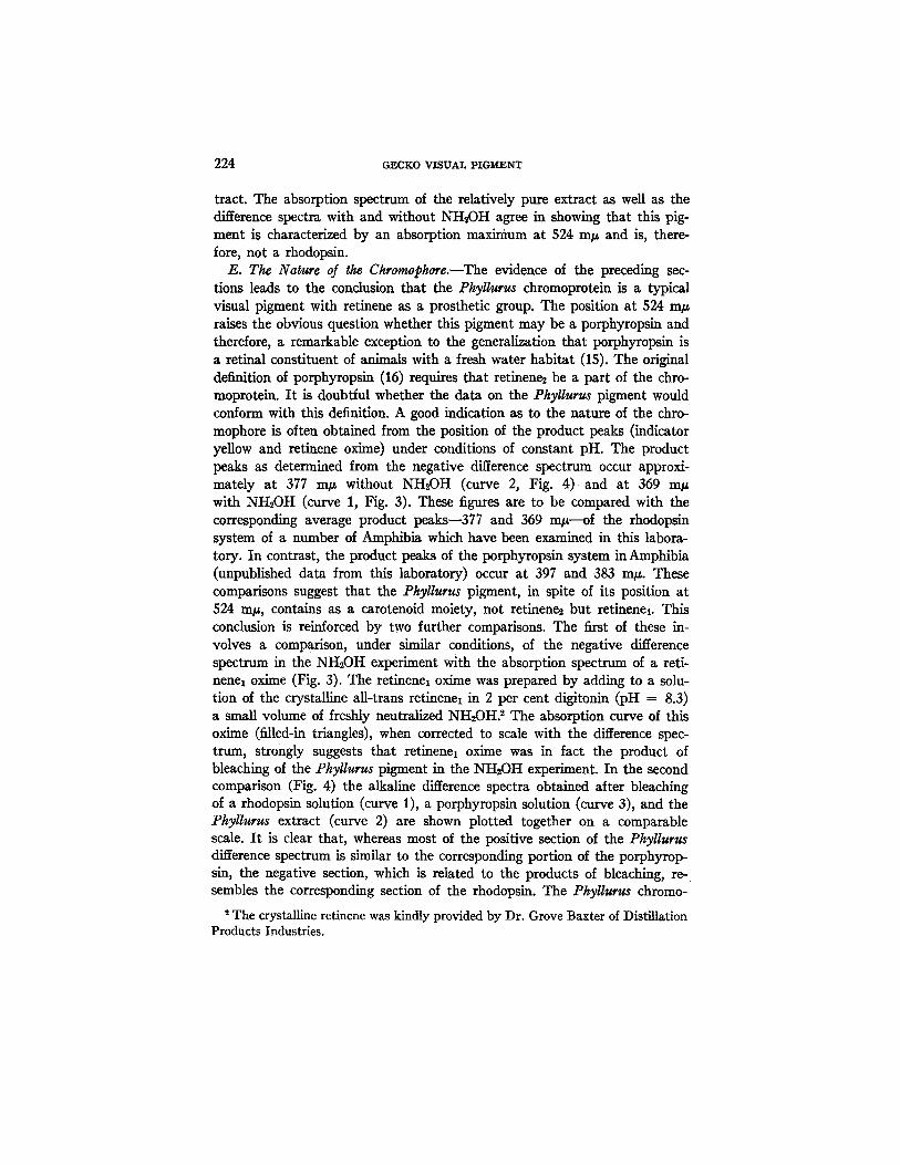
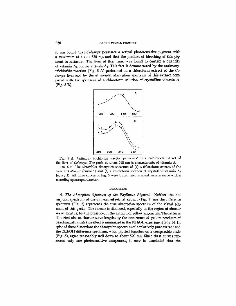
it was found that Coleonyx possesses a retinal photosensitive pigment with a maximum at about 520 m~ and that the product of bleaching of this pig- ment is retinenex. The liver of this lizard was found to contain a quantity of vitamin AI but no vitamin As. This fact is demonstrated by the antimony- trichloride reaction (Fig. $ A) performed on a chloroform extract of the Co- leonyx liver and by the ultraviolet absorption spectrum of this extract com- pared with the spectrum of a chloroform solution of crystalline vitamin A1 (Fig. 5 B).
| .~,.f..........~: A
, . f - L ' j
560 600 640 680
B
i
"'. i'"%. J °°o°°..,.."
260 3 0 0 3 4 0 380
FIG. 5 A. Antimony trichloride reaction performed on a chloroform extract of the liver of Coleonyx. The peak at about 618 m~ is characteristic of vitamin At.
Fro. 5 B. The ultraviolet absorption spectrum of (a) a chloroform extract of the liver of Coleonyx (curve 1) and (b) a chloroform solution of crystalline vitamin A~ (curve 2). All three curves of Fig. 5 were traced from original records made with a recording spectrophotometer.
DISCUSSION
A. The Absorption Spectrum of the Pkyllurus Pigmenl.--Neither the ab- sorption spectrum of the unbleached retinal extract (Fig. 1) nor the difference spectrum (Fig. 2) represents the true absorption spectrum of the visual pig- ment of this gecko. The former is distorted, especially in the region of shorter wave lengths, by the presence, in the extract, of yellow impurities. The latter is distorted also at shorter wave lengths by the occurrence of yellow products of beaching, although this effect is minimized in the NH~OH experiment (Fig. 3). In spite of these distortions the absorption spectrum of a relatively pure extract and the NH:OH difference spectrum, when plotted together on a comparable scale (Fig. 6), agree reasonably well down to about 520 m#. Since these curves rep- resent only one photosensitive component, it may be concluded that the
F R E D E R I C K CRESCITELLI 227
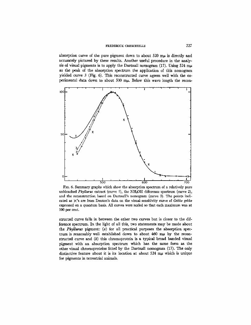
absorption curve of the pure pigment down to about 520 m/z is directly and accurately pictured by these results. Another useful procedure in the analy- sis of visual pigments is to apply the Dartnall nomogram (17). Using 524 m/z as the peak of the absorption spectrum the application of this nomogram yielded curve 3 (Fig. 6). This reconstructed curve agrees well with the ex- perimental data down to about 500 m/~. Below this wave length the recon-
!
50
3."* t • /
500 600 700
FIo. 6. Summary graphs which show the absorption spectrum of a relatively pure unbleached Phytlurus extract (curve 1), the NH20H difference spectrum (curve 2), and the reconstruction based on Dartnall's nomogram (curve 3). The points indi- cated as X's are from Denton's data on the visual sensitivity curve of Gekko gekka expressed on a quantum basis. All curves were scaled so that each maximum was at 100 per cent.
structed curve falls in between the other two curves but is closer to the dif- ference spectrum. In the light of all this, two statements may be made about the Phyllurus pigment: (a) for all practical purposes the absorption spec- trum is reasonably well established down to about 460 m~ by the recon- structed curve and (b) this chromoprotein is a typical broad banded visual pigment with an absorption spectrum which has the same form as the other visual chromoproteins fitted by the Dartnall nomogram (17). The only distinctive feature about it is its location at about 524 m/z which is unique for pigments in terrestrial animals.
228 GECKO VISUAL P I G M E N T
B. Physiological Correlations.--It is now possible to correlate the justified portions of the absorption curves (Fig. 6) with Denton's spectral sensitivity data. Through the kindness of Dr. Denton, who made his data available to the writer, this correlation is shown in Fig. 6. The points marked with X's represent the sensitivity data. There is no reasonable doubt that the physio- logical and biochemical data are in accord. This agreement leads to two mu- tually related conclusions: (a) the visual sensitivity function of the gecko is satisfactorily explained in terms of the retinal pigment and (b) the Phil- lurus chromoprotein is undoubtedly a visual pigment and is, therefore, of physiological interest.
C. The Visual Pigment of Other Geckos.--The general description of the PhyUurus pigment as given here is not unique for this species alone. The reti- nal extracts of six other species of nocturnal geckos were examined by the methods applied to the PhyUurus preparations. Though differences were noted in the spectral positions of the pigments from the different species, the two notable characteristics of the Phyllurus visual protein--location in the region, 518 to 528 m#, and the presence of retinenez as a chromophore --also apply to the pigments from the other geckos. I t is likely that these two characteristics also apply to other nocturnal geckos and to Gekko gekko, the species employed by Denton. I t should be emphasized, however, that when applied to geckos as a whole, generalizations cannot yet be made. The geckos are a diverse group of animals and this is especially true from an oph- thalmological point of view. Thus far only an insignificant number of genera have been examined and one important family, the Sphaerodactylidae, is missing from the results.
D. Tke Visual Pigment of Other Reptiles.--A few attempts have been made already to extract, and to identify, the visual pigments of reptiles (18, 19). These attempts have led to little success probably because of the presence of these proteins in such low concentrations in these cone-rich retinae. By a direct visual method Walls (20) noted the occurrence of a photolabile pig- ment in the retinae of several reptiles. He referred to it as visual purple. The finding of an unusual pigment in geckos raises the question whether a unique pigment, and not rhodopsin, is also a retinal constituent of other reptilian groups. Using the present methods, the successful extraction and identifica- tion of visual pigments have been accomplished in the case of two reptiles: the alligator (Alligator mississippiensls) and the Pacific rattlesnake (Crotalus viridis heUeri), a In both cases rhodopsins were found. The difference spectrum of the alligator pigment is reproduced in Fig. 4 (curve 1). A similar difference spectrum was obtained as the result of bleaching the retinal extract of the rattlesnake. The gecko results need not be extrapolated to apply to other
8 Dr. R. B. Cowles kindly provided the rattlesnakes and was most helpful in the risky experiment of handling these animals in the dark room.
FREDERICK CRESCITELLI 229
reptiles. In the light of present information geckos appear to be unique among terrestrial animals in the possession of an atypical pigment system.
1~. The Biological Signif~anc, e of the Gecko Pigment.--A discussion of the significance of this unusual visual chromopmtein involves phylogenetic con- siderations. The basis for this statement is the transmutation theory of Walls (3, 4) which has received support from the studies of Underwood (5). Ac- cording to this concept the ancestral gecko was a diurnal lizard with retinal cones, In the course of evolution a transmutation of cones to rods occurred in association with the development of the secretive, nocturnal habit. At some time during this evolution the high concentration of visual pigment in the outer segments is assumed to have developed. Underwood's interesting study of retinal cytology has brought to light a number of transition forms among living genera of geckos which support the transmutation theory in a convincing manner. Perhaps the most unexpected support for Walls' theory, in so far as geckos are concerned, is the finding by Crozier and "Wolf (21) that the critical fusion frequency contour of the gecko, Sphe~roda~tyl~s in- aquae Noble and Klingel, is similar to that obtained from Pseudemys, a turtle with a predominantly pure cone retina. Except for these few studies the transmutation theory has stood almost alone while overwhelming support has been marshalled in favor of the duplicity theory and no one, except for Walls' qualification about nomenclature, already referred to,' seems to have questioned the view that the gecko visual pigment is anything but a typical rhodopsin. The present results, demonstrating, as they do, that this view is untenable, may be interpreted as supporting the transmutation theory. The so called rods of nocturnal geckos are characterized biochemically by a pig- ment of the retinenel class but one which is roughly intermediate in spectral position between the other two retinenel chromoproteins: the typical rod con- stituent, rhodopsin and the cone pigment, iodopsin. Is it possible that this arrangement is of evolutionary significance possibly representing substances which had their origin from a common ancestral retinenet pigment?
The data for the seven, species of geckos from which retinal extracts were prepared suggest that the visual pigments are not spectroscopically identical in all these lizards. Minor, but significant, variations appear to exist in the position of the pigment from each of the seven species. It remains to be dis- covered whether a systematic, taxonomically significant variation occurs in the pigment system of geckos as a whole. Underwood (5) has recently pro- posed a classification of geckos into three families: (a) Eublepharidae, (b) Sphaerodactylidae, and (c) Gekkonidae. Transmutation is supposed to have occurred independently in these three gecko stocks. It will be of interest in future work to inquire whether the evolution of the visual pigment also oc- curred independently in these three families or whether the biochemical pat- tern was fixed in the common ancestral form. It would not be too surprising
230 GECKO VISUAL PIGMENT
to discover in future work, gecko pigments in several other spectral positions and all members of the retinenel system. Even typical rhodopsin may turn out to be present in some species. It will be of special interest to determine which, if any, visual pigment can be extracted from the retinae of those diur- nal geckos whose retinal cells consist of cones presumably derived tertlarily from rods. The geckos appear to offer fruitful prospects for future physio- logical and biological queries.
F. The Matter of Noz~/a~ure.--The discovery of a visual pigment at the position of porphyropsin yet containing retinenel, raises the question of a proper nomenclature for this group of pigments. The classical system of describing them according to their color (rhodopsin, porphyropsin, iodopsin, or visual purple, visual violet) is obviously insufficiently informative in the case of the Phyllur~ pigment which has the same color as the porphyropsins but which differs from them in the nature of its chromophore. The same ob- jection can be made to Dartnall's system (22) of naming each pigment nu- mericaUy according to the spectral location of the absorption maximum. There are several alternative methods which might be adopted including modification of the present systems by the addition of the subscript 1 or 2 according to whether retinenel or retinene2 is obtainable from the products of bleaching. In view of the possibility of further discoveries in this field the present author is not yet prepared to offer suggestions for a definitive system of nomenclature even though it is clear that present systems are unsatisfac- tory in a number of respects.
SI.rMrM~Ry
Retln~l extracts of the Australian gecko, Phyllurus railii (White), have revealed the presence of a photosensitive pigment, unusual for terrestrial animals, because of its absorption maximum at 524 m/~. This pigment has an absorption spectrum which is identical in form with that of other visual chromoproteins. It is not a porphyropsin, for bleaching revealed the presence, not of retinene2, but of retinenel as a chromophore. Photolabile pigments with characteristics similar to those of the Phyllur~.s visual pigment were also detected in retinal extracts of six other species of nocturnal geckos.
The presence of this retinal chromoprotein adequately accounts for the unusual visual sensitivity curve described by Denton for the nocturnal gecko. This pigment may have special biological significance in terms of the unique phylogenetic position of geckos as living representatives of nocturnal animals which retain some of the characteristics of their diurnal ancestors. The occur- rence of this retinenel pigment, intermediate in spectral position between rhodopsin and iodopsin, is interpreted in support of the transmutation theory of Walls. The results and interpretation of this investigation point up the fact that, from a phylogenetic point of view, too great an emphasis on the
FREDERICK CKESCITELLI 231
duplicity theory may serve to detract attention from the evolutionary history of the retina and the essential unltarianism of the visual cells.
REFERENCES
1. Denton, E. J., The spectral sensitivity of a nocturnal gecko, Abstr. Communi- cations, 19th International Physiological Congress, 1953, 306.
2. Detwiler, S. R., Studies on the retina. An experimental study of the gecko retina, J. Comp. Neurol., 1923, 36, 125.
3. Walls, G. L., The reptilian retina, Am. J. Opl~h., 1934, 17, 892. 4. Walls, G. L., The visual cells and their history, Biol. Syrup. Visual Mechanisms,
1942, 7, 203. 5. Underwood, G., On the classification and evolution of geckos, Proc. Zool. Sot.,
London, 1954, 124, 469. 6. Krause, A. C., Visual purple, Tabulae Biologicae, 1951, 22, 200. 7. Crescitelli, F., The nature of the lamprey visual pigment, Y. Gen. Physiol., 1956,
39, 423. 8. Hubbard, R., and Wald, G., Cis-trans isomers of vitamin A and retinene in the
rhodopsin system, J. Gon. Physiol., 1952, 86, 259. 9. Wald, G., and Brown, P. K., The molar extinction of rhodopsin, J. Gen. Physiol.,
1953, 37, 189. 10. Crescitel]i, F., and Dartnall, H. J. A., A photosensitive pigment of the carp
retina, J. Physiol., 1954, 125, 607. 11. Crescitelli, F., and Dartnall, H. J. A., Human visual purple, Nature, 1953, 172,
195. 12. Lythogoe, R. J., The absorption spectra of visual purple and of indic&tor yellow,
J. Physiol., 1937, 89, 331. 13. Lythgoe, R. J., and Quilliam, J. P., The relation of transient orange to visual
purple and indicator yellow, J. Physiol., 1938, 94, 399. 14. Wald, G., On thodopsin in solution, J. G6m. Physiol., 1938, 21, 795. 15. Wald, G., The photoreceptor process in vision, Am. J. Ophth., 1955, 40, 18. 16. Wald, G., The porphyropsin visual system, J. Gan. Physiol, 1939, 22, 775. 17. Dartnall, H. J. A., The interpretation of spectral sensitivity curves, Brit. Med.
Bug., 1953, 9, 24. 18. Bliss, A. F., The chemistry of daylight vision, J. G~. Physiol, 1946, 29, 277. 19. Wald, G., Brown, P. K., and Smith, P. H., Cyanopsin, a new pigment of cone
vision, Science, 1953, ].18, 505. 20. Walls, G. L., Visual purple in snakes, Science, 1932, 75, 467. 21. Crozier, W. J., and Wolf, E., The flicker response contour for the gecko (rod
retina), J. Gen. Physiol., 1939, 22, 555. 22. Dartnall, H. J. A., Visual pigment 467, a photosensitive pigment present in