Gellan gum is a polysaccharide manufacturedby microbial fermentation of the Sphingomonas paucimobilismicroorganism, being commonly used in the food andpharmaceutical industry. It can be dissolved in water, andwhen heated and mixed with mono or divalent cations,forms a gel upon lowering the temperature under mildconditions. In this work, gellan gum hydrogels wereanalyzed as cells supports in the context of cartilage regen-eration
Gellan gum: A new biomaterial for cartilage tissue engineering applications J. T. Oliveira, 1,2 L. Martins, 3 R. Picciochi, 1,2 P. B. Malafaya, 1,2 R. A. Sousa, 1,2 N. M. Neves, 1,2 J. F. Mano, 1,2 R. L. Reis 1,2 1 3B’s Research Group - Biomaterials, Biodegradables and Biomimetics, University of Minho, Headquarters of the European Institute of Excellence on Tissue Engineering and Regenerative Medicine, AvePark, 4806-909 Taipas, Guimara ˜es, Portugal 2 IBB - Institute for Biotechnology and Bioengineering, PT Associated Laboratory, Guimara ˜es, Portugal 3 Life and Health Sciences Research Institute, School of Health Sciences, University of Minho, 4710-057 Braga, Portugal Received 26 September 2008; revised 5 May 2009; accepted 6 May 2009 Published online 5 August 2009 in Wiley InterScience (www.interscience.wiley.com). DOI: 10.1002/jbm.a.32574 Abstract: Gellan gum is a polysaccharide manufactured by microbial fermentation of the Sphingomonas paucimobilis microorganism, being commonly used in the food and pharmaceutical industry. It can be dissolved in water, and when heated and mixed with mono or divalent cations, forms a gel upon lowering the temperature under mild conditions. In this work, gellan gum hydrogels were analyzed as cells supports in the context of cartilage regen- eration. Gellan gum hydrogel discs were characterized in terms of mechanical and structural properties. Transmission electron microscopy revealed a quite homo- geneous chain arrangement within the hydrogels matrix, and dynamic mechanical analysis allowed to characterize the hydrogels discs viscoelastic properties upon compres- sion solicitation, being the compressive storage and loss modulus of 40 kPa and 3 kPa, respectively, at a frequency of 1 Hz. Rheological measurements determined the sol-gel transition started to occur at approximately 368C, exhibiting a gelation time of 11 s. Evaluation of the gellan gum hydrogels biological performance was performed using a standard MTS cytotoxicity test, which showed that the leachables released are not deleterious to the cells and hence were noncytotoxic. Gellan gum hydrogels were afterwards used to encapsu- late human nasal chondrocytes (1 3 10 6 cells/mL) and culture them for total periods of 2 weeks. Cells viability was confirmed using confocal calcein AM staining. Histological observations revealed normal chondrocytes morphology and the obtained data supports the claim that this new biomaterial has the potential to serve as a cell support in the field of cartilage regeneration. Ó 2009 Wiley Periodicals, Inc. J Biomed Mater Res 93A: 852–863, 2010 Key words: hydrogel; natural origin; polysaccharide; cartilage; tissue engineering INTRODUCTION Tissue engineering has been proposed as a new method to address problems such as organ failure and tissue regeneration, being widely studied nowa- days as a tool to tackle problems in a diverse range of tissues. 1–4 Such conditions pose serious health problems, being responsible for a decrease in people quality of life. Cartilage is one of the most studied tissues in this field giving the importance it has on mobility and locomotion. Because of its limited capacity for self repair, cartilage becomes an enor- mous constraint to normal everyday life once degen- erated or traumatized. Structures that can provide support for specific cells to develop and generate a functional cartilaginous tissue are an important sub- ject of study. Different types of natural and synthetic biomaterials have been processed using different techniques for this purpose. We are proposing in this work a new biomaterial-Gellan gum to be used in the engineering of cartilaginous tissues, even though its application may not be restricted to this tissue only, as it will be shown by the different 3D structures that can be obtained. Recent work per- formed by Smith et al. has also suggested the use of this biomaterial for tissue engineering applications. 5 Correspondence to: J. T. Oliveira; e-mail: joao.oliveira@ dep.uminho.pt Contract grant sponsor: The Portuguese Foundation for Science and Technology (FCT); contract grant number: SFRH/BD17135/2004 Contract grant sponsor: The European NoE EXPERTIS- SUES; contract grant number: NMP3-CT-2004-500283 Contract grant sponsor: The European Project HIPPO- CRATES; contract grant number: STRP 505758-1 Ó 2009 Wiley Periodicals, Inc.
Transcript
Gellan gum: A new biomaterial for cartilage tissueengineering applications
J. T. Oliveira,1,2 L. Martins,3 R. Picciochi,1,2 P. B. Malafaya,1,2 R. A. Sousa,1,2 N. M. Neves,1,2
J. F. Mano,1,2 R. L. Reis1,213B’s Research Group - Biomaterials, Biodegradables and Biomimetics, University of Minho,Headquarters of the European Institute of Excellence on Tissue Engineering and Regenerative Medicine,AvePark, 4806-909 Taipas, Guimaraes, Portugal2IBB - Institute for Biotechnology and Bioengineering, PT Associated Laboratory, Guimaraes, Portugal3Life and Health Sciences Research Institute, School of Health Sciences, University of Minho, 4710-057 Braga, Portugal
Received 26 September 2008; revised 5 May 2009; accepted 6 May 2009Published online 5 August 2009 in Wiley InterScience (www.interscience.wiley.com). DOI: 10.1002/jbm.a.32574
Abstract: Gellan gum is a polysaccharide manufacturedby microbial fermentation of the Sphingomonas paucimobilismicroorganism, being commonly used in the food andpharmaceutical industry. It can be dissolved in water, andwhen heated and mixed with mono or divalent cations,forms a gel upon lowering the temperature under mildconditions. In this work, gellan gum hydrogels wereanalyzed as cells supports in the context of cartilage regen-eration. Gellan gum hydrogel discs were characterizedin terms of mechanical and structural properties.Transmission electron microscopy revealed a quite homo-geneous chain arrangement within the hydrogels matrix,and dynamic mechanical analysis allowed to characterizethe hydrogels discs viscoelastic properties upon compres-sion solicitation, being the compressive storage and lossmodulus of !40 kPa and 3 kPa, respectively, at afrequency of 1 Hz. Rheological measurements determinedthe sol-gel transition started to occur at approximately
368C, exhibiting a gelation time of !11 s. Evaluation ofthe gellan gum hydrogels biological performance wasperformed using a standard MTS cytotoxicity test,which showed that the leachables released are notdeleterious to the cells and hence were noncytotoxic.Gellan gum hydrogels were afterwards used to encapsu-late human nasal chondrocytes (1 3 106 cells/mL) andculture them for total periods of 2 weeks. Cells viabilitywas confirmed using confocal calcein AM staining.Histological observations revealed normal chondrocytesmorphology and the obtained data supports the claimthat this new biomaterial has the potential to serve as acell support in the field of cartilage regeneration.! 2009 Wiley Periodicals, Inc. J Biomed Mater Res 93A:852–863, 2010
Tissue engineering has been proposed as a newmethod to address problems such as organ failureand tissue regeneration, being widely studied nowa-days as a tool to tackle problems in a diverse rangeof tissues.1–4 Such conditions pose serious healthproblems, being responsible for a decrease in people
quality of life. Cartilage is one of the most studiedtissues in this field giving the importance it has onmobility and locomotion. Because of its limitedcapacity for self repair, cartilage becomes an enor-mous constraint to normal everyday life once degen-erated or traumatized. Structures that can providesupport for specific cells to develop and generate afunctional cartilaginous tissue are an important sub-ject of study. Different types of natural and syntheticbiomaterials have been processed using differenttechniques for this purpose. We are proposing inthis work a new biomaterial-Gellan gum to be usedin the engineering of cartilaginous tissues, eventhough its application may not be restricted to thistissue only, as it will be shown by the different 3Dstructures that can be obtained. Recent work per-formed by Smith et al. has also suggested the use ofthis biomaterial for tissue engineering applications.5
Correspondence to: J. T. Oliveira; e-mail: [email protected] grant sponsor: The Portuguese Foundation for
Science and Technology (FCT); contract grant number:SFRH/BD17135/2004Contract grant sponsor: The European NoE EXPERTIS-
SUES; contract grant number: NMP3-CT-2004-500283Contract grant sponsor: The European Project HIPPO-
CRATES; contract grant number: STRP 505758-1
! 2009 Wiley Periodicals, Inc.
Gellan gum is a linear anionic polysaccharidecomposed of tetrasaccharide (1,3-b-D-glucose, 1,4-b-D-glucuronic acid, 1,4-b-D-glucose, 1,4-a-L-rhamnose)repeating units, containing one carboxyl side group,and was initially described by Moorhouse et al.6,7
This material has a broad use in the food industryand biomedical fields, mostly due to its processinginto transparent gels that are resistant to heat andacid stress. Two gellan gum forms exist, acetylatedand deacetylated, being the latter the most commonand commercially available form. Both form thermor-eversible gels, varying in their mechanical propertiesfrom soft and elastic for the acetylated form to hardand brittle for the fully deacetylated polysaccha-ride.8,9 Gellan gum can form gels in the followingway: at high temperatures, Gellan gum is in the coilform; upon temperature decrease, a thermally-reversible coil to double-helix transition occurs, whichis a prerequisite for gel formation. Afterwards, astructure composed of anti-parallel double helices selfassembled to form oriented bundles, called junctionzones, is formed. Untwined regions of polysaccharidechains, in the form of extended helical chains, link thejunction zones, leading to the formation of a threedimensional network, that creates the gel.10 Thesestructural changes occurring to gellan gum moleculeshave been shown by different techniques. During thecooling process, for example, rheological and differ-ential scanning calorimetry (DSC) studies revealed afirst step increase of loss modulus that corresponds tothe coil-helix transition, and a second step increase ofloss modulus due to sol-gel transition.11 The gelationof gellan gum solutions is strongly influenced by thechemical nature and quantity of cations present in thesolution. The presence of cations is critical when astructurally stable gel is to be prepared.10,12,13 In fact,at low Gellan gum concentrations, the helix formationand its partial aggregation may form an orderedstructure, but this does not lead to gel formationbecause the number of helical aggregates does notgive rise to a continuous network in the wholevolume.11 The main barrier are the carboxyl sidegroups that repulse each other by electrostatic interac-tion, therefore hindering the tight binding of helicesand their cohesive aggregation.9,14–16 The introduc-tion of cations shields the electrostatic repulsion andthereby allows the tight binding and aggregation ofhelices.11,17,18
The gelation properties of Gellan gum are alsoinfluenced by the nature of the cations used, inwhich divalent cations promote the gelation muchmore strongly than monovalent cations.11,12 In mono-valent cations, the gelation is mainly a result of thescreening of the electrostatic repulsion between theionized carboxylate groups on the Gellan gumchains. In the case of divalent cations, the gelationand aggregation of Gellan occurs via a chemical
bonding between divalent cations and two carboxy-late groups belonging to glucuronic acid moleculesin the Gellan chains, in adittion to the screeningeffect.19 It was also suggested that different types ofmono or divalent cations also influenced the visco-elastic behavior of Gellan gum solutions. K" wasmore remarkable than Na", and Ca2" more thanMg2".11 Gellan gum structures have excellent heatresistance properties because the formed junctionsupon gelation can only be unzipped on heating at1208C.11 In the initial state, a junction zone in Gellangum is estimated to be four double helices wide andfive repeat units long, its length being increased toseven repeat units upon annealing.10 In the solidstate, the double helix structure adopted by Gellangum has a similar arrangement to the double helixstructure of iota carrageenan.20 Previous studiesindicate that solutions of deacetylated gellan gumbehave as a pseudoplastic liquids, as evidenced bycreep testing, and have little thixotropy.13 Gellangum advantageous use in the context of biomedicalapplications includes its lack of toxicity, processingunder mild conditions, the ability to used as aninjectable system in a minimally invasive manner,and also the structural similarity it presents withnative cartilage glycosaminoglycans by the presenceof glucuronic acid residues in their repeatingunit.21,22 The presence of this carbohydrate residue,which contains carboxylic groups, may confer addedfunctions to this material. Some intellectual propertyassociated with the application of this material in themedical field has already been disclosed, as its usefor ophthalmologic purposes.23,24
This work tested for the first time gellan gum as anew biomaterial to be used in cartilage regenerationapproaches. As shown here, gellan gum hydrogelsare quite versatile in terms of processing and itsmaterials properties reveal good prospects for theiruse as a cell encapsulating agents. Biological evalua-tion of their cytotoxicity and in vitro culturing ofhuman nasal chondrocytes generated interestingresults indicating that this new biomaterial may playa potential role in cartilage regeneration approaches.
Note: Unless otherwise stated the reagents werepurchased from Sigma-Aldrich.
Gellan gum (G1910, Sigma, St. Louis, MO) wasprocessed in different ways giving rise to various struc-tures, therefore evidencing the versatility of this natural bio-material. The processing involved temperature-dependent
GELLAN GUM FOR CARTILAGE TISSUE ENGINEERING APPLICATIONS 853
Journal of Biomedical Materials Research Part A
and pH-dependent reactions. Regarding gellan gum discsand membranes production, the following methodologywas used. Gellan gum powder was mixed with distilledwater under constant stirring at room temperature to obtaina final concentration of 0.7% (w/v). The solution wasprogressively heated to 908C, under which complete andhomogeneous dispersion of the material was obtained. Thesolution was kept at this temperature during 20–30 min.Afterwards, CaCl2 (Merck, DE) was added to obtain a finalconcentration of 0.03% (w/v) in the gellan gum solutionand the temperature was progressively decreased to 508C.Gellan discs were produced by casting the solution intocylindrical moulds and allowing it to rest at room tempera-ture for 2–5 min and form a solid gel. The discs were thencut using a borer for final discs dimensions of Ø 6 6 0.01mm 3 5.5 6 0.46 mm height. Gellan gum membranes wereproduced by casting the solution into Petri dishes andallowing it to stand at room temperature for 2–5 min andform a solid gel. The Petri dishes were kept in an oven at378C for 90 min. Concerning the production of Gellan gumfibers and particles the methodology was as follows. Gellangum powder was mixed with a NaOH 0.10M solution andstirred at room temperature with a final concentration of4% (w/v). Gellan gum fibers were produced by extrudingthe gellan gum solution into a L-ascorbic acid 20% (v/v)solution under a constant flow rate of 0.2 mL/min, using a21G needle. The gellan gum fibers formed were thenwashed in distilled water, pressed into cylindrical moulds,and dried overnight at 378C. Gellan gum particles wereproduced by extruding the Gellan gum 4% (w/v) solutiondropwise to an L-ascorbic acid 20% (v/v) solution under aconstant flow rate of 0.8 mL/min, using a 21G needle.Gellan gum scaffolds were produced by immersing gellangum 0.7% (w/v) (Ø 6 6 0.01 mm 3 5.5 6 0.46 mm height)discs in liquid nitrogen for 1–2 min and quickly transferringthem to a lyophilizator (Telstar Cryodos-80, Telstar, Spain)where they were lyophilized during 2 days. Lyophilizedgellan gum 0.7% discs were further analyzed under micro-computed tomography (l-CT) using a high-resolution l-CTSkyscan 1072 scanner (Skyscan, Kontich, Belgium) using aresolution of 6.76 lm pixel size and integration time of 1.7ms. The X-ray source was set at 70 keV of energy and 142lA of current. Approximately 500 projections were acquiredover a rotation range of 1808 and a rotation step of 0.458.Data sets were reconstructed using standardized cone-beamreconstruction software (NRecon v1.4.3, SkyScan). The out-put format for each sample was a 500 serial of 1024 3 1024bitmap images. Representative data sets of 150 slices weresegmented into binary images (CT Analyser, v1.5.1.5, Sky-Scan) with a dynamic threshold of 70–255 (gray values) thatwas applied to build the 3D models. 3D virtual models(height 1 mm 3 Ø 3 mm) of representative regions in thebulk of the hydrogels were created, visualized, and regis-tered using image processing software (CT Analyser,v1.5.1.5 and ANT 3D creator, v2.4, both from SkyScan).
Transmission electron microscopy
Gellan gum discs were prepared for transmission mi-croscopy analysis in the following way. Briefly, sections of1 mm3 were fixed in formalin-glutaraldehyde-osmium
tetroxide for 2 h at room temperature and then washed3 times in PBS. Semithin sections (1 lm) were cut fromepon-embedded blocks and stained with toluidine blue.Ultrathin sections (600 A) were cut in a ultratome (ReichertUltranova Leica), mounted onto copper grids, stained withuranyl acetate (7 min) and lead citrate (5 min), andobserved on a Zeiss 902A (50 Kv) electron microscope.
Dynamic mechanical analysis
Dynamic mechanical analysis (DMA) was conducted tocharacterize the mechanical behavior of Gellan gum hydro-gel discs. Gellan gum 0.7% (w/v) discs (Ø 6 6 0.01 mm 35.5 6 0.46 mm height) discs were subjected to compressioncycles of increasing frequencies ranging from 0.1 to 10 Hzwith constant amplitude displacements of 0.1 mm using aTritec 2000 DMA (Triton Technology, UK). Storage andloss modulus were measured and experiments wereconducted at room temperature. The total number of discsper assay were n 5 3. The described values for the com-pression modulus were collected at a frequency of 1 Hz.Statistical analysis was performed using confidence inter-vals based on the experimental results, with a confidencelevel of 99%.
Rheological studies
Cone-Plate rheometry was conducted for gellan gumhydrogels in order to assess their rheological behaviordependence of temperature and time. For this purpose,gellan gum powder was mixed at room temperature withdistilled water at a concentration of 0.7% (w/v) underconstant stirring. The solution was heated to 908C andkept at this temperature for 30 min. Afterwards, CaCl2was added to the Gellan gum solution at concentration of0.03% (w/v) and rheological measurements were per-formed using a controlled stress cone-plate rheometer(Reometer Reologica, StressTech, Sweden). For each mea-surement, a volume of 2 mL of the Gellan gum solutionwas placed in the bottom plate of the rheometer andheld at a constant temperature of 708C. The polymer so-lution was allowed to rest for 1 min before starting theexperiments. Measurements were performed by coolingeach sample from 708C to 258C (at a cooling rate of268C/min) applying a constant shear stress of 0.1 Pa.Temperature, time, shear rate, and viscosity were con-stantly measured. The total number of repeats was n 5 3and confidence intervals were estimated, with a confi-dence level of 99%.
Cytotoxicity evaluation
To assess the possible cytotoxicity of the processedgellan gum hydrogels, MTS (3-(4,5-dimethylthiazol-2-yl)-5(3-carboxymethoxyphenyl)-2(4-sulfofenyl)-2H-tetrazolium)test was used according to ISO/EN 10993 part 5 guide-lines, which determines whether cells are metabolicallyactive.25 This cytotoxicity test is based on the bioreduc-tion of the substrate, 3-(4,5-dimethylthiazol-2-yl)-5(3-
854 OLIVEIRA ET AL.
Journal of Biomedical Materials Research Part A
carboxymethoxyphenyl)-2(4-sulfofenyl)-2H-tetrazolium(MTS) (Cell Titer 961 Aqueous Solution Cell ProliferationAssay, Promega), into a brown formazan product by dehy-drogenase enzymes in metabolically active cells, and iscommonly used for cell viability evaluation. Latex rubberwas used as positive control for cell death, due to its highcytotoxicity to cells, and culture medium was used as anegative control. A rat lung fibroblasts cell line-L929,acquired from the European Collection of Cell Cultures(ECACC), was used for the studies. The cells were grownas monolayers in Dulbecco’s modified Eagle’s medium(DMEM) supplemented with 10% foetal bovine serum (Bio-chrom, Berlin, Germany; Heat Inactivated) and 1% of anti-biotic-antimycotic mixture.
The gellan gum hydrogel discs were incubated inculture medium for 24 h at 378C with constant shaking,as well as latex. Cultured L929 cells were trypsinizedusing trypsin-EDTA (Gibco, Invitrogen Corporation) andplated at a density of 6.6 3 104 cells/well into 96-wellmicrometer plates (200 lL/well). The plates were incu-bated for 24 h at 378C in a humidified atmosphere of5% CO2 in air. Afterwards, the medium was replacedby the extracts previously obtained, using culture me-dium as a negative control. After 72 h, the cell culturewas incubated with MTS (using culturing medium with-out phenol red) for further 3 h at 378C in a humidifiedatmosphere of 5% CO2 in air. Culture medium withMTS was then transferred to new wells. The opticaldensity (OD) which is directly proportional to the cellu-lar activity, being a measure of mitochondrial acitivity,was read on a multiwell microplate reader (Synergy HT,Bio-TeK Instruments) at 490 nm. Statistical analyses wereconducted using a two-sample t-test assuming unequalvariances for n 5 3.
Isolation and expansion of humannasal chondrocytes
Nasal cartilage was harvested from the nasal septumof adult patients (40–65 years) undergoing reconstructivesurgery. This was performed within the scope of a proto-col established with the Hospital de S. Marcos, Braga,Portugal, approved by its Ethical Committee and alwayssampled upon patient informed consent. The humannasal septum cartilage free from all surrounding tissuewas placed in a Petri dish containing sterile phosphatebuffered saline (PBS) and cut into square slices of 5 mmand thickness between 2–3 mm. The pieces were washedin sterile PBS solution, immersed in 20 mL of trypsin-EDTA solution, and incubated for 30 min at 378C on arotator. Trypsin was removed and the pieces washedwith basic DMEM. Then, 20 mL of filter sterilized colla-genase type II solution (2 mg/mL) in basic medium wasadded, and the mixture incubated for !12 h at 378C on arotator. The digested tissue and cell suspension solutionwas centrifuged at 200g for 7 min and the supernatantremoved. The cell pellet was washed with PBS and thecells centrifuged as before. The procedure was repeatedand the cells were ressuspended in PBS and countedusing a hemocytometer. They were again centrifuged, thesupernatant removed, and ressuspended in expansion
medium consisting of Dulbecco’s Modified Eagle’sMedium, containing 10 mM HEPES buffer pH 7.4, 10,000units/mL penicillin/10,000 lg/mL streptomycin, 20 mML-alanyl glutamine, 13 MEM nonessential amino acids,and 10% (v/v) foetal bovine serum (FBS; Biochrom, Berlin,Germany; Heat Inactivated), supplemented with 10 ng/mLbasic fibroblast growth factor (bFGF) (PeproTech, UK).Human nasal chondrocytes were plated into tissue cultureflasks and incubated at 378C in a humidified atmosphere of5% CO2 in air for expansion.26
Human nasal chondrocytes encapsulation in gellangum and agarose hydrogels
Human nasal chondrocytes were expanded until anadequate cell number was obtained for cells encapsulation.Cells were encapsulated at passage 1 in gellan gum hydro-gels and in agarose type VII (A6560; Sigma, St. Louis, MO)hydrogels, the latter being used as controls.
Regarding gellan gum the procedure was the follow-ing. Gellan gum powder was mixed with sterile distilledwater under constant stirring at room temperature toobtain a final concentration of 0.7% (w/v). The solutionwas progressively heated to 908C and kept at this tem-perature for 20–30 min. A sterile CaCl2 solution wasadded to obtain a final concentration of 0.03% (w/v). Thetemperature was progressively decreased to 408C andstabilized at this stage always under constant stirring.Human nasal chondrocytes were detached by trypsiniza-tion, mixed with expansion medium, and centrifuged at200g for 7 min. The supernatant was removed and thecells were ressuspended in warm sterile PBS solution,counted using and hemocytometer, and finally centri-fuged at 200g for 7 min. The supernatant was discardedand the cells pellet kept at the bottom of the falcon tube.The gellan gum 0.7% (w/v) with CaCl2 0.03% (w/v)solution was added to the cells pellet and the mixtureressuspended for complete homogenization of cells withinthe matrix with a final concentration of 1 3 106 cells/mL.Gellan discs with encapsulated human nasal chondrocyteswere produced by casting this mixture into sterile cylin-drical polystyrene moulds, allowing it to rest at roomtemperature for 1–2 min to form a solid gel, and thendiscs of Ø 6 6 0.01 mm 3 5.5 6 0.46 mm height werecut using a borer.
Regarding the agarose hydrogels, the procedure isdetailed elsewhere.27 Briefly, a sterile agarose type VII lowTm 4% (w/v) solution prepared in sterile PBS was heatedto 708C for 30 s, until complete dissolution. The solutionwas stabilized at 408C and added to a human nasal chon-drocytes pellet prepared as described for the gellan gumencapsulation and the mixture ressuspended for completehomogenization of cells within the matrix with a finalconcentration of 1 3 106 cells/mL. Agarose discs withencapsulated human nasal chondrocytes were producedby casting this mixture into sterile cylindrical polystyrenemoulds, allowing it to rest at room temperature for 20 minto form a solid gel, and then discs were cut using a borer.Both gellan gum and agarose hydrogels with encapsulatedcells were cultured for 2 weeks with expansion mediumunder orbital rotation (50 rpm). Afterwards, expansion
GELLAN GUM FOR CARTILAGE TISSUE ENGINEERING APPLICATIONS 855
Journal of Biomedical Materials Research Part A
medium was replaced for 6 weeks by differentiationmedium to promote the formation of a chondrogenic phe-notype. This medium presents the same composition asthe expansion medium except for the bFGF which isreplaced with 1 lg/mL of insulin and 50 lg/mL ofL-ascorbic acid. The cells-hydrogel systems were returnedto the orbital shaker and the culture medium was replacedevery 2–3 days. The experiments were repeated three timesindependently.
Human nasal chondrocytes encapsulated in gellangum and agarose hydrogels: Cell viability tests andhistological analysis
Human nasal chondrocytes morphology in the twohydrogels used, gellan gum and agarose, was observed at2 weeks of culture under optical microscopy. Onerepresentative sample of each type of support wasobserved at different magnifications using an opticalmicroscope (Axiovert 40 CFL, Zeiss).
Cells viability at 2 weeks of culturing was assessedusing calcein AM staining. Calcein AM (C3099, InvitrogenCorp.) is a fluorescence-based method for assaying cell via-bility and cytotoxicity in which the reagent is retained incells that have intact membrane. Briefly, a calcein AM so-lution of 1/1000 was prepared in culture medium. Onedisc of each type of hydrogel with encapsulated humannasal chondrocytes was collected from the culturing platesand incubated in the calcein AM solution for 15–30 min at378C and afterwards washed in sterile PBS. The sampleswere observed under fluorescent microscopy (Zeiss HAL100/HBO 100; Axiocam MRc5 (Zeiss)).
Concerning the histological analysis, hematoxylin-eosinand alcian blue staining were performed on 8 lm thick-ness sections of gellan gum and agarose discs collected at2 weeks of culture using in-house methodology. The discswere fixated by immersion for 30–40 min in glutaralde-hyde 2.5% (v/v) at 48C, and washed in PBS. Histologicalprocessing was performed using Tecnhovit 71001
(Heraeus Kulzer GmbH, DE) and the technical details andprocedure can be found in the commercial package. Sec-tions were cut using a microtome Leica RM2155 (LeicaMicrosystems, Nusslock GmbH).
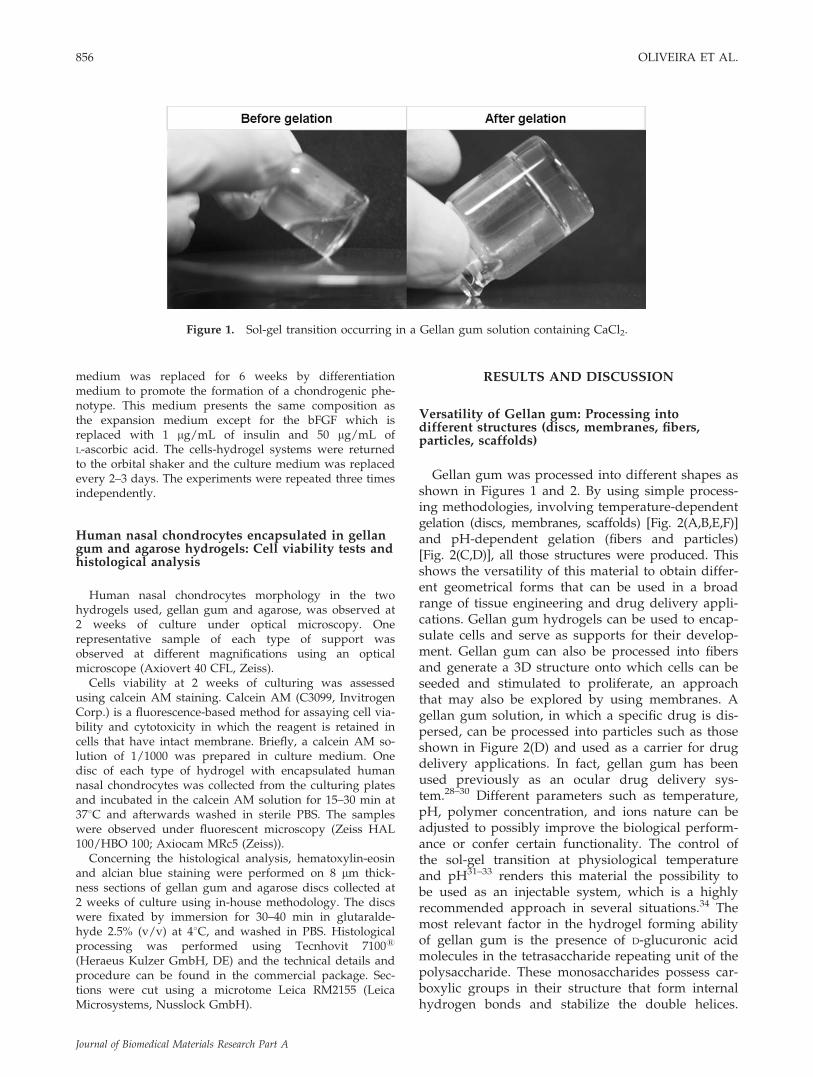
Gellan gum was processed into different shapes asshown in Figures 1 and 2. By using simple process-ing methodologies, involving temperature-dependentgelation (discs, membranes, scaffolds) [Fig. 2(A,B,E,F)]and pH-dependent gelation (fibers and particles)[Fig. 2(C,D)], all those structures were produced. Thisshows the versatility of this material to obtain differ-ent geometrical forms that can be used in a broadrange of tissue engineering and drug delivery appli-cations. Gellan gum hydrogels can be used to encap-sulate cells and serve as supports for their develop-ment. Gellan gum can also be processed into fibersand generate a 3D structure onto which cells can beseeded and stimulated to proliferate, an approachthat may also be explored by using membranes. Agellan gum solution, in which a specific drug is dis-persed, can be processed into particles such as thoseshown in Figure 2(D) and used as a carrier for drugdelivery applications. In fact, gellan gum has beenused previously as an ocular drug delivery sys-tem.28–30 Different parameters such as temperature,pH, polymer concentration, and ions nature can beadjusted to possibly improve the biological perform-ance or confer certain functionality. The control ofthe sol-gel transition at physiological temperatureand pH31–33 renders this material the possibility tobe used as an injectable system, which is a highlyrecommended approach in several situations.34 Themost relevant factor in the hydrogel forming abilityof gellan gum is the presence of D-glucuronic acidmolecules in the tetrasaccharide repeating unit of thepolysaccharide. These monosaccharides possess car-boxylic groups in their structure that form internalhydrogen bonds and stabilize the double helices.
Figure 1. Sol-gel transition occurring in a Gellan gum solution containing CaCl2.
856 OLIVEIRA ET AL.
Journal of Biomedical Materials Research Part A
Nevertheless, carboxyl side groups that repulse eachother by electrostatic interaction, hinder the tightbinding of helices and their cohesive aggregation,affecting the formation of stable gels. The mono ordivalent ions present in the solution play a key rolein this matter. Their presence diminishes the repul-sive energy between the carboxylic groups allowingthe hydrogels to be formed. The variation in pH alsoaffects the solubility of the material being this themain factor in the processing of gellan gum fibersand particles [Fig. 2(C,D)]. At a basic pH, such asthe NaOH solution used in the experiments, the car-boxylic groups present in each D-glucuronic acid res-idue should be in the anionic form, COO2, andtherefore soluble in solution. Once the pH is low-ered, as upon extrusion into an L-ascorbic acid solu-tion, the carboxylic groups become protonated,COOH, and the material turns insoluble.
Transmission electron microscopy
Transmission electron microscopy (TEM) wasperformed to have an insight on the ultrastructuralmorphology of the gellan gum hydrogels (Fig. 3). Asdescribed before for the gel state, gellan gum hydro-gels constitute a matrix where double helices thatoriginated from the coil form rearrangement in solu-tion are widely present and distributed in a ratherhomogeneous fashion.35 These give rise to junctionzones by linking to a neighbor double helical mole-cule. The overall stability of the hydrogel networkderives from the loose ends within the double helicalmolecules. These, together with the cationic antirepulsive effect allow obtaining a stable hydrogelwhen the temperature is decreased below the settingpoint. Previous work has already used TEM as a
Figure 2. The versatility of Gellan gum structures that can be formed using simple polymer processing technologies:(A) discs; (B) membranes; (C) fibers; (D) particles; (E) and (F) 3D lyophilized scaffolds.
GELLAN GUM FOR CARTILAGE TISSUE ENGINEERING APPLICATIONS 857
Journal of Biomedical Materials Research Part A
tool to characterize the ultrastructural properties ofgellan-based hydrogels.36 The authors showedthat gellan gum forms strong gels at low ionicconcentrations, being these highly homogeneous andconstituted by a dense fibrous network structure. Thework presented here confirmed this, being observedthat gellan gum hydrogels provide a uniform matrixat a nanoscale throughout which cells could be encap-sulated in a rather homogeneous way (Fig. 3).
Dynamic mechanical analysis
Living tissues exhibit clear viscolelastic propertiesand therefore it is important to characterize thesolid-state rheological features of materials that aremeant to be in contact with them. DMA has beenused in our group to assess the viscoelastic proper-ties of biomaterials, including natural-based hydro-gels or highly hydrated systems.37–40 In this work,gellan gum hydrogels were analyzed in the wet statethroughout a physiological relevant frequency range.Both the storage (elastic) and loss (viscous) compo-nents of the complex modulus are shown in Figure4. The storage modulus (E0) is about one order ofmagnitude higher than the loss modulus (E@) indi-cating a clear elastic nature of the gel. However, itpossesses some damping capability that may be use-ful to dissipate some cyclic mechanical energy that isimposed in an implantation scenario. Although someincrease in E0 is observed for increasing frequencies,the elastic properties of the biomaterial are quite sta-ble, when compared with the viscous component. Infact, a clear increase in E@ is observed between 0.4and 10 Hz, which suggests that the material exhibitshigher dissipation capability for high frequencies. At
a frequency of 1 Hz, the compression modulus ofthe gels was estimated to be of 38.3 6 6.3 kPa [38.2,38.4 t(0.01,2)] at room temperature. Even though thisvalue is not optimal in terms of mimicking humanarticular cartilage mechanical properties,41 it ishigher or within the range of values found forhydrogels used in similar cartilage regenerativeapproaches.42,43 The gellan gum support is conceivedin this initial work to serve as a cell support due toits features, even though it may be optimized forbeing applied as an injectable system.31–33 Cellsencapsulation and extracellular matrix depositionmay result in progressive increase of the mechanicalproperties of the 3D structures, as shown before forother systems.42
Rheological Studies
Rheological measurements were performed todetermine the temperature range at which the
Figure 3. Transmission electron microscopy micrographof a Gellan gum hydrogel showing a dense and homoge-neous network structure at the ultrastructural level.
Figure 4. Dynamic mechanical analysis of Gellan gumhydrogels showing the storage (E0) and loss (E@) modulusupon compression solicitation using different frequencies.
Figure 5. Rheological measurements of Gellan gum solu-tions. The upper x axis shows the relation between temper-ature and viscosity, while the bottom x axis shows therelation between time-length and viscosity.
858 OLIVEIRA ET AL.
Journal of Biomedical Materials Research Part A
sol-gel transition occurred and the time-scale for gel-ling. Regarding gelation temperature, it is possibleto state from the rheological measurements thatit happens around 378C (36.6 6 0.058C) [36.586,36.588 t(0.01,2)] (Fig. 5).
Concerning the time-scale for gelling, it is possibleto observe from the graph on Figure 5 that it is of!11 s (11.27 6 0.40 s) [11.258, 11.275 t(0.01,2)]. Theresults obtained for both temperature and time ofgelation provide important information concerningsubsequent experiments for cells encapsulation. Thetemperature at which the sol-gel transition occurs,and the overall residence time, is similar to otherhydrogels used for the same purpose.27,44 Gellan gumhydrogels allowed for a homogeneous cell suspensionto be prepared at a temperature above the settingtemperature of the gels. At such temperatures, theviscosity of the solution presents values near to zero,which enable it to be mixed with the cells, ressus-
pended to generate a uniform cells distribution, andthen lower the temperature to allow gel formationand cells entrapment within the newly formed ma-trix. The quick gelling time may be useful in the useof gellan gum as an injectable system that coulddeliver cells through a minimally invasive procedure,although these kinetics can be modified.
Cytotoxicity evaluation
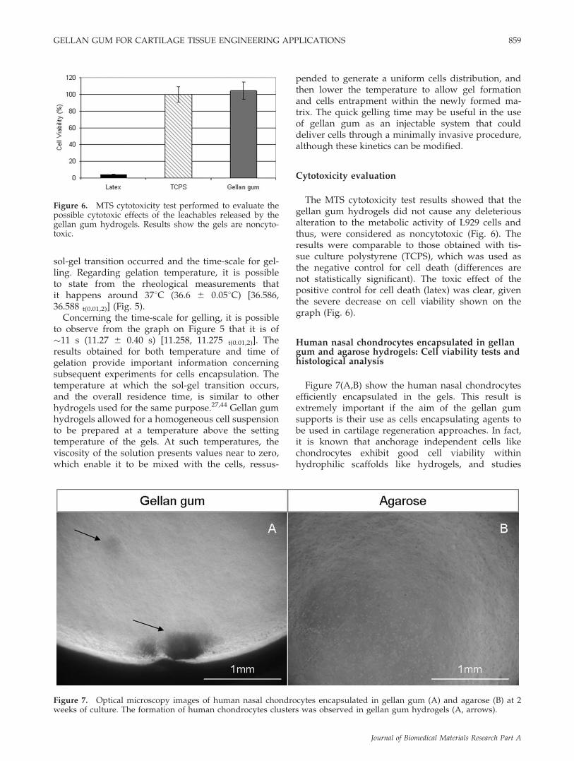
The MTS cytotoxicity test results showed that thegellan gum hydrogels did not cause any deleteriousalteration to the metabolic activity of L929 cells andthus, were considered as noncytotoxic (Fig. 6). Theresults were comparable to those obtained with tis-sue culture polystyrene (TCPS), which was used asthe negative control for cell death (differences arenot statistically significant). The toxic effect of thepositive control for cell death (latex) was clear, giventhe severe decrease on cell viability shown on thegraph (Fig. 6).
Human nasal chondrocytes encapsulated in gellangum and agarose hydrogels: Cell viability tests andhistological analysis
Figure 7(A,B) show the human nasal chondrocytesefficiently encapsulated in the gels. This result isextremely important if the aim of the gellan gumsupports is their use as cells encapsulating agents tobe used in cartilage regeneration approaches. In fact,it is known that anchorage independent cells likechondrocytes exhibit good cell viability withinhydrophilic scaffolds like hydrogels, and studies
Figure 6. MTS cytotoxicity test performed to evaluate thepossible cytotoxic effects of the leachables released by thegellan gum hydrogels. Results show the gels are noncyto-toxic.
Figure 7. Optical microscopy images of human nasal chondrocytes encapsulated in gellan gum (A) and agarose (B) at 2weeks of culture. The formation of human chondrocytes clusters was observed in gellan gum hydrogels (A, arrows).
GELLAN GUM FOR CARTILAGE TISSUE ENGINEERING APPLICATIONS 859
Journal of Biomedical Materials Research Part A
using human nasal chondrocytes revealed that thishydrophilicity facilitated the redifferentiation ofde-differentiated chondrocytes.34 This evidenceopens interesting prospects for the performance ofthese new supports in cartilage regeneration alongwith the rather homogeneous distribution of thechondrocytes throughout the gellan gum hydrogelsmatrix (Fig. 7), which showed a round-shapedmorphology typically present in the native humancartilaginous tissue. An interesting result wasobserved in gellan gum but not in agarose. In thefirst, the formation of chondrocytes clusters was fre-quently observed near 2 weeks of culture [Fig. 7(A)],a feature that was not noticed on the early periodsof culture. Such structures may be indicative of cellproliferation in these clusters which may give a posi-tive contribution towards the production of a hya-line-like cartilage matrix45–47 The cells may use thegellan gum as a source of carbohydrates due to itspolysaccharidic nature, a fact that may be even moreinteresting to study in an in vivo scenario. Thescenario of cells using the matrix as a source ofenergy is possible although such hypothesis demandproper validation testing. Also, the formation of thechondrocytes clusters has been previously describedin the literature as osteoarthritis related events.47,48
This seems however unlikely because no typicallyhypertrophic cells with increased cell size wereobserved, but these assumptions should be con-firmed in further studies.
Calcein AM fluorescence-based method was con-ducted to confirm chondrocytes viability. Resultsfrom samples collected after 2 weeks of culture arepresented on Figure 8 showing the cells were viablein both hydrogels. These are also indicative of theadequacy of gellan gum for cartilage regeneration,
because no apparent difference is noticeable whencompared to agarose hydrogels.
Concerning the histological analysis, it is clearfrom the images that cells distribution within thetwo supports are similar, presenting uniform distri-bution and active states of division (Fig. 9, arrows).This indicates that gellan gum allowed adequatechondrocytes encapsulation while its network matrixpermits cells to encompass active division. More-over, alcian blue staining indicates that proteogly-cans are being deposited to a small extent in thepericellular regions of some groups of cells, whilemaintaining the typical chondrocyte round-shapedphenotype (Fig. 10, arrows).
The data collected so far with gellan gum hydrogelsshowed that they possess suitable materials proper-ties to be used as supports for chondrocytes develop-ment, such as the gelling at physiological conditionsand their tested noncytotoxicity. Furthermore, theywere able to efficiently encapsulate human nasalchondrocytes with a homogeneous distribution andmaintain their viability for at least 2 weeks of culture.The overall data analysis suggests that this newbiomaterial can have a high potential application incartilage regeneration approaches and work isongoing to further corroborate this hypothesis.Another aspect to look into is their potential use forother types of tissues or strategies, given its versatilityin terms of processing and materials properties.
CONCLUSIONS
In this work, gellan gum has been presented as anew biomaterial for cartilage tissue engineering
Figure 8. Calcein AM viability test of human nasal chondrocytes encapsulated in gellan gum (A) and agarose(B) hydrogels at 2 weeks of culture.
860 OLIVEIRA ET AL.
Journal of Biomedical Materials Research Part A
approaches. Gellan gum was shown to be veryversatile in terms of processing, which can becontrolled by both temperature and pH, formingstructures with different shapes using simple polymerprocessing technologies. Discs, membranes, fibers,particles, and scaffolds were produced demonstratingthe range of possible applications for this biomaterial.These may range from cell encapsulation technologiesto drug delivery strategies, for example. An extensivecharacterization of Gellan gum discs to be used as cellsupports indicated that they are suitable for fulfillingsuch functions, since a solution combining nonharshreagents (gellan gum, calcium chloride and water) can
be prepared, mixed uniformly with human nasalchondrocytes, and gelled near the body temperaturein few seconds, enabling a high cell entrapment yieldand homogeneous distribution. Gellan gum hydrogelspresented viscoelastic properties within the range ofother hydrogels used for cells encapsulation42,43 andwere shown to be noncytotoxic. Calcein AM stainingshowed cells were viable during the time of theexperiments. Hematoxylin-eosin staining revealedthat active cells division was occurring and alcian bluestaining indicated that extracellular matrix compo-nents are being deposited to a small extent in somepericellular regions.
Figure 9. Hematoxylin-eosin staining of histological sections of gellan gum (A) and agarose (B) hydrogels at 2 weeks ofculture. Human nasal chondrocytes present a typical round-shaped morphology and active cell division can be observedin both supports (arrows).
Figure 10. Alcian blue staining of histological sections of gellan gum (A) and agarose (B) hydrogels at 2 weeks of culture.Proteoglycans were detected in some pericellular regions in both supports (arrows).
GELLAN GUM FOR CARTILAGE TISSUE ENGINEERING APPLICATIONS 861
Journal of Biomedical Materials Research Part A
The authors would like to thank the patients at Hospitalde S. Marcos, Braga, Portugal for the donation of the bio-logical samples, and the medical staff for their help andsupport. The authors would also like to thank theAdvanced Tissue Analysis Facility (ATAF) at the Instituteof Cellular and Molecular Biology (IBMC) University ofPorto, namely Rui Fernandes, the Institute for Health andLife Sciences (ICVS), University of Minho, Braga, Portugal,for allowing the use of their research facilities, namelyAntonio Salgado; as well as Jorge Teixeira and EvaBarroso at the Department of Polymer Engineering, Cam-pus de Azurem, University of Minho, for their help withthe rheological measurements.
References
1. Salgado AJ, Gomes ME, Chou A, Coutinho OP, Reis RL,Hutmacher DW. Preliminary study on the adhesion and pro-liferation of human osteoblasts on starch-based scaffolds.Mater Sci Eng C-Biomimetic Supramol Syst 2002;20:27–33.
2. Chen G, Sato T, Ohgushi H, Ushida T, Tateishi T, Tanaka J.Culturing of skin fibroblasts in a thin PLGA-collagen hybridmesh. Biomaterials 2005;26:2559–2566.
3. Holmes TC, de Lacalle S, Su X, Liu G, Rich A, Zhang S.Extensive neurite outgrowth and active synapse formation onself-assembling peptide scaffolds. Proc Natl Acad Sci USA2000;97:6728–6733.
5. Smith AM, Shelton RM, Perrie Y, Harris JJ. An initial evalua-tion of gellan gum as a material for tissue engineering appli-cations. J Biomater Appl 2007;22:241–254.
6. Jansson P-E, Lindberg B, Sandford PA. Structural studies ofGellan gum, an extracellular polysaccharide elaborated byPseudomonas elodea. Carbohydr Res 1983;124:135–139.
7. Moorhouse R, Colegrove GT, Sandford PA, Baird JK, Kan KS.PS-60: A new gel-forming polysaccharide. In: Brandt DA,editor. Washington DC: American Chemical Society; 1981.
8. Kang KS, Colegrove T, Veeder GT. US Patent 4326052, 1982.9. Grasdalen H, Smidsrod O. Gelation of Gellan gum. Carbo-
hydr Polym 1987;7:371–393.10. Quinn FX, Hatakeyama T, Yoshida H, Takahashi M, Hata-
keyama H. The conformational properties of Gellan gumhydrogels. Polym Gels Networks 1993;1:93–114.
11. Miyoshi E, Takaya T, Nishinari K. Rheological and thermalstudies of gel-sol transition in Gellan gum aqueous solutions.Carbohydr Polym 1996;30:109–119.
12. Ogawa E, Takahashi R, Yajima H, Nishinari K. Effects ofmolar mass on the coil to helix transition of sodium-typeGellan gums in aqueous solutions. Food Hydrocolloids 2006;20:378–385.
13. Deasy PB, Quigley KJ. Rheological evaluation of deacetylatedgellan gum (Gelrite) for pharmaceutical use. Int J Pharm1991;73:117–123.
14. Crescenzi V, Dentini M, Coviello T, Rizzo R. Comparativeanalysis of the behavior of Gellan gum (S-60) and welan gum(S-130) in dilute aqueous solution. Carbohydr Res 1986;149:425–432.
15. Dentini M, Coviello T, Burchard W, Crescenzi V. Solutionproperties of exocellular microbial polysaccharides. III. Lightscattering from Gellan and from the exocellular polysaccha-ride of Rhizobium trifolii (strain TA-1) in the ordered state.Macromolecules 1988;21:3312–3320.
16. Milas M, Shi X Rinaudo, M. On the physicochemical proper-ties of Gellan gum. Biopolymers 1990;30:451–464.
17. Morris ER, Rees DA, Robinson G. Cation-specific aggregationof carrageenan helices: Domain model of polymer gel struc-ture. J Mol Biol 1980;138:349–362.
18. Nakajima K, Ikehara T, Nishi T. Observation of gellan gumby scanning tunneling microscopy. Carbohydr Polym 1996;30:77–81.
19. Singh BN, Kim KH. Effects of divalent cations on drugencapsulation efficiency of deacylated gellan gum. J Microen-capsul 2005;22:761–771.
20. Arnott S, Scott WE, Rees DA, McNab CGA. i-Carrageenan:Molecular structure and packing of polysaccharide doublehelices in oriented fibres of divalent cation salts. J Mol Biol1974;90:253–256.
21. Ruoslahti E. Proteoglycans in cell regulation. 1989;13369–13372.
22. Jen AC, Wake MC, Mikos AG. Review: Hydrogels for cellimmobilization. Biotechnol Bioeng 1996;50:357–364.
23. Claude Mazuel M-C F. Ophthalmological composition of thetype which undergoes liquid-gel phase transition. US Patent48617, 1986.
24. Tacey VX, Reeve LE, Henry RL. Medical uses of in situformed gels. US Patent 5,318,780, 1993.
25. ISO10993, d., Biological compatibility of medical devices. 5.Test for citotoxicity: In vitro methods, December 1992.
26. Crawford A, Dickinson SC. Methods in Molecular Biology:Biopolymer Methods in Tissue Engineering. Totowa, NJ:Humana Press Inc.; 2004.
28. Mundorf TK, Ogawa T, Naka H, Novack GD, StephensCrockett, R. A 12-month, multicenter, randomized, double-masked, parallel-group comparison of timolol-LA once dailyand timolol maleate ophthalmic solution twice daily in thetreatment of adults with glaucoma or ocular hypertension.Clin Ther 2004;26:541–551.
29. Shedden A, Laurence J, Tipping R. Efficacy and tolerabilityof timolol maleate ophthalmic gel-forming solution versustimolol ophthalmic solution in adults with open-angle glau-coma or ocular hypertension: A six-month, double-masked,multicenter study. Clin Ther 2001;23:440–450.
30. Rozier A, Mazuel C, Grove J, Plazonnet B. Gelrite(R): Anovel, ion-activated, in-situ gelling polymer for ophthalmicvehicles. Effect on bioavailability of timolol. Int J Pharm1989;57:163–168.
31. Kang KS, Veeder GT, Mirrasoul PJ, Kaneko T, Cottrell IW.Agar-Like polysaccharide produced by a Pseudomonas spe-cies-production and basic properties. Appl Environ Microbiol1982;43:1086–1091.
32. Ogawa E, Matsuzawa H, Iwahashi M, Sagara Y, Shioya T,Kimura T. Effects of metal ions on the conformational changeof gellan gum in aqueous solutions. Trans Mater Res Soc Jpn2001;26:613–616.
33. Ogawa EFT, Matsuzawa H, Iwahashi M. Effects of sugars onthe conformational change of Gellan gum in aqueous solu-tions. Trans Mater Res Soc Jpn 2001;26:617–620.
35. Nickerson MT, Paulson AT, Speers RA. Rheological propertiesof Gellan solutions: Effect of calcium ions and temperature onpre-gel formation. Food Hydrocolloids 2003;17:577–583.
36. Amici E, Clark AH, Normand V, Johnson NB. Interpenetrat-ing network formation in agarose-sodium gellan gel compo-sites. Carbohydr Polym 2001;46:383–391.
37. Elvira C, Mano JF, San Roman J, Reis RL. Starch-based biode-gradable hydrogels with potential biomedical applications asdrug delivery systems. Biomaterials 2002;23:1955–1966.
862 OLIVEIRA ET AL.
Journal of Biomedical Materials Research Part A
38. Boesel LF, Mano JF, Reis RL. Optimization of the formulationand mechanical properties of starch based partially degrad-able bone cements. J Mater Sci-Mater Med 2004;15:73–83.
39. Tuzlakoglu K, Alves CM, Mano JF, Reis RL. Production andcharacterization of chitosan fibers and 3-D fiber mesh scaf-folds for tissue engineering applications. Macromol Biosci2004;4:811–819.
40. Silva RM, Silva GA, Coutinho OP, Mano JF, Reis RL.Preparation and characterisation in simulated body condi-tions of glutaraldehyde crosslinked chitosan membranes.J Mater Sci-Mater Med 2004;15:1105–1112.
41. Froimson MI, Ratcliffe A, Gardner TR, Mow VC. Differencesin patellofemoral joint cartilage material properties and theirsignificance to the etiology of cartilage surface fibrillation.Osteoarthritis Cartilage 1997;5:377–386.
42. Kisiday J, Jin M, Kurz B, Hung H, Semino C, Zhang S,Grodzinsky AJ. Self-assembling peptide hydrogel fosterschondrocyte extracellular matrix production and cell division:Implications for cartilage tissue repair. Proc Natl Acad SciUSA 2002;99:9996–10001.
43. Nettles DL, Vail TP, Morgan MT, Grinstaff MW, Setton LA.Photocrosslinkable hyaluronan as a scaffold for articularcartilage repair. Ann Biomed Eng 2004;32:391–397.
44. Hu JC, Athanasiou KA. Low-density cultures of bovine chon-drocytes: Effects of scaffold material and culture system.Biomaterials 2005;26:2001–2012.
45. Schulze T, Schulze-Tanzil G, de S, Souza Pd, Villegas C,Castrejon HV, John John T, Merker Merker HJ, Scheid ScheidA, Shakibaei Shakibaei M. Redifferentiation of dedifferenti-ated human chondrocytes in high-density cultures. Cell Tis-sue Res 2002;308:371–379.
46. Kuettner KE, Pauli BU, Gall G, Memoli VA, Schenk RK.Synthesis of cartilage matrix by mammalian chondrocytesin vitro. I. Isolation, culture characteristics, and morphology.J Cell Biol 1982;93:743–750.
47. Quintavalla J, Uziel-Fusi S, Yin J, Boehnlein E, Pastor G,Blancuzzi V, Singh H, Kraus K, Byrne E Pellas T. Fluores-cently labeled mesenchymal stem cells (MSCs) maintainmultilineage potential and can be detected following im-plantation into articular cartilage defects. Biomaterials 2002;23:109.
48. Mankin HJ, Dorfman H, Lippiello L, Zarins A. Biochemicaland metabolic abnormalities in articular cartilage from osteo-arthritic human hips: II. Correlation of morphology with bio-chemical and metabolic data. J Bone Joint Surg Am 1971;53:523–537.
GELLAN GUM FOR CARTILAGE TISSUE ENGINEERING APPLICATIONS 863


![BIOMATERIAL [XRD and FTIR analysis]nuristianah.lecture.ub.ac.id/files/2016/09/Biomaterial-13.pdf · BIOMATERIAL [XRD and FTIR analysis] ... • Historical retrospective CHAPTER 3:](https://static.documents.pub/doc/80x56/5b0d75de7f8b9a952f8d8c05/biomaterial-xrd-and-ftir-analysis-xrd-and-ftir-analysis-historical-retrospective.jpg)


