47
General signs and symptoms of abdominal diseases Jánoskuti, Lívia

General signs and symptoms of abdominal diseasesabdominal diseases
Jánoskuti, Lívia

Symptoms
• A. Abdominal pain
• B.Vomiting
• C.Gastrointestinal hemorrhage• C.Gastrointestinal hemorrhage
• D.Diarrhea,constipation
• E.Jaundice

Abdominal pain/Origin
• Stretching of a hollow organ or tension in the wall of an organ
• Inflammation• Inflammation• Ischemia• Reffered pain to extraabdominal sites
(sympathetic pathways-spinal sensory neurons also receive input from peripheral nonpain neurons)

Abdominal pain/Patterns
• Visceral-dull poorly localized
• Parietal peritoneum inflammation-intense, well localizedwell localized
• Reffered- superficial, inervated by the same spinal segment

Abdominal pain/Acute

Acute abdominal pain/ Management
• Potential lethal problems- need for prompt surgical or medical intervention
• Rule out extraabdominal causes:
Thorax- pneumonia, inferior myocardial infarction Thorax- pneumonia, inferior myocardial infarction Spine- radiculitis
Genitalia-torsion of the testis
Metabolic causes: uremia,diabetic ketoacidosis,porphyria, lead poisoning
Neurogenic causes: herpes zooster, tabes dorsalis

Abdominal pain/Management
• History, associated symptoms• Observation:restlessness, or immobile• Palpation: tenderness-guarding, rigidity-• Palpation: tenderness-guarding, rigidity-
signs of peritoneal irritation, presence of masses or incarcerated hernias
• Percussion: fluid in the abdomen, bowel distension
• Auscultation:bowel sounds

Abdominal pain/ Management
• Rectal digital examination• Laboratory tests:Ht,wbc,differential,
glucose,bilirubin,electrolytes,BUN,transaminase, amylase,lipase,urinalysis,stool for occult blood or amylase,lipase,urinalysis,stool for occult blood or pus
• Imaging procedures:plain films-free air, intestinal gas pattern, stonesUS, TcHida
endoscopicprocedures

Free abdominal air

Bowel obstruction

Necrotizing colitis

Obstructive uropathy

Gallbladder stones

Acute appendicitis with stone

Acute appendicitis US

Choledochus stone and sludge by US

Pancreas pseudocyst by CT

B.Vomiting-characteristics/1
History• Early morning- pregnancy, uremia,alcoholism• Without nausea- elevated intracranial pressureInspection of vomitus:Inspection of vomitus:• Undigested food- pylorus stenosis• Bile constantly present in large quantities-
obstruction below the ampulla of Vater • Feculent or putrid-distal intestinal obstruction• bloody –upper GI cause

B.Vomiting-associated symptoms/2
Diarrhea-gastroenteritis
Meningism, headache-increased intracranial pressurepressure
Colic – biliary,- kidney stone
Visual disturbance- glaucoma
Confusion-intoxication
Amenorrhoea-pregnancy

Management of vomiting patient
Physical examination
• Signs of hypovolaemia (blood pressure and pulse rate)
• Examination of the abdomen –signs of abdominal diseases
• Neurological examination-consciousness, reflexs, edema of papillae,visual field defectspapillae,visual field defects
Other examinations:
• As at acute abdomen, if you suspect abdominal disease
• Neurologic consultation- brain CT /MR
• Toxicologic examinations

Abdominal pain and nausea, vomiting
• Obstruction
• Motility disorders
• Peritoneal irritation• Peritoneal irritation
• Drugs, gastric mucosa irritants
• Other-intracranial pressure increase, psychogenic,pregnants alcoholics

Case of a 42 years old man/ 1
• 1988.Dg.:Obesity. HLP.IGT.Cholelithiasis
• 1990. After some hours of dinner,abdominal pain and vomiting pain and vomiting
• Physical exam.: distended abdomen, moderate ,diffuse tenderness, dimidished bowel sounds.RD:empty ampulla, brown colored stool.

Case of a 42 years old man/2
• Chest X ray: Fleischner atelectasis
• Plain abdominal: no characteristic abnormalities
• Abd. US: pseudocyst of the pancreas• Abd. US: pseudocyst of the pancreas
• Lab. Values: Sed.,Hb, leukocytes,liver enzymes were normal, Se bi 22 uM/l (n:19) gammaGT 101U/l( n:28), Se amilaz 836U/l (n:121),vizelet amilaz 13376( n:530)
• Dg.Acute pancreatitis.

Case of a 40 years old woman
• Smoker, but never had any illness.• In the morning she squattingly cleaned the stove,
stood up, when acute sharp, epigastric pain appeared.appeared.
• Phys. exam.: distended abdomen, defense in the epigatsrium. No bowel sounds.No liver dullness on percussion. RD: neg.
• Chest and abdominal X ray: free abdominal air • Urgent surgery:perforation of a duodenal ulcer.

Case of a 50 years old man/1
• Moderate obesity, and smoking
• After some hours of dinner epigastric/chest pain and vomiting.pain and vomiting.
• Ambulance doctor did an ECG.

50 years old man, ECG 2

Case of an 50 years old man/3
• Urgent coronary angio:no coronary disease• Transportation to our department.• Physical ex.: fever 37,8 C, moderate abdominal
distension, right upper abdominal tenderness distension, right upper abdominal tenderness • Lab.values: leukocytosis, moderately elevated
Sebi, SGOT GPT, SAP• Abd. US: cholecystolithiasis, inflammed, thick-
walled gall- bladder.• Dg: Acute cholecystitis.

Case of a 65 years old man/1
• Gradually increasing abdominal pain in the last some days.
• At admission tachypnoea.Exsiccosis.No • At admission tachypnoea.Exsiccosis.No abdominal distension, slight diffuse tenderness.No mass, no defense. RD: neg.
• ECG: Sinus tachycardia.
• Blood and urine specimens for lab.
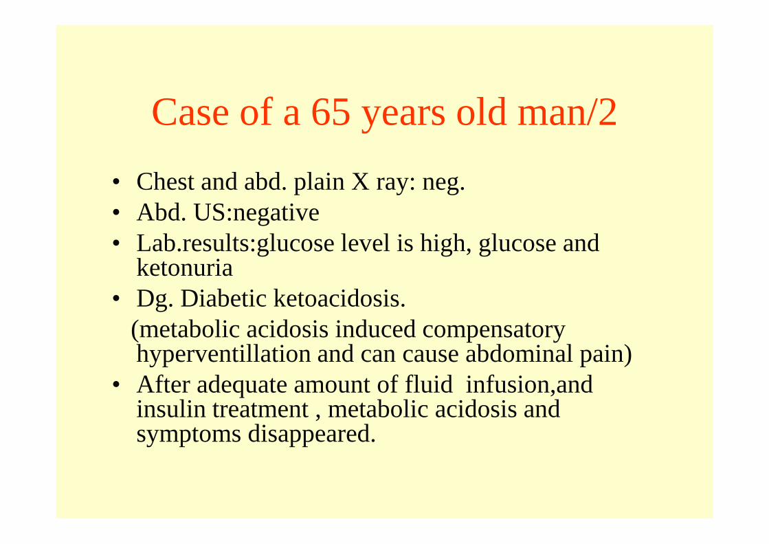
Case of a 65 years old man/2
• Chest and abd. plain X ray: neg.• Abd. US:negative• Lab.results:glucose level is high, glucose and
ketonuriaketonuria• Dg. Diabetic ketoacidosis.
(metabolic acidosis induced compensatory hyperventillation and can cause abdominal pain)
• After adequate amount of fluid infusion,and insulin treatment , metabolic acidosis and symptoms disappeared.

C.Gastrointestinal hemorrhage
• Acute, chronic• Hematemesis:vomiting of bright red blood or
coffee grounds gastric contents-bleeding site is proximal to the lig. of Treitzproximal to the lig. of Treitz
• Melena: passage of black tarry stool-blood loss is greater than 500ml- cause most often upper GI bleeding
• Hematochezia: passage of bright red or maroon-colored stools- cause most often lower GI bleeding

Gastrointestinal hemorrhage/ Causes


Gastric ulcer bleeding

Bleeding from colonic diverticulum

Mesenteric angio.: jejunális dysplasia,bleeding

D./1Diarrhea
• Increase in stool liquidity and weight ( more than 200gm/day)
• Associated with increased stool frequency , • Associated with increased stool frequency , urgency perianal discomfort and/or fecal incontinence

Diarrhea/ Classification

Management of diarrhea
• Acute: -Fluid replacement (electrolytes p os,or iv.) -If no fever,symptomatic th.- loperamid -In most milder cases there is no need for AB -In most milder cases there is no need for AB treatment.-Fever, blood or pus in the stool-culture and antibiotic treatment
• Chronic: stool culture for bacter, parasites, tests for malabsorption/maldigestion,inflammatory bowel diseases,endocrine disease, and tumor.

D.2. Constipation
• Less than two bowel movements a week, less than 50gr/ day
• History: for years/or recent onset, • History: for years/or recent onset, abdominal or defecations pain, stool color, mucus or blood in/on the stool, after constipation spontaneous diarrhea( colonic obstruction), weight loss etc…

Causes of constipationRecent onset: • Colon obstruction- tu, inflammation,strictures, impactation • Sphincter ani spasm- inflammation, fissures,fistulasChronic:• Alimentari causes- decreased dietary fibers and fluids• Alimentari causes- decreased dietary fibers and fluids• Irritable bowel sy. • Drugs, toxins: Ca chan.inhibitors,opiats, iron
drugs,diuretics,lead poissoning...• Endocrine/metabolic: hypothyroidism, hypoK, hyperCa, diab.
mell. ...• Neuromuscular: megacolon, Parkinson disease. Spinal
medullar compression…• Psychiatric: depression, drug, immobility

Management of constipation
• Physical examination::general and focus on the abdomen (tumor), RD.
• Labor.:blood smear and test for occult blood • Labor.:blood smear and test for occult blood in the stool. TSH.
• Imaging: abd. US, ano-sigmoideo-colonoscopy, (irrigoscopia).

D. Jaundice
• Hyperbilirubinemia causes skin and sclera yellow discoloration.
• Other pigments (caroten, urochrom) can cause mainly skin discoloration-overdigestion of carots, pumpkin, orange

Classification of jaundice
• Prehepatic (haemolitic)increased production (indirect bi)
• Hepatocellular • Hepatocellular decrease uptake ,or conjugation
(indirect bi)impaired excretion of conjugated bi
(direct bi)• Posthepatic (obstructive) (direct bi)
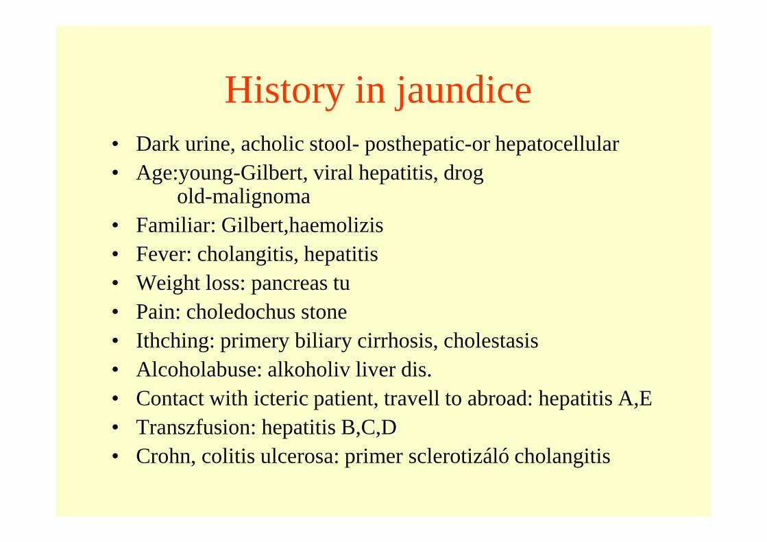
History in jaundice• Dark urine, acholic stool- posthepatic-or hepatocellular• Age:young-Gilbert, viral hepatitis, drog
old-malignoma• Familiar: Gilbert,haemolizis• Fever: cholangitis, hepatitis• Weight loss: pancreas tu• Weight loss: pancreas tu• Pain: choledochus stone• Ithching: primery biliary cirrhosis, cholestasis• Alcoholabuse: alkoholiv liver dis.• Contact with icteric patient, travell to abroad: hepatitis A,E• Transzfusion: hepatitis B,C,D • Crohn, colitis ulcerosa: primer sclerotizáló cholangitis

Management of jaundice
• Rule out posthepatic/obstructive causes!• Urine bilirubin negative-hemolysis
• Urine bilirubin positive (brown • Urine bilirubin positive (brown discoloration) + acholic stool-choledochus obstrucion of the choledochus or Vater papillae

Choledochus stone and sludge by US

Tc-HIDA


