Generation of Micronuclei during Interphase by Coupling between Cytoplasmic Membrane Blebbing and Nuclear Budding Koh-ichi Utani ¤ , Atsushi Okamoto, Noriaki Shimizu* Graduate School of Biosphere Science, Hiroshima University, Higashi-hiroshima, Hiroshima, Japan Abstract Micronucleation, mediated by interphase nuclear budding, has been repeatedly suggested, but the process is still enigmatic. In the present study, we confirmed the previous observation that there are lamin B1-negative micronuclei in addition to the positive ones. A large cytoplasmic bleb was found to frequently entrap lamin B1-negative micronuclei, which were connected to the nucleus by a thin chromatin stalk. At the bottom of the stalk, the nuclear lamin B1 structure appeared broken. Chromatin extrusion through lamina breaks has been referred to as herniation or a blister of the nucleus, and has been observed after the expression of viral proteins. A cell line in which extrachromosomal double minutes and lamin B1 protein were simultaneously visualized in different colors in live cells was established. By using these cells, time- lapse microscopy revealed that cytoplasmic membrane blebbing occurred simultaneously with the extrusion of nuclear content, which generated lamin B1-negative micronuclei during interphase. Furthermore, activation of cytoplasmic membrane blebbing by the addition of fresh serum or camptothecin induced nuclear budding within 1 to 10 minutes, which suggested that blebbing might be the cause of the budding. After the induction of blebbing, the frequency of lamin- negative micronuclei increased. The budding was most frequent during S phase and more efficiently entrapped small extrachromosomal chromatin than the large chromosome arm. Based on these results, we suggest a novel mechanism in which cytoplasmic membrane dynamics pulls the chromatin out of the nucleus through the lamina break. Evidence for such a mechanism was obtained in certain cancer cell lines including human COLO 320 and HeLa. The mechanism could significantly perturb the genome and influence cancer cell phenotypes. Citation: Utani K-i, Okamoto A, Shimizu N (2011) Generation of Micronuclei during Interphase by Coupling between Cytoplasmic Membrane Blebbing and Nuclear Budding. PLoS ONE 6(11): e27233. doi:10.1371/journal.pone.0027233 Editor: Joanna Mary Bridger, Brunel University, United Kingdom Received August 29, 2011; Accepted October 12, 2011; Published November 2, 2011 Copyright: ß 2011 Utani et al. This is an open-access article distributed under the terms of the Creative Commons Attribution License, which permits unrestricted use, distribution, and reproduction in any medium, provided the original author and source are credited. Funding: This work was supported in part by a Grant-in-Aid for Scientific Research (B) [grant number 17370002] and a Grant-in-Aid for Challenging Exploratory Research [grant number 21657051] both from the Japan Society for the Promotion of Science to NS and a Grant-in-Aid for Scientific Research on Priority Areas — Nuclear dynamics [grant number 19038016] from the Ministry of Education, Science, Sports and Culture of Japan to NS. The funders had no role in study design, data collection and analysis, decision to publish, or preparation of the manuscript. Competing Interests: The authors have declared that no competing interests exist. * E-mail: [email protected]¤ Current address: Laboratory of Molecular Pharmacology, National Cancer Institute, National Institutes of Health, Bethesda, Maryland, United States of America Introduction Growing mammalian cells often form secondary nuclei that are smaller than the main nucleus and that are referred to as micronuclei. Usually, micronuclei are generated from acentric chromosomal fragments or malsegregated whole chromosomes after mitosis. Such chromatin is left behind the separating chromosomes during anaphase, and generates micronuclei independently from the main nucleus at the following interphase. Acentric chromosomal fragments may be derived from unrepaired or miss-repaired chromatin after DNA double strand breakage, while malsegregated whole chromosomes can arise from chromosomes that are not bound to the spindle. The latter can occur by several mechanisms including changes in the DNA methylation level at the centromeric region (reviewed in ref. [1]). The malsegregation of chromosomes may also occur when they are merotelically bound to microtubules coming from both spindle poles [2]. In addition, the micronucleus may be formed from the chromatin bridge between segregating sister chromatids, if the bridge breaks at multiple sites during the anaphase to cytokinesis transition [3-5]. Chromatin bridge formation can be caused by the miss-repair of DNA damage, and is involved in the breakage-fusion-bridge (BFB) cycle that destabilizes the chromosome arm and amplifies the genes critical to cancer cell growth [6,7]. The appearance of micronuclei is closely linked to the DNA damage- repair process and genome instability, and monitoring the frequency of micronuclei is therefore widely used to assess the environmental or endogenous stresses that damage the genome and cause cancer (for a review, see the special issue of Mutagenesis, vol 26, no. 1, 2011). Micronuclei are generated not only from chromosomal materials, but also from extrachromosomal elements. Extrachro- mosomal elements called double minutes (DMs) are cytogenetic manifestations of gene amplification that are detected in many human cancer cells. The amplified genes on DMs determine the malignant phenotype of cancer cells; therefore the elimination of DMs from cancer cells results in the loss of malignant phenotypes [8–10]. The elimination of DMs is mediated by their specific incorporation into micronuclei, and the purification of such micro- nuclei yielded highly purified DM DNA [11]. The mechanism of the generation of such DM-type micronuclei is related to the intracellular behavior of DMs during cell cycle progression PLoS ONE | www.plosone.org 1 November 2011 | Volume 6 | Issue 11 | e27233
Transcript
Generation of Micronuclei during Interphase byCoupling between Cytoplasmic Membrane Blebbing andNuclear BuddingKoh-ichi Utani¤, Atsushi Okamoto, Noriaki Shimizu*
Graduate School of Biosphere Science, Hiroshima University, Higashi-hiroshima, Hiroshima, Japan
Abstract
Micronucleation, mediated by interphase nuclear budding, has been repeatedly suggested, but the process is stillenigmatic. In the present study, we confirmed the previous observation that there are lamin B1-negative micronuclei inaddition to the positive ones. A large cytoplasmic bleb was found to frequently entrap lamin B1-negative micronuclei, whichwere connected to the nucleus by a thin chromatin stalk. At the bottom of the stalk, the nuclear lamin B1 structureappeared broken. Chromatin extrusion through lamina breaks has been referred to as herniation or a blister of the nucleus,and has been observed after the expression of viral proteins. A cell line in which extrachromosomal double minutes andlamin B1 protein were simultaneously visualized in different colors in live cells was established. By using these cells, time-lapse microscopy revealed that cytoplasmic membrane blebbing occurred simultaneously with the extrusion of nuclearcontent, which generated lamin B1-negative micronuclei during interphase. Furthermore, activation of cytoplasmicmembrane blebbing by the addition of fresh serum or camptothecin induced nuclear budding within 1 to 10 minutes,which suggested that blebbing might be the cause of the budding. After the induction of blebbing, the frequency of lamin-negative micronuclei increased. The budding was most frequent during S phase and more efficiently entrapped smallextrachromosomal chromatin than the large chromosome arm. Based on these results, we suggest a novel mechanism inwhich cytoplasmic membrane dynamics pulls the chromatin out of the nucleus through the lamina break. Evidence for sucha mechanism was obtained in certain cancer cell lines including human COLO 320 and HeLa. The mechanism couldsignificantly perturb the genome and influence cancer cell phenotypes.
Citation: Utani K-i, Okamoto A, Shimizu N (2011) Generation of Micronuclei during Interphase by Coupling between Cytoplasmic Membrane Blebbing andNuclear Budding. PLoS ONE 6(11): e27233. doi:10.1371/journal.pone.0027233
Editor: Joanna Mary Bridger, Brunel University, United Kingdom
Received August 29, 2011; Accepted October 12, 2011; Published November 2, 2011
Copyright: � 2011 Utani et al. This is an open-access article distributed under the terms of the Creative Commons Attribution License, which permitsunrestricted use, distribution, and reproduction in any medium, provided the original author and source are credited.
Funding: This work was supported in part by a Grant-in-Aid for Scientific Research (B) [grant number 17370002] and a Grant-in-Aid for Challenging ExploratoryResearch [grant number 21657051] both from the Japan Society for the Promotion of Science to NS and a Grant-in-Aid for Scientific Research on Priority Areas —Nuclear dynamics [grant number 19038016] from the Ministry of Education, Science, Sports and Culture of Japan to NS. The funders had no role in study design,data collection and analysis, decision to publish, or preparation of the manuscript.
Competing Interests: The authors have declared that no competing interests exist.
¤ Current address: Laboratory of Molecular Pharmacology, National Cancer Institute, National Institutes of Health, Bethesda, Maryland, United States of America
Introduction
Growing mammalian cells often form secondary nuclei that
are smaller than the main nucleus and that are referred to as
micronuclei. Usually, micronuclei are generated from acentric
chromosomal fragments or malsegregated whole chromosomes after
mitosis. Such chromatin is left behind the separating chromosomes
during anaphase, and generates micronuclei independently from the
main nucleus at the following interphase. Acentric chromosomal
fragments may be derived from unrepaired or miss-repaired
chromatin after DNA double strand breakage, while malsegregated
whole chromosomes can arise from chromosomes that are not
bound to the spindle. The latter can occur by several mechanisms
including changes in the DNA methylation level at the centromeric
region (reviewed in ref. [1]). The malsegregation of chromosomes
may also occur when they are merotelically bound to microtubules
coming from both spindle poles [2]. In addition, the micronucleus
may be formed from the chromatin bridge between segregating sister
chromatids, if the bridge breaks at multiple sites during the anaphase
to cytokinesis transition [3-5]. Chromatin bridge formation can be
caused by the miss-repair of DNA damage, and is involved in the
breakage-fusion-bridge (BFB) cycle that destabilizes the chromosome
arm and amplifies the genes critical to cancer cell growth [6,7]. The
appearance of micronuclei is closely linked to the DNA damage-
repair process and genome instability, and monitoring the frequency
of micronuclei is therefore widely used to assess the environmental or
endogenous stresses that damage the genome and cause cancer (for a
review, see the special issue of Mutagenesis, vol 26, no. 1, 2011).
Micronuclei are generated not only from chromosomal
materials, but also from extrachromosomal elements. Extrachro-
mosomal elements called double minutes (DMs) are cytogenetic
manifestations of gene amplification that are detected in many
human cancer cells. The amplified genes on DMs determine the
malignant phenotype of cancer cells; therefore the elimination of
DMs from cancer cells results in the loss of malignant phenotypes
[8–10]. The elimination of DMs is mediated by their specific
incorporation into micronuclei, and the purification of such micro-
nuclei yielded highly purified DM DNA [11]. The mechanism of
the generation of such DM-type micronuclei is related to the
intracellular behavior of DMs during cell cycle progression
PLoS ONE | www.plosone.org 1 November 2011 | Volume 6 | Issue 11 | e27233
(reviewed in ref. [12,13]). Namely, acentric DMs are segregated to
daughter cells by sticking to the centric chromosome arm during
mitosis [14–16]. These DMs localize to the nuclear periphery
during the G1 phase, and move to the interior during the early S
phase, when DMs themselves are replicated [17,18]. During the
early S phase, the presence of low concentrations of hydroxyurea
(HU) induces DNA damage at the replication site. The damage at
the chromosome arm is repaired rapidly, but difficulties encoun-
tered in the repair of DNA damage at DMs induce their aggregation
[19]. The aggregated DMs lag behind the separating chromosomes
at anaphase and generate DM-type micronuclei. This mechanism
may be applied to a broad spectrum of extrachromosomal elements,
because many kinds of viral nuclear plasmids stick to the
chromosome arm during mitosis (reviewed in ref. [12,15]).
Micronuclei resemble nuclei in structure, and a large portion of
them has nuclear lamina. However, lamin-negative micronuclei
were reported for both chromosome-type [3,20–22] and DM-type
micronuclei [16,22]. The presence or absence of lamin around
micronuclei has important implications for the phenotypes of
cells, because it is correlated with transcription [22] or replication
(Okamoto et al., unpublished data) inside micronuclei. Investiga-
tion of the origin of micronucleus-heterogeneity is therefore an
important task. This heterogeneity could be attributed to
differences in the origin of micronuclei, as chromosome-type
lamin B-positive micronuclei are generated from the anaphase
laggards, while lamin B-negative micronuclei are generated from
the anaphase chromatin bridge [3]. The present report describes
another mechanism involved in the generation of lamin-negative
micronuclei, namely interphase nuclear budding.
In addition to the mitotic generation of micronuclei, the formation
of micronuclei during interphase through nuclear budding has been
repeatedly hypothesized based on the detection of nuclear buds or
protrusions in cytogenetic preparations (reviewed in ref. [23]), and the
close resemblance of some of them to micronuclei, with the exception
of their connection to the nucleus through a chromatin stalk.
Furthermore, a cell-cycle synchronization experiment suggested that
a portion of DM-type micronuclei might be generated through
nuclear budding [24]. The generation of micronuclei through a
budding process in the mammalian nucleus, which is reinforced by
the nuclear lamina, is surprising. Prior work based on the
simultaneous visualization of DMs and the lamin protein did not
support the protrusion of a portion of the nucleus with lamina as a
mechanism for the generation of buds/micronuclei [16]. Instead,
lamin-negative DM aggregates were detected on the outside of the
nuclear lamina in a shape resembling a bud. The present study shows
that the budding structure was generated through a break in the
lamina, which resembled the nuclear blister induced by the HIV Vpr
protein [25] or the herniation induced by the reoviral sigma 1s
protein [26]. Extrachromosomal small DMs frequently localize to the
nuclear periphery [17], and can pass more easily through the break in
the lamina than the large chromosomal arm. Furthermore, time-
lapse live cell imaging of DMs, lamin B1 and DNA revealed that
budding is coupled to cytoplasmic membrane blebbing. Blebbing is
frequently associated with cell locomotion, and is activated during
mitosis or apoptotic cell death. The present study is the first to link
two previously unrelated phenomena, namely the elimination of
nuclear materials and cytoplasmic membrane blebbing.
The human COLO 320DM-GFP cell line, in which LacO-
tagged DMs were visualized by the expression of the LacR-GFP
fusion protein, was previously established (see the methods
section). This cell line enables the detection of DMs without the
use of FISH, which requires heat denaturation and may disrupt
the 3-D structure of the cells. Fixation of these cells by PFA
followed by immunofluorescence-based detection of lamin B1
protein revealed the presence of DM-enriched micronuclei
surrounded by lamina (Figure 1A) and those without lamina
(Figure 1B, C), as previously reported [16,22]. The use of a cell
line bearing visible DMs enabled the application of time-lapse
imaging to examine the formation of DM-enriched micronuclei.
As shown in Figure 1E, the aggregated DMs detached from the
chromosomes during the metaphase to anaphase transition, and
formed micronuclei after the end of mitosis. The time-lapse
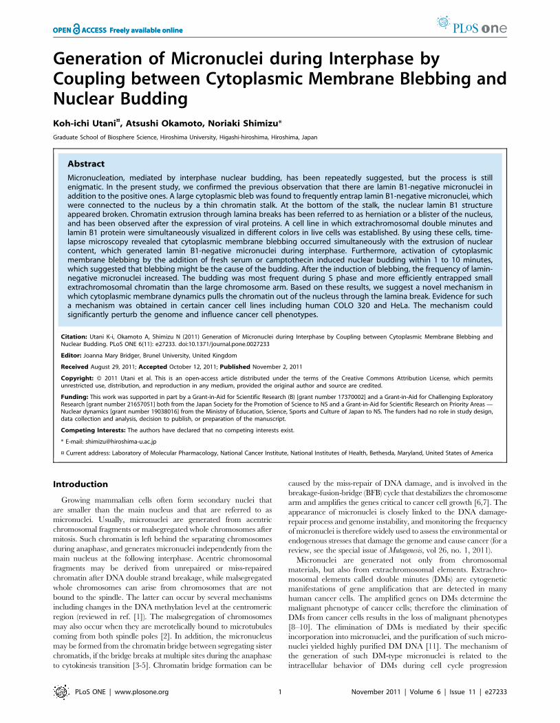
Figure 1. Lamin B1-negative micronuclei and postmitotically-generated lamin B1 positive micronuclei were detected. (A to D)COLO 320DM-GFP cells were fixed with PFA and the lamin B1 protein wasdetected by immunofluorescence. Representative confocal images ofthe DM-type micronucleus with lamin B1 (A; arrow) or without lamin B1(B and C; arrow). The image of lamin B1 is shown in gray-scale, and it wasshown in red in merged panels. The micronucleus in B is attached to thenucleus whereas the one in C is detached from the nucleus. In mitoticcells in telophase, the aggregated DMs were left behind the separatingchromatids, and lamin B1 was detected at the rim (D; arrowheads).(E) Living COLO 320DM-GFP cells were stained with Hoechst 33342 andanalyzed by time-lapse microscopy (E). DM aggregates were locatedseparate from chromosomes at metaphase and anaphase, and generatedthe DM-type micronuclei after mitosis (arrowheads). Elapsed time (inhours:minutes:seconds) is shown in each images.doi:10.1371/journal.pone.0027233.g001
Nuclear Budding and Cytoplasmic Membrane Blebbing
PLoS ONE | www.plosone.org 2 November 2011 | Volume 6 | Issue 11 | e27233
observation confirmed the hypothesis derived from fixed cell
observation that micronuclei arise from aggregated DMs after
mitosis [16,19]. Furthermore, simultaneous visualization of lamin
B1 and DMs in fixed cells showed that micronuclei generated
through this mechanism are surrounded by Lamin B1 (Figure 1D).
Large cytoplasmic blebs are associated with nuclearbudding through breaks in the lamina
Cultured COLO 320DM cells and COLO 320DM-GFP cells did
not spread on the substratum, but rather attached weakly to the
tissue culture coated dish. The surface of these round-shaped cells
had many protrusions, which are commonly referred to as
cytoplasmic membrane blebbing. Imaging of the blebbing of
rounded cells in addition to labeled DMs, lamin B1 and DAPI-
stained DNA by DIC microscopy revealed that, surprisingly, the
large cytoplasmic membrane protrusions entrapped the lamin B1-
negative micronuclei. For simplicity, the large cytoplasmic mem-
brane protrusion is hereafter referred to as a ‘‘large (cytoplasmic)
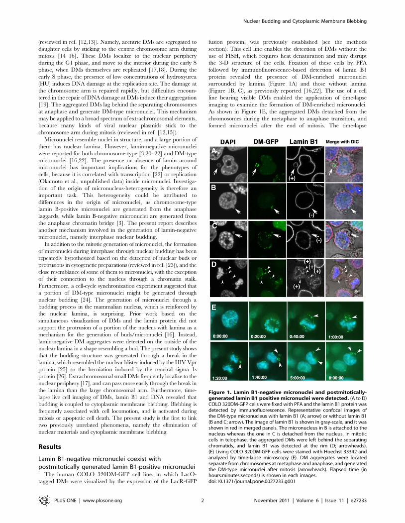
bleb’’. The micronuclei in the large bleb contained highly
concentrated DMs (Figure 2A and B) or DMs with other
chromosomal material (Figure 2C). Notably, a DAPI-stained thin
chromatin stalk connected a portion of this micronucleus to the
nucleus (Figure 2B and C). These types of micronuclei are referred
to as ‘‘the nuclear buds’’ during the micronucleus test (for a review,
see, ref. [27]). The lamin B1 protein was not detected at these
micronuclei, but it showed dense staining at the stalk between the
micronucleus and the nucleus, regardless of whether or not the
chromatin was microscopically visible at the stalk (Figure 2A to C).
There were large buds showing the protrusion of large amounts of
chromatin from the nucleus (Figure 2D), and these buds were
associated with obvious breaks in the nuclear lamina. The
morphology of the nuclear bud was strikingly similar to that of
the phenomenon reported as nuclear herniation [26] and to the
nuclear blister [25], which are induced by viral proteins.
Furthermore, careful observation of the cells by confocal micros-
copy showed that the lamin-negative micronuclei inside the large
cytoplasmic blebs were frequently associated with small lamina
breaks, as shown in Figure 2E. These results indicate that the large
cytoplasmic bleb is correlated with the extrusion of nuclear content
through lamina breaks, which may explain the so-called ‘‘nuclear
budding’’ phenomenon previously described [24,27].
Fresh serum efficiently induced both the largecytoplasmic blebs and the nuclear budding/micronucleation
The frequency of the large cytoplasmic blebs was low among
the logarithmically growing cells (see below). The addition of fresh
serum has been reported to induce cytoplasmic membrane
Figure 2. The large cytoplasmic bleb entraps the nuclear bud/micronucleus that is devoid of lamin B1. (A to D) COLO 320DM-GFP cellswere fixed, and the lamin B1 protein was detected as in Figure 1. Representative confocal images of lamin-negative micronuclei (white arrows)connected to the nucleus by thin chromatin are shown. These micronuclei were inside the large cytoplasmic bleb. (E) The left two panels show themerged images of lamin B1 (red), DMs (green), DAPI (blue in left, gray in right) and DIC (gray in left). The rectangle region was enlarged in the right threepanels showing the serial confocal images taken at 0.8 mm intervals in the Z-axis. The break in the lamin B1 envelope is indicated by a white arrow.doi:10.1371/journal.pone.0027233.g002
Nuclear Budding and Cytoplasmic Membrane Blebbing
PLoS ONE | www.plosone.org 3 November 2011 | Volume 6 | Issue 11 | e27233
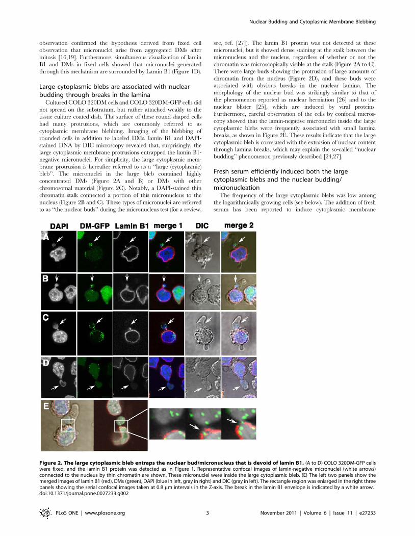
blebbing [28,29]. In the present study, the addition of fresh fetal
calf serum (FCS) to the logarithmically growing cells efficiently
induced the formation of large cytoplasmic blebs in the COLO
320DM-GFP cells within a few minutes (Figure 3A and B), which
was then followed by a decrease in their frequency up to 60 min.
(Figure 3B). The bleb-inducing activity was detected in the fresh
serum, but not in the serum-containing medium used for cell
growth (the ‘‘conditioned medium’’ in Figure 3C). The activity was
associated with the heat-resistant non-dialyzable fraction, and it
was not associated with bovine serum albumin (BSA; Figure 3C).
Because serum induced the large cytoplasmic blebs, the
frequency of micronuclei per large bleb decreased after the serum
addition (Figure 3D). On the other hand, measurement of the
frequency of the large cytoplasmic blebs that entrapped ‘‘the bud-
shaped micronuclei’’, namely those connected to the nucleus by a
thin chromatin stalk, during the course of serum activation
(Figure 3D) showed that the frequency increased until 10 min after
serum addition and declined until 60 min (Figure 3D). As shown
in the graph, these micronuclei with stalks were always lamin B1-
negative, which is shown in the representative image in Figure 2.
Figure 3. Fresh serum induced the formation of large blebs and lamin B1-negative micronuclei. (A) DIC images of living COLO 320DM-GFP cells are shown. The incubation time (min:sec) after the addition of fresh serum is shown in each panel. The arrows indicate the large blebs.(B) The cells were fixed with PFA and the frequency of the large blebs among the total cells was measured. (C) Cells cultured for two days in amedium containing 10% serum rarely showed the large bleb (-; ‘‘before stimulation’’), whereas addition of fresh serum or treated serum at the finalconcentration of 10% induced extensive blebbing (+) after 10 min. (D) Cells were stimulated with fresh serum for the indicated time and fixed fordetection of the lamin B protein. The frequency of the formation of the large bleb containing the micronuclei that were apart from the nuclei or thebud-shaped micronuclei that was connected to the nuclei among the total number of large blebs was calculated in 300 to 1,000 cells. The incidencesamong the counted blebs were noted in the graph. (E) In the same slides used in D, the frequencies of the micronuclei with/without DMs or with/without lamin B1 among the total cells were measured by examining 1,000 cells in three replicates. Error bars represent mean +/- SEM.doi:10.1371/journal.pone.0027233.g003
Nuclear Budding and Cytoplasmic Membrane Blebbing
PLoS ONE | www.plosone.org 4 November 2011 | Volume 6 | Issue 11 | e27233
The frequency of the total intracellular micronuclei was measured
independently of their connection to the nucleus by the stalk or
their entrapment in cytoplasmic blebs (Figure 3E). The results
showed that the frequency of lamin B1-negative micronuclei
increased after fresh serum addition, whereas the frequency of
lamin B1-positive micronuclei did not show a change. This
increase was observed for both DM-type and chromosome-type
(DM-negative) micronuclei. Taken together, these results indicate
that nuclear budding is associated with the formation of
micronuclei during interphase.
Although cytoplasmic membrane blebbing is known to become
active during the initial phase of apoptosis, the present results show
that fresh serum induced blebbing but did not cause the
appearance of apoptotic cells (Figure S1). Therefore, the
cytoplasmic blebbing and nuclear budding induced by fresh
serum were not related to apoptosis.
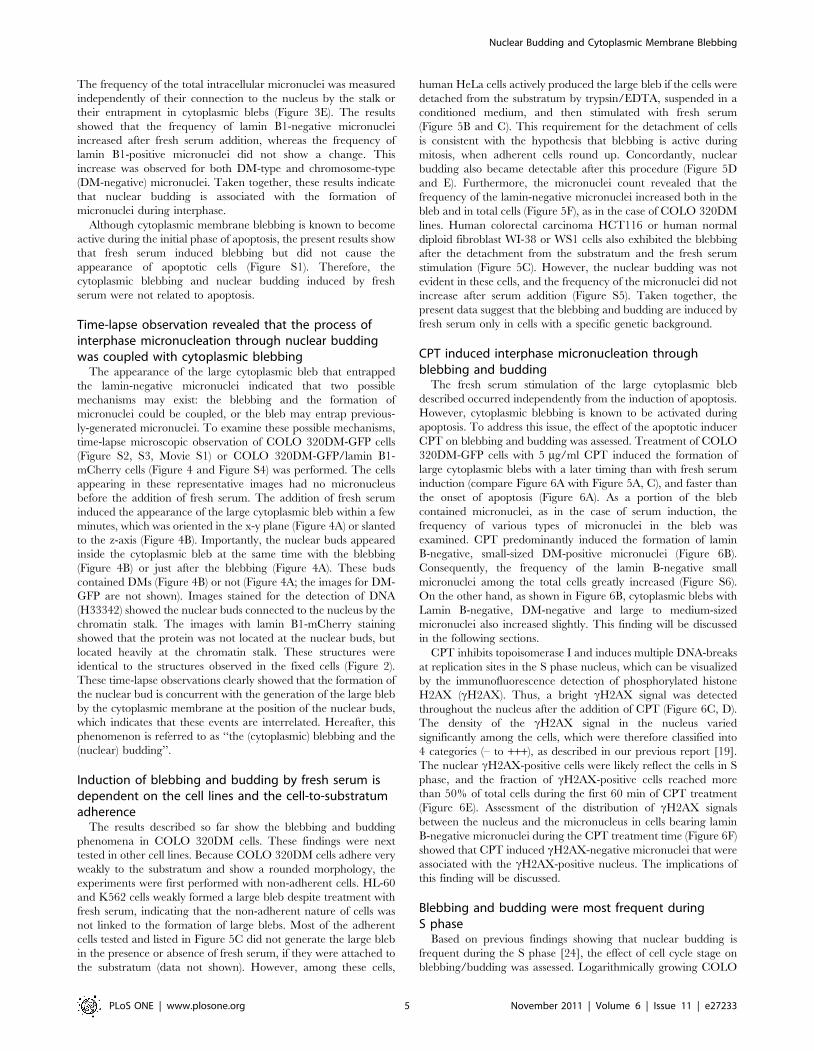
Time-lapse observation revealed that the process ofinterphase micronucleation through nuclear buddingwas coupled with cytoplasmic blebbing
The appearance of the large cytoplasmic bleb that entrapped
the lamin-negative micronuclei indicated that two possible
mechanisms may exist: the blebbing and the formation of
micronuclei could be coupled, or the bleb may entrap previous-
ly-generated micronuclei. To examine these possible mechanisms,
time-lapse microscopic observation of COLO 320DM-GFP cells
(Figure S2, S3, Movie S1) or COLO 320DM-GFP/lamin B1-
mCherry cells (Figure 4 and Figure S4) was performed. The cells
appearing in these representative images had no micronucleus
before the addition of fresh serum. The addition of fresh serum
induced the appearance of the large cytoplasmic bleb within a few
minutes, which was oriented in the x-y plane (Figure 4A) or slanted
to the z-axis (Figure 4B). Importantly, the nuclear buds appeared
inside the cytoplasmic bleb at the same time with the blebbing
(Figure 4B) or just after the blebbing (Figure 4A). These buds
contained DMs (Figure 4B) or not (Figure 4A; the images for DM-
GFP are not shown). Images stained for the detection of DNA
(H33342) showed the nuclear buds connected to the nucleus by the
chromatin stalk. The images with lamin B1-mCherry staining
showed that the protein was not located at the nuclear buds, but
located heavily at the chromatin stalk. These structures were
identical to the structures observed in the fixed cells (Figure 2).
These time-lapse observations clearly showed that the formation of
the nuclear bud is concurrent with the generation of the large bleb
by the cytoplasmic membrane at the position of the nuclear buds,
which indicates that these events are interrelated. Hereafter, this
phenomenon is referred to as ‘‘the (cytoplasmic) blebbing and the
(nuclear) budding’’.
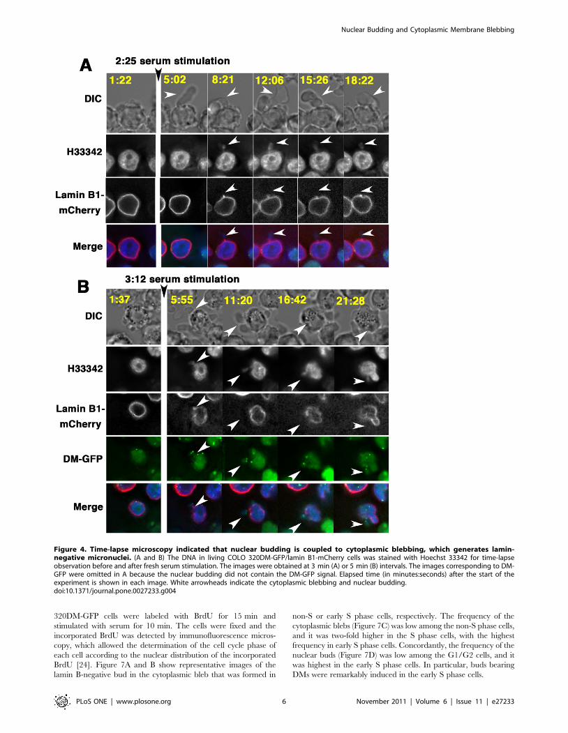
Induction of blebbing and budding by fresh serum isdependent on the cell lines and the cell-to-substratumadherence
The results described so far show the blebbing and budding
phenomena in COLO 320DM cells. These findings were next
tested in other cell lines. Because COLO 320DM cells adhere very
weakly to the substratum and show a rounded morphology, the
experiments were first performed with non-adherent cells. HL-60
and K562 cells weakly formed a large bleb despite treatment with
fresh serum, indicating that the non-adherent nature of cells was
not linked to the formation of large blebs. Most of the adherent
cells tested and listed in Figure 5C did not generate the large bleb
in the presence or absence of fresh serum, if they were attached to
the substratum (data not shown). However, among these cells,
human HeLa cells actively produced the large bleb if the cells were
detached from the substratum by trypsin/EDTA, suspended in a
conditioned medium, and then stimulated with fresh serum
(Figure 5B and C). This requirement for the detachment of cells
is consistent with the hypothesis that blebbing is active during
mitosis, when adherent cells round up. Concordantly, nuclear
budding also became detectable after this procedure (Figure 5D
and E). Furthermore, the micronuclei count revealed that the
frequency of the lamin-negative micronuclei increased both in the
bleb and in total cells (Figure 5F), as in the case of COLO 320DM
lines. Human colorectal carcinoma HCT116 or human normal
diploid fibroblast WI-38 or WS1 cells also exhibited the blebbing
after the detachment from the substratum and the fresh serum
stimulation (Figure 5C). However, the nuclear budding was not
evident in these cells, and the frequency of the micronuclei did not
increase after serum addition (Figure S5). Taken together, the
present data suggest that the blebbing and budding are induced by
fresh serum only in cells with a specific genetic background.
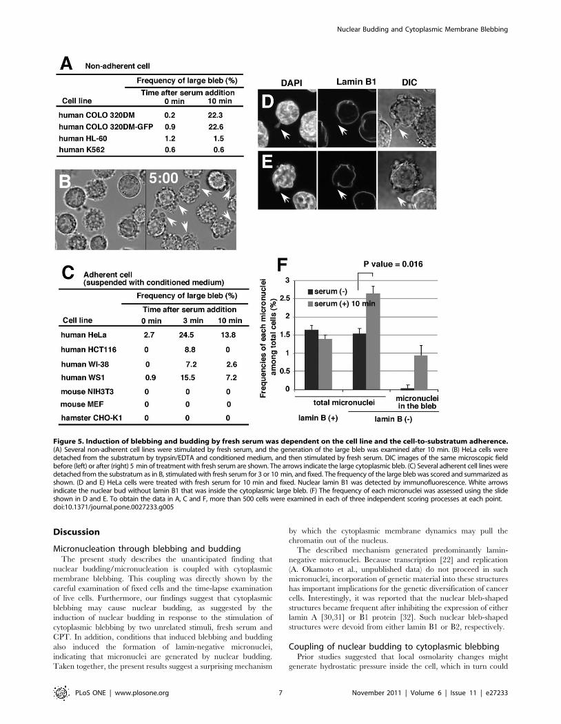
CPT induced interphase micronucleation throughblebbing and budding
The fresh serum stimulation of the large cytoplasmic bleb
described occurred independently from the induction of apoptosis.
However, cytoplasmic blebbing is known to be activated during
apoptosis. To address this issue, the effect of the apoptotic inducer
CPT on blebbing and budding was assessed. Treatment of COLO
320DM-GFP cells with 5 mg/ml CPT induced the formation of
large cytoplasmic blebs with a later timing than with fresh serum
induction (compare Figure 6A with Figure 5A, C), and faster than
the onset of apoptosis (Figure 6A). As a portion of the bleb
contained micronuclei, as in the case of serum induction, the
frequency of various types of micronuclei in the bleb was
examined. CPT predominantly induced the formation of lamin
Consequently, the frequency of the lamin B-negative small
micronuclei among the total cells greatly increased (Figure S6).
On the other hand, as shown in Figure 6B, cytoplasmic blebs with
Lamin B-negative, DM-negative and large to medium-sized
micronuclei also increased slightly. This finding will be discussed
in the following sections.
CPT inhibits topoisomerase I and induces multiple DNA-breaks
at replication sites in the S phase nucleus, which can be visualized
by the immunofluorescence detection of phosphorylated histone
H2AX (cH2AX). Thus, a bright cH2AX signal was detected
throughout the nucleus after the addition of CPT (Figure 6C, D).
The density of the cH2AX signal in the nucleus varied
significantly among the cells, which were therefore classified into
4 categories (– to +++), as described in our previous report [19].
The nuclear cH2AX-positive cells were likely reflect the cells in S
phase, and the fraction of cH2AX-positive cells reached more
than 50% of total cells during the first 60 min of CPT treatment
(Figure 6E). Assessment of the distribution of cH2AX signals
between the nucleus and the micronucleus in cells bearing lamin
B-negative micronuclei during the CPT treatment time (Figure 6F)
showed that CPT induced cH2AX-negative micronuclei that were
associated with the cH2AX-positive nucleus. The implications of
this finding will be discussed.
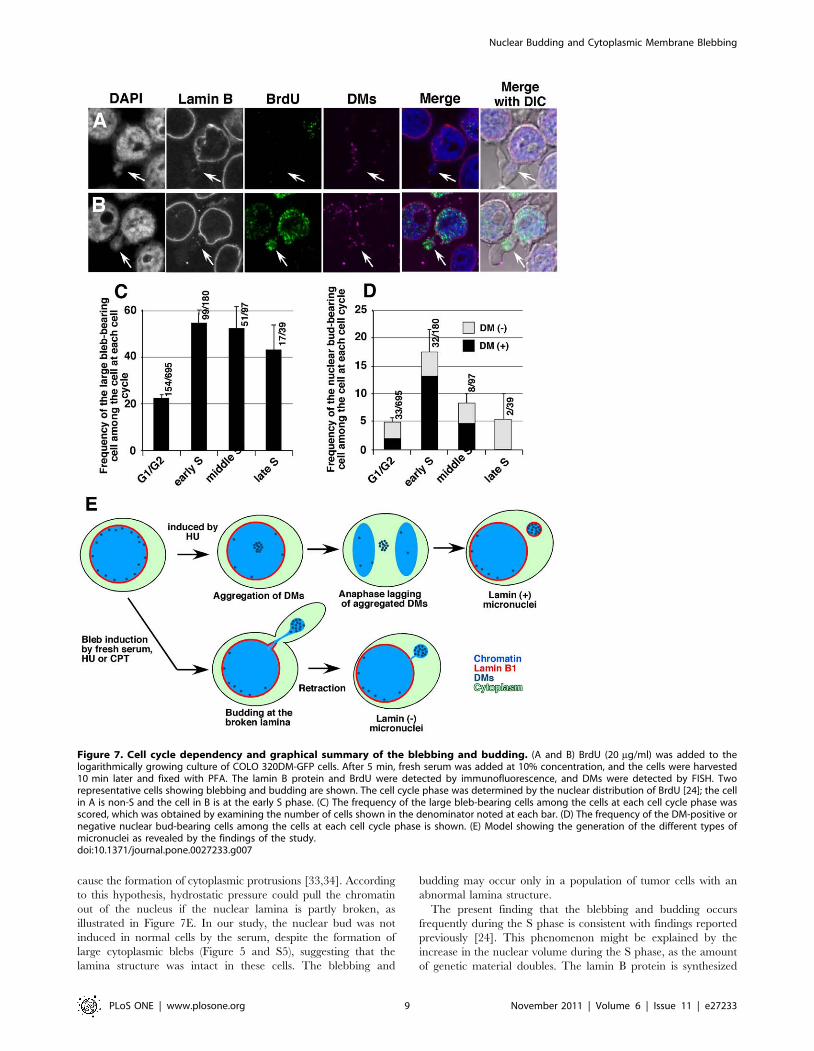
Blebbing and budding were most frequent duringS phase
Based on previous findings showing that nuclear budding is
frequent during the S phase [24], the effect of cell cycle stage on
blebbing/budding was assessed. Logarithmically growing COLO
Nuclear Budding and Cytoplasmic Membrane Blebbing
PLoS ONE | www.plosone.org 5 November 2011 | Volume 6 | Issue 11 | e27233
320DM-GFP cells were labeled with BrdU for 15 min and
stimulated with serum for 10 min. The cells were fixed and the
incorporated BrdU was detected by immunofluorescence micros-
copy, which allowed the determination of the cell cycle phase of
each cell according to the nuclear distribution of the incorporated
BrdU [24]. Figure 7A and B show representative images of the
lamin B-negative bud in the cytoplasmic bleb that was formed in
non-S or early S phase cells, respectively. The frequency of the
cytoplasmic blebs (Figure 7C) was low among the non-S phase cells,
and it was two-fold higher in the S phase cells, with the highest
frequency in early S phase cells. Concordantly, the frequency of the
nuclear buds (Figure 7D) was low among the G1/G2 cells, and it
was highest in the early S phase cells. In particular, buds bearing
DMs were remarkably induced in the early S phase cells.
Figure 4. Time-lapse microscopy indicated that nuclear budding is coupled to cytoplasmic blebbing, which generates lamin-negative micronuclei. (A and B) The DNA in living COLO 320DM-GFP/lamin B1-mCherry cells was stained with Hoechst 33342 for time-lapseobservation before and after fresh serum stimulation. The images were obtained at 3 min (A) or 5 min (B) intervals. The images corresponding to DM-GFP were omitted in A because the nuclear budding did not contain the DM-GFP signal. Elapsed time (in minutes:seconds) after the start of theexperiment is shown in each image. White arrowheads indicate the cytoplasmic blebbing and nuclear budding.doi:10.1371/journal.pone.0027233.g004
Nuclear Budding and Cytoplasmic Membrane Blebbing
PLoS ONE | www.plosone.org 6 November 2011 | Volume 6 | Issue 11 | e27233
Discussion
Micronucleation through blebbing and buddingThe present study describes the unanticipated finding that
nuclear budding/micronucleation is coupled with cytoplasmic
membrane blebbing. This coupling was directly shown by the
careful examination of fixed cells and the time-lapse examination
of live cells. Furthermore, our findings suggest that cytoplasmic
blebbing may cause nuclear budding, as suggested by the
induction of nuclear budding in response to the stimulation of
cytoplasmic blebbing by two unrelated stimuli, fresh serum and
CPT. In addition, conditions that induced blebbing and budding
also induced the formation of lamin-negative micronuclei,
indicating that micronuclei are generated by nuclear budding.
Taken together, the present results suggest a surprising mechanism
by which the cytoplasmic membrane dynamics may pull the
chromatin out of the nucleus.
The described mechanism generated predominantly lamin-
negative micronuclei. Because transcription [22] and replication
(A. Okamoto et al., unpublished data) do not proceed in such
micronuclei, incorporation of genetic material into these structures
has important implications for the genetic diversification of cancer
cells. Interestingly, it was reported that the nuclear bleb-shaped
structures became frequent after inhibiting the expression of either
lamin A [30,31] or B1 protein [32]. Such nuclear bleb-shaped
structures were devoid from either lamin B1 or B2, respectively.
Coupling of nuclear budding to cytoplasmic blebbingPrior studies suggested that local osmolarity changes might
generate hydrostatic pressure inside the cell, which in turn could
Figure 5. Induction of blebbing and budding by fresh serum was dependent on the cell line and the cell-to-substratum adherence.(A) Several non-adherent cell lines were stimulated by fresh serum, and the generation of the large bleb was examined after 10 min. (B) HeLa cells weredetached from the substratum by trypsin/EDTA and conditioned medium, and then stimulated by fresh serum. DIC images of the same microscopic fieldbefore (left) or after (right) 5 min of treatment with fresh serum are shown. The arrows indicate the large cytoplasmic bleb. (C) Several adherent cell lines weredetached from the substratum as in B, stimulated with fresh serum for 3 or 10 min, and fixed. The frequency of the large bleb was scored and summarized asshown. (D and E) HeLa cells were treated with fresh serum for 10 min and fixed. Nuclear lamin B1 was detected by immunofluorescence. White arrowsindicate the nuclear bud without lamin B1 that was inside the cytoplasmic large bleb. (F) The frequency of each micronuclei was assessed using the slideshown in D and E. To obtain the data in A, C and F, more than 500 cells were examined in each of three independent scoring processes at each point.doi:10.1371/journal.pone.0027233.g005
Nuclear Budding and Cytoplasmic Membrane Blebbing
PLoS ONE | www.plosone.org 7 November 2011 | Volume 6 | Issue 11 | e27233
Figure 6. CPT induced interphase micronucleation through blebbing and budding. COLO 320DM-GFP cells were cultured in the presenceof 5 mg/ml of CPT for the indicated time (A, E and F) or 3 hours (B to D) and fixed with PFA. Lamin B1 and DM-GFP (B), or lamin B1 and cH2AX (A, C toF) were simultaneously detected by immunofluorescence microscopy. (A) The frequencies of apoptotic cells and cells with large blebs among thetotal cells were scored and plotted. (B) The frequency of several kinds of micronuclei that were trapped in the large bleb among the cells with largeblebs (85 non-treated and 246 CPT-treated) was scored and plotted. (C and D) Representative images of the large bleb trapping the lamin B-negativemicronuclei. These micronuclei were either cH2AX-negative (C) or positive (D), while the neighboring nucleus was cH2AX-positive. (E) Time course ofthe appearance of cH2AX-positive cells. The intensities of the nuclear cH2AX-signal were classified from – to +++ according to a previous report [19].(F) The lamin B-negative micronuclei were classified according to the distribution of the cH2AX-signal between the nucleus and the micronucleus. Toobtain these data, more than 500 (A, E and F) or 1,000 (B) cells were examined in each of three independent scoring processes at each point.doi:10.1371/journal.pone.0027233.g006
Nuclear Budding and Cytoplasmic Membrane Blebbing
PLoS ONE | www.plosone.org 8 November 2011 | Volume 6 | Issue 11 | e27233
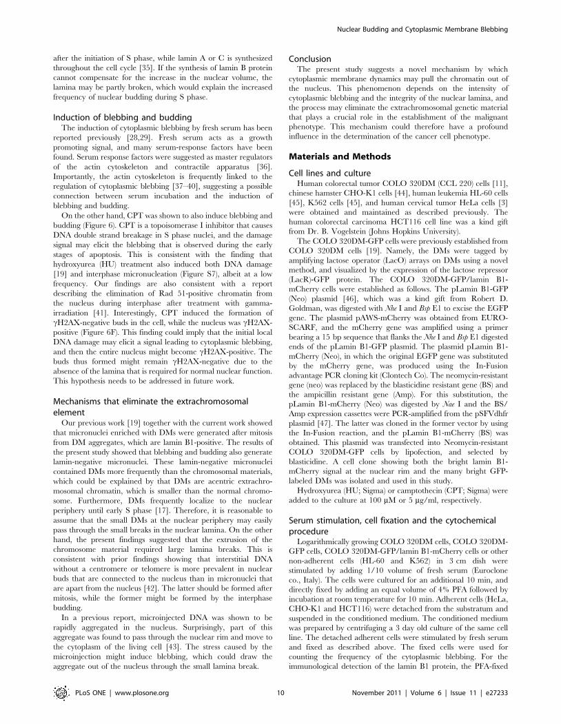
cause the formation of cytoplasmic protrusions [33,34]. According
to this hypothesis, hydrostatic pressure could pull the chromatin
out of the nucleus if the nuclear lamina is partly broken, as
illustrated in Figure 7E. In our study, the nuclear bud was not
induced in normal cells by the serum, despite the formation of
large cytoplasmic blebs (Figure 5 and S5), suggesting that the
lamina structure was intact in these cells. The blebbing and
budding may occur only in a population of tumor cells with an
abnormal lamina structure.
The present finding that the blebbing and budding occurs
frequently during the S phase is consistent with findings reported
previously [24]. This phenomenon might be explained by the
increase in the nuclear volume during the S phase, as the amount
of genetic material doubles. The lamin B protein is synthesized
Figure 7. Cell cycle dependency and graphical summary of the blebbing and budding. (A and B) BrdU (20 mg/ml) was added to thelogarithmically growing culture of COLO 320DM-GFP cells. After 5 min, fresh serum was added at 10% concentration, and the cells were harvested10 min later and fixed with PFA. The lamin B protein and BrdU were detected by immunofluorescence, and DMs were detected by FISH. Tworepresentative cells showing blebbing and budding are shown. The cell cycle phase was determined by the nuclear distribution of BrdU [24]; the cellin A is non-S and the cell in B is at the early S phase. (C) The frequency of the large bleb-bearing cells among the cells at each cell cycle phase wasscored, which was obtained by examining the number of cells shown in the denominator noted at each bar. (D) The frequency of the DM-positive ornegative nuclear bud-bearing cells among the cells at each cell cycle phase is shown. (E) Model showing the generation of the different types ofmicronuclei as revealed by the findings of the study.doi:10.1371/journal.pone.0027233.g007
Nuclear Budding and Cytoplasmic Membrane Blebbing
PLoS ONE | www.plosone.org 9 November 2011 | Volume 6 | Issue 11 | e27233
after the initiation of S phase, while lamin A or C is synthesized
throughout the cell cycle [35]. If the synthesis of lamin B protein
cannot compensate for the increase in the nuclear volume, the
lamina may be partly broken, which would explain the increased
frequency of nuclear budding during S phase.
Induction of blebbing and buddingThe induction of cytoplasmic blebbing by fresh serum has been
reported previously [28,29]. Fresh serum acts as a growth
promoting signal, and many serum-response factors have been
found. Serum response factors were suggested as master regulators
of the actin cytoskeleton and contractile apparatus [36].
Importantly, the actin cytoskeleton is frequently linked to the
regulation of cytoplasmic blebbing [37–40], suggesting a possible
connection between serum incubation and the induction of
blebbing and budding.
On the other hand, CPT was shown to also induce blebbing and
budding (Figure 6). CPT is a topoisomerase I inhibitor that causes
DNA double strand breakage in S phase nuclei, and the damage
signal may elicit the blebbing that is observed during the early
stages of apoptosis. This is consistent with the finding that
hydroxyurea (HU) treatment also induced both DNA damage
[19] and interphase micronucleation (Figure S7), albeit at a low
frequency. Our findings are also consistent with a report
describing the elimination of Rad 51-positive chromatin from
the nucleus during interphase after treatment with gamma-
irradiation [41]. Interestingly, CPT induced the formation of
cH2AX-negative buds in the cell, while the nucleus was cH2AX-
positive (Figure 6F). This finding could imply that the initial local
DNA damage may elicit a signal leading to cytoplasmic blebbing,
and then the entire nucleus might become cH2AX-positive. The
buds thus formed might remain cH2AX-negative due to the
absence of the lamina that is required for normal nuclear function.
This hypothesis needs to be addressed in future work.
Mechanisms that eliminate the extrachromosomalelement
Our previous work [19] together with the current work showed
that micronuclei enriched with DMs were generated after mitosis
from DM aggregates, which are lamin B1-positive. The results of
the present study showed that blebbing and budding also generate
lamin-negative micronuclei. These lamin-negative micronuclei
contained DMs more frequently than the chromosomal materials,
which could be explained by that DMs are acentric extrachro-
mosomal chromatin, which is smaller than the normal chromo-
some. Furthermore, DMs frequently localize to the nuclear
periphery until early S phase [17]. Therefore, it is reasonable to
assume that the small DMs at the nuclear periphery may easily
pass through the small breaks in the nuclear lamina. On the other
hand, the present findings suggested that the extrusion of the
chromosome material required large lamina breaks. This is
consistent with prior findings showing that interstitial DNA
without a centromere or telomere is more prevalent in nuclear
buds that are connected to the nucleus than in micronuclei that
are apart from the nucleus [42]. The latter should be formed after
mitosis, while the former might be formed by the interphase
budding.
In a previous report, microinjected DNA was shown to be
rapidly aggregated in the nucleus. Surprisingly, part of this
aggregate was found to pass through the nuclear rim and move to
the cytoplasm of the living cell [43]. The stress caused by the
microinjection might induce blebbing, which could draw the
aggregate out of the nucleus through the small lamina break.
ConclusionThe present study suggests a novel mechanism by which
cytoplasmic membrane dynamics may pull the chromatin out of
the nucleus. This phenomenon depends on the intensity of
cytoplasmic blebbing and the integrity of the nuclear lamina, and
the process may eliminate the extrachromosomal genetic material
that plays a crucial role in the establishment of the malignant
phenotype. This mechanism could therefore have a profound
influence in the determination of the cancer cell phenotype.
orientation versus chromosome mono-orientation in the origin of lagging
chromosomes in human primary cells. J Cell Sci 115: 507–515.
3. Utani K, Kohno Y, Okamoto A, Shimizu N (2010) Emergence of Micronuclei
and Their Effects on the Fate of Cells under Replication Stress. PlosONE 5:
e10089.
4. Pampalona J, Soler D, Genesca A, Tusell L (2010) Telomere dysfunction and
chromosome structure modulate the contribution of individual chromosomes in
abnormal nuclear morphologies. Mutat Res 683: 16–22.
5. Hoffelder DR, Luo L, Burke NA, Watkins SC, Gollin SM, et al. (2004)
Resolution of anaphase bridges in cancer cells. Chromosoma 112: 389–397.
6. Shimizu N, Shingaki K, Kaneko-Sasaguri Y, Hashizume T, Kanda T (2005)
When, where and how the bridge breaks: anaphase bridge breakage plays a
crucial role in gene amplification and HSR generation. Exp Cell Res 302:
233–243.
7. Harada S, Sekiguchi N, Shimizu N (2011) Amplification of a plasmid bearing a
mammalian replication initiation region in chromosomal and extrachromosomal
contexts. Nuc Acids Res 39: 958–969.
8. Von Hoff DD, McGill JR, Forseth BJ, Davidson KK, Bradley TP, et al. (1992)
Elimination of extrachromosomally amplified MYC genes from human
tumor cells reduces their tumorigenicity. Proc Natl Acad Sci U S A 89:
8165–8169.
Nuclear Budding and Cytoplasmic Membrane Blebbing
PLoS ONE | www.plosone.org 11 November 2011 | Volume 6 | Issue 11 | e27233
9. Shimizu N, Nakamura H, Kadota T, Kitajima K, Oda T, et al. (1994) Loss of
amplified c-myc genes in the spontaneously differentiated HL-60 cells. CancerRes 54: 3561–3567.
10. Eckhardt SG, Dai A, Davidson KK, Forseth BJ, Wahl GM, et al. (1994)
Induction of differentiation in HL60 cells by the reduction of extrachromoso-mally amplified c-myc. Proc Natl Acad Sci U S A 91: 6674–6678.
11. Shimizu N, Kanda T, Wahl GM (1996) Selective capture of acentric fragmentsby micronuclei provides a rapid method for purifying extrachromosomally
amplified DNA. Nat Genet 12: 65–71.
12. Shimizu N (2009) Extrachromosomal double minutes and chromosomalhomogeneously staining regions as probes for chromosome research. Cytogenet
Genome Res 124: 312–326.13. Shimizu N (2011) Molecular mechanisms of the origin of micronuclei from
extrachromosomal elements. Mutagenesis 26: 119–123.14. Levan A, Levan G (1978) Have double minutes functioning centromeres?
Hereditas 88: 81–92.
15. Kanda T, Wahl GM (2000) The dynamics of acentric chromosomes in cancercells revealed by GFP- based chromosome labeling strategies. J Cell Biochem 79:
107–114.16. Tanaka T, Shimizu N (2000) Induced detachment of acentric chromatin from
mitotic chromosomes leads to their cytoplasmic localization at G1 and the
micronucleation by lamin reorganization at S phase. J Cell Sci 113: 697–707.17. Itoh N, Shimizu N (1998) DNA replication-dependent intranuclear relocation of
double minute chromatin. J Cell Sci 111: 3275–3285.18. Shimizu N, Ochi T, Itonaga K (2001) Replication timing of amplified genetic
regions relates to intranuclear localization but not to genetic activity or G/Rband. Exp Cell Res 268: 201–210.
19. Shimizu N, Misaka N, Utani K (2007) Nonselective DNA damage induced by a
replication inhibitor results in the selective elimination of extrachromosomaldouble minutes from human cancer cells. Genes Chromosomes Cancer 46:
865–874.20. Willingale-Theune J, Schweiger M, Hirsch-Kauffmann M, Meek AE, Paulin-
Levasseur M, et al. (1989) Ultrastructure of Fanconi anemia fibroblasts. J Cell
Sci 93: 651–665.21. Paulin LM, Blake DL, Julien M, Rouleau L (1996) The MAN antigens are non-
lamin constituents of the nuclear lamina in vertebrate cells. Chromosoma 104:367–379.
22. Utani K, Kawamoto JK, Shimizu N (2007) Micronuclei bearing acentricextrachromosomal chromatin are transcriptionally competent and may perturb
the cancer cell phenotype. Mol Cancer Res 5: 695–704.
nuclear localization of a viral protein. J Virol 78: 6360–6369.27. Fenech M (2009) A lifetime passion for micronucleus cytome assays—reflections
from Down Under. Mutat Res 681: 111–117.
28. Dixon SJ, Aubin JE (1987) Serum and alpha 2-macroglobulin induce transienthyperpolarizations in the membrane potential of an osteoblastlike clone. J Cell
Physiol 132: 215–225.
29. Dixon SJ, Pitaru S, Bhargava U, Aubin JE (1987) Membrane blebbing is
associated with Ca2+-activated hyperpolarizations induced by serum and alpha
2-macroglobulin. J Cell Physiol 132: 473–482.
30. Sullivan T, Escalante-Alcalde D, Bhatt H, Anver M, Bhat N, et al. (1999) Loss of
A-type lamin expression compromises nuclear envelope integrity leading to
muscular dystrophy. J Cell Biol 147: 913–920.
31. Muchir A, van Engelen BG, Lammens M, Mislow JM, McNally E, et al. (2003)
Nuclear envelope alterations in fibroblasts from LGMD1B patients carrying
nonsense Y259X heterozygous or homozygous mutation in lamin A/C gene.
Exp Cell Res 291: 352–362.
32. Shimi T, Pfleghaar K, Kojima S, Pack CG, Solovei I, et al. (2008) The A- and B-
type nuclear lamin networks: microdomains involved in chromatin organization