8

J. Microbiol. Biotechnol. (2008), 18(2), 199–206
Genetic and Physiological Characterization of Oxytetracycline-ResistantBacteria from Giant Prawn Farms
Heepngoen, Pimpak1, Kannika Sajjaphan
1*, John A. Ferguson
2, and Michael J. Sadowsky
2
1Department of Soil Science, Kasetsart University, 50 Phahon Yothin Rd., Cha-tuchak, Bangkok 10900, Thailand2Department of Soil, Water, & Climate, and BioTechnology Institute, University of Minnesota, St. Paul, MN 55108, U.S.A.
Received: May 25, 2007 / Accepted: August 8, 2007
Four hundred and thirteen oxytetracycline-resistant bacteria
were recovered from six freshwater giant prawn farms
with a history of oxytetracycline use. Most oxytetracycline-
resistant isolates were Gram-negative bacteria. Six groups
of oxytetracycline-resistant bacteria were classified using
cluster analysis based on a comparison of levels of
oxytetracycline resistance. Complex fingerprint patterns
were obtained for 71 isolates studied. In general, the band
patterns of isolates from different ponds were very similar,
and the data indicated that the isolates were closely related.
The exploration for cross-resistance found that most of
the 71 oxytetracycline-resistant isolates were also resistant
to tetracycline and chlortetracycline, but had a relatively
low resistance to doxycycline. Many isolates showed higher
chlortetracycline resistance than oxytetracycline resistance.
Additionally, the oxytetracycline-resistant isolates were
examined for the presence of tetracycline resistance (tet)
genes. Fifty percent of the isolates carried one of the 14
known tet genes examined. The most common determinants
were TetA and TetD. However, TetB, TetC, TetE, TetK,
TetL, and TetM were also found with various frequencies.
Keywords: Oxytetracycline-resistant bacteria, freshwater
giant prawn, antibiotics
The significant increase in global demand for shrimp has
encouraged many developing countries to enter into the
practice of shrimp farming. This has made Thailand the
world’s leader in shrimp exports. Thai shrimp farming
has rapidly developed during the 1980s. Since 1993,
Thai shrimp farmers produced 235,000-275,000 tons of
cultured shrimps annually [10]. From shrimp exports,
Thailand has earned more than 2 billion USD annually,
which corresponds to 3-4% of the country’s total export
value in 2000 and 2001 [2-4].
Like all food production sectors, aquaculture requires
external inputs for successful production, including chemicals
and antibiotics. Antibiotics are extensively used in shrimp
aquaculture to treat bacterial infections. Some of the antibiotics
widely used in Thailand include erythromycin, nitrofurans
(furacin, furanace), oxytetracycline, sulfamonomethoxine
(Dimeton), and oxolinic acid [25]. They are applied through
feed additions or by simple addition to the water. Most of
the unused antibiotics end up in sediments, where they are
either degraded or slowly leached back into the surrounding
water.
Antibiotics and other chemicals used in aquaculture
may be toxic not only to the target pathogen, but also to
nontarget populations such as the cultured species, wild
flora and fauna, and human consumers. Many antibiotics
mixed with feed tend not to be absorbed by fish, and many
studies have reported that about 60-85% of antibiotics can
be excreted through feces in an unchanged form [1, 22, 26].
In addition, a great deal of the antibiotic-treated feed falls,
uneaten, to pond beds, where it accumulates in the sediments.
Antibiotics vary in their persistence in sediments, which
can range from a day to 1.5 years. The most commonly
used antibiotics, oxytetracycline (OTC) and oxolinic acid,
can persist in sediments for 6 months [26]. Jacobsen and
Berglind [15] studied the persistence of oxytetracycline in
fish farms. They indicated that OTC is relatively persistent
in anoxic sediments. OTC concentrations found in sediments
can vary from 0.1 to 4.9 mg/kg dry matter. OTC may
remain in concentrations capable of causing antibacterial
effects for 12 weeks within the sediments after the cessation
of treatment. Coyne et al. [8] investigated the concentration
of OTC in the sediment of two cages at a fish farm site,
and found half-lives of 16 and 13 days. Hektoen et al. [13]
found that oxytetracycline, oxolinic acid, flumequine, and
sarafloxacine were very persistent in sediments. In the deeper
layer of the sediment, hardly any degradation occurred
after 180 days with a calculated half-life of more than 300
days. However, antibiotic residues in the top layer of the
sediment rapidly disappeared. The removal of these substances
*Corresponding authorPhone: 66-942-8104-5; Fax: 662-942-8106;E-mail: [email protected]

200 Heepngoen et al.
from the sediment is most probably due to leaching and
redistribution rather than degradation.
Oxytetracycline, furazolidone, erythromycin, and kanamycin
have been found to be health hazards associated with
digestive disorders and allergies [23]. Lutzhøft et al. [18]
found that cyanobacteria have a greater sensitivity towards
antibacterial agents compared with crustaceans and fish.
Moreover, widespread antibiotic applications have the
potential to cause the development of drug resistance
among pathogens. Antibiotic resistance has been identified
in strains of Aeromonas salmonicida, the bacteria responsible
for furunculosis [5, 12]. Antibiotic resistance has also been
reported in natural sediment bacteria from antibiotics
accumulating below net pens [7, 14, 17, 21]. The presence of
antibiotics in the bottom sediments may affect the natural
bacterial composition and activity, and thereby change the
ecological structure of benthic microbial communities. The
accumulation of antibiotics in pond sediment can also
lead to decreased or inhibited microbial activity in the
sediment.
Antibiotic-resistant bacteria isolated from animal farms
or aquaculture are raising concerns that antibiotics used
in agriculture may play an important role in selecting
for antibiotic resistance among foodborne bacteria. The
environmental fate of veterinary drugs and the factors that
influence the persistence and biodegradation of antibiotics
used in agriculture is not yet well understood. Metabolites
resulting from the biotransformation of these drugs may
have either enhanced or reduced biological activity compared
with the parent compound, and may affect the microbial
ecology of these systems.
Fig. 1. The chemical structure of the compounds used in thisstudy.
Fig. 2. Dendrogram prepared from the comparison of level of oxytetracycline resistance.

OXYTETRACYCLINE-RESISTANT BACTERIA IN GIANT PRAWN FARMS 201
To better understand the fate and effect of antibiotics in
the aquaculture system, oxytetracycline was chosen for
this study. Oxytetracycline is not only one of the most
commonly used antibacterials in aquaculture, but is also
used in animal farms. Therefore, the main purpose of this
study was to examine resistance profiles of bacteria in an
aquaculture system.
MATERIALS AND METHODS
Chemicals
The tetracycline class antibiotics used in this study, oxytetracycline,
chlortetracycline, doxycycline, and tetracycline, were obtained from
Sigma (St. Louis, MO, U.S.A.). The chemical structures of these
compounds are shown in Fig. 1.
Freshwater Giant Prawn Farms and Sampling Sites
This study was initiated with soil, sediment, and water samples
obtained from six freshwater giant prawn (Macrobrachium
rosenbergii) ponds, on farms located in Nakhon Pathom Province
in Thailand, which is approximately 80 km northwest of Bangkok.
The ponds were chosen based on their histories of oxytetracycline
applications. The sizes of the ponds varied between 0.3 and
1.6 ha.
Isolation of Oxytetracycline-Resistant Bacteria
Soil and sediment samples were diluted 10-fold on 0.85% NaCl
and agitated using a vortex mixer. Extracts were serially diluted
10-fold in 0.85% NaCl, and 0.1-ml aliquots were plated on
Mueller-Hinton agar supplemented with 10 µg/ml of oxytetracycline.
Medium without antibiotic was used as a positive control.
The plates were incubated at 30oC for 24 to 48 h. Total and
oxytetracycline-resistant colony counts were acquired. Oxytetracycline
resistant bacteria were purified and stored in 96-well microtiter
plates containing freezing medium [27] and kept at -80oC until used
for later study.
Identification and Characterization
The resistance profiles of oxytetracycline-resistant bacteria were
determined for each of the isolates. Strains were grown at 28oC for
18-36 h in Mueller-Hinton (MH) broth with six concentrations of
oxytetracycline (10, 20, 40, 60, 100, and 120 µg/ml) using 96-well
microtiter plates. Resistance was defined as showing bacterial
growth that was 70% of OD600 compared with growth of the same
strain in the control MH broth without oxytetracycline. Strains were
categorized as being resistant or sensitive to each concentration of
oxytetracycline, and assigned a value of 1 or 0, respectively. The
resistance profiles of 413 oxytetracycline-resistant isolates were used
for dendrogram analyses. The dendrogram was produced using
Jaccard similarity coefficients.
HFERP DNA Fingerprinting
Seventy-one isolates were further characterized by DNA fingerprint
analysis. Isolates were chosen based on profiles of resistance to
the tested antibiotics. DNA fingerprints were obtained by using the
horizontal, fluorophore-enhanced, rep-PCR (HFERP) method as
described by Johnson et al. [16]. Fingerprint data were normalized
and analyzed using BioNumerics v.3.5 software (Applied Maths,
Sint-Martens-Latem, Belgium). DNA fingerprint similarities were
calculated by using Pearson’s product-moment correlation coefficient,
with 1% optimization. A binary band-matching character table was
generated by using the HFERP-derived PCR DNA fingerprint data,
and results were analyzed, accounting for the covariance structure,
by using the multidimensional scaling (MDS) and multivariate
analysis of variance (MANOVA), forms of discriminant analysis,
subroutines of the Bionumerics software.
Fig. 3. Dendrogram showing the oxytetracycline resistance isolatesobtained from soil, sediment, or water from six prawn ponds andirrigation system.
202 Heepngoen et al.
Determination for Cross-resistance to Antibiotic of Tetracycline
Class
Seventy-one oxytetracycline-resistant bacterial isolates were examined
for cross-resistance to chlortetracycline, doxycycline, or tetracycline
using 96-well microtiter plates as described above.
Tetracycline-Resistant Gene Determination
The 71 isolates were examined further using a multiplex PCR for
the presence of the 14 tetracycline resistance genes: tetA, tetB, tetC,
tetD, tetE, tetG, tetK, tetL, tetM, tetO, tetS, tetA(P), tetQ, and tetX
[20]. Pairs of primers were multiplexed in groups as described by
Ng et al. [20]: Group I: tetB, tetC, tetD; Group II: tetA, tetE, tetG;
Group III: tetK, tetL, tetM, tetO, tetS; Group IV: tetA(P), tetQ, tetX.
PCR reactions were prepared as described by Bryan et al. [6].
Single-colony isolates were streaked onto MH agar supplemented
with 10 µg/ml oxytetracycline and picked using sterile loops and
suspended in 50 µl of sterile H2O. One µl of the standardized cell
suspension served as a template DNA for colony-based multiplex
PCR. The primers used for PCR amplification of the 14 tetracycline
resistance genes were as described by Ng et al. [20]. The primers
were aliquoted into four groups: group I contained primers for tetB,
tetC, and tetD; group II contained primers for tetA, tetE, and tetG;
group III contained primers for tetK, tetL, tetM, tetO, and tetS; and
group IV contained primers for tetA(P), tetQ, and tetX. PCR was
performed with an MJ Research (Waltham, MA, U.S.A.) model
PTC100 thermocycler, by using the following conditions as described
previously: 5 min of initial denaturation at 94oC, followed by 35 cycles
of 94oC for 1 min, 55oC for 1 min, and 72oC for 1.5 min. The PCR
products were separated by gel electrophoresis in 1% (w/v) agarose
gels in 1× Tris-acetate-EDTA buffer, stained with ethidium bromide,
and visualized under UV illumination. The validity of multiplex PCRs
and product sizes was ascertained by using the following positive
control plasmids: pSL18, pRT11, pBR322, pSL106, pSL1504,
pJA8122, pAT102, pVB.A15, pJ13, pUOA1, pAT451, pJIR39,
pNFD13-2, and pBS5, for the genes tetA, tetB, tetC, tetD, tetE, tetG,
tetK, tetL, tetM, tetO, tetS, tetA(P), tetQ, and tetX, respectively. The
sizes of the PCR products were determined by comparison with the
migration of a GeneRuller 100-bp ladder (MBI Fermentas).
RESULTS AND DISCUSSION
Isolation, Identification, and Characterization of
Oxytetracycline-Resistant Bacteria
A total of 413 bacterial colonies were isolated from six
freshwater giant prawn ponds and the percentage of
oxytetracycline-resistant isolates was different from each
pond (data not shown). These isolates were further purified
and examined for oxytetracycline resistance and dendrogram
analysis (Fig. 2). Of the 413 isolates, 37.0% (153 isolates),
22.3% (92), 21.3% (88), 3.4% (14), 5.8% (24), and 8.5%
(35) were resistant to >120, 100, 60, 40, 20, and 10 µg/ml
oxytetracycline, respectively. However, 1.7% (7) were not
resistant to 10 µg/ml oxytetracycline, and not used in
further studies.
Dendrogram analysis (Fig. 2) indicated that the strains
could be divided into two major subgroups, I and II, which
diverged at a similarity value of 50%. The group I isolates
contained the majority of strains, and could be further
divided into two subgroups (A and B) that diverged at a
similarity value of 70%. Each of these subgroups could be
further divided into two subsubgroups (1 and 2). Overall
strains in subgroups A1 and A2, and B1 and B2, were
related to each other, with similarity values of 85%. In
contrast, Group II consisted of far fewer strains. There was
no apparent relationship between isolation pond and
subgroup status.
The HFERP DNA fingerprinting technique was conducted
to differentiate oxytetracycline-resistant isolates. Complex
fingerprint patterns were obtained for the 71 isolates studied.
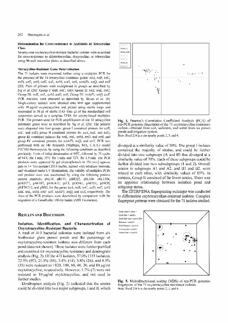
Fig. 4. Pearson’s Correlation Coefficient Analysis (PCA) ofrep-PCR genomic fingerprint of the 71 oxytetracycline resistanceisolates obtained from soil, sediment, and water from six prawnponds and irrigation system.Note: Pond 234 is a site nearby ponds 2, 3, and 4.
Fig. 5. Multidimensional scaling (MDS) of rep-PCR genomicfingerprint of the 71 oxytetracycline resistance isolates.Note: Pond 234 is a site nearby ponds 2, 3, and 4.
OXYTETRACYCLINE-RESISTANT BACTERIA IN GIANT PRAWN FARMS 203
There was a very high proportion of genetically identical
clones found, shared in all of the ponds (Figs. 3 and 4).
Almost half of the isolates present in the analysis were
clones of some isolates present in another pond. We
define a clone in HFERP as any isolate sharing 92% or
more similarity based on Pearson’s Correlation Coefficient
(Fig. 4). This condition is usually found in areas of low
genetic diversity in which all of the sampling points share
a common source of contamination. Multidimensional
scaling (MDS) was performed to visualize the large clusters
of clones within the dataset. The MDS indicated that a
large number of strains in different ponds were related and
clustered together in MDS analysis (Fig. 5). Multivariate
analysis of variance (MANOVA) was then performed to
confirm that there were no significant variables affecting
genetic similarity from site to site (Fig. 6). This indicates
that there was little to no change in the genetic diversity
between sites.
Additionally, all of these 71 isolates were further tested
for Gram-stain reaction. The result showed that most
oxytetracycline-resistant isolates were Gram-negative bacteria
with rod shape. Only a few of them were Gram-positive
bacteria.
Determination for Cross-resistance to Antibiotics of
Tetracycline Class
The 71 isolates were randomly selected based on sample
sites and classification using dendrogram analysis. These
71 isolates were determined for cross-resistance to
antibiotics of tetracycline class. Isolates were resistant to
oxytetracycline, tetracycline, chlortetracycline, and doxycline.
Most of the isolates in the ponds had high resistance
to three antibiotics (i.e., oxytetracycline, tetracycline, and
chlortetracycline), but relatively low resistant to doxycycline.
Of all isolates, 72% were resistant to all four antibiotics,
16% were resistant to oxytetracycline, tetracycline, and
chlortetracycline, 6% were resistant to only oxytetracycline,
3% were resistant to oxytetracycline and tetracycline, and
1.5% were resistant to oxytetracycline and chlortetracycline.
Fig. 6. Multivariate analysis of variance (MANOVA) of rep-PCR genomic fingerprint of the 71 oxytetracycline resistanceisolates.Note: Pond 234 is a site nearby ponds 2, 3, and 4.
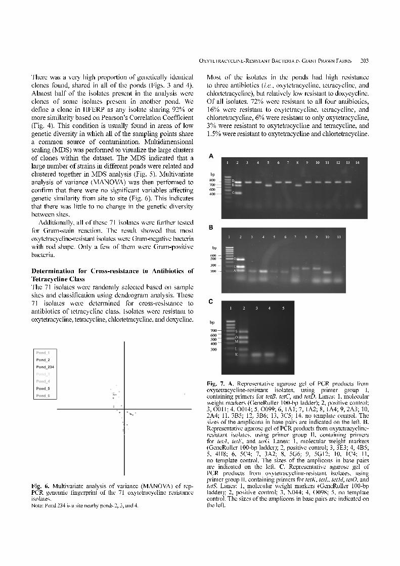
Fig. 7. A. Representative agarose gel of PCR products fromoxytetracycline-resistant isolates, using primer group I,containing primers for tetB, tetC, and tetD. Lanes: 1, molecularweight markers (GeneRuller 100-bp ladder); 2, positive control;3, O011; 4, O014; 5, O099; 6, 1A1; 7, 1A2; 8, 1A4; 9, 2A3; 10,2A4; 11, 3B5; 12, 3B6; 13, 3C5; 14, no template control. Thesizes of the amplicons in base pairs are indicated on the left. B.Representative agarose gel of PCR products from oxytetracycline-resistant isolates, using primer group II, containing primersfor tetA, tetE, and tetG. Lanes: 1, molecular weight markers(GeneRuller 100-bp ladder); 2, positive control; 3, 3E3; 4, 4B5;5, 4H8; 6, 5C4; 7, 3A2; 8, 5G6; 9, 5G12; 10, 1C4; 11,no template control. The sizes of the amplicons in base pairsare indicated on the left. C. Representative agarose gel ofPCR products from oxytetracycline-resistant isolates, usingprimer group II, containing primers for tetK, tetL, tetM, tetO, andtetS. Lanes: 1, molecular weight markers (GeneRuller 100-bpladder); 2, positive control; 3, N044; 4, O098; 5, no templatecontrol. The sizes of the amplicons in base pairs are indicated onthe left.

204 Heepngoen et al.
Table 1. Tetracycline-resistant genes in oxytetracycline-resistant isolates.
Pond Source IsolationTetracycline-resistant genes
Group 1 (B, C, D) Group 2 (A, E, G) Group 3 (K, L,M, O, S) Group 4 (A(P), Q, X)
1
Soil
3E3 - A - -
3G7 D - - -
4A4 D - - -
4A12 - A - -
4B5 - A - -
4E10 - A - -
4H8 - A - -
5C4 - A - -
5D2 D - - -
5E4 D - - -
5G6 D E - -
5G12 D E - -
O011 D - -
O013 - E - -
O014 D - -
O015 - A - -
Water
1A1 D - - -
1A2 D - - -
1A4 B - - -
1A6 - A - -
2 Sediment
6F11 - A S -
6D6 - A - -
6D8 - - K -
6H6 B - - -
K022 - A - -
K026 - A - -
3Sediment
3A2 - E - -
3B5 D - - -
3B6 D - - -
3C5 D - - -
3C12 D - - -
T031 - A - -
T032 - A - -
T033 - A - -
Water 1C4 - A - -
4 Sediment
N041 - A, E - -
N042 - - L -
N044 - - K -
2, 3, 4Drainage outlet
1G1 - - L -
2A3 D - - -
2A4 D - - -
Irregation system 2B6 D - - -
5 SedimentT051 - E - -
T054 - E - -
6 Sediment
O096 - A - -
O097 - A - -
O098 - - K -
O099 B, C - M -

OXYTETRACYCLINE-RESISTANT BACTERIA IN GIANT PRAWN FARMS 205
Surprisingly, it was found that most of the isolates showed
higher resistance to chlortetracycline than oxytetracycline.
This is because the freshwater giant prawn farmers had
been using oxytetracycline for diseases disinfection for
long periods of time and only until recently has the
government tried to control the use of oxytetracycline in
the prawn farm. Then, the prawn farmers changed from
using oxytetracycline to chlortetracycline.
Distribution of Tetracycline-Resistant Genes
Results of the distribution of the tet genes using
PCR showed that 67.6% of the 71 isolates contained at
least 1 of 14 tetracycline resistance genes. The most
common determinants were TetA (26.8% of isolates)
and TetD (23.9% of isolates) (Figs. 7A, 7B, and 7C and
Table 1). However, TetB, TetC, TetE, TetK, TetL, and
TetM were also found with various frequencies. The
presence of tetA through tetG genes had previously
been reported in bacteria isolated from freshwater fish
farms [9, 11, 19, 24]. Moreover, TetK, TetL, and TetM
were also found in this study. However, the presence of
any tetracycline-resistant gene was not found in group 4
(tetA(P), tetQ, tetX).
Acknowledgments
This study was supported by the Institute of Science and
Technology for Sustainability (UNU & GIST Joint
Programme), Korea. We would like to thank Prof. Hor-Gil
Hur for help with discussions.
REFERENCES
1. Alderman, D. J., H. Rosenthal, P. Smith, J. Stewart, and D.
Weston. 1994. Chemicals used in mariculture, ICES Cooperative
Research Report No. 202. ICES, Copenhagen.
2. Bangkok Post. 2001. Shrimp raisers want state help, March 15.
3. Bangkok Post. 2001. 2000 Year-End Economic Review.
Available from <http://www.bangkokpost.com/yereview2000/
exports.html>.
4. Bangkok Post. 2002. 2001 Year-End Economic Review.
Available from <http://bangkokpost.net/yearend2001/trade.html>.
5. Barnes, A. C., T. S. Hastings, and S. G. B. Amyes, 1994.
Amoxycillin resistance in Scottish isolates of Aeromonas
salmonicida. J. Fish Dis. 17: 357-363.
6. Bryan, A., N. Shapir, and M. J. Sadowsky. 2004. Frequency and
distribution of tetracycline resistance genes in genetically diverse,
nonselected, and nonclinical Escherichia coli strains isolated from
diverse human and animal sources. Appl. Environ. Microbiol.
70: 2503-2507.
7. Capone, D. G., D. P. Weston, V. Miller, and C. Shoemaker.
1996. Antibacterial residues in marine sediments and invertebrates
following chemotherapy in aquaculture. Aquaculture 145: 55-
75.
8. Coyne, R., M. Hiney, and P. Smith. 1997. Transient presence of
oxytetracycline in blue mussels (Mytilus edulis) following its
therapeutic use at a marine Atlantic salmon farm. Aquaculture
149: 175-181.
9. DePaola, A., J. T. Peeler, and G. E. Rodrick. 1995. Effect of
oxytetracycline medicated feed on antibiotic resistance of Gram-
negative bacteria in catfish ponds. Appl. Environ. Microbiol. 61:
2335-2340.
10. FAO. 2001. FAO Yearbook. Fishery Statistics. Aquaculture
Production, Vol. 88/2. FAO, Rome.
11. Guardabassi, L., A. Dalsgaard, M. Raffatellu, and J. E. Olsen.
2000. Increase in the prevalence of oxolinic acid resistant
Acinetobacter spp. observed in a stream receiving the effluent
from a freshwater trout farm following the treatment with
oxolinic acid-medicated feed. Aquaculture. 188: 205-218.
12. Hawkins, L., H. Hariharan, K. Whitman, G. Johnson, and
J. Bryenton. 1997. Drug resistance of a typical Aeromonas
salmonicida from Atlantic salmon and rainbow trout in
Newfoundland. Bull. Aquacult. Assoc. Can. 2: 39-41.
13. Hektoen, H., J. A. Berge, V. Hormazabal, and M. Yndestad.
1995. Persistence of antibacterial agents in marine sediments.
Aquaculture 133: 175-184
14. Husevåg, B., B. T. Lunestad, P. J. Johannessen, Ø. Enger, and
O. B. Samuelsen. 1991. Simultaneous occurrence of Vibrio
salmonicida and antibiotic resistant bacteria in sediments at
abandoned aquaculture sites. J. Fish Dis. 14: 631-640.
15. Jacobsen, P. and L. Berglind. 1988. Persistence of oxytetracycline
in sediments from fish farms. Aquaculture 70: 365-370.
16. Johnson, L. K., M. B. Brown, E. A. Carruthers, J. A. Ferguson,
P. E. Dombek, and M. J. Sadowsky. 2004. Sample size, library
composition, and genotypic diversity among natural populations
of Escherichia coli from different animals influence accuracy of
determining sources of fecal pollution. Appl. Environ. Microbiol.
70: 4478-4485.
17. Kerry, J., R. Coyne, D. Gilroy, M. Hiney, and P. Smith. 1996.
Spatial distribution of oxytetracycline and elevated frequencies
of oxytetracycline resistance in sediments beneath a marine
salmon farm following oxytetracycline therapy. Aquaculture
145: 31-39.
18. Lutzhøft, H.-C., B. Halling-Sørensen, and S. E. Jørgensen.
1999. Algal toxicity of antibacterial agents applied in Danish
fish farming. Arch. Environ. Contam. Toxicol. 36: 1-6.
19. Miranda, C. D., C. Kehrenberg, C. Ulep, S. Schwarz, and M. C.
Roberts. 2003. Diversity of tetracycline resistance genes in bacteria
from Chilean salmon farms. Antimicrob. Agents Chemother. 47:
883-888.
20. Ng, L. K., I. Martin, M. Alfa, and M. Mulvey. 2001. Multiplex
PCR for the detection of tetracycline resistant genes. Mol. Cell.
Probes 15: 209-215.
21. Nygaard, K., B. T. Lunestad, H. Hektoen, J. A. Berge, and
V. Hormazabal. 1992. Resistance to oxytetracycline, oxolinic
acid and furazolidone in bacteria from marine sediments.
Aquaculture 104: 31-36.
22. Samuelsen, O. B., B. T. Lunestad, B. Husevåg, T. Hølleland,
and A. Ervik. 1992. Residues of oxolinic acid in wild fauna
following medication in fish farms. Dis. Aquat. Org. 12: 111-
119.
23. Schnick, R. A. 1991. Chemicals for worldwide aquaculture, pp.
441-466. In: Fish Health Management in Asia-Pacific. Report

206 Heepngoen et al.
on a Regional Study and Workshop on Fish Disease and Fish
Health Management. Asian Development Bank and Network of
Aquaculture Centres in Asia, Bangkok, Thailand.
24. Sørum, H., M. C. Roberts, and J. H. Crosa. 1992. Identification
and cloning of a tetracycline resistance gene from the fish pathogen
Vibrio salmonicida. Antimicrob. Agents Chemother. 36: 611-615.
25. Tonguthai, K. 2000. The use of chemicals in aquaculture in
Thailand, pp. 207-220. In: Use of Chemicals in Aquaculture in
Asia. Proceedings of the Meeting on the Use of Chemicals in
Aquaculture in Asia, Tigbauan, Iloilo, Philippines, 20-22 May
1996.
26. Weston, D. P. 1996. Environmental considerations in the use of
antibacterial drugs in aquaculture. In: D. J. Baird (ed.).
Aquaculture and Water Resource Management. Blackwell
Science, Oxford, U.K.
27. Woo, S. S., J. Jiang, B. S. Gill, A. H. Paterson, and R. A. Wing.
1994. Construction and characterization of a bacterial artificial
chromosome library of Sorghum bicolor. Nucl. Acids Res. 22:
4922-4931.


![Amultiplexoligonucleotideligation-PCRasacomplementarytool ... · Materials and methods Bacterial isolates All S. Typhimurium and S. 1,4,[5],12:i:- isolates were re-ceived from the](https://static.documents.pub/doc/80x56/5fc667b777944d3b580d6a67/amultiplexoligonucleotideligation-pcrasacomplementarytool-materials-and-methods.jpg)