156
Genetic epidemiology of osteoarthritis Studies of familial aggregation and candidate genes

Genetic epidemiology of osteoarthritis
Studies of familial aggregation and candidate genes
This study was financially suppolled by the Netherlands Organization fOf Scientific Research (N\'VO), the "Dutch League against Rheumatism", the NESTOR siimulation program for geriatric research in the Netherlands (Ministries of Health and Education), the municipality of Rotterdam ;md the Looseo foundation.
The author gratefully acknowledges the collaboration with the Depal1ment of Rheumatology, Leiden University Medical Center, Leiden, The Netherlands (F.e. Breedveld), TNO Prevention and I Iealth, Gorter location, Leiden, The Netherlands (T.M. te Koppele, B.S. Miedema) and the Depal1ment of Internal IvIedicine Ill, Erasmus University Medical School, Rotterdam, The Netherlands (I LA.P. Pols).
The Depa11ment of Epidemiology & Biostatistics, Erasmus University Medical School, Rotterdam, TNO Prevention and Health, Gaubius LaLoratory, Lciden, the Netherlands Organization for Scientific Research (NWO), and UCB Pharma BV granted additional financial support [or the publication of this thesis.
Layout: Bon Mot, Rotterdam Printed by: Print Pallners lpskamp
ISBN 90-9012489-6
© e. Bijkerk, 1999 No pall of this book may be reproduced, stored in a retrieval system or transmitted in any form or by any means, without permission of the author, or, when appropriate, of the publishers of the publications.
Genetic epidemiology of osteoarthritis
Studies of familial aggregation and candidate genes
Genetische epidemiologie van artrose
Onclerzoek naar familie aggregatie en kandidaat genen
Proefschrift
ter verkrijging van de graad van cloctor
aan de Erasmus Universiteit Rotterdam
op gezag van de rector magnificus
Prof. Dr. P.W.C. Akkermans, M.A.
en volgens het besluit van het College voor Proffioties.
De openbare verdediging zal plaatsvinden op
woensdag 3 maalt 1999 om 13.45 Ullr
door
Casper Bijkerk
geboren te Breda

Promotiecommissie
Promotor
CO-pr0111otores
Overige leden
Prof. Dr. A. Hofman
Dr. C.M. van Duijn
Dr. P.E. Slagboolll
Prof. Dr. F.e. Ereeclveld
Prof. Dr. B.A. Oostra
Prof. Dr. H.A.P. Pols
Contents
1. Introduction
2. Methods 2.1. Methodological considerations concerning the genetic
epidemiology of osteoarthritis
2.2. Assessment of radiological osteoarthritis in peripheral joints and
of disk degeneration of the spine. A population-based sample
1
7
aged 55 to 70 years 17
2.3. Assessment of radiological osteoarthritis in peripheral joints and
of disk degeneration of the spine. A sibling pair sample 27
3. Osteoarthritis in the general population 3.1. Pattern of joint involvement and deten11inants of osteoarthitis at
multiple sites in a population-based study
3.2. Heritabilities of radiological osteoalthritis in peripheral joints and
of disk degeneration of the spine
4. Collagen genes and osteoarthritis 4.1. Association of the COL2Al gene with radiological osteoalthritis in
35
47
a population-based study. 'The Rotterdam Study 61
4.2. The COL9Al gene and C0L11A2 region and radiographically
assessed osteoarthritis in subjects from a population-based study 77
4.3. A sibling pair study on the role of the COL2Al and COL9Al genes
in radiological osteoarthritis 91
5, Gene interaction in osteoarthritis 5.1. The IGF-l gene and radiological osteoarthritis in a population
based study
5.2. Interaction between the IGP-l and COL2Al genes in the
association with radiological osteoalthritis
6, General discussion
7. Summary
8. Samenvatting
Epiloog
Curriculum vitae
103
113
121
135
141
147
149
Manuscripts based on the studies described in this thesis
Chapte,' 3 Bljkerk C, Slagboom PE, Valkenburg HA, Miedema lIS, Hofman A, Breedveld FC, Pols HAP, van Duijn eN!. Pattern of joint involvement and determinants of osteoaI1hrilis at multiple sites in a population-based study. Submitted.
l3ijkel'k C, HOLlwing-Duistermaat .II, Valkenburg HA, Mculenbelt I, Hofman A, Breedveld FC, Pols HAP, van Duijn eM, Slagboom PE. Heritabilities of radiological osteoaltilritis in peripheral joints and of disk degeneration of the spine. Al1hritis Rheum (accepted for publication).
Chapter 4 Bijkerk C, ivleulenbelt It Odding E, Valkcnburg IIA, Miedema HS, Brcedveld FC, Hofman A, te Koppe1e JlvI, Pols HAP, van Duijn ctvl, Slagboom PE. Association of the COL2Al gene with radiological osteoanhritis in a population-based study. The Rolterdam Study. Submitted.
Bijkerk C, Meulenbelt I, de Wildt SC, Pols HAP, Breedve1d FC, Hofman A, le Koppele JM, van Duljn CM, Slagboom PE. The COL9Al gene and C0L11A2 region and radiographically assessed ostcoal1hrilis in subjects from a population-based study. Submitted.
Bijkerk C, Meulenbe1t I, Breedveld FC, Hofman A, Pols I1AP, Sandkuyl L, van DUljn Ov', Slagboom PE. A sibling pair study on the role of the COL2A1 and COL9A1 genes in radiological osleoal1hritis. To be submitted.
Chapter 5 Meulenbclt I, Bijkerk C, Miedema HS, Breedveld FC, Hofman A, Valkenburg HA, Pols HAP, Slagboom PE, van Duijn CM. A genetic association study of the IGF-1 gene and radiological osteoatthrltis in a population-based cohort study (the Rotterdam Study). Ann Rheum Dis 1998;57:371-4.
Bijkcrk C, Meulenbelt I, Hofman A, 13reedveld FC, Pols HAP, van Duijn CM, Slagboom PE. Interaction between the [GF-1 and COLZA1 genes in the association with radiological oste031111rilis. Submitted.
Other publications by the author
rVfculcnbelt I, Bijkerk C, Breedveld FC, Slagboom PE. Genetic linkage analysis of 1r1 candidate gene loci in a [amil)' with autosomal dominant osteoatthritis withollt dysplasia. J Med Genet 1997;31:1021-7.
IVleulenbelt I, I3ijkerk C, de Wildt SC, .I'vIiedema HS, Val ken burg J lA, 13reedvcld FC, Pols HAP, te Koppcle JlvI, Sloos VF, Hofman A, Slagl.Joom PE, van Duijn C.LVI. Investigation of the association of the CRT!vI and CRn1 genes with radiographically evident osteoa11hrilis in subjects from the Rotterdam Study. AI1hrllis Rheum 1997;40:1760-5.

1
Introduction

Introduction
O steoalthritis eO A) is the most common rheumatic disease and an i.mpor
tant cause of disability in the elderly (l,2). It is characterized by a progres
sive degeneration of articular cartilage of diarthrodial joint::; without synovial
inflammation or bone erosions. Il leads in a minority of subjects to clinical OA,
Le. joint pain, limited range of motion of the affected jOint, joint effusion, local
inflanul1atory reaction or crepitus. The dinical diagnosis of OA is confirmed by
radiographic evidence, reflecting deterioration of cal1ilage with narrowing of
joint space, formation of osteophytes at the joint margins, development of scle
rosis of subchondral bone and development of pseudocystic areas in subchon
dral bone.
OA is a chronic disease with a multifactorial etiology that includes genetic
factors (e.g. skeletal disorders, heritable forms of obesity\ other systemic factors
(e.g. age, sex, race, bone mineral density), biomechanical factors (e.g. trauma,
joint deformity, muscle weakness) and environmental factors (e.g. nutrition,
spons, estrogen replacement therapy). The genetic influence on the etiology of
OA has long been recognized for women with Heberden's nodes and for pa
tients with generalized OA 0,4). There is growing evidence from POFulation
based studies, that comnlOn forms of OA, such as hand and knee OA, are also
heritable (5-7). Various mutations in several genes have been detected in fami
lies with severe early-onset OA associated with heritable disorders as osteo
chondrodysplasia, Stickler syndrome, chondrocalcinosis or epiphyseal dysplasia
(8,9). It remains largely unclear which genes are involved in causing common
forms of OA that occur in an elderly population. Finally, genetic susceptibility to
OA could also result from genetic influences on risk factors for OA, like obesity
and increased bone mineral density.
This thesis first describes some issues of consideration when studying the
genetic epidemiology of a complex disease as osteoarthritis (Chapter 2.1). Next,
the methods of the studies presented in this thesis are described. Radiological
OA (ROA) in knees, hips, and hands and disk degeneration of the spine was
assessed in a large population-based sample (Chapter 2.2) and in a selected
sample of siblings (Chapter 2.3). Subsequently, four main issues in the genetic
epidemiology of OA are addressed. Firstly, the clustering of OA at multiple joint
sites is examined and risk [actors for generalized OA are studied (Chapter 3.1).
Secondly, in a sibling pair study, the familial aggregation of knee, hip) and hand
ROA, of disk degeneration of the spine, and of the combined presence of ROA
and disk degeneration at mUltiple sites was examined (Chapter 3.2). Thirdly,
three collagen genes were sntdied as candidate genes for OA, as they encode
structural proteins of articular cartilage, in an association and a sibling pair study
(Chapter 4). Fourthly, the role of a non-collagen candidate gene, the insulin-like
growth facto1'-1 (IGf-1) gene, was examined, together with the possible interac
tion with the procollagen type II (COL2A1) gene (Chapters 5.1 and 5.2).
3

Chapter 1
Finally, a general discussion concerning the validity and implications of the
reponed results is given in Chapter 6, together with suggestions for future re
search.
References
1. Mast AT, IVIedsger TA. Epidemiology of the rheumatic diseases. 10, Al1hritis and allied conditions. McCatthy D] (ed). London. Lea and Febiger. 1989; 16-54.
2. Odding E, Valkcnburg HA, Algra D, van den Ouwcland FA, Grobbee DE, Hofman A. Associations of radiological osteoal1hritis of the hip and knee with locomotor disability in the Rotterdam Study. Ann Rheum Dis 1998;57:203-8 .
. J. Stecher RM. Hcbcrdcn's nodes. I leredity in hypel1rophic arthritis of the finger joints. Am} Mcd Science 1911;201:801.
1. Kellgren JH, Lawrence JS, Bier F. Genetic factors in generalized osteoaI1hwsis. Ann Rheum Dis 1963;22:237-55.
5. Spector TD, Cicuttini F, Baker .1, Loughlin J, Hart D. Genetic influences on osteoarthritis in women: a t"\vin study. 13MJ 1996;.312:940-3.
6. Fe1son DT, Couropmitree NN, Chaisson CE, Hannan MT, Zhang Y, .rv1cAlindon TE, La Valley lvI, Levy D, Myers RI l. Evidence for a mendelian gene in a segregation analysis of generalized radiographic osteoarthritis. A11hritis Rheum 1998;41:1061-71.
7. Hirsch R, Lethbridge-Cejku .rvl, Hanson R, Scott \Y/\Y/, Reichle R, Pbto ce, Tobin .1D, Hochberg Me. Familial aggregation of osteoalthritis. AI1hritis Rheum 1998;11:1227-32.
8. Spranger .1, \X1interpacht A, Zabel B. Thc type II collagenopathics: a spectrum o/" chondrodysplasias. Em.1 Pediatr 1994;151:407-15.
9. Vikkula M, Ivlariman ECM, Lui ve, Zhidkova N, THier G, Goldring 11'1, van Becrsum S, de \'VaallVlalcfijt IVI, van den IIoogen F, Ropers HH, et a1. Autosomal dominant and recessive ostcochondrodysplasias associated with the C0L11A2 locus. Cel! 1995;80:131-7.
4

2
Methods

2.1
Methodological considerations
concerning the genetic epidemiology of
osteoarthritis
Introduction
In recent years, considerable progress has been made in unraveling the etiology
of several important monogenetic heritable diseases such as Huntington's dis
ease and cystic fibrosis 0,2), In these disorders there is a direct causal relation
ship between the genetic eldect anel the occurrence of disease. However, a
growing number of studies at present are focused on complex disorders. These
studies have become possible through the availability of saturated rnarker maps
of the human genome, improved :::>tatistical methods for genetic studies, and high
throughput genotyping technology (3, 4). Characteristic for these complex dis
ease.::; is that multiple genetic factors may playa role in their etiology. With the
growing attention directed towards complex disorders in studies concerning
genetic factors, the methodology of these studies and the analysis of the ac
quired data have changed dramatically. In this chapter the methodological diffi
Cliities in the genetic epidemiology of complex disorders will be discussed, in
light of our studies on osteoarthritis eOA).
OA is a heritable disease, with a substantial influence of genetic factors in
the occurrence of hand and knee ROA in the general population (sib-sib corre
lations range for hand ROA from 0.24 to 0.65 and for knee ROA from 0.06 to
0.17) (5-7). Which genes are involved in the development of conunon forms of
OA is not clear at the moment. A number of genes were identified as being as
sociated with the occurrence of OA, e.g. the pro collagen type II (COI.2Al) gene
7
Chapter 2.7
(8), and the genes encoding the caltilage matrix protein (CRTM) and the carti
lage link protein (CRTII) (9). However, these associations need to be contlnned
and fUl1her investigated. Ne:A't to mUltiple systemic and environmental factors
that have been identified as risk factors for OA, e.g. age, sex, race, and nutrition,
some other risk factors for OA are also genetically determined, e.g. body mass
index and bone mineral density (10). Moreover, interaction probably exists be
tween genetic factors and environmental factors in cau!::iing OA or in influencing
the progression of OA.
Methodology in genetic epidemiological research of osteoarthritis
Linkage studies
In families, segregalion of genetic markers can be studied through linkage
analysis, a likelihood method that correlates the segregation of a disease with
that of well-localized polymorphic markers (11,12). If the alleles from two loci
tend to be transmitted together to offspring they are considered linked and lo
cated close to each other on the same chromosome. 111e genetic distance be
tween two loci can be expressed as the recombination rate and two loci at a
distance of less than 50%) recombination are considered genetically linked. The
basics of linkage analysis is to test the hypothesis that a polymorphic locus is
linked to a disease locus at a recombination rate smaller than 500;(), i.e. that the
polymorphic locus is transmitted more often to affected subjects than expected
by chance.
Family studies have contributed substantially to the detection of heritable
causes in human disorders. Linkage studies within families in whom the disease
segregates according to a Mendelian pattern, e.g. dominant or recessive, have
also played a major role in the detection of the candidate genes that are in
volved in ~A. More than 20 different mutations in the COL2A1 gene have been
described, mainly leading to severe early-onset OA associated with osteochon
drodysplasias (13). Also, a few mutations in the COL2A1 gene have been re
ported that cause familial generalized OA with relatively mUd chondrodysplasia
(14) or without signs of dysplasia (15), Several other genes, including the
COL9A1, COLl1A1, COLl1A2 genes, have been identilled as candidate genes for
OA panly through linkage studies in families and partly through findings in
studies using animal models (16-18). The pedigrees with familial generalized
GA, in whom a mutation was detected, repre.':ient only a small part of the OA
occurring in the population. In fact, only for a minority of families OA could be
attributed to a mutation in one of the known candidate genes OS).
8

Methodological considerations
In case of late-on.set forms of OA, large pedigrees extending over several
generations are seldom at hand clue to normallllOltality in the elderly. Classical
linkage studies arc ba::;ed on the assumption that one gene or at most a few
genes playa role in the etiology of a disease within the families studied (19).
Further, it is assumed that the inheritance pattern is known within affected fanli
lies. For late-onset [OIms of OA, multiple genes and environmental factors are
involved in the etiology. As a consequence, the statistical power of classical
linkage studies for such a complex trait is low and should therefore be regarded
of limited value.
Sibling pair studies
The Sibling pair study is an alternative method of research within the concept of
family studies. Sibling pairs for studies on late-onset disorders Jre in general
easier to recruit than extensive pedigrees with affected family members. Differ
ent strategies are available in sibling pair studies. Firstly, recruitment of siblings
that are both affected with OA according to preset criteria, e.g. knee OA absent
or present, meaning the disorder is regarded a qualitative trait. Secondly, an ap
proach in which the disorder is treated as a quantitative trait. In case of OA, the
sum of the total number of joint.s affected with ROA could be regarded a quan
titative trait. In this J pproach, all available siblings in a family contribute to the
analyses.
The affected sibling pair method uses the concept of allele sharing to de
termine whether a certain locus plays a role in the occurrence of the disease
under study. The hypothesis is that two Siblings, who are both affected with OA,
share more of their genetic information for the locus or loci that are involved in
the etiology of OA than according to chance. In the quantitative trait locus (QTL)
approach, the assumption is that for loci determining the trait, the variance
between siblings decreases as they share alleles of a marker at that locus. To
consider OA a quantitative trait is a relatively new concept (6,7). This concept is
based on radiological clata derived from multiple joints, e.g. hands, knees, hips,
feet, and spine.
'I11e number of sibling pairs that is required to detect a causal gene in the
case of a complex disorder is large, i.e. estimates range fr0111 250 to over 800
sibling pairs (20). Many studies up to date have been conducted with a consid
erably smaller number of affected sibling pairs, which has had serious repercus
sions for their reproducibility. For OA, only one sibling pair study of lirnited size
(n ~ 76) has been published, concerning tbe genes COL2Al, CRTM, and CRTI,-l
(21). No excess allele sharing was found in affected sibling pairs. Recently, a
study has been reponed showing no involvement of the COL2A1 and Vitamin D
receptor (VDR) loci in a sibling pair study on hane! ROA and knee ROA (22).
Two other recent reports, concerning genomic screens in respectively sibships
9
Chapter 2. 1
with ROA from the genetically isolated Finnish population, and affected sibling
pairs who had undelwent joint replacement, showed evidence [or linkage at the
chromosomal regions 2q and llq (23,24). The OA susceptibility genes in these
regions remain to be determined.
Genetic association studies
Association studies are based on the assumption that linkage disequilibrium ex
ists between a disease-susceptibility locus and a chosen marker locus, which
must be present for an extensive number of meioses. In genetic association
studies, unrelated individuals are studied instead of families to test candidate
genes. This facilitates clata collection substantially. However, in genetically het
erogeneous disorders multiple mutation.s in different genes may exist or different
mutations within one gene derived from several different founders may exist.
1nis reduces the statistical power of genomic screens. Genetically isolated popu
lations are more suitable for such studies, because both the number of founders
and the size of these populations is limited, which has reduced the number of
mutations residing in the population considerably. Which candidate genes are to
be studied in a genetic a.').')ociation study can for example be determined by the
role of the gene product in the pathophysiologic mechanism OJ by findings of
previou.s studies.
TI1e COL2Al gene is one of the candidate genes for osteoalthritis (OA) as it
encodes the ai-chain of collagen type II, which constitutes 90% of the collagen
pJesent in articular caJtilage. Up to date, four association studies have focused
on the COL2Al gene with conflicting results (8)5-27). Two studies reported an
association between OA and rare alleles of the COL2Al gene (frequencies re
spectively 0.03 and 0.04) (8,25). Two other studies did not confirm this finding
(26,27). Each of these studies was of limited size and dealt with subjects with
symptomatic OA, leaving the role of the COL2A1 gene in the general population
unapprised. Other association studies found evidence for an association be
tween different subgroups of OA and respectively the CRTM and CRTLl genes
(9), the human aggrecan gene (AGN) (28), the alpha l-antichymotlYpsin gene
(AlACT) (29), and the Vitamin D receptor gene (30,31).
Validity In genetic epidemiological studies
External validity
Genetic research concerning complex disorders is prone to bias. A small sample
10
Methodological considerations
.size can lead to a false negative result. The use of less stringent criteria for
appliance of a significance level can lead to false positive conclusions. Repetitive
testing with different markers could give rise to fabe positive results in both
family .studie.s zl11d association studies. In general, a false positive result will
eventually be recognized as fabe, whereas a false negative result could remain
unrecognized. Genomic screening is necessaIY when no or little knowledge i.':i
available about putative risk or susceptibility loci of a disease. In a genomic
screen 300-1000 marker loci may be tested. When multiple marker loci or
marker loci with multiple alleles are used, it is sensible to apply a much higher
level of statistical significance than the normal p-value of 0.05. However, what
level of significance should be applied is an issue of debate. Lander and
Kruglyak suggested that the significance level should be corrected [or the total
number of possible marker loci tested, i.e. an infinite number (2). This renders
very conselvative levels of Significance, probably giving rise to false negative
results. More straightfolward would be to use the actual number of marker loci
used in the study to correct for multiple testing (p-value = al number of tests
performed, in which a is the type I error, which is usually 0.05). A similar
approach can be followed when exarnining a candidate gene. However, when
assessing the risk on disease for an allele that has previously been shown to be
associated with the dbease, a different situation emerges in which the need for
adjustment is not evident (3).
'I11e possibility of generalizing the results of a genetic epidemiological
study is determined in part by the choice of the study population. Family studies
are usually conducted in highly selected populations. No bias will be present
when the study aims at localizing a gene. However, the families used may not
be representative for the general population. This was illustrated for OA by the
finding that, although mutations in the COL2A! gene are the most common
known cause o[ early-onset generalized familial OA, only in about 2% of families
with more common forms of generalized OA, mutations in the COL2Al gene
could be detected (15). Although findings in selected popUlations are biologi
cally relevant, only an approach in a population-based sample renders results
that can be generalized to other populations.
Internal validity
Misclassificatioll
Large-scale genotyping is prone to misclassification. In family studies, these er
rors are usually easily recognized because of the limited possible genetic varia
tion within families. In association studies, where only affected, not related,
subjects are tested, such errors will not be detected by testing for Mendelian
segregation. When misclassification is independent from the disease stahls, the
11
Chapter 2. 1
errors will be randomly distributed across cases and controls. This will probably
lead to a dilution of the effects found and in the worst case to a false negative
result. To prevent systematic errors, it is imp011Jnt that cases and controls are
distributed randomly across gels and that reading of the gels is done blind to the
disease status. Also testing for Hardy-Weinberg eqUilibrium (HWE) gives an
indication about the probability of systematic genotyping errors.
Another issue related to misclassification in genetic epidemiological studies
is linkage disequilibrium (LO) between a marker locus and a causative mutation
or a linked polymorphism. The weaker the LD between marker and mutation or
risk associated polymorphism, the greater the chance that a false negative asso
ciation will occur. Another issue is the causal inference of a detected association.
Even when a marker locus is located within the gene that is being studied, the
marker locus could be in LD with another, known or unknown, gene in the vi
cinity. This is in pal1icular t!ue for association studies using a single marker lo
cus. For OA, this issue has emerged in Glse of the COL2Al gene and the Vitamin
D receptor gene, at a distance of 750 kb, on chromosome] 2 (30,31). The Vita
min D receptor gene encodes the receptor of the hormonally active fonn of Vi
tamin D (l,25-dihyclroxyD3). '111is gene is associated with bone density, and
bone density is associated with radiological OA (ROA), Le. knee and hip ROA
(30,31,34). 111erefore, both the COL2A1 gene and the Vitamin D receptor gene
are candidate genes for OA and it is difficult to determine which of both genes is
causally related to ROA.
Selection bias
Selection bias may occur when the probability of being included in the study
population is dependent upon the genetic factor that is being examined. An ex
ample of this is ascel1ai11111ent bias in family studies. Families who are detected
through an index case with a certain disease are mostly not representatives for
all the patients in the population with this disease. Firstly, these families always
have a positive family histOly, which will lead to overestimation of the familial
component, which would lead to bias in a study examining the heritability of a
disease in the general population. Secondly, the chance of inclu.sion will in
crease with the number of family members that are affected.
In association studies, selection bias can occur when carriership of a ce1tain
genotype of a disease gene is associated with sUlvival, through a different rate of
mortality from the disease under investigation. This type of bias makes it difficult
to distinguish whether an allele is associated with the risk for disease or with
disease progression.
Population stratification
This is a special form of confounding bias in association studies, also known as
12

Methodological considerations
admixture, that occurs when the population that is examined is a mixture of
several populations with a different genetic background and differing risk.s of
disease in the contributing populations OS)' Especially in the population of the
USA, where a considerable amount of admixture has occulTed, this issue calls for
attention. The best known example in the litera ture concerns a study on diabe
tes in Pirna Indians (36). In this study the amount of aclmbnure between Indians
and Caucasians was responsible for the association that was observed between a
genetic marker and diabetes. Population stratification may also occur in stuciies
on ~A. For example, prim~llY hip OA, i.e. hip OA not caused by any known
Cluse, is three times more frequent in white Americans than in African-Ameri
cans, whereas in Asians pritllalY hip OA is virtually unknown (37). In case the
allele frequencies of a particular marker locus are associated with ethnicity) this
could give rise to a fabe positive association when the actual study population
exists of different ethnic subgroups.
Conclusion
In search for genes involved in the etiology of late-onset OA, several study de
signs can be applied. Each of the designs discussed has its limitations. Therefore,
confirmation of findings through different approaches is crucial. 'I11c role of cbs
sicallinkagc studies is limited because of the multifactorial etiology of late-onset
OA and the difficulties of recruiting extended families with slJfficient numbers of
affected individuals. In the present thesis we aim to apply an integrated ap
proach of populJtion-basect association studies and sibling pair studies to study
common 1 late-onset 1 forms of OA. Given the lack of knowledge concerning the
geneticS of OA in the general population, the first aim of this thesis is to study
the phenotype and heritability of late-onset OA in unbiased population series.
Secondly, in these popl11ations we will study candidate genes, including the
genes COLZA1, COL9Al, COLlIA2, and insulin-like growth faclor-l CIGF-l).
Thirdly, a targeted genomic screen will be conducted based on the findings of
previous studies. Finally, one would like to find the functional defect in a dis
ease-susceptibility locus and understand by which mechanism the deleterious
protein or its altered expression contributes to the pathogenesis of OA. There
fore, follow-up research of the genetic epidemiological findings is needed
through mutation analysis and subsequent functional moleculJr genetic research
in cell cultures, and transgenic animals. Genomic screening and functional stud
ies are outside the scope of this thesis. The studies presented here concern the
identification of the trait of interest (Chapter 3) and candidate gene studies in the
general population and in sibling pairs (Chapters :1 and 5).
13

Chapter 2. 1
References
1. Baxendale S, j-dcDonald IvIE, Ivlott R, Francis F, Lin C, Kirby SF, James lvI, Zchctner G, Huml11cricil H, Valdes j, ct al. A cosmid cloning and high resolution restriction map of the 2 megabase region containing the Huntington's disease gene. Nat Genet 1994;4; 181-6.
2. Riordan JR, Rommens JlVI, Kerem B, Alon N, Rozmahel R, Grzelczak Z, Ziclcnski J, Lok S, Plavsic N, Chou JL, et al. Identification of the cystic fiLrosis gene: Cloning and characterization of complementary DNA. Science 1989;245:1066-T3.
3. McKusick V. l\tlapping and sequencing the human genome. N Engl ] .!\lied 1990;320;910-5.
4. \'i7atson ]D. The human genome project: past, present and future. Science 1990;248;44-91.
5. Spector TD, Cicuttini F, Baker ], Loughlin J, Hart D. Genetic influences on osteoarthritis in women: a twin study. BMJ 1996;312:940-3.
6. Fe!.son DT, COllropmitree NN, Chaisson CE, Hannan IvlT, Zhang Y, McAlindon TE, LaValley M, Levy D, Myers RH. Evidence for a mendelian gene in a segregation analysis of generalized radiographic osleoal1hritis. Althritis Rheum 1998;41: 1064-71.
7. Hirsch R, Lethbridge-Cejku lvI, Hanson R, Scott W/\Y/, Reichle R, Plato CC, Tobin JD, Hochberg l\11C. Familial aggregation of osteoal1hritis. Arthritis Rheum 1998;-11:1227-32.
8. Hull R, Pope Fl'd. Osteoal1hritis and cartilage collagen genes. Lancet 1989;i:1337-8. 9. IvIeulenbelt I, 13ijkerk C, de Wildt SC, Miedema HS, ValkenLurg I lA, Breedveld FC,
Pols HAP, te Koppelc jM, SloGS VF, Hofman A, Slagboom PE, van Duijn CM. Investigation of the association of the CRTM and CRTI1 genes with radiographically evident osteoal1hritis in subjects from the Rotterdam Study. Al1hritis Rheum 1997;40:1760-5.
10. Cerhan JR, Wallace RB, El-Khollry GY, [,vIoore TE. Risk factors for progression to new sites of radiographically defined osteoaJ1hritis in women . .1 Rheumatol 1996;23: 1565-78.
11. Lander ES. Ivlapping complez genetic traits in humans. In: Davies KE (ed), Genome analysis: a practical approach. Oxford: IHL Press, 1988;171-89.
12. Ott j. Analysis of human genetic linkage: Revised edition. Baltimore & London: The John Hopkins UniverSity Press, 1991.
13. Prockop DJ, Kuivaniemi H, Tromp G. Heritable disorders of connective tissue. In, Harrison's principles of internal medicine, thil1eenth edition. Wilson JD, Braunwald E, Issclbachcr K.1, Petersdor!" ltG, Martin JB, Fauci AS, Root RK, editors. New York, ivfcGraw-Hill. 1993.
14. Pun YL, Moskowitz R\v, Lie S, Sundstrom \y/R, Block SR, McEwen C, Williams I'U, Uleasel .IF, Holderbaum D, Haqqi TM. Clinical correlations of osteoanhritis associated with a Single-base mutation (Arginine519 10 Cysteine) in type II procol1agen gene. Arthritis Rheum 1991;37:261-9.
15. Ritvanicmi P, Kbrkkb .1, Bonaventure .1, Vik.kula M, Hyland .I, Paassilra P, KaitHa 1, Kaariainen H, Sokolov BP, Hakala M. Identification of COL2A1 gene mutations in patients with chondrodysplasias and familial osteoa11hritis. Al1hritis Rheum 1995;38;999-1001.
16. Fassler R, Schnegelsberg P, Dausman], Shinya T, Muragaki Y, McCal1hy IvlT, Olsen BR. Mice lacking ul(IX) collagen develop non-inDammatOlY degenerative joint disease. Proc Nat] Acad Sci USA 1991r;91:5070-1r.
17. Richard AJ, Yates JH.\v, \villiams R, Pa"yne S.1, Pope Fl'vl, Scott JD, Snead MP. A family \"\Iith Stickler syndrome type 2 has a mutation in the C0L11Al gene resulting in the substitution o/" glycine 97 by valine in al(Xl) collagen. IIum Mol Genet 1996;5:"l339-'i3.
14
Methodological considerations
18. Brunner JIG, Bccrsum SEC, \'{Tarman .rvIL, Olsen BR, Ropers HH, Mariman ECM. A Stickler syndrome gene is linked to chromosome 6 near the COLlIA2 gene. Hum Mol Genet 1991;3;1561-4.
19. Khouty MJ, Beaty 'fIr, Cohen BH. Fundamentals of genetic epidemiology. New York & Oxford, Oxford University Press, 1993;164-99.
20. Fulker DW, Cardon LR. A sib-pair approach to intelval mapping of quantitative trait loci. Am] lIum Genet 1991;51:1092-103.
21. Loughlin], lrvcn C, Fergusson C, Sykes B. Sibling pair analysis shows no linkage of generalized osteoat1hritis to the loci encoding type II collagen, cartilage link protein or cal1ilage matrix protein. 13r J Rheumatol 1991;33:1103-6.
22. Baldwin CT, Joost 0, Chaisson C, McAlindoll '1', Farrer L, Ordovas .1, Schaefer E, Levy D, Ivlyers R, Felson D. The type II collagen/Vitamin D receptor locus and osteoaIthritis: The Framingham Osteoal1hritis Study. Al1hritis Rheum 1998;41:S352.
23. Leppavuori JK, Kujala UM, Kaprio .1, Nissila M, Heliovaara M, Kinnunen J, Koskenvuo M. Genome scan for predisposing loci of distal interphalangeal jOint osteoalthritis. AmJ Hum Genet 1998;63:A297.
24. Chapman K, Ivlustafa Z, Itven C, Smith A, Carr A, Clipsham K, Chitnavis J, Sinsheimer J, Sykes B, Loughlin j. A comprehensive genome-wide screen for susceptibility loci in osteoal1hritis: linkage to chromosomes 2q and llq. Am) Hum Genet 1998;63:A16.
25. Loughlin .1, lIven C, Athanasou N, et a1. Differential allelic expression of the type II collagen gene (COL2A!) in osleoaIthritic cal1ilage. AmJ Hum Genet 1995;56:1186-93.
26. Vikkula NI, NissiJa lVI, Hirvensalo E, et a1. MultialleJic polymorphism of the cal1ilage collagen gene: no association with osteoal1hritis. Ann Rheum Dis 199.);52:762-4.
27. Aerssens .1, Dequeker .1, Peeters J, Breemans S, Boonen S. Lack of association between osteoarthritis of the hip and gene polymorphisms of VDR, COLlAl, COL2Al in postmenopausal women. At1hritis Rheum 1998/11:1946-50.
28. Holton WI, Balakir R, Precht P, Plato CC, Tobin JD, Lethbridge-Cejku M, Hochberg MC, Meek L, Doege K. Association bet.veen an aggrecan polymorphic allele and bilateral hand osteoal1hritis in elderly white men. AIihritis Rheum 1996;39(Suppl):S167.
29. Sakkas Ll, Macfarlane DC, Bird I-I, \X1clsh KI, Panayi CS. Association of osteoaIthritis with homozygosity for a 5.8 kb Taq I fragment of the alpha l-chymotIypsin gene. Br J Rheumatol 1990;29;245-8.
30. Uitterlinden AG, Burger H, Huang Q, Odding E, van Duijn CM, Hofman A, Birkenager .1C, van Leeuwen JPTM, Pols HAP. Vitamin D receptor genotype is associated with radiogr3phic osteoa11hritis at the knee. J Clin Invest 1997;100:259-63.
31. Keen RW, Hal1 DJ, LanchbUlY JS, Spector 'I'D. Association of early osteoal1hritis of the knee with a Taq 1 polymorphism of the Vitamin D receptor gene. Al1hritls Rheum 1997;40:1444-49.
32. Lander E, Kruglyak L. Genetic dissection of complex traits: gUidelines for interpreting and repol1ing linkage results. Nat Genet 1995;11:241-7 .
. 3.3. Rothman K]. Causes. Am.1 Epidemiol1976;104:587-92. 34. Eisman JA. Vitamin D Receptor gene alleles and osteoporosis: an affirmative view. J
Bone Miner Res 1995;10:1289-95. 35. Lander ES, Schork NJ. Genetic dissection in complex traits. Science 1994;265:2037-48. 36. Knowler WC, \VilIiams RC, Pettitt DJ, Steinberg AG. Gm 3;5,13,14 and type 2 diabetes
mellitus: an association in American Indians with genetic admixture. Am .1 Hum Genet 1988;13;520-6.
37. Hoaglund PT, Oishi CS, Cialamis GG. Extreme variations in racial rates of total hip arhtroplasty for primary coxaJ1hmsis: a population-based study in San Francisco. Ann Rheum Dis 1995;54:107-10.
15


2.2
Assessment of radiological osteoarthritis
in peripheral joints and of
disk degeneration of the spine
A population-based sample aged 55 to 70 years
Subjects
To estimate the prevalence of radiological osteoarthritis (ROA) and disk degen
eration of the spine in the general population) a random sample was drawn from
the Rotterdam Study. The Rotterdam Study is a prospective population-based
cohort shlCly of determinants and prognosis of chronic diseases in the elderly
(1). For this study, all inhabitants of a suburb of Rotterdam, aged 55 years and
over, including instiHltionalized persons were invited to participate. In total 7983
palticipants (response rate 78 percent) were examined for the first time at the
research center between 1990 and 1993.
We focused in this thesis on ROA and disk degeneration of relatively early
onset (55 to 70 years), as in the early-onset forms of ROA genetic influences are
expected to be more prominent. Late-onset ROA is more likely the result of ag
ing and/or the accumulation of environmental influences on ROA. Institutional
ized persons were excluded (n = 16)) because no radiographs were available. Of
the non-institutionalized persons below 70 years of age (n = 3908, 1713 men and
2195 women) radiographs of the knees and hips had previously been scored for
ROA in 1701 individuals (43.3 percent). Tllis was performed as palt of studies
concerning the association between ROA and locomotor disability in the elderly
(2). For the present thesis, available radiographs of the hands and spine were
scored for respectively ROA and disk degeneration in these subjects. Of the 1701
17

Chapter 2.2
individuals, radiographs could be tracked down for 1583 persons, 666 men anel
917 women. In Table 1, baseline characteristics, including the major risk factor::;
for OA Cage, body mass index, anel bone mineral density), for all 3908 non-in
stitutionalized individuals below 70 years of age of the Rotterdam Study and the
sample of 1583 individuals, derived from the total sample of 3908, used in the
present thesis are shown.
Radiographic measurements
Weightbearing anterior-posterior pelvic radiographs with both feet in 100 enclo
rotation, weightbearing anterior-posterior knee radiographs with the patellae in
central position, posteroanterior radiographs of both hands and three lateral ra
diographs of the thoracolumbar spine were obtained. Radiographic data was
complete for 1542 individuals (97.4 percent). Radiographs of the knees and hips
were in both instances for 15 persons not available. Lateral radiographs of the
thoracic, lumbar and lumbosacral spine were missing for respectively 15,13 and
27 individuals. At the start of the Rotterdam Study only radiographs of the right
hand were made, causing that radiographic data of the left hand was missing for
50 persons.
ROA in the knees, hips, and hands was assessed by means of the Kellgren
grading system in five grades (0-4) using the figures and legends of the original
atlas 0). Definite ROA at a particular joint site is defined as a Kellgren-score two
or over. The definition of grades in the Kellgren grading system is different for
the hip joints as compared to the knee and hand joints, as is outlined in Table 2.
Both osteophytes and joint space narrowing need to be present in a hip joint for
a Kellgren-score of two or higher, whereas in the knee and hand osteophytes
accompanied by possible joint space narrowing is sufficient (see Table 2). Disk
degeneration of the spine was also scored using the Kellgren grading system,
based on the definitions outlined in Table 3),
Two independent readers, blinded to all other data of the participant
scored all radiographs. After each set of 100-150 radiographs the scoreS of the
two readers were evaluated. Whenever the scores were two or more points dif
ferent, or, was two for one reader but one for the other, a consensus score was
agreed upon. For the knees only the tibiofemoral joint could be assessed. ROA
of the hand was assessed in the distal interphalangeal (DIP) joints, the interpha
langeal joint of the thumb (IP), the proximal interphalangeal (PIP) joints, the
metacarpalphalangeal (MCP) joints, the first carpometacarpal (CMC 1) joints, the
trapezoscaphoideal (TS) joints, the radionavicular eRN) joints and the distal ra
dioulnar (RU) joints. Disk degeneration of the spine was assessed at three levels,
18

Table 1 Baseline characteristics of the Rottedam Study and randomly drawn study population.
The Rotterdam Study
Total Men n ~ 3908 n ~ 1713
Age' (SO) 62.7 (4.2) 62.9 (4.1) BMI' (SO) 26.2 (4.0) 25.8 (4.0) BM03 (SO) 0.86 (0.13) 0.89 (0.13) % Heberden's nodes 17.4 12.2 Smoking 4
: % current 27.9 31.5 % former 44.2 60.6 % never 28.0 80
% with OA5 27.6 21.6 % with RA6 3.7 2.5
1 Age in years. 2 BM! = Body mass index in kg/m2. 3 BMD = Bone mineral density of the femoral neck in g/cm 2
•
4 Cigarette smoking. 5 Self reported diagnosis of osteoarthritis {~Al. 6 Self reported diagnosis of rheumatoid arthritis (RA).
Women Total n ~ 2195 n ~ 1583
62.6 (4.2) 63.1 (4.1) 26.6 (4.0) 26.3 (3.7)
0.84 (0.13) 0.86 (0.13) 21.5 21.9 25.1 30.2 31.3 44.1 43.7 25.7 31.1 24.5
4.3 3.1
Study population
Men Women n ~ 666 n ~ 917
63.4 (4.1) 62.9 (4.1) 25.9 (3.0) 26.6 (42)
0.89 (0.13) 0.83 (0.13) 15.9 26.3 34.5 27.1 59.3 33.0
6.2 39.9 17.2 29.8
2.3 3.7

Chapter 2.2
Table 2 Kellgren grading system for radiological osteoarthritis (ROA).
Grade
Knee and Hand
o 1
2
3
4
Hip
None
Doubtful
Minimal
Moderate
Severe
o None
Doubtful
2 Minimal
3 Moderate
4 Severe
Description
Possible osteophytic lipping or doubtful narrowing of joint space
Definite osteophytes and possible narrowing of joint space
Multiple osteophytes, definite narrowing of joint space and some sclerosis and possible deformity of bone ends
Large osteophytes, marked narrowing of joint space, severe sclerosis and definite deformity of bone ends
Possible osteophytes around femoral head and possible narrowing of joint space medially; or osteophytes alone
Definite osteophytes, definite narrowing of joint space inferiorly and slight sclerosis
Definite osteophytes, marked narrowing of joint space, some sclerosis and cyst formation and deformity of femoral head and acetabulum
Large osteophytes, gross loss of joint space with sclerosis and cysts, marked deformity of femoral head and acetabulum
All features are scored left and right separately. Definite ROA is defined as Kellgren-score two or over.
Table 3 Kellgren grading system for disk degeneration of the spine.
Grade
0 None
Doubtful
2 Minimal
3 Moderate
4 Severe
Description
Doubtful intervertebral disk space narrowing or possible osteophytes
Definite osteophytes and possible narrowing of intervertebral disk space
Definite osteophytes, definite narrowing of intervertebral disk space and some sclerosis of vertebral end plates
Large ("bridging") osteophytes, marked narrowing of intervertebral disk space and sclerosis of vertebral endplates
Definite disk degeneration is defined as Kellgren-score two or over.
20

Population-based sample
i.e. thoracic (Th4 to '1'hI2), IUlnbar (Ll to L1 or LS) andlulllbosacral (L5-S1 or L5-
L6)
Prevalence of RDA and disk degeneration
The prevalence of ROA in the knees, hips, and h::l11ds and of disk degeneration
of the spine in 666 men and 917 women aged 55 to 70 years is shown in respec
tively Tables 4 and 5. For this purpose hand ROA was defined as Kellgren-score
two or over in any of the hand joints that was scored, i.e. 18 for each hane!.
Definite disk degeneration was defined as Kellgren-score two or over in any of
the three levels that was scored.
ROA in peripheral joints, i.e. knee, hip, and hand, was clearly more preva
lent in women as compared to men in all age categories, except for hip ROA
before the age of 64 years. 111e prevalence of disk degeneration of the spine wa.')
.sLmiL:u for men and women. Hand ROA is almo.')t without exception more fre
quent at the right-hand .side than at the left-hand side. This difference is most
marked in women with hand ROA. All joint sites show an increase of the per
centage of affected individuals with age. In women, the increase with age was
statistically significant for the hip, hand and spine (p < O.OOJ), with hip ROA
showing the shJrpest increase. In men, the increase of peripheral joint ROA with
age leveled off and was only statistically significant for hand ROA (p ~ 0.01).
Furthermore, it is important to note that at the age of 70 years more than 80 per
cent of women and more than 55 percent of men have one or more joint.s in the
bands affected with ROA. 'I11e thoracolumbar spine is affected with disk degen
eration at one or more levels in more than 80 percent of both men and WOlllen.
111is suggests that hand ROA and dbk degeneration of the spine are typically
aging disorder.s.
The number of men or women that have joint complaints together with the
radiological signs of ROA or disk degeneration is considerably lower. Data on
joint complaint.s are derived from the Rotterdam Study and apply to nlen and
women aged 55 to 70 years. For the hands, one in three women and one in
seven men have joint complJints in the hands together with hand ROA. For the
hip:;, one in two women and one in four men have complaint.s in conjunction
with ROA. Fifty percent of women and just 7 percent of men have both knee
ROA and knee complaints.
In Table 6 the number of joint sites that is affected with ROA Of disk de
generation is shown for men and women separately. At the age of 70 only 2.2
percent of women and 8.4 percent of men have not a single joint site affected
with ROA or disk degeneration. The number of persons with three or more joint
21

Table 4 Prevalence of knee, hip, and hand ROA and of disk degeneration of the spine in 666 men aged 55-70 years from the Rotterdam Study.
Age category (in years) Total
55·58 58·61 61·64 64·67 67·70
Number with knee ROA (%)
Left 6 (6.5) 5 (4.6) 14 (9.8) 14 (8.8) 11 (7. 1 ) 50 (7.6) Right 8 (8.6) 3 (2.8) 15 (10.5) 17 (10.6) 20 (12.8) 63 (9.5) Left and/or right 10 (10.8) 7 (6.4) 23 (16.1) 24 (15.1) 23 (14.7) 87 (13.2)
Number with hip ROA (%) Left 6 (6.5) 8 (7.3) 14 (9.9) 14 (8.7) 8 (51) 50 (7.6) Right 6 (6.5) 10 (9.2) 17 (12.0) 15 (9.3) 15 (9.6) 63 (9.5) Left and/or right 8 (8.7) 12{11.0) 19 (13.4) 20 (12.4) 20{12.7) 79 (12.0)
Number with hand ROA (%) Left 22 (23.4) 29 (26.4) 49 (34.0) 71 (44.1) 67 (42.7) 238 (35.7) Right 25 (26.6) 38 (34.5) 58 (40.3) 75 (46.6) 71 (45.2) 267 (40.1) Left and/or right 35 (37.2) 42 (38.2) 71 (49.3) 89 (55.3) 89 (56.7) 326 (48.9)
Number with disk degeneration of the spine (%) 55 (59.8) 61 (56.0) 102 (72.3) 116 (73.0) 125 (80.1) 459 (69.9)
Numbers rlsted are number of individuals affected, with in brackets the percentage within the according age category. Defin"lte ROA or disk degeneration was defined as Kellgren-score two or over.
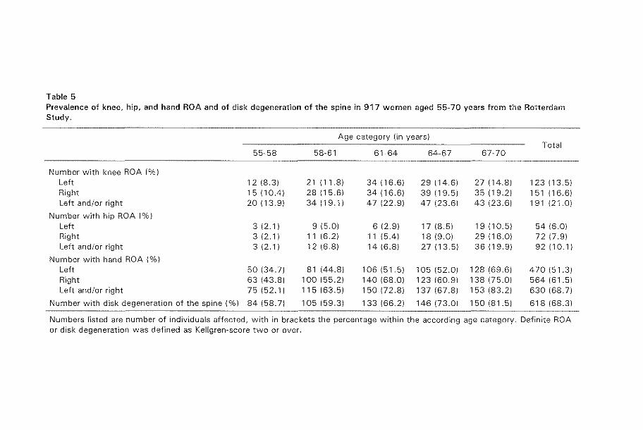
Table 5 Prevalence of knee, hip, and hand ROA and of disk degeneration of the spine in 917 women aged 55-70 years from the Rotterdam Study.
Age category (in years) Total
55-58 58-61 61-64 64-67 67-70
Number w"lth knee ROA (%)
Left 12 (8.3) 21(11.8) 34 (16.6) 29 (146) 27 (14.8) 123 (13.5) Right 15 (10.4) 28 (15.6) 34 (16.6) 39 (19.5) 35 (19.2) 151 (16.6) Left and/or right 20 (13.9) 34 (19.1) 47 (22.9) 47 (23.6) 43 (23.6) 191 (21.0)
Number with hip ROA (%)
Left 3 (2.1) 9 (5.0) 6 (2.9) 17 (8.5) 19 (10.5) 54 (6.0) Right 3 (2.1) 11 (6.2) 11(5.4) 18 (9.0) 29(16.0) 72 (7.9) Left and/or right 3 (2.1) 12 (6.8) 14 (6.8) 27 (13.5) 36 (19.9) 92 (10.1)
Number with hand ROA (%)
Left 50 (34.7) 81 (44.8) 106 (51.5) 105 (52.0) 128 (69.6) 470 (51.3) Right 63 (43.8) 100 (55.2) 140 (68.0) 123 (60.9) 138 (75.0) 564 (61.5) Left and/or right 75 (52.1) 115 (63.5) 150 (72.8) 137 (67.8) 153 (83.2) 630 (68.7)
Number with disk degeneration of the spine (%) 84 (58.7) 105 (59.3) 133 (66.2) 146 (73.0) 150 (81.5) 618 (68.3)
Numbers listed are number of individuals affected, with in brackets the percentage within the according age category. Definite ROA or disk degeneration was defined as Kellgren-score two or over.

Chapter 2.2
sites affected with ROA and/or disk degeneration increases 2.2 times in men and
3.8 times in women between the ages of 55 and 70 years.
Earlier [be prevalence of band ROA was shown (TaiJles 4 and 5). In this
case the hand was regarded a single joint site. In Figure 1 the prevalence of ROA
at different sites in the hand is shown for men and women separately. Three
interphalangeal jOint sites are distinguished (DIP-, IP- and PIP-joint.s), next to the
first carpometacarpal (CMC 1) and the trapezoscaphoideal (TS) jOints. The ra
dionavicular (RN) and distal radioulnar (RU) joints represent the jOints of the
wrists. In women all joint sites in the hands, except for the wrist joints, show a
statistical significant increased frequency with increasing age (adjusted p-value
for trend < 0.01). In men. only the IP-, [be CMC 1-, the PIP-, and the MCr joints
show a statistical Significant increase with age (adjusted p-value for trend '$
0.01). The DIP-joint.s are most often affected with ROA in both men and women.
In women the CMC 1 joint shows the strongest increase with age as compared to
all other hand joint.s) in men this holds true for the MCP-joints. Differences be
rnreen men and women in the prevalence of ROA are observed for the inter
pbalangeal joints and the first carpometacarpal joint. The prevalences of ROA in
MCP-) TS-) and wrist joints are similar in men and women.
'I11e prevalence of disk degeneration is shown in Figure 2 for men and
women separately. In none of the age categories statistically significant differ-
Table 6 Number of joint sites (knee, hip, hand, and spine) affected in 650 men and 892 women aged 55 to 70 years.
Age Number of joint sites affected Total category 0 2 3 4
Men 55-58 21123.1) 41145.1) 23 125.3) 5 15.5) 111.1) 91 58-61 26 123.9) 50 145.9) 27 124.8) 6 15.5) 0 109 61-64 20114.5) 46 133.3) 55 139.9) 17110.8) 0 138 64-67 15 19.6) 57 136.3) 67 142.7) 17110.8) 1 10.6) 157 67-70 13 18.4) 55 135.5) 64141.3) 21113.5) 2 11.3) 155
Women 55-58 30 121.0) 56 139.2) 46 132.2) 10 17.0) 1 10.7) 143 58-61 30117.4) 58 133.7) 58 133.7) 24114.0) 2 11.2) 172 61-64 20110.0) 58 129.0) 92 146.0) 27 113.5) 3 11.5) 200 64-67 16 18.1) 62131.5) 80 140.6) 29114.7) 10 15.1) 197 67-70 4 12.2) 40 122.2) 84146.7) 42 123.3) 10 15.6) 180
Numbers listed are numbers of individuals affected, with in brackets the per-centage within the according age category. Definite ROA or disk degeneration was defined as Kellgren-score two or over in the left andlor right corresponding joint.
24

70
60
50 > u c 40 0
" ~ l' 30
20
10
0
70
60
55-58
>50 u ~40
" ~ ~30
20
Hand RDA in 666 men aged 55 to 70
years
58-61 61-64 64-67 67-70
> 0 c v
" ~ l'
70
60
50
40
30
20
10
0 55-58
Population-based sample
Hand RDA in 917 women aged 55 to
70 years
58-61 61-64 64-67 67-70 age in 3-year age categories age in 3-year age categories
• DIP .,P 0 PIP 0 Mep
Hand ROA in 666 men aged 55 to 70 Hand ROA in 917 women aged 55 to
years 70
60
>50 u ~40
" ~ ~30
20
70 years
': t;;;;~:;:;;;;~::::::::::::=~ l:~~~~c=====~=====:=====:: 55-58 58-61 61-64 64-67 67-70
age in 3-year age categories
• CMel
Wrist RDA in 666 men aged 55 to 70 10 years
8
> 0
6 c 0
" ~ l' 4
2
55-58
o TS
10
B
> u 6 c
0
" ~ l' 4
2
58-61 61-64 64-67 67-70
age in 3-year age categories
Wrist RDA in 917 women aged 55 to
70 years
O~~~r----.----__ ---. o~~?=~===~--~ 55-58 58-61 64-67 67-70 55-58 58-61 61-64 64-67 67-70
age in 3-year age categori'lS age in 3-year age categories
• RN 0 RU
Figure 1 Prevalences of hand ROA and wrist ROA for 666 men and 917 women aged 55 to 70 years from the Rotterdam Study.
25

Chapter 2.2
> u c • 0 ~
~
70
60
50
40
30
Disk degeneration in 658 men
aged 55 to 70 years
)t(-)t(
/ ~
20/ )t(
-;:«- Thoracic
1~ j ----..- Lumbar
-0- Lumbosacral
55-58 58-61 61-64 64-67 67-70 age in 3-year age categories
Figure 2
> u
70
60
50
~ 40 o ~
~ 30
20
10
Disk degeneration in 898 women
aged 55 to 70 years
-::t(- Thoracic
----+- Lumbar
-0-- Lumbosacral o +--~--~-~~-~ 55-58 58-61
age in 3-year age categories
Prevalences of disk degeneration of the spine for 658 men and 898 women aged 55 to 70 years from the Rotterdam Study.
ences were founel between men and women. A statistically significant increase
with age was observed for disk degeneration of the thoracic and lumbar spine in
both men and women (p-value for trend < 0.001), but neither for men nor for
women for disk degeneration of the lumbosacral joint.
References
1. Hofman A, Grobbee DE, Dc jong PTVI'vI, van den Ouweland FA. Determinants of disease and disability in the elderly. Eur j Epidernio11991;7:403-22.
2. Odding E, Valkenburg HA, Algra D, van den Omvcland FA, Grobbee DE, I lorman A. Associations of radiological osteoanhrltis of the hlp and knee with locomotor disability in the Rotterdam Study. Ann Rheum Dis 1998;57:20.3-8.
3. Kellgren JI l, Jeffrey MR, BallJ (eds). The epidemiology of chronic rheumatism. Volllme II: Atlas of standard radiographs of arthritis. Oxford: Blackwell ScientiHc Publications, 1963.
26
2.3
Assessment of radiological osteoarthritis
in peripheral joints and of
disk degeneration of the spine
A sibling pair sample
Subjects
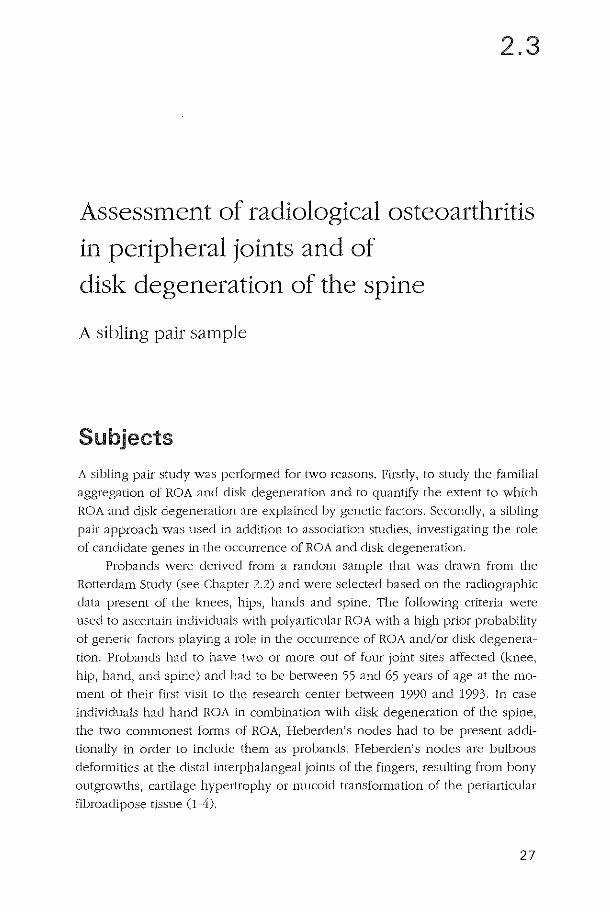
A sibling pair study was perf01l11ed for two reasons. Firstly, to study the familial
aggregation of ROA and disk degeneration and to quantify the extent to which
ROA and dbk degeneration are explained by genetic factors. Secondly, a sibling
pair approach was used in addition to association studies, investigating the role
of candidate genes in the occurrence of ROA and disk degeneration.
Probands were derived from a random sample that was drawn from the
Rotterdam Study (see Chapter 2.2) and were selected based on the radiographic
data present of the knees, hips, hands and spine. The following criteria were
used to ascertain individuals with polyarticular ROA with a high prior probability
of genetic factors playing a role in the occurrence of ROA and/or disk degenera
tion. Probands had to have two or more out of four joint sites affected (knee,
hip, hand, and spine) and bad to be between 55 and 65 years of age at the mo
ment of their first visit to the research center between 1990 and 1993. In case
individuals had hand ROA in combination with disk degeneration of the spine,
the two commonest forms of ROA, Heberden's nodes had to be present addi
tionally in order to include them as probands. Heberden's nodes are bulbolls
deformities at the distal interphalangeal joints of the fingers, resulting from bony
outgrowths, cartilage hypertrophy or mucoid transformation of the perial1icuiar
fibroadipose tissue (1-4).
27
Chapter 2.3
Probands
29 non-response of proband
24 did not have
siblings
79 did not contribute
siblings
250
221
197
Siblings
708
368
340 not eligible
111
~ non-response
of sibling
257
118
Figure 1 Participation of pro bands and siblings in sibling pair study.
no contact
deceased
168 (49.4%)
~ 63
(18.5%)
unknown
emigrated
disease of sib
Figure 2 Reasons for non-eligibility of 340 siblings.
28
Sibling pair sample
A flow chart outlining the participation of pro bands and siblings in the sib
ling pair .'::iwdy is shown in Figure 1. In total 273 persons met the criteria for in
clusion, of whom at the time of the present study 10 were deceased and 13 hJd
moved over long distance. The remaining 250 persons received a letter in which
they were invited to participate in a family study concerning ~A. Of the 221
probands whom agreed to take part in the study (response rate 88 percent) 24
had no siblings. The remaining 197 proband.') were visited at home and asked to
supply the names and, if available, addresses of all their siblings born alive. The
mean number of siblings per pedigree was 4.1 (including the proband), corre
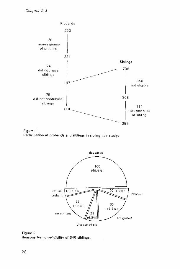
sponding to a total of 708 sibling.s born alive. Of these 708 siblings, 340 were not
eligible. The main reasons were death of the sibling, emigration and the absence
of contact between siblings. This i,s shown in detail in Figure 2. The 368 siblings
that were eligible also received a letter, in which they were invited to pal1ici
pate, which was refused by 111 sibling.s (response rate 70 percent). A majority
(64 percent) indicated that they had no particular reason to refuse.
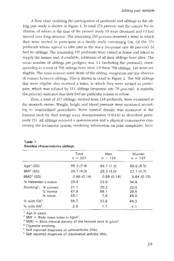
Thus, a total of 257 siblings, derived from 118 probands) were examined at
the research center. Weight) height and blood pressure were measured accord
ing to standardized procedures. Bone mineral density was measured at the
femoral neck by dual energy x-ray absorptiometIy (DEXA) as described previ
ously (5). All siblings received a questionnaire and a physical examination con
cerning the locomotor system, rendering information on joint complaints, loco-
Table 1 Baseline characteristics siblings.
Total Men n ~ 257 n ~ 124
Age 1 ISD) 65.317.9) 64.717.3)
BMI'ISD) 26.714.0) 26.3 13.0)
BMD'ISD) 0.8610.14) 0.88 10.14)
% Heberden's nodes 29.4 23.6
Smoking 4; % current 21.1 20.2
% former 47.9 68.1 % never 28.1 7.6
% with OA 5 39.7 32.8
% with RAG 2.9 1.7
1 Age in years. 2 BMI = Body mass index in kg/m 2
,
3 BMD = Bone mineral density of the femoral neck in g/cm 2.
4 Cigarette smoking. 5 Self reported diagnosis of osteoarthritis (OA). e Self reported diagnosis of rheumatoid arthritis (RA).
Women n ~ 133
65.8 18.5)
27.1 14.7)
0.84 10.15)
34.8
22.0 28.5 48.0
46.3
4.1
29
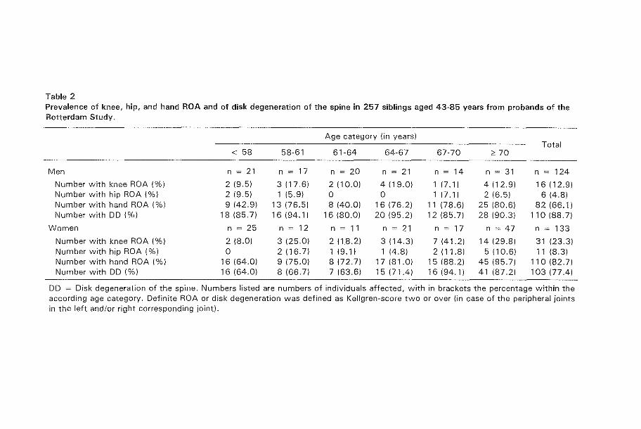
Table 2 Prevalence of knee, hip, and hand ROA and of disk degeneration of the spine in 257 siblings aged 43-85 years from probands of the Rotterdam Study.
Age category (in years) Total
< 58 58-61 61-64 64-67 67-70 ;> 70
Men n ~ 21 n ~ 17 n ~ 20 n ~ 21 n ~ 14 n ~ 31 n ~ 124
Number with knee ROA (%) 2 (9.5) 3 (17.6) 2 (10.0) 4 (19.0) 1 (7.1) 4 (12.9) 16(12.9) Number with hip ROA (%) 2 (9.5) 1 (5.9) 0 0 1 (7.1) 2 (6.5) 6 (4.8) Number with hand ROA (%) 9 (42.9) 13 (76.5) 8 (40.0) 16 (76.2) 11 (78.6) 25 (80.6) 82 (66.1) Number with DO (%) 18 (85.7) 16 (94.1) 16 (80.0) 20 (95.2) 12(85.7) 28 (90.3) 110(88.7)
Women n ~ 25 n ~ 12 n ~ 11 n ~ 21 n ~ 17 n ~ 47 n ~ 133
Number with knee ROA (%) 2 (8.0) 3 (25.0) 2 (18.2) 3 (14.3) 7 (41.2) 14 (29.8) 31 (23.3) Number with hip ROA (%) 0 2 (16.7) 1 (9.1) 1 (4.8) 2 (11.8) 5 (10.6) 11 (8.3) Number with hand ROA (%) 16 (64.0) 9 (75.0) 8 (72.7) 17 (81.0) 15 (88.2) 45 (95.7) 110 (82.7) Number with DO (%) 16 (64.0) 8 (66.7) 7 (63.6) 15(71.4) 16 (94.1) 41 (87.2) 103 (77.4)
DO = Disk degeneration of the spine. Numbers listed are numbers of individuals affected, with in brackets the percentage within the according age category. Definite ROA or disk degeneration was defined as Kellgren-score two or over (in case of the peripheral joints in the left and/or right corresponding joint).

Sibling pair sample
motor disability, Heberden's nodes, [cHnily histOlY of rheumatic diseases, occu
pation and work load, injuries and trauma's and comorbidity. As described ear
lier in Chapter 2.2 for the random sample from the Rotterdam Study, radiographs
of the knees, hips, hands and spine were taken using identical .':itanclarclized
conditions. Two independent readers used the Kellgren grading system to score
all radiographs (6), based on the protocol described in Chapter 2.2. Baseline
characteristics of the 257 siblings that participated in the sibling pair study are
shown in Table 1.
Prevalence of ROA and disk degeneration
The prevalence of ROA in the knees, hips, and hands and of disk ciegeneralion
of the spine in 124 men and 133 women aged 43 to 85 years is shown in Table
2. Hand ROA was defined as Kellgren-score two or over in any of the hand
joints that was scored, i.e. 18 for each hanel. Definite disk degeneration was de
fined as Kellgren-score two or over in any of the three levels that was scored.
In siblings of probands with ROA and disk degeneration at multiple joint
sites ROA in peripheral joints is more frequent in female siblings as compared to
male siblings. This was in keeping with the findings from the Rotterdam Study
(see Tables 4 anel 5, Chapter 2.2). In siblings, the ti'equency of disk degeneration
Table 3 Number of joint sites (knee, hip, hand, and spine) affected in 124 men and 133 women aged 43 to 85 years, siblings of probands with RDA at multiple sites.
Age Number of joint sites affected category Total
0 2 3 4
Men <58 2 {9.5} 10 {47.6} 6 {28.6} 3 {14.3} 0 21 58-61 1 {5.9} 2 {11.8} 11 {64.7} 3 {17.6} 0 17 61-64 2 {10.0} 12 {60.0} 4 {20.0} 2 {lO.O} 0 20 64-67 1 {4.8} 4 {19.0} 12 {57.1} 4 {19.0} 0 21 ~67 1 {2.2} 12 {26.7} 25 {55.6} 6 {13.3} 1 {2.2} 45
Women <58 4 {16.0} 8 {32.0} 13 {52.0} 0 0 25 58-61 0 5 {41.7} 5 {41. 7} 1 {8.3} 1 {8.3} 12 61-64 0 6 {54.5} 3 {27.3} 2 {18.2} 0 11 64-67 1 {4.8} 7 {33.3} 10 {47.6} 3 {14.3} 0 21 67-70 0 7 {I O.g} 37 {57.8} 16 {25.0} 4 {6.3} 64
Numbers listed are numbers of individuals affected, with in brackets the percentage within the according age category. Definite ROA or disk degeneration was defined as a Kellgren-score two or over in the left and/or right corresponding joint.
31
Chapter 2.3
of the spine was higher in men as compared to women, which was overall C88.7
percent in men versus 77.4 percent in women, see Table 2) statistically signifi
cant (p ~ 0.02). Within the hand, the DIP-joints were most often affected with
ROA. The MCP-, CMC-l- and IP-joints were in both men and women about
equally often affected with ROA, while in comparison herewith the frequency of
PIP-joint ROA was slightly lower. Relatively rare were ROA in the carpal trape
zoscaphoideal joint (TS) and the wrist joints (RN- and RU-joints).
In Table 3 the number of joint sites that is affected with ROA or disk de
generation i::; shown for men and women separately. Four different joint sites are
considered, i.e. the knee, hip, hand, and spine. Out of the total number of sib
lings 15.3 percent of men and 20.3 percent of women have three or more joint
sites affected with ROA and/or disk degeneration. However, 37.9 percent of all
male siblings and 28.6 percent of all female siblings have zero or one joint site
affected with ROA or disk degeneration. The latter percentages are substantially
lower as cOll1pared to the frequencies obselved in the Rotterdam Study (respec
tively 52.9 percent in men and 41.9 percent in women). In Chapter 3.2 we will
study the familial clustering and heritability of ROA and disk degeneration based
on these data.
References
1. Charcot ]M. Clinical lectures on senile and chronic diseases. London, Payne T, 1803,148.
2. Nichols·EH, Richard.son FL. AI1hritis deformans.] Med Res 1909;16:149-221. 3. Fassbender IIG. Pathology of rheumatic diseases. New York, Springer-Verlag, 1975. 4. Begg l\.1\X7, Scott]E. Hyaluronic acid and protein in simple ganglia and I Ieberden's
nodes. Ann Rheum Dis 1966;25:145-8. 5. Burger H, van Dae1e PLA, Algra D, van den Ouweland FA, Grobbee DE, Hofman A,
van Kuijk C, SchUtte lIE, Birkenhager ]C, Pols IIAP. The association between age and bone mineral density in men and women aged 55 years and over. The Rotterdam StUdy. Bone ['diner 1994;25:1-13.
o. Ke!Jgren JI r, Jeffrey IvIR, Ball J (eds). The epidemiology of chronic rheumatism. Volume II: Atlas of standard radiographs of a11hritis. Oxford: Blackwell Scientific Publications, 1963.
32

3
Osteoarthritis in the General Population


Pattern of joint involvement and
determinants of osteoarthritis at
multiple sites in a population-based
study
Abstract
3.1
Objective To investigate the determinants for radiological osteoarthritis
(ROA) and disk degeneration at multiple sites and to examine the cluster
ing of ROA in the knees, hips, and hands and disk degeneration of the
spine in the general population.
Methods A random sample of 1583 individuals, aged 55 to 70 years,
was drawn from the Rotterdam Study. Radiographs of the knees, hips,
hands, and thoracolumbar spine were scored for ROA and disk degenera
tion by means of the Kellgren grading system. Heberden's nodes were
assessed in both hands. Multiple logistic regression analysis was used to
estimate the odds ratio (OR) for the association of ROA and disk degen
eration at multiple sites with the most important risk factors for OA. In
separate multiple logistic regression analyses the clustering of joint in
volvement at the four different joint sites was examined with adjustments
for the effects of age, body mass index (BM/), and bone mineral density
(BMD) and stratified according to sex.
Results In women, BMI, BMD, and Heberden's nodes were besides age
all statistically significant risk factors of polyarticular disease. The pres
ence of knee ROA was significantly associated with radiological abnor-
35

Chapter 3. 1
malities at the hip, hand and spine. In men, only 8MI was in addition to
age significantly associated with three or more affected joint sites and
only polyarticular hand ROA was associated with disk degeneration of the
spine. Conclusion Our results strongly support the existence of a subset of
polyarticular OA in women. This is in agreement with a genetically deter
mined susceptibility for cartilage degradation, which is modified by sys
temic factors. In men, our results were less equivocal.
Introduction
Osteoanhritis eOA) is the most prevalent rheumatic disease. OA is not only a
lnajar cause of disability in the elderly but also the principal cause of knee and
hip replacements (1,2). It is characterized by a progressive degeneration of hya~
line cartilage and accompanying subchondral bone reaction of diarthrodial
joints. The exact pathogenesis of OA is unknown, but exogenous as well as en
dogenou.s factors playa role in its etiology. Although some factors, such as age
and sex, are determinants of OA at all joint !::iites, the role of most other factors
has been reponed to depend on the joint site of interest 0,4). TI1is implies that
OA is a clinically heterogeneous disorder that can be subdivided into discrete
subsets, primarily based on descriptive definitions.
Kellgren and Moore suggested that the occurrence of OA in multiple joints,
design;:lted primary generalized OA, could be identified as a specific subtype of
OA (5), They found evidence of a distinct pattern of joint involvement at multi
ple sites that was associated with Heberden's nodes. Other studies have focused
on the effect of a distinct risk factor or of OA at a particular joint site on the oc
currence of generalized OA (6-8). Generalized OA has a multifactorial etiology,
involving hormonal, metabolic, mechanical and genetic influences. Women are
more likely to have OA at multiple joint sites than men and in women there is
evidence for a polyarticular subset of hand OA (7,9). Heberden's nodes may be
predictors of OA in multiple joints, although their pathogenesis is not well un
derstood. It has been suggested that these nodal deformities represent an in
flammatOlY component in the etiology of generalized OA that, given their pre
dominant presence in women, may be controlled by h01"monal factors (10).
TIle classification criteria for generalized OA are a matter of debate and at
the population level, the pattern of joint involvement in generalized OA has not
been quantified in te1"ms of prevalence of affected joint sites. Also, the role of
risk factors that playa role in the occurrence of OA at individual jOint sites is
unclear in the etiology of generalized ~A. The present study examines whether
36

Clustering and determinants of OA
the most important risk faclOfs for OA are also determinants for radiological OA
(ROA) and disk degeneration at mUltiple sites. Furthermore, we investigated the
clustering of ROA in the knees, hips and hands and disk degeneration of the
spine in a population based study.
Materials and Methods
Study population
11w study was part of the Rotterdam Study; a prospective population based co
hort study of occurrence and determinants of disease and disability in the eld
erly. Objectives and methods of the Rotterdam Study have been described in
detail elsewhere (11). Briefly, all inhabitants of Ommoord, a district of the city of
Rotterdam, aged 55 years or over were invited to panicipate. In total 7983 par
ticipants (response rate of 78 percent) were interviewed at home and examined
extensively at the research center between 1990 and 1993. The medical ethics
committee of Erasmus University Medical School granted permiSSion to this
study. \'Vritten informed consent was obtained from all paltidpants.
A random sample of 1583 individuals, aged 55 to 70 years, was drawn from
the total cohort of the Rotterdam Study. Radiographs of the peripheral joints, Le.
the knees, hips and hands, and of the thoracolumbar spine were scored for re
spectively ROA and disk degeneration in all individuals. Heberden's nodes were
assessed in both hands separately, classified as absent or present, by trained
investigators at the research center. Bone mineral density measurements of the
femoral neck were performed using dual energy X-ray absorptiometlY (Lunar
DPX-L densitometer) as described previously (12). Height and weight were
measured, with the participants in standing position without shoes. At the base
line intelview, c1ata was collected on joint complaints and morning stiffness.
Radiological OA and disk degeneration
Weight-bearing anterior-posterior radiographs of the pelviS and knees were ob
tained with respectively both feet in 10 0 endorotation and the patellae in central
pOSition. Furthermore, posteroanterior radiographs of the hands and wrists and
lateral radiographs of the thoracolumbar spine (T114-S1) were obtained.
Radiological determined criteria were used in accordance with other epi
demiological studies concerning OA (3,9). We applied the Kellgren grading
system (13), which incorporates the classic features of radiological OA, osteo
phyte formation and joint space narrowing. 111ese radiological abnormalities
reflect the pathophysiological changes in OA. 111e five point Kellgren grades (0-
4), according to the figures and legends of the original atlas, were grade 0 =
37

Chapter 3. 1
normal; grade 1 = doubtful; grade 2 = minimal; grade 3 = moderate; grade 4 =
severe. In the analysis definite ROA in a joint was defined as Kellgren-score t\Vo
or over. Two trained obselvers, who had no knowledge of the other data of the
palticipant, independently assessed all radiogra phs. After each set of about 150
radiographs the scores of the two observers were evaluated. Whenever the
scores differed two or more points, Of, was two for one obselver but one for the
other, a consensus score was agreed upon. Radiographs of the knees and hips
had previously been scored in a similar fashion (J4). ROA of the knee was as
sessed in the tibiofemoral joint. ROA of the hand was assessed in each inter- and
metacarpalphalangeal, the first carpometacarpal, the trapezonavicular) the 1'a
dionaviclliar and distal radioulnar joints.
ROA of the spine is confined to the apophyseal joints) but these joints
could not be assessed at the lateral radiographs that were available. Although
disk degeneration is not con.sidered to be ROA of the spine) these radiological
changes may be associated with ROA. Disk degeneration was scored using the
Kellgren grades (0-4), in which a grade 0 or 1 denotes no or doubtful disk de
generation, a grade 2 denotes vertebral osteophytosis only and grades 3 and 4
vertebral osteophytosis accompanied by moderate or severe disk space nar
rowing. Three separate levels were scored, i.e. thoracic) lumbar and lumbosac
ral. Definite disk degeneration was defined as a Kellgren-score two or over in at
least one level.
Statistical analysis
The prevalence of ROA at individual joint sites was calculated in three 5-year
age strata for men and women separately. For examining ROA and disk degen
eration at multiple sites we used data from four different joint sites, i.e. the knee)
hip, hand and spine. In our study on familial aggregation of ROA and disk de
generation) we found hand ROA and disk degeneration of the spine to cluster in
families (see Chapter 3.2). This suggests a common genetic origin in which car
tilage degeneration is tbe unifying pathophySiologic ballmark. Knee ROA, hip
ROA and disk degeneration of the spine were considered as dichotomous vari
ables according to the absence or presence of this condition. To study polyar
ticular hand ROA) we categorized hand ROA according to the number of sites
affected within the hands. Six separate sites within the hands were considered)
i.e. the dist;:li and proximal interphalangeal joints) the interphalangeal joint of the
thumb, the metacarpal joints, the first carpometacarpal joint and the wrist joints.
Firstly) multiple logistic regression analysis was used to estimate the odds
ratio (OR) for the association of ROA and disk degeneration at multiple sites
with the most important risk [actors for ~A. 111e reference group in these analy
ses was the individuals free from ROA and disk degeneration. In gender specific
analyses we compared individuals with one, wo or three or more joint sites af-
38

Clustering and determinants of OA
feeted with this reference group. The independent variables were age (continu
ous), bone mineral density of the femoral neck (continuous), body mass index
(C0l1til111011S\ IIeberc\en's nodes (dichotomous), morning stiffness (dichoto
mous) and joint complaints. Joint complaints were recorded in the knee::;, hips,
hands, and lower back as absent or present. In the analyses joint complaints
were categorized in three categories: no joint complaints (reference group),
complaints in one joint site and compbints in !\Va or more joint .':iites. Joint com
plaint.s should not be regarded as a determinant of ROA and disk degeneration,
but rather as :111 indicator of the presence of clinical signs once ROA or disk de
generation has occurred.
Secondly, we investigated in separate multiple logistic regre.s.sion analyses
the clustering of joint involvement at the four different joint sites. 'I11e strength of
the association between different joint sites WJS for each pair of joint sites sum
marized by an OR, which denotes the relative odds of having one site affected if
the other site is also affected. To render these associations between pairs of joint
sites independent of the effects of both other joint sites, all four joint sites were
considered simultaneollsly in the model. All ORs were adjusted for age, bone
mineral density, and body mass index and were stratified according to gender.
Logistic regression analyses were performed using SPSS statistical software.
All ORs are presented with 95 percent confidence intervals (Cl).
Results
Table 1 shows the prevalence of grade 2+ ROA at the knee, hip and hand joints
and the prevalence of grade 2+ disk degeneration of the spine among 666 men
and 917 women, stratified in 5 year strata. At all sites in both men and women
the prevalence of ROA and disk degeneration increased with age. 111is increase
was roughly equal in men and women, except for hip ROA, in which joint in
women the prevalence increased fourfold between ages 55 and 70 years. The
prevalence of knee ROA and hand ROA was at any age between 55 and 70 years
higher in women than in men. Hip ROA was below the age of 65 years more
prevalent in men, but after this age the prevJlence of hip ROA rose steeply in
women. Disk degeneration of the spine was the most prevalent condition, oc
curring in similar frequencies in men and women.
Table 2 shows the association between the number of joint sites affected
with ROA and disk degeneration and its most impOltant determinants among
women. In women, in case that one of four joint sites is affected, only age was
statistically significantly associated with the presence of ROA and disk degen
eration. Age, bone mineral density, and Heberclen's nodes were stati.stically sig
nificant determinants of two affected joint sites. In CJse three or more joint sites
39

Table 1 Distribution of knee ROA, hip ROA, disk degeneration of the spine and hand ROA per age stratum of 5 years in 666 men and 917 women, aged 55-70 years, from the Rotterdam Study_
Men Women
55-60 years 60-65 years 65-70 years Total 55-60 years 60-65 years 65-70 years Total
frequency (n) frequency (nJ frequency (n) n frequency (n) frequency (n) frequency (n) n
Knee ROA* No 92.2 (141) 87.0 (208) 83.6 (224) 573 85.0 (210) 77.5 (265) 75.9 (242) 717 Yes 7.8 (12) 13.0 (31) 16.4 (44) 87 15.0 (37) 22.5 (77) 24.1 (77) 191
Hip RDA' No 89.5 (136) 88.2 (210) 87.1 (236) 582 95.5 (236) 93.3 (319) 81.8 (260) 815 Yes 10.5 (16) 11.8{28) 12.9 (35) 79 4.5 (11) 6.7 (23) 18.2 (58) 92
DD Spinet No 40.1 (61) 30.1 (71) 24.5 (66) 198 40.1 (99) 36.5 (123) 20.2 (65) 287 Yes 59.9 (91) 69.9 (165) 75.5 (203) 459 59.9 (148) 63.5 (214) 79.8 (256) 618
Hand RDA No 61.9 (96) 50.8 (122) 45.0 (122) 340 44.0{110) 29.6 (102) 23.3 (75) 287 1 joint site 27.7 (43) 25.4 (61) 26.6 (72) 176 35.2 (88) 31.3 (108) 22.4 (72) 268 2 joint sites 8.4 (13) 12.1 (29) 15.5 (42) 84 11.6{29) 18.8 (65) 23.6 (76) 170 ;::: 3 joint sites 1.9 (3) 11.7 (28) 12.9 (35) 66 9.2 (23) 20.3 (70) 30.7 (99) 192
* Knee radiographs were missing for 15 subjects. t Hip radiographs were missing for 15 subjects. t Radiographs of the spine were missing for 21 subjects.

Clustering and determinants of OA
are affected also body mass index became a statistically significant determinant.
111e prevalence of poly;:uticular disease in women, defined as three or more joint
sites affected, increased with EvelY year increase in age by 24 %, with EvelY 0.01
glcm2 increase in bone mineral density this prevalence increased with 8 %.
EvelY 1.0 kg/m2 increase in body mass index increased the prevalence of poly
Jlticular disease with 10 %. \Vhen Heberclen's nodes are present this prev~dencc
increased threefold (OR ~ 3.01,95 % CI 1.29.7.(0). Joint complaints at two or
more joint sites were 2.59 titnes C95°;iJ CI 1.03-6.53) more likely to be present in
women with three or more joint sites affected as compared to women with no
ROA ancino disk degeneration. All risk factors that were statistically significantly
associated with polyarticular disease showed an increasing OR with increasing
number of joint sites affected.
Table 3 shows the association between the number of joint sites affected
with ROA and disk degeneration and its most important determinants among
men. In men, as was the case in women, only age was associated with one af
fected joint site. Body mass index was a statistically significant determinant for
men with tw"o or more joint sites affected with ROA and disk degeneration. For
Heberden's nodes a significant association was only observed in men with two
Table 2 Determinants of ROA and/or disk degeneration according to the number of joint sites affected in 917 women (numbers shown are odds ratios with 95 % el).
Determinant
Age in years
BMD in cg/cm2 ~
BMI in kg/m'
Heberden's nodes (present vs absent)
Morning stiffness (present vs absent)
Complaints at: 1 joint site ~ 2 joint sites
* P < 0.05.
Number of joint sites affected r
1
1 joint site (n ~ 274)
.08 (1.02·1.16)'
1.01 (0.99·1.04)
1.03 (0.96·1.10)
1.36 (0.69·2.71)
1.10 (0.61·1.98)
0.78 (0.43·1.44) 0.95 (0.47·1.92)
2 joint sites (n ~ 360)
1.21 (1.13·1.30)'
1.03 (1.01·1.06)'
1.03 (0.95·1.11)
2.30 (1.17·4.52)*
1.33 (0.74·2,39)
1.14 (0.61·2.13) 0.83 (0.40·1.71)
~ 3 joint sites (n ~ 158)
1.24 (1.13·1 .36)*
1.08 (1.04·1 .11 ) *
1.10 (1,01·1.21)*
3.01 (1.29·7.00)*
1.05 (0.50·2.19)
2.40 (1.00·5.78) 2.59 (1.03·6.53)*
t Four different joint sites were considered, i.e. the knee, hip, hand and spine. The reference group in these analyses was the individuals free from ROA and disk degeneration at all four joint sites (n = 100). t Bone mineral density was expressed in cg/cm2 . BMD = bone mineral density. 8MI = body mass index.
41

Chapter 3. 1
joint sites affected. Neither bone mineral density nor morning stiffness was sig
nificantly associated with the presence of ROA and disk degeneration in men.
Joint complaints at two or Illore joint sites were 3.92 times (95% CI 1.17-13.2)
more likely to be present in men with polyal1icular disease as compared to men
with no joint sites affected. In men, the ORs for all factors, except Heberden's
nodes and morning stiffness, increased with increasing number of joint sites af
fected.
The associations between the four individual joint sites, showing the pat
tern of clustering for different joint sites, are shown in Tables 4 and 5 for respec
tively women and men. In women, the presence of knee ROA was statistically
significantly associated with the presence of hip ROA, hand ROA anel disk de
generation of the ~pine. The strongest association was found between knee ROA
and hand ROA in three or more joint sites (OR ~ 2.94, 95 % CI 1.72-5.03); see
Table 4. Di~k degeneration of the spine in women wa~ associated with hand
ROA in three or more joint sites (OR ~ 1.76, 95 % CI 1.09-2.83). 111is latter asso
ciation was also obselved in men (OR ~ 2.63, 95 % CI 1.19-5.82); see Table 5. In
men) there was no statistically signifICant evidence for clustering of ROA and/or
dbk degeneration at other ~ites.
Table 3 Determinants of ROA and/or disk degeneration according to the number of joint sites affected in 666 men (numbers shown are odds ratios with 95 % CD.
Number of joint sites affected t
Determinant 1 joint site 2 joint sites ;;:; 3 joint sites (n ~ 249) (n ~ 236) (n ~ 70)
Age in years 1.08 (1.02-1.16)' 1.16 (1.08-1.25)* 1.26 (1.13-1.40)*
BMD in cg/cm2 t 1.00 (0.98-1.02) 1.01 (0.98-1.03) 1.02 (0.99-1.05)
8MI in kg/m' 1.05 (0.96-1.16) 1.15 (1.03-1.27)* 1.20 (1.04-1.39)*
Heberden's nodes 1.67 (0.64-4.31) 3.08 (1.22-7.82)* 2.97 (0.86-10.3)* (present vs absent)
Morning stiffness 0.95 (0.51-1.79) 0.87 (0.44-1.70) 2.05 (0.86-4.86) (present vs absent)
Complaints at: 1 joint site 1.15 (0.64-2.06) 1.53 (0.82-2.84) 1.92 (0.78-4.76) ;;:; 2 joint sites 1.82 (0.75-4.44) 1.95 (0.78-4.86) 3.92 (1.17-13.2)*
* p < 0.05. T Four different joint sites were considered, i.e. the knee, hip, hand and spine. The reference group in these analyses was the individuals free from ROA and disk degeneration at all four joint sites (n = 95). t Bone mineral density was expressed in cg/cm2 . BMD = bone mineral density. BMI = body mass index.
42

Clustering and determinants of OA
Table 4 Pattern of joint involvement in radiological osteoarthritis (ROA) of the knee, hip, and hand and disk degeneration of the spine in women.
Knee ROA Hip ROA DO Spine OR (95 % ell OR (95 % ell OR (95 % ell
I(nee ROA
Hip ROA 1.84 (1.07-3.181'
DO Spine 1.62 (1.05-2.501' 1.58 (0.84-2.971
Hand ROA: 1 site 1.60 (0.95-2.681 1.47 (0.73-2.961 1.07 (0.74-1.551 2 sites 1.84 (1.05-3.231' 0.87 (0.38-1.971 1.28 (0.82-2.001 ;:: 3 sites 2.94 (1.72-5.031* 1.68 (0.82-3.401 1.76 (1.09-2.831'
• p < 0.05. All ORs are adjusted for age, body mass index and bone mineral density. DO Spine = Disk degeneration of the spine.
Table 5 Pattern of joint involvement in radiological osteoarthritis (ROA) of the knee, hip, and hand and disk degeneration of the spine in men.
Knee ROA Hip ROA DO Spine OR (95 % ell OR (95 % ell OR (95 % ell
Knee ROA
Hip ROA 1.62 (0.83-3.181
DO Spine 0.98 (0.56-1.721 1.24 (0.69-2.231
Hand ROA: 1 site 0.7810.42-1.451 1.58 (0.87-2.881 1.03 (0.68-1.561 2 sites 1.11 (0.53-2.301 1.84 (0.88-3.851 1.34 (0.75-2.401 ;:: 3 sites 1.19 (0.55-2.571 1.40 (0.58-3.341 2.63 (1.19-5.821'
* p < 0.05. All ORs are adjusted for age, body mass index and bone mineral density, DO Spine = Disk degeneration of the spine,
Discussion
Our remits strongly support the existence of a subset of polyarticular OA in
women. In women, body mass index, bone mineral density, and Heberclen's
nodes were besides age all stati.stically Significant risk factors of ROA and disk
degeneration in three or more joint sites. In women, the presence of knee ROA
was statistically Significantly associated with radiological abnornlalities at the
hip, hand and spine. 111is latter finding was independent of the effects of age,
43

Chapter 3.1
body mass index and bone mineral density. In men our results were less
equivocal. In men, only body mass index was in addition to age statistically sig
nificantly associated with three or more affected joint sites. Moreover, only poly
articular hand ROA was associated with disk degeneration of the spine.
A limitation of our study is the lack of data on the patellofemoral joint, the
celvica] spine, the apophyseal joints of the spine, and joints of the feet. This re
sulted most likely in an undereslimation of the clustering of ROA and/or disk
degeneration and may have diminished the detected associations. As all radio
graphs were scored irrespective of the other data of the participant studied, mis
classification wi1l most likely have been randomly distributed across the partici
pants, making the occurrence of spurious associations due to these omissions
unlikely. Several studies have suggested that in the occurrence of OA at different
joint sites different risk factors playa role (15,16). In the present study, age was
the only risk factor associated with the presence of ROA or disk degeneration at
one joint site. In women, the most important known risk factors for OA, i.e. age,
body mass index and bone mineral density were all stati:.itically significantly as
sociated with polyarticular disease. In men, polyal1icular disease was associated
with age and body mass index. These findings suggest a generalized effect, i.e.
not acting joint site specific, of body mass index on the occurrence of ROA and
disk degeneration. In women, bone mineral density was found to have a similar
generalized effect. Our findings are compatible with the hypothesis of a suscep
tibility of genetic origin, which gives rise to OA whenever additionally one or
more systemic risk factors are present. 'n1e clustering of knee ROA with ROA at
the hip and hand and with disk degeneration of the spine, adjusted for age,
body mass index, and bone mineral density, SUpp0l1S this hypothesis of a gen
eralized susceptibility in polyal1icular ~A. The association of disk degeneration
of the spine with knee ROA in women and with polyarticular hand ROA in both
men and women suggests that peripheral ROA and disk degeneration share a
common pathogenesis. As these associations were independent of the influence
of systemic risk factors for OA, this common pathogenesis may be genetically
determined. To our knowledge this is the first study suggesting that disk degen
eration of the spine can be included in the concept of polyal1icular OA in
women.
The role or meaning of Heberden's nodes in generalized OA has been un
clear. 111e present study shows that in both men and women Heberden's nodes
are strong predictors of polyarticular disease, although in men with three or
more joint sites affected this was not statistically significant. In an analysis in
which we stratified according to the presence or absence of Heberden's nodes,
the associations with age, body mass index, and bone mineral density were not
essentially different in individuals with Heberden's nodes as compared to incli-
44
Clustering and determinants of OA
vicluals withOllt Heberclen's nodes (data not shown), However, in those with
Heberclen's nodes we found a strong association betvleen polyarticular disease
(three or rnore joint sites affected) and morning stiffness in both men and
women. 11115 finding suggests that the presence of Heberc!en's nodes is associ
ated with an inflammatOlY component in the pathogenesis of OA, giving rise to
a more aggressive progression of the disease and/or complaints. This is in line
with the high prevalence of Heberden' s nodes that is usually obsclvecl in clinical
OA.
Two earlier population-based studies have examined the pattern of joint
involvement in generalized OA (7,17). A British study by Cooper et a1. suggested
the existence of polyarticular OA in women, based on the Jssociation of knee
ROA with hip and hand ROA (17). Neither men nor the role of putative risk fac
tors were examined. An American study by Hirsh et a1. showed evidence for the
existence of polyarticular disease in both men and women based on the asso
ciation of knee and hand ROA (7). For women the findings of the present. study
were consistent with both previous studies. For men our findings were less out
spoken I as clustering of ROA was restricted to polya111cular hand ROA and disk
degeneration of the spine.
In conclusion I our results strongly SUppOlt the existence of a subset of
polyarticular OA in women. In women I age, body mass index, and bone mineral
density were all statistically significantly associated with polyal1icular disease. In
men, this was only observed for age and body mass index. For women, our
findings are in keeping with a genetically determined susceptibility for caltilage
degradation, which is modified by systemic factors.
References
1. Felson DT. Epidemiology of hip and knee osteoarthritis. Epidemiol Rev 1988;10:1-28. 2. I3ulstrode C. Keeping up with 011hopaedic epidemicS. I3NIJ 1987;295:511 . . 1. Cerhan JR, Wallace RB, El-Khoury GY, Moore TE. Risk factors for progression to new
sites of radiographically dellned osteo;uthritis in women. J Rheumatol 1996;23:1565-78.
1. Peyron JG. The epidemiology of osteoanhritis. In: NIoskowitz RW, Howell DS, Goldberg VM, .Mankin I-IS, eds. Osteoal1hritis: diagnosis and management. Saunders: Philadelphia and London, 1981:9-27.
5. Kellgren JI-I, Moore R. Generalised osteoarthritis and fIeberden's nodes. BM] 1952;USI-7.
6. Kellgren Jll, Lawrence JS, Bier F. Genetic factors in generalized osteo-arthrosis. Ann Rheum Dis 1963;22:237-54.
7. Hirsh R, Lelhbridge-Cejku NI, Scott Jr \'VW, Reichle R, Plato CC, Tobin], IIochberg Me. Association of hand and knee osteoallhritis: evidence for a polyallicubr disease subset. Ann R.heum Dis 1996;55:25-9.
45

Chapter 3. 1
8. Croft P, Cooper C, Wickham C, Coggon D. Is the hip involved in generallzed osteoarthritis? Br J Rheumatol 1992;31:325-8.
9. Egger P, Cooper C, Halt DJ, Doyle DV, Coggon D, Spector TD. Patterns of joint involvement in ostcoal1hritis of the hand: the Chingford Study. ] Rheumatol 1995;22; 1509-13.
10. Spector 'I'D, Campion GD. Generalized osteoanhritis: a honnona!ly mediated disease. Ann Rheum Dis 1989;18:523-7.
11. Hofman A, Grobbee DE, Dc long PTV?vf, Van de Ouweland FA. Determinants of disease and disability in the elderly. Em] EpidemioI1991;7:403-22.
12. Burger H, van Daele PLA, Algra D J van den Ouweland FA, Grobbee DE, Hofman A, van Kuljk C, SchUtte I-IE, Birkenhager .Ie, Pols HAP. The association between age and bone mineral density in men and women aged 55 years and over: the Rotterdam Study. Bone l'diner 1991/;25:1-13.
n. Kellgren JH, Jeffrey IvIR, Ball J (eds). The epidemiology of chronic rheumatism. Volume II: Atlas of standard radiographs of a11hritis. Oxford: Blackwell Scientific Publications, 1963.
14. Odding E, Valkenburg IIA, Algra D, Van den Ouweland FA, Grobbee DE, Hofman A. Associations of radiological osteoal1hritis of the hip and knee with locomotor disability in the Rotterdam Study. Ann Rheum Dis 1998;57:203-8.
15. Felson DT, Zhang Y. An update on the epidemiology of knee and hip osteoa11hritis with a view to prevention. Althritis IU1eum 1998;4] :1343-55.
16. Creamer P, Hochberg Ivrc, Osteoa11hritis. Lancet 1997;350:503-8. 17. Cooper C, Egger P, Coggon D, Hal1 DJ, IvIasud T, Cicuttini F, Doyle DV, Spector TD.
46
Generalized osteoalthritis in women: pattern of joint involvement and approaches to definition for epidemiologic:ll studies. J Rheumatol 1996;23:1938-42.
3.2
Heritabilities of radiological
osteoarthritis in peripheral joints and of
disk degeneration of the spine
Abstract
Objective To estimate the genetic influence on the occurrence of radio
logical osteoarthritis (ROA) in the knees, hips and hands and disk degen
eration of the spine in the general population.
Methods A random sample of 1583 individuals was drawn to estimate
the prevalence of ROA and disk degeneration in the general population. Of
118 probands with multiple affected joint sites, derived from this sample,
we were able to recruit 257 siblings. The variance of ROA and disk de
generation within sibling pairs was compared with the variance between
pairs of siblings. Heritabilitv estimates for ROA in the knees, hips, and
hands and for disk degeneration of the spine were calculated. Osteoarthri
tis (OA) was defined on radiological criteria, using Ke/lgren's grading svs
tem.
Results We observed that hand ROA and disk degeneration of the spine
were statisticallv significantlv more frequent in siblings as compared with
the random sample, whereas the prevalence of knee and of hip ROA was
similar and lower, respectivelV. Heritabilitv estimates for hand ROA and
disk degeneration were statisticallv significant, respectivelv 0.56 (95 per
cent CI 0.34-0.76) and 0.75 (95 percent CI 0.30-1.00), For knee and hip
47
Chapter 3.2
ROA no evidence for a genetic effect in the general population was found.
Finally, the heritability estimate for a score summing the number of joints
affected in the knees, hips, hands and spine was O. 78 (95 percent CI
0.52-0.98). All heritability estimates were adjusted for age, sex, body
mass index, and bone mineral density.
Conclusion The present study shows that in the general population a
strong genetic effect exists for hand ROA and disk degeneration of the
spine. The findings on the total number of joints affected at multiple sites
suggest genetic susceptibility to generalized OA.
Introduction
A genetic effect on osteoalthritis eOA) was initially recognized in 1941 by
Stecher, who showed that Heberden's nodes of the fingers were more common
in sisters af affected subjects than in the papulatian (1). In 1963, Kellgren et al
(2) reponed, in a study of subjects derived from an out-patient clinic, that first
degree relatives of subjects with generalized radiological OA (ROA) were twice
as likely ta be affected with ROA than expected in the papulatian. In 1996,
Spector et al (3) measured ROA in hands and knees in female twins and sug
gested that genetic factors might explain up to 65 percent of the variability in
ROA af the hand and knee. 111is familial aggregatian af hand and knee ROA was
confirmed recently in two population-based studies (4,5). Flilthermore, Felson et
al (4) found evidence, in a segregation analYSiS, for effects of a major recessive
gene with a residual multifactorial component.
With the exception of the study by Kellgren et al (2), previous studies con
cerning the role of genetic factors in the occurrence of ROA were limited to OA
of the hands and knees 0-5). The contribution of genetic factors to other com
mon forms of OA, including hip ROA and disk degeneration of the spine, has
not been studied. Two studies addressed the role of body mass index in the fa
nlilial aggregation of ROA (4,5), but none of the previous studies examined the
role of bone mineral denSity. This is impOltant because bone mineral density,
like body mass index, i.s a risk factor for OA ZI11e1 is strongly influenced by ge
netic faclars (6,7).
We performed a population-based study on the contribution of genetic
factors to the occurrence of ROA in the knees, hips, and hands and of disk de
generation in the spine. Heritability estimates for ROA anel disk degeneration
were calculated, using data af a randam sample af 1583 individuals and 257 sib
lings of subjects with I\OA and disk degeneration at mUltiple joint sites.
48

Heritability
Methods
Subjects
The study WZI.':i embedded in the Rotterdam Study; a prospective population
based follow-up study of determinants and prognosis of chronic diseases in the
elderly (8). All persons living in Ommoorci, a suburb of Rotterdam, aged 55
years and over were invited to participate. In total 7983 participants (re::;ponse
rate of 78 percent) were examined. The medical ethics committee of Erasmus
University Medical School has approved the study and written informed consent
was obtained from all patticipants.
'In order to quantify the occurrence of ROA anel disk degeneration in the gen
eral population, we have drawn a random sample of 1583 individuals aged 55 to
70 years fro111 the Rotterdam Study. Radiographs of the peripheral joints, Le. the
knees, hips and hands, and of the thoracolumbar spine were scored [or respec
tively ROA and disk degeneration in all individuals. To estimate the genetic
component in the etiology of ROA and disk degeneration, we studied the sib
lings of a subsample of 250 pro bands Caged between 55 and 65 years) with ROA
and disk degeneration in at least tvvo joint sites. In this relatively young age
categ01Y, genetic predisposition may playa more predominant role than in the
elderly, in whom environmental factors and aging may be more important de
terminants. Probands were selected based on the radiological abnonnalities
present on the radiographs of the knees, hips, hands, and spine. Probancb had
to have tvvo or more of these four joint sites affected. Except when individuals
had hand ROA in combination with disk degeneration of the spine, both highly
prevalent conditions, which are likely to occur in combination by chance in high
frequency. In that case, a proband had to have additional Heberden's nodes in
order to reduce the number of siblings to be studied. This selection of probands
was carried out in order to ascertain the group with the highest a priori prob
ability of genetic factors playing a role in the occurrence of ROA.
Of the 221 probands that were willing to participate (response rate of 88
percent), 24 had no siblings. The remaining 197 probands had a total of 708 sib
lings born alive. Of these 708 siblings, 168 were deceased, 63 lived abroad, 65
could not be contacted, 24 were not able to participate because of a disease
other than OA, and for 20 individuals the reason for not participating was un
known. Fronl the 368 siblings that could be contacted, we were able to recnlit
257 siblings (70 percent). These 257 siblings were derived from 118 pro bands
and were examined at the research center. A majority (64 percent) of the 111
sibling.s that upon request refused to enter into the study indicated that they had
no particular reason to do so.
49

Chapter 3.2
Measurements
For all individuals the following radiographs were obtained: weight bearing an
terior-posterior pelvic radiograph.') with both feet in IOoendorotatioll, weight
bearing knee radiographs with the patellae in central position, anteropo.sterlor
radiographs of the hands and wrists and lateral radiographs of the spine (1114-
Sl).
111e exact definition of OA remains a matter of debate, but the use of radi
ologically determined changes as can be seen on radiographs is widely accepted
in epidemiological research concerning OA (9,10). We used the Kellgren grading
system (I 1) for all joint sites, since it incorporates both classic features of radio
logical OA, osteophyte formation and joint space narrowing and to our opinion
up until now no convincing evidence has been brought fOlward to regard these
features as completely independent markers of disease. Thus) ROA was assessed
by means of the Kellgren grading system in five grades (0-4), using the figures
and legends of the original atlas. Two independent readers) who had no know
ledge of the other data of the palticipant) scored all radiographs. After each set
of about 150 radiographs the scores of the two readers were evaluated) in order
to reduce bias related to int1'a- and inter-rater agreement. Whenever the scores
were two or more points different) or) was two for one reader but one for the
other) a consensus score was agreed upon. Previous validation studies con
cerning the use of the Kellgren grading system showed that full agreement be
tween obselvers varied from 82 percent in case of hip ROA to 60 percent in case
of disk degeneration of the spine (12). ROA of the knee was only assessed in the
tibiofemoral joint. ROA of the hand was assessed in each inter- and metacarpal
phalangeal joint, the first carpometacarpal) the trapezoscaphoideaC the radiona
vicular and distal radioulnar joints.
By definition) ROA of the spine is confined to the apophyseal joints, but
these joints could not be assessed on the lateral radiographs of the spine that
were available. In stead) we assessed disk degeneration of the spine, of which
the genetic etiology may be associated with the occurrence of ROA in the pe
ripheral joints (13). Disk degeneration was scored using the Kellgren grades (0-
4), in which a grade zero or one denotes no or doubtful disk degeneration, a
grade two denotes vertebral osteophytosis only and grades three and four velte
bral osteophytosis accompanied by moderate or severe disk space narrowing.
Three levels were scored separately, i.e. thoracic, lumbar and lumbosacral.
The presence of Heberden's nodes was determined by an examination of
the hands) which was performed by trained investigators at the research center.
Heberden's nodes were scored in both hands separately, classified as absent or
present, without knowledge of the radiographic findings. Bone mineral density
was rneasured at the femoral neck by dual energy x-ray absorptiometIy as de-
50
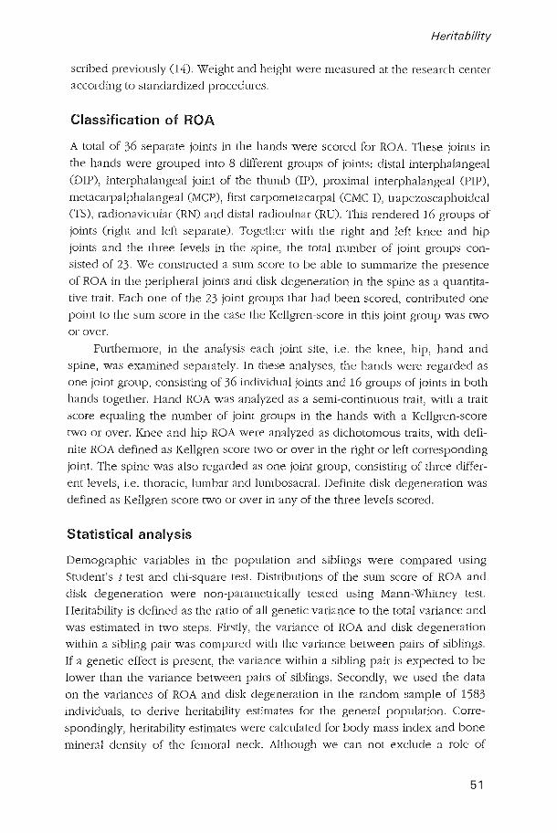
Heritabilitv
scribed previously (14). Weight ~mcl height were measured at the research center
according to standardized procedures.
Classification of ROA
A total of 36 separate joint.s in the hands were scored for ROA. These joints in
the hand.s were grouped into 8 different groups of joints: distal interphalangeal
(DIP), interphalangeal joint of the tbumb (Ip), proximal interphalangeal (PIP),
metacarpalphalangeal (MC!'), first carpometacarpal (CMC I), trapezmcapboideal
(TS), radionavicular (RN) and elistal radioulnar (RU). Tbis rendered 16 groups of
joints (rigbt and len separate). Together witb the right and left knee and bip
joint.s and the three leveb in the spine, the total number of joint groups (011-
si.sted of 23. We constructed a sum score to be able to summarize the presence
of ROA in the peripheral joints and dbk degeneration in the spine as a quantita
tive trait. Each one of the 23 joint groups that had been scored, contributed one
point to the sum score in the case the Kellgren-score in this joint group was two
or over.
Furthermore, in the analysis each joint site, I.e. the knee, hip, hand and
spine, was examined separately. In these analyses, the hands were regarded as
one joint group, consisting of 36 individual joints and 16 groups of joints in both
hands together. Hand ROA was analyzed as a semi-continuous trait, with a trait
score equaling the number of joint groups in the hands with a Kellgren-score
two or over. E:l1ee and hip ROA were analyzed as dichotomous traits, with defi
nite ROA defined as Kellgren score two or over in the right or left corresponding
joint. The spine was also regarded as one joint group, consisting of three differ
ent levels, i.e. thoracic, lumbar and lumbosacral. Definite disk degeneration was
defined as Kellgren score two or over in any of the three levels scored.
Statistical analysis
Demographic variables in the population and siblings were compared using
Sttldent's t test and chi-square test. Distrihutions of the sum score of ROA and
disk degeneration were non-parametrically tested using Mann-Whitney test.
Herita bility is defined as the ratio of all genetic variance to the total variance and
was estimated in two ;;tep;;. Firstly, the variance of ROA and disk degeneration
within a sibling pair was compared with the variance between pairs of siblings.
If a genetic effect is present, the variance within a Sibling pair is expected to be
lower than the variance between pairs of sibling;;. Secondly, we used the data
on the variances of ROA and disk degeneration in the random sample of 1583
individuals, to derive heritability estimates for the general population. Corre
spondingly, herita bility estimates were calculated for body mass index and bone
mineral density of the femoral neck. Although we can not exclude a role of
51

Chapter 3.2
shared environmental factors early in life, the influence of shared environment
on the occurrence of ROA at late middle age is expected to be limited, given that
sibling.s lead separate lives. Flllthennore, the correlation of OA in spouses has
been found to be low (4), suggesting the absence of a strong environmental
factor. At the individual level adjustments were made for known genetic risk
factors for ROA, which siblings may share, including age, sex, body mass index
and bone mineral density.
To calculate the heritability estimates, a random effects model, in which
random effects represent genetic effects (15), was fitted using maximum likeli
hood estimation (16). For the normally distributed outcomes, Le. sum score of
ROA and disk degeneration (log transformed), hand ROA (log transformed),
body mass index and bone mineral density of the femoral neck, a linear model
was used. A logistic model was used for the binaIY outcomes, Le. knee ROA, hip
ROA and disk degeneration of the spine. Heritability estimates are presented
with 95 percent confidence intervals eel). The heritability estimates for ROA and
disk degeneration are applicable to the general population under the assump
tions that, the sample drawn from the Rotterdam Study is a random sample from
the population, the genetic variance within a sibling pair i:::; independent of ROA
status, and the influence of shared environment on this late onset disease is lim
ited.
Results
Characteristics of the 1] 8 probands, their 257 corresponding siblings, and the
random population based sample of 1583 individuals, from which the probands
were derived, are shown in Ta ble 1. Four joint sites were affected in 5 probands,
47 probands had three affected joint sites, and 66 probands had a combination
of two joint sites affected. Of the 76 probands with at least hand ROA and disk
degeneration of the spine, the combination most frequently affected, 28 had ad
ditionally Heberden's nodes. Sibling.s were recruited about four years after the
probands had been examined at the research center (see Table 1). 111e fre
quency of knee ROA in siblings was similar to the frequency found in the total
sample. The frequency of hip ROA in siblings was lower than would be ex
pected based on the data from the total .':.lJmple. Hand ROA and disk degenera
tion was significantly more frequent in siblings as compared to the total sample.
The distributions of the sum score of ROA and disk degeneration, expressing the
total number of joints affected, for probands, siblings, and the random sample
derived from the RotterdzUll Study are shown in Figure 1. This figure shows that
the distribution for siblings is positioned in between the distributions of the pro
band.::; Jnd the total sample. The median of the sum score in siblings was signifi-
52

Table 1 Characteristics of probands (n ::::: 118) and their corresponding siblings {n (n = 1583)' from which the probands were derived.
257) in relation to a random population sample
Male Male Men Female Female probands siblings total sample probands siblings n ~ 33 n ~ 124 n ~ 666 n ~ 85 n ~ 133
Age (SO) in years 61.1 (2.71 64.7 (7 31 63.4 (4.1) 60.8 (2.7) 65.8 (8.5)-Range 56 - 65 45 - 82 55-70 55 - 65 43 - 85
Body mass "Index (SO) in kg/m2 27.2 (2.4) 26.3 (3.0) 25.9 (3.0) 27.6 (4.7) 27.1 (4.7)
Bone mineral density (SO) in g/cm2 0.93 (0.13) 0.88 (0.14) 0.89 (0.13) 0.90 (0.14) 0.84 (0.15)
Number with knee ROA (%) 14 (42.4) 16(12.9) 87 (13.1) 56 (65.9) 31 (23.3)
Number with hip ROA (%) 12 (36.4) 6 (4.8)' 79(11.9) 18 (21.2) 11 (8.3)
Number with hand ROA (%) 24 (72.7) 82(66.1)- 326 (48.9) 73 185.9) 110(82.7)-
Number with disk degeneration (%) 28 (84.8) 110 (88.7)- 456 (68.5) 67 (78.8) 103 (77.4)'
Median of sum score .- 3.0 (2,8) 3.0 (1,9)' 2.0 (0,6) 5.0 (2,9) 5.0 (1,10)'
• p ::s; 0.001 and T 0.01 > P < 0.05 (siblings compared with total cohort for men and women separately) . .- Sum score of ROA and disk degeneration with in brackets the 101h and 90 th percentile values. 9 p < 0.001 (siblings compared with total sample using Mann-Whitney test). ROA = radiological osteoarthritis.
Women total sample
n ~ 917
62.9 (4.1) 55-70
26.6 (4.2)
0.83 (0.13)
191 (20.8)
92 (10.0)
630 (68.7)
607 (66.2)
3.0 (0,7)
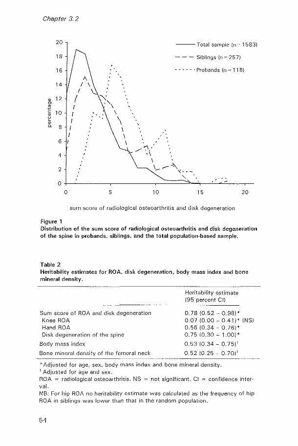
Chapter 3.2
20
18
16
14
ID 12
'" ro ~ c 10 ID 0
ID D. 8
6
4
2
( I I I I I I
o
" / \ / \
~ ,,\
5
\
\ \ \ \
--- Total sample (n = 1583)
- - - Siblings (n = 257)
- - - . - - Probands (n = 118)
10 15 20
sum score of radiological osteoarthritis and disk degeneration
Figure 1 Distribution of the sum score of radiological osteoarthritis and disk degeneration of the spine in pro bands, siblings, and the total population-based sample.
Table 2 Heritability estimates for ROA, disk degeneration, body mass index and bone mineral density.
Sum score of ROA and disk degeneration Knee ROA Hand ROA Disk degeneration of the spine
Body mass index
Bone mineral density of the femoral neck
Heritability estimate (95 percent CI)
0.78 (0.52 - 0.98)* 0.07 (0.00 - 0.41)* (NS)
0.56 (0.34 - 0.76)* 0.75 (0.30 - 1.00)*
0.53 (0,34 - 0.75)'
0.5210.25 - 0.70)'
* Adjusted for age, sex, body mass index and bone mineral density. 1 Adjusted for age and sex. ROA = radiological osteoarthritis. NS = not significant. CI = confidence interval. NB: For hip ROA no heritability estimate was calculated as the frequency of hip ROA in siblings was lower than that in the random population.
54

Heritability
cantly higher than the median of the sum score found in the random sample and
was similar to that in probands.
111C heritability estimate of the sum score of ROA and disk degeneration
was 0.78 (95 percent CI 0.52-0.98), after adjustment for age, sex, body mass in
dex and bone mineral density Cfable 2). This implies that up to 78 percent of the
variance in the sum score of ROA and disk degeneration is explained by genetic
factors, independent of the influences of age, sex, body mass index and bone
mineral density. For the individual joint sites, heritability estimates are given in
Table 2. Tn these analyses, hand H.GA was analyzed as a normally distributed
trait, according to the number of joint groups affected in both hands (at maxi
mum 16). Knee and hip ROA, and disk degeneration of the spine were analyzed
as dichotomous traits. Table 2 shows that disk degeneration had the highest
heritability (0.75 with 95 percent CI 0.30-1.00). Hanel ROA was also statistically
significantly correlated in siblings (heritability estimate: 0.56 with 95 percent CI
0.34-0.76). However, ROA in the knee was not statistically significantly corre
lated in family members. The frequency of hip ROA in siblings was even lower
than that in the random sample. Only higher than expected concordance rates in
siblings can be explained biologically, therefore no heritability estimate was cal
culated for hip ROA.
Heritability estimates for body mass index and bone mineral density of the
femoral neck were abo statistically significant, independent of age and sex, 0.53
(95 percent CI 0.34-0.75) and 0.52 (95 percent CI 0.25-0.70)' respectively.
Discussion
The present study suggests that genetic factors playa substantial role in the oc
currence of radiological OA in the general population. Siblings of subjects with
ROA and disk degeneration at multiple joint sites had higher frequencies of
hand ROA and disk degeneration. However, the frequencies of knee ROA and
hip ROA were equal and lower, respectively, as compared to a random sample
of individuals derived from the Rotterdam Study. When considering the total
number of joints affected at four separate sites, Le. the knees, hips, hands and
spine, we found that IIp to 78 percent of the total variance of this sum score of
ROA and disk degeneration was explained by genetic factors. In particular hand
ROA and disk degeneration of the spine showed a statistically significant aggre
gation in siblings. Interestingly, the genetic influence on ROA established here is
independent of the well-known genetic influences present in body mass index
and bone mineral density, which are also obselved in this study. Since heritabil
ity estimates were calculated with the use of both the data from the sibling pairs
and a random population based sample from which the probands were selected,
55
Chapter 3.2
these estimates are applicable to the general population (16). Our data suggest
the existence of a genetic susceptibility to generalized cartilage degeneration,
independent of the genetic influences in body mass index and bone mineral
density.
We compared the findings of the three previous studies on the heritability
of OA, including a twin study and two population-based studies 0-5), with the
results of the present shiely. For the sake of comparability we also calculated sib
sib correlations. Except for the twofold higher correlation of hand ROA between
siblings in the study by Hirsch et a1. (5), the sib-sib correlations in all four studies
were reasonably similar, despite differences bet ween studies in type of study
population and definition criteria for ROA. The present study is the first to report
an increased frequency of disk degeneration in siblings of probands with ROA
and disk degeneration, suggesting a shared genetic etiology for ROA and disk
degeneration in humans. Furthermore, the present study suggests a genetic pre
disposition for ROA and disk degeneration at multiple sites as measured by the
sum score. Although two earlier studies have examined familial aggregation of
ROA by sununing the number of joints affected, this is the first study to include
hip ROA and disk degeneration of the spine in the sum score (4,5). Disk degen
eration in addition to hand ROA showed the highest heritability estimates,
whereas the evidence for familial aggregation was weakest for the weight-bear
ing joints, i.e. the knee and hip joints, as repOlted earlier (2,3,5). For the knee
joint, environmental factors, e.g. former trauma, may playa major role in the
development of ROA. Several studies of autosomal dominant hip ROA suggest a
genetic defect underlying hip ROA due to dysplasia, findings of the present
study suggest that such major genes contribute little to hip ROA in the general
population 07,18). It should be noted that the power of the present study was
most likely lower for knee and hip ROA, dichotomous traits, than for hand ROA,
disk degeneration of the spine and the sum score of ROA and disk degeneration,
that could be analyzed as quantitative traits.
There are several potential biases associated with our study. Selection bias
might playa role when response rates were associated with ROA. Of the 368
siblings that were eligible for our study III refused to participate. In most in
stances there was no pal1icular reason for not participating. Seven subjects indi
cated that they were known to have OA and did not participate for that reason.
Although in general, non-response has most likely been at random, disability in
elderly subjects with hip OA may have led to an underestimate of the number of
hip ROA cases in siblings. Lateral knee radiographs were not available, which
means no data could be presented on patellofemoral ~A. Finally, heritability
estimates were not calculated for men and women separately due to insufficient
numbers of sibling pairs of the same sex.
56

Heritabilitv
It is a priori unlikely that environmental factors shared by siblings early in
life will influence the development of ROA and disk degeneration at late middle
age such that it could explain the c1ata found in the present study. Moreover, we
adjusted for possible shared factors by siblings, sllch as age and sex, body mass
index anel bone mineral density. This did not change the results of the analyses
essentially and the evidence for familial aggregation of ROA anel disk degenera
tion remained statistically Significant. Fll11hennorc, our result on hand ROA and
to a lesser extent on knee ROA was comparable to the results of Spector et a1 in
a tvlin study [3], in which adjustments were made for environnlental factors
shared by twins, by comparing monozygotic and dizygotic twin pairs. 111is sug
gests that the influence of shared environment on the familial aggregation of
ROA may be velY limited, which was recently suppOlted by rejection of an envi
ronmental model in a segregation analysis of knee and hand ROA,( 4).
In conclusion, the present study shows a strong genetic effect for ROA and
disk degeneration at multiple sites. Up to 78 percent of the total variance in ROA
and disk degeneration is explained by genetic factors, independent of the (ge
netic) influence of body mass index and bone mineral denSity. In particular, a
strong familial aggregation was found for hand ROA and disk degeneration of
the spine. We found no evidence for a statistically significant genetic effect on
the occurrence of knee and hip ROA at the population level. Previous findings
suggest that there may be two possible genetic pathways, firstly the existence of
a com111on recessive allele (frequency 0.45) and secondly a polygenic form of
inheritance, perhaps in interaction with environmental factors (4). The two dif
ferent mechanisms involved may explain the strong clustering of ROA in fami
lies. However, the genes underlying the familial aggregation of ROA in the
population remain to be determined.
References
I. Stecher iUvl. Heberden's nodes. I reredity in hypertrophic al1hrilis of the finger joints. Am J Med Science 1941 ;20"1 :801.
2. Kellgren JH, Lawrence ]S, Bier F. 1963. Genetic factors in generalized osteoarthrosis. Ann Rheum Dis 196.1;22:237-55.
3. Spector TD, Cicuttini F, Baker .J, Loughlin j, IIan D. Genetic influences on osteoarthritis in women: a twin study. BM] 1996;312:940-.1.
4. Fclson DT, COllroprnitree NN, Chaisson eE, Hannan MT, Zhang Y, McAlindon TE, et al. Evidence for a mendel ian gene in a segregation analysis of generalized radiographic osteoal1hritis. Al1hritis Rheum 1998;41:1064-71.
5. Hirsch R, Lethbridge-Cejku lvI, Hanson R, Scott W\y/, Reichle R, Plato cc, ct a1. Familial aggregation of osteoarthritis. Arthritis Rheum 1998;41:1227-32.
57
Chapter 3.2
6. Burger H, van Dade PLA, Odding E, Valkenburg HA, Hofman A, GroLbee DE, et al. Osteo::111hritis is associated with higher bone mineral density and increased bone loss with age: The Rotterdam Study. Allhritis Rheum 1996;39:81-6.
7. Pocock NA, Eisman .lA, Hopper .Te, Yeates MG, Sambrook PN, Eberl S. Genetic determinants of bone mass in adults. A twin study. ] CUn Inves11987;80:706-1O.
8. Hofman A, Grobbce DE, Dc Jong FVfM, van den ouweland FA. Determinants of disease and disability in the elderly. Ellr) Epidemlol1991;7:403-22.
9. Silman AJ, Hochberg Me. 1993. The epidemiology of the rheumatic diseases. Oxford, Oxford University Press 257-88.
10. Peyron .IT, AJtman R. 1992. The epidemiology of osteoarthritis. In Osteoarthritis: pathogenesis and management. Moscowitz R\X1, Howell DS,Goldberg VM, Mankin HJ, editors, 2nd edition. London, Saunders-Harcolllt. 15-37.
11. Kellgren JB, Jeffrey MR, Ball J. (editors). 1963. The epidemiology of chronic rheumatism. Volume II: Atlas of standard radiographs of althritis. Oxford: Blackwell Scientific Publications.
12. Haanen HCM. 1984. An epidemiological study on low-back pain. Thesis. Erasmus University Rotterdam, The Netherlands.
13. Kimura T, Nakata K, Tsumaki N, Miyamoto S, Matsui Y, Ebara S, ct al. Progressive degeneration of articular cal1ilage and interveltebral discs. An experimental study in transgenic mice bearing a type IX collagen mutation. Int Ol1hopaed 1996;20:177-81.
14, Burger H, van Dacle PLA, Algra D, van den Ouweland FA, Grobbee DE, Hofman A, et al. The association between age and bone mineral density in men and women aged 55 years and over: The Rotterdam Study. Bone Miner 1991;25:1-13.
15. Houwing-Duistermaat JJ, Derkx BHP, Rosendaal PR, van Houwelingen ne. Testing familia! aggregation. l3iometrics 199551:1292-301.
16. HOllwing-Duistermaat JJ, van I-Iouwelingen HC, Terhell A. Modelling the cause of dependency with application to filaria infection data. Statistics in Medic. In press.
17. Sher C, Ramesan R, Maltel! R, Learmoth I, Tsipouras P, Beighton P. Mild spondyloepiphyseal dysplasia (Namaqualand type): genetic linkage to the type II collagen gene COL2A1. Am J Hum Cen 1991;48:518-24.
18. Kaitila I, Korkko J, Mautinnen E, Ala-Kokko L. Phenotypic expressions of a Gly 151 Arg !Dutation in type II collagen in two unrelated patients with spondyloepimetaphyseal dysplasia (SEMD). Am] Med Gen 1996;63;111-22.
58

4
Collagen Genes and Osteoarthritis

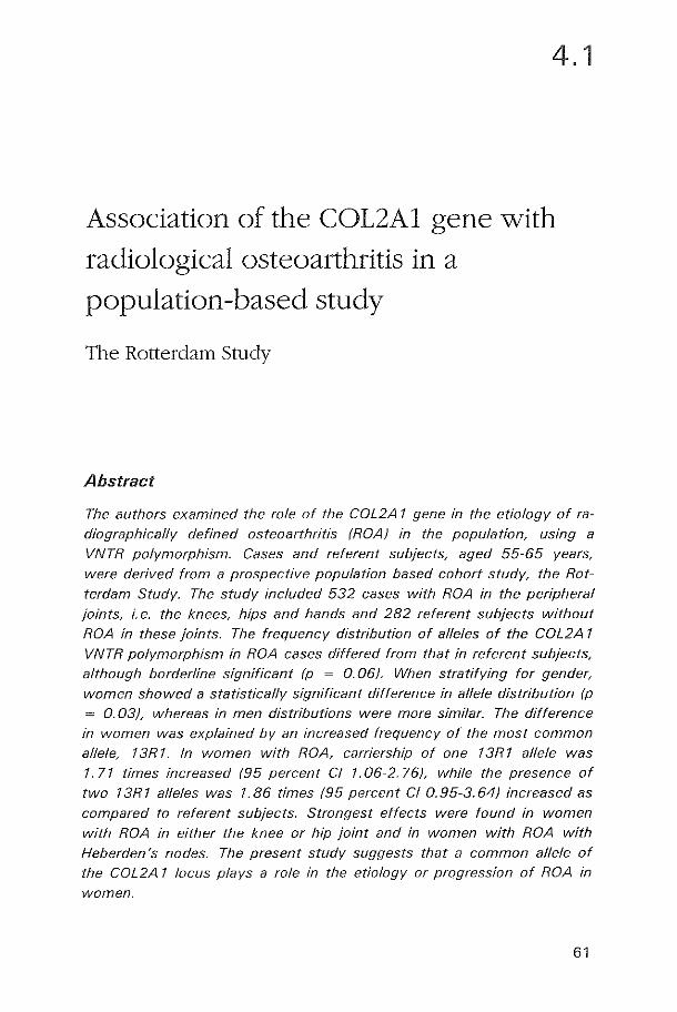
4.1
Association of the COL2Al gene with
radiological osteoarthritis in a
population-based study
The Rotterdam Study
Abstract
The authors examined the role of the COL2A 1 gene in the etiology of ra
diographically defined osteoarthritis (ROA) in the population, using a VNTR polymorphism, Cases and referent subjects, aged 55-65 years,
were derived from a prospective population based cohort study, the Rot
terdam Study, The study included 532 cases with ROA in the peripheral
joints, i. e, the knees, hips and hands and 282 referent subjects without
ROA in these joints, The frequency distribution of alleles of the COL2A 1
VNTR polymorphism in ROA cases differed from that in referent subjects,
although borderline significant (p ~ 0,06), When stratifying for gender,
women showed a statistically significant difference in allele distribution (p
~ 0,031- whereas in men distributions were more similar, The difference
in women was explained by an increased frequency of the most common allele, 13Rl, In women with ROA carriership of one 13Rl allele was
1,71 times increased (95 percent CI 1,06-2,76), while the presence of
two 13Rl alleles was 1,86 times (95 percent CI 0,95-3,64) increased as
compared to referent subjects, Strongest effects were found in women
with ROA in either the knee or hip joint and in women with ROA with
Heberden's nodes, The present study suggests that a common allele of
the COL2A 1 locus plays a role in the etiology or progression of ROA in
women.
61

Chapter 4.1
Introduction
Osteoalthritis eOA) is the most common disease of the musculoskeletal system.
The prevalence of OA rises from 4 percent in 18-24 year old individuals to 85
percent in subjects of ages over 74 years 0)). 111e disease can lead to pain and
a limited range of motion in the affected joint, and is an irnpOl1ant cause of dis
ability in the elderly (3). n,e radiographic changes in OA reflect a progressive
deterioration of al1icular (hyaline) cartilage of diarthrodial joints with narrowing
of the joint space, formation of osteophytes and development of sclerosis and
pseudocystic areas in subchondral bone.
Genetic factors are involved in the rare forms of early-onset (before 30
years of age) familial OA, which have shown to be transmitted as an autosomal
dominant trait (4,5). Over 40 mutations in the COLZA1 gene have indeed been
identified in sllch families (6-8), In the 19505, an influence of genetic factors on
the etiology of OA was recognized in individuals with generalized GA, defined
as OA in at least three different joint groups, in the presence of Heberden's
nodes (9). Heberden's nodes are bulbous deformities at the distal interphalan
geal joints of the fingers, that result from bone and soft tissue enlargement. Tbe
clinical expression of the genetic predisposition to generalized OA was found to
be modified by gender and age (10-12). At the population level one recent study
(13) has addressed the qtle.:;tion of the role of genetic factors in the etiology of
OA. This study of female twins suggested that genetic factors may play an iJu
portant role in the occurrence of knee and hand OA in the population.
As caltilage deterioration is the hallmark of the pathologic process in OA,
genes encoding collagenous (14) and non-collagenous (15) proteins of the ex
tracellular luatrix in cartilage have been proposed to be implicated in the etiol
ogy of OA. In particular, the COLZA1 gene (1Zq13), encoding the predominant
caltilage collagen) type II) is a major candidate gene for OA. Mutations in the
COLZAl gene give rise to various grades of matrix failure in articular cartilage
resulting in OA (16,17).
Findings on the role of the COL2Al gene in OA in population studies have
been controversial. An association of COLZAl alleles and OA was found in two
hospital based British studies (18,19). In contrast, an affected sibling-pair analy
sis (20) has failed to show a significant association with the COL2Al locus. Nei
ther did a study of patients with generalized OA or hand OA in the Finnish
population (21). Each of these studies was of limited size and studied patients
with symptomatic OA I I.e. patients visiting a clinic with joint pain. Thus the role
of the COL2Al gene in OA in the general population has not been addressed
and has not been examined before in a population based study using radiologi
cal OA as outcome measurement.
62

Association of COL2A 1 gene with ROA
We conducted an association study on the relation between the COL2Al
gene and radiograpbically defined OA (ROA) in a random subset of 814 subjects
aged 55-65 years, derived from a single population based study (the Rotterdam
Study), Cases with ROA in the peripheral joints, i.e. the knee, hip or hand joints
were compared with referent subjects without ROA in the peripheral joints.
Materials and Methods
Subjects
The present study was pan of the Rotterdam Study, a prospective population
based cohort study of determinants and prognosis of chronic diseases in the
elderly (22). For thb study, aU inhabitants of a suburb of Rotterdam, aged 55
years or over, including institutionalized persons, were invited to participate. In
total 7983 participants were examined between 1990 and 1993. The response
rate was 78 percent. From all subjects informed consent was obtained and the
study was approved by the medical ethics committee of Erasmus University
Medical School.
In order to ascertain ROA of relatively early onset and to target genetic pre
disposition to early ROA fro111 other determinant.s, the current study was re
stricted to non-institutionalized pal1icipants aged between 55 and 65 years
(n~2593). Radiographs of the peripheral joints, i.e. the knees, hips and hands,
and of the thoracolumbar spine have been scored in a random .)ubset of 944
individuab in this age categ01y. Radiographs from other joints were not avail
able. DNA was obtained in 814 subjects (86 percent) out of the subset of 944
individuals.
Measurements
Weight bearing anterior-posterior pelvic radiographs with both feet in 10° enclo
rotation were obtained. Correspondingly weight bearing knee films were made
with the patellae in central position. Finally, anteroposterior radiographs of the
hands and wris," and lateral radiographs of the spine (TM-Sl) were obtained.
ROA was assessed by means of the Kellgrcn grading system (23) in five
grades (0-4), using the figures and legends of the original atlas. Within the
Kellgren grading system, the definition of the five grades is different for the hip
joints as compared to the knee and hand joints. In the analysis definite ROA at a
particular site was defined as Kellgren-score !\VA or over in the left and/or right
corresponding joint.
All radiographs were scored by two independent readers, blinded to all
other data of the participant. After each set of about 150 radiogra phs the scores
63

Chapter 4, 1
of the two reader~ were evaluated. Whenever the scores differed two or more
points, OJ, was two for one reader but one for the other, a consensus score was
agreed upon. Radiographs of the knees and hips had previously been scored
(24). ROA of the band was assessed in each inter- and metacarpalphalangeal
joint individually, and the first carpometacarpal and trapezoscaphoideal joints.
ROA of the wrist was assessed at the radiocarpal and distal radioulnar joint.s. In
the analysis hand ROA was defined as a Kellgren-score two or over in at least
one of the 36 joint.s that were scored.
'I11e presence of Heberden's nodes was determined by an examination of
the hands as part of a general physical examination of the locomotor system.
This was performed by trained investigators during the first visit of each paltici
pant of the Rotterdam Study to the research center. Heberden's nodes were
scored in both hands separately, classified into two categories, i.e. absent or
present. In the analysis definite Heberden's nodes are defined as Heberden's
nodes present in the right and/or left hand.
Age, body mass index (BMJ), bone mineral density (BMD) and disk degen
eration of the spine were considered as possible confounders. Although disk
degeneration is not considered to be ROA of the spine, these abnormalities may
be associated with ROA. By definition, ROA of the spine is confined to the apo
physeal joints, but these joints could not be assessed at the radiographs that
were available. Disk degeneration was scored using the Kellgren grades (0-4), in
which a grade zero or one denotes no or doubtful disk degeneration, a grade
two denotes vettebral osteophytosis only and grades three and four ve11ebral
osteophytosis accompanied by moderate or severe disk space narrowing. Three
levels were scored, i.e. thoracic, lumbar and lumbosacral. Definite disk degen
erat.ion was defined as a Kellgren-score two or over in at least one of the three
levels that were scored. Finally, bone density (g/cnl) at the femoral neck was
measured by dual energy x-ray absorptiometry as described previously (25).
VNTR polymorphism
Genotypes of the VNTR polymorphism, located 1.35 kilobases to the 3' end of
the COL2Al gene were determined in all 814 subjects of whom DNA was avail
able. In order to genotype this nrultiallelic VNTR locus, PCR reactions were per
formed in 25 MI containing 25-50 ng genornic DNA, 2.5 pmoles of each primer
(fcnward primer: 5'-CAA CTG ATA AAA CAG AGA GC- 3' and reverse primer:
5'-CTC crr TGT CAT GAA CTA GC- 3' [26]), 1 'Super Taq buffer, 2 flCi
a["P]-dCTP, 200 >LM each of dCTP, dGTP, dTTP, and dATP, and 0,05 U of su
perTaq DNA polymerase (HT Biotechnology, Cambridge, UK), Amplification
was initiated with 3 min denaturation at 94°C followed by 35 cycles with 93°C (1
min) denaturation, S6°C (1 min) annealing step, and a 72°C (2 min) elongation
64
Association of COL2A 7 gene with ROA
step in a Hybaicl OmniGene thermal cycler using tube contro1. The amplification
was finished by a final incubation at 72°C for 4 min. Alleles were separated by
high resolution gel electrophoresis through a denaturing 3.5 percent polyaCIy
lamide gel, and analyzed by autoradiography. The nomenclature of alleles and
allelic ladder was used from Berg and Olaisen (27).
Statistical analysis
Demographic variables were compared by using the Student's t test and ch1-
square test. A two step strategy of analysis was followed. Allele frequencies for
C3.'::ieS and referent silbjects were assessed by counting alleles and calculating
sample propOltions (with each individual contributing two alleles). Allele distri
butions were compared between cases and referent subjects using J likelihood
ratio test (28). The rationJle of this analysis is that, in a population in Hardy
Weinberg equilibrium, under the null hypothesi.':;, the probability that one parent
transmits a certain allele to the subject studied is (statistically) independent from
the probability that the other parent transmits the allele. nlUS, from a genetic
point of view, the presence of the two alleles of an individual can be considered
as two independent observations. This approach is powerful as the number of
obse1vations in N subjects i.':; doubled to 2N alleles transmitted from the parents.
A drawback is that this approach does not yield meaningful estimations for
population risks. Therefore, in the second step of the analysis, the strength of
the association between the COL2Al allele(s) associated to the presence of ROA
in a subject was estimated by classical epidemiological methods, i.e. Hlllltiple
logistic regreSSion. Odds ratios (ORs), derived from the logistic regression equa
tions, are presented with 95 percent confidence intelvals (CT). All analyses were
carried out while adjusting for age. The analyses were additionally adjusted for
disk degeneration, BMI and BMD, in order to examine whether the relationship
is independent of osteophytosis and/or disk space narrowing in the spine, body
mass index and bone density. Because each of the statistical tests are invalid if
strata comprise less than five observations, alleles with a frequency lower then
0.05 were pooled.
Results
The baseline characteristics of our study population are shown in table 1. Out of
814 individuals 532 (65.4 percent) had ROA in the peripheral joints, i.e. the
knee, hip or hand joints. These will be referred to as ROA cases. The remaining
282 04.6 percent) individuals will be referred to as referent subjects. In these
subjects ROA in the peripheral joints was absent on all radiographs assessed.
ROA cases were on average 1.1 year older (p < 0.001) and had on average a 1.1
65

Table 1 Characteristics and radiographic readings from 814 subjects aged 55-65 years, Rotterdam. The Netherlands, 1990-1993
Men Women
Total 328 486
ROA cases (% of total}t 183 (55.8) 349 171.8) Age (SO) in years 60.8 12.7)' 60.7 12.6)* Body mass index (SO) in kg/m2 26.2 12.9) 26.9 (4.2)* Bone mineral density femoral neck (SOl in g/cm2 0.9010.12) 0.85 10.13)** Number with hand ROA (% of total) 144 143.9) 321 166.0) Number with knee ROA (% of total) 42 (12.8) 106 121.8) Number with hip ROA (% of total) 43 (13.1) 33 16.8) Number with disc degeneration (% of ROA cases) 123 (67.2) 221 (63.3)* Number with Heberden's nodes (% of ROA cases) 32 117.5) 108 (30.9)*
Referent subjects = No ROA (% of total) =I: 145 144.2) 137 (28.2) Age (SD) in years 59.9 12.7) 59.4 13.0) Body mass index (SO) in kg/m2 25.7 12.7) 25.4 13.7) Bone mineral density femoral neck (SO) in g/cm2 0.91 10.13) 0.83 10.12) Number with disc degeneration (% of referent subjects) 83 (57.2) 65 147.4) Number with Heberden's nodes (% of referent subjects) 16 (11.0) 20 114.6)
t ROA cases had radiological osteoarthritis in the peripheral joints, e.g. the knees, hips and hands +- Referent subjects had no radiological osteoarthritis in the peripheral joints * p:::;: 0.001 (ROA cases are compared to referent subjects). ** p = 0.03 (ROA cases are compared to referent subjects).
All
814
532 (65.4) 60.7 12.6)* 26.7 13.8)*
0.87 10.13) 465 157.1 ) 148 (18.2)
76 (9.3) 344 (64.7)* 140 (26.3) *
282 134.6) 59.6 12.9) 25.6 13.2)
0.87 10.13) 148 (52.5)
36 (12.8)
Table 2 Allele frequencies of the COL2A 1 VNTR polymorphism in 814 subjects aged 55-65 years. Rotterdam. The Netherlands. 1990-1993
Total Allele 11 R 1 13Rl 13R2 14Rl 14R2 Others number of p-value
alleles
Overall 171 (0.111 686 (0.42) 98 (0.06) 439 (0.27) 98 (0.06) 136 (0.08) 1628
Referent subjects = No ROA 62(0.11) 218 (0.39) 44 (0.08) 159 (0.28) 36 (0.06) 45 (0.08) 564
ROA cases 109 (0.10) 468 (0.44) 54 (0.05) 280 (0.26) 62 (0.06) 91 (0.09) 1064 0.06
Women Referent subjects 32 (0.12) 100 (0.36) 24 (0.09) 76 (0.28) 16 (0.06) 26 (0.09) 274 ROA cases 71 (0.10) 313 (0.45) 33 (0.05) 176 (0.25) 45 (0.06) 60 (0.09) 698 0.03 Hand ROA 68{0.11) 282 (0.44) 30 (0.05) 164 (0.26) 44 (0.07) 54 (0.08) 642 0.06 Knee ROA 19 (0.09) 97 (0.46) 15 (0.07) 45 (0.21) 19 (0.09) 17 (0.08) 212 0.07 Hip ROA 8 (0.12) 31 (0.47) 2 10.03) 15 (0.23) 3 (0.05) 7 (0.11) 66 0.25
Men Referent subjects 30 (0.10) 118 (0.41) 20 (0.07) 83 (0.29) 20 (0.07) 19 (0.07) 290 ROA cases 38 (0.10) 155 (0.42) 21 (0.06) 104 (0.28) 17 (0.05) 31 (0.08) 366 0.50 Hand ROA 30 (0.10) 125 (0.43) 13 (0.05) 79 (0.27) 16 (0.06) 25 (0.09) 288 0.50 Knee ROA 6 (0.07) 34 (0.40) 4 (0.05) 27 (0.32) 4 (0.05) 9 (0.11) 84 0.50 Hip ROA 10 (0.12) 37 (0.43) 10{0.12) 21 (0.24) 3 (0.03) 5 (0.06) 86 0.50
Numbers listed are number of alleles, with each subject contributing two alleles to the table (alleles with individual frequencies lower than 0.05 are summed in the category others). P-value is based on the likelihood ratio test by Terwilliger (28).

Chapter 4.7
kg/m2 higher (p < 0.001) EMI as compared to referent subjects. In women with
ROA, BMD was significantly higher and the number of women with disk degen
eration and with Heberden's nodes was significantly increased as conlpared to
referent subjects. In this age categOlY, hip ROA and disk degeneration was more
frequent in men, whereas knee ROA, hand ROA and Heherden's nodes were
more frequent in women. In this study population 14 out of 23 reported alleles of the COL2AI VNTR
polymorphism were detected. In table 2, allele frequencies in all 814 genotyped
subjects, as well as the allele frequencies in ROA cases and referent subjects,
both overall and stratified by gender and site of ROA, are shown. In the overall
analysis, the frequency distribution of alleles in ROA cases and referent subjects
was different, although this difference was borderline significant (p ~ 0.06). In
women the allele distribution in ROA cases as compared to referent subjects was
statistically significantly different (p ~ 0.03). 11,e difference in allele distribution
between female ROA cases and referents was explained by an increased fre
quency of allele 13Rl, wbile the frequencies of all other alleles, except allele
14R2, were found to be decreased or similar in cases and referent subjects. In
lnen no significant differences were found between ROA cases and referent
subjects, although the allele frequency of allele 13RI in male cases was slightly
elevated (except for male cases with knee ROA).
To estimate the strength of the association between the presence of the
13Rl allele in an individual and ROA, we perfonned a logistic regression analy
sis. We adjusted for age and other (putative) risk factors for ROA. Analyses were
conducted for men and women separately. In women (table 3), the presence of
one 13RI allele (13Rl heterozygosity) in ROA cases was 1.71 times increased (95
percent CI 1.06-2.76) and the frequency of two 13Rl alleles (J3RI homozygos
ity) 1.86 times (95 percent CI 0.95-3.64) as compared to referent subjects.
Strongest effects were found, although not Significant, in the case groups of
women, homozygote for allele 13RI, witb either knee ROA or hip ROA (OR 2.06
with 95 percent CI 0.81-5.21 and 2.31 with 95 percent CI 0.63-8.50, respectively).
In men, no significant increase in OR for ROA was found associated with the
13RI allele (table 4). Only the OR for male cases with hand ROA tended to be
increased (OR ~ 1.52 with 95 percent CI 0.73-3.17).
As in women Heberden's nodes were associated with ROA and have been
described as part of the genetically determined forms of ROA earlier, we exam
ined the effect of Heberden's nodes on the association between 13R1 genotypes
and ROA. For this purpose, ROA cases with Heberden's nodes (n = 140) were
compared with referent subjects without Heberden's nodes (n = 246). In par
ticular in women, who were homozygote for allele 13R1, we found a slrong as
sociation with ROA (table5). In female ROA cases the prevalence of 13RI homo
zygotes was 3.04 times increased (95 percent CI 1.22-7.61). This effect was fur-
68

Table 3 Association between ROA and carriership of one or two copies of allele 13R1 in 486 women aged 55~65 years, Rotterdam, The Netherlands. 1990-1993
Genotype 13Rl-/13Rl- 13Rl +/13Rl- 13Rl +/13Rl +
OR* ORt OR* ORt n n
(95% el) (95% el) n
(95% ell (95% el)
Referent subjects = No ROA 56 Ref 62 19
ROA cases 101 Ref 183 1.62 1. 71 65 1.99 1.86 (1.04-2.541 (1.06-2.761 (1.07-3.701 (0.95-3.641
Hand ROA 96 Ref 168 1.57 1.68 57 1.77 1.73 ( 1.00-2.461 (1.04-2.721 (0.94-3.321 (0.73-3.401
Knee ROA 30 Ref 55 1.50 1.48 21 2.32 2.06 (0.83-2.721 (0.76-2.891 (1.05-5.121 (0.81-5.211
Hip ROA 9 Ref 17 1.60 1.64 7 2.79 2.31 (0.65-3.961 (0.61-4.421 (0.88-8.921 (0.63-8.501
13Rl-/13Rl- denotes "Individuals carrY"lng no copy of alle[e 13R 1, 13Rl + 113R 1- (13Rl heterozygotes) denotes indiv"lduals carrying one copy of allele 13Rl and 13Rl +/13Rl + {13Rl homozygotes) denotes individuals carrying two copies of allele 13Rl. * OR adjusted for age, t OR adjusted for age, body mass index, bone mineral density and disk degeneration.

Table 4 Association between ROA and carriership of one or two copies of allele 13R1 in 328 men aged 55-65 years, Rotterdam, The Netherlands, 1990-1993
Genotype 13Rl-/13Rl- 13Rl +/13R1- 13R1 +/13R1 +
OR* ORt OR* ORt n n
(95% CII (95% CI) n
(95% CI) (95% CI)
Referent subjects = No ROA 50 Ref 72 23
ROA cases 61 Ref 89 1.09 1.01 33 1.24 1.39 (0.66-1.79) (0.60-1.69) (0.64-2.41) (0.69-2.82)
Hand ROA 48 Ref 67 1.04 0.94 29 1.40 1.52 (0.61-1.77) (0.54-1.63) (0.70-2.79) (0.73-3.17)
Knee ROA 14 Ref 22 1 .11 1.00 6 0.96 1.14 (0.51-2.39) (0.44-2.27) (0.32-2.86) (0.36-3.57)
Hip ROA 14 Ref 21 1.08 0.99 8 1.26 1.19 (0.50-2.34) (0.45-2.18) (0.46-3.45) (0.41-3.51)
13Rl~!13Rl- denotes individuals carrying no copy of allele 13Rl, 13Rl +!13Rl- (13Rl heterozygotes) denotes individuals carrying one copy of allele 13Rl and 13Rl +/13Rl + (13Rl homozygotes) denotes individuals carrying two copies of allele 13Rl. * OR adjusted for age, t OR adjusted for age, body mass index, bone mineral density and disk degeneration.
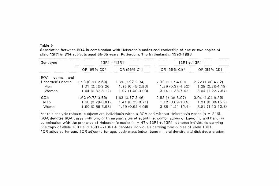
Table 5 Association between ROA in combination with Heberden's nodes and carriership of one or two copies of allele 13R1 in 814 subjects aged 55-65 years, Rotterdam, The Netherlands, 1990-1993
Genotype 13Rl +/13Rl- 13R1 +/13Rl +
OR 195% CII* OR 195% Cllt OR 195% CII* OR 195% Cllt
ROA cases and Heberden's nodes .5310.91-2.601 .69 10.97-2.941 2.3311.17-4.631 2.22 11.06-4.621
Men 1.31 10.53-3.261 1.16 10.45-2.961 1.2910.37-4.501 1.09 10.28-4.181 Women 1.64 10.87-3.121 1.9711.00-3.901 3.1411.33-7.421 3.0411.22-7.611
GOA 1.62 10.73-3.591 1.63 10.67-3.461 2.93 11.06-8.071 3.0411.04-8.89) Men 1.60 10.29-8.81 I 1.4110.23-8.711 1.121009-13.51 1.21 10.09-15.91 Women 1.60 10.65-3.931 1.59 10.62-4.091 3.8811.21-12.41 3.8711.13-13.31
For this analysis referent subjects are individuals without ROA and without Heberden's nodes (n = 246). GOA denotes ROA cases with two or three jO'lnt sites affected (i.e. comb'lnations of knee, h"lp and hand) in combination with the presence of Heberden's nodes (n = 47). 13R1 +113Rl- denotes individuals carrying one copy of allele 13R1 and 13Rl +!13R1 + denotes individuals carrying two copies of allele 13R1. *OR adjusted for age. tOR adjusted for age, body mass index, bone mineral density and disk degeneration

Chapter 4.7
ther increased in women with Heberden's nodes and generalized ROA, Le. ROA
in two or three joint ,ites (OR ~ 3.87 with 95 percent CI 1.13-13.3). In men, no
substantial increase in ORs was obselved (table 5),
In all logistic regression analyses, we examined whether disk degeneration
in the spine, body mass index or bone mineral density of the femoral neck had
an effect on these findings (see tables 3-5), Although some small differences
were obselved in OR or confidence interval, neither one of these analyses
changed the findings materially.
Discussion
We found an association between the COL2Al locus and ROA. A statistically
significant association was obselved in women, especially in those with either
knee or hip ROA and those with ROA in the presence of Heberden's nodes. The
association with the 13Rl allele was strongest for 13Rl homozygotes, suggesting
a dme dependent effect of this allele. In women, the effect of allele 13Rl could
not be explained by differences between cases and referent subjects in age, EMI,
BMD or disk degeneration. In men, no significant evidence for an association
between tbe COL2A1 locu, and ROA was found.
Regarding the validity, and the comparability of our study to previous
studies, the prevalence of ROA at different joint sites as obselved in our study
population is in keeping with earlier repOlts on Caucasian populations, taking
into account the considerable variance in the prevalence of ROA in different
populatiom (2). To increase the validity of ROA measurements all radiographs
were scored by two trained readers. The grading system according to Kellgren
(23) that was used, io a valid tool for scoring ROA in the knee and hand (29).
The diagnosis of hip ROA is an issue of debate. According to Kellgren, ROA of
the hip is defined as definite joint space narrowing accompanied by slight scle
rosis and definite osteophytes. However, others have argued that joint space
narrowing alone can be a sufficient 'ign for the diagnosis of hip ROA (30). Thus,
the use of the Kellgren-score may have introduced misclassification, but as all
radiographs were scored blind to genotyping and genotyping was performed
blind to the disease status, this misclassification has most likely been non-cliffer
entizll. The number of alleles as well as the allele frequencies in our study
population are comparable with those previously reponed in Caucasian popula
tions (26,27,31).
Case-referent studies are liable to confounding. Spurious associations lllay
arise in genetic studies when a population consists of several subpopulations,
that differ in allele and disease frequencies. This type of confounding occurs if
the population studied is not homogeneous, but comprises several (ethnic) sub-
72
Association of COL2A 1 gene with ROA
populations anel is referred to as population adrnL'\.1:ure (32). To prevent this
problem, it is essential that cases and referent subjects are sampled from the
same source population. An advantage of the present study is that cases and
referent subjects were derived from a well-defined Single population, making
the occurrence of bias due to admixture unlikely.
Although the present study suggests an association between the COL2Al
gene anel ROA, it does not confirm the findings of two previous studies (18,19)
that showed evidence for such an association. Both previous studies suggested
that relatively rare clinical phenotypes of generalized OA were associated with
an increased frequency of a rare allele of the COL2Al gene. In contrast, we
found common types of peripheral ROA at the knee, hip and hand to be associ
ated with a frequent allele of the COL2Al gene, the 13Rl allele. The only rare
allele in our study that tended to be increased in ROA cases, was the 14R2 allele
in women. Despite the size of the present study, the statistical power to examine
differences in frequencies of rare alleles in phenotypes like generalized OA was
low and therefore this study does not allow for fIrm conclusions on this issue.
Two previous studies (20,21) using the same VNTR polymorphism as in our
study did not show an association of OA with the COL2Al gene. Both studies
were of limited size and compared a subset of relatively rare and severe OA
cases with a reference group. One study (20) selected as reference group sub
jects with an unknown OA status, which, given the high frequency of ROA in
the population, may have led to a substantial underestimation of the effects. The
other (21), canied out in the Finnish population, detected only 4 out of 23 re
ported alleles of the COL2Al VNTR polymorphism. TI,e Finnish population is
genetically isolated) i.e. limited number of immigrants) and relatively smalL
Thus) the variety in alleles i.s limited as compared to other populations and the
allele frequencies are subject to fluctuation. It can be shown that) through this
random fluctuation, (rare) alleles can disappear easily from these populations) in
favor of a few predominant common alleles. Because of this process) called ge
netic drift) allele frequencies in the Finnish population differ Significantly from
those in other European countries. It is conceiva ble that, due to genetic drift, the
COL2Al gene does not playa role in the etiology of OA in the Finnish popula
tion) because disease associated alleles) such as the 13R1 allele) have disap
peared by chance.
As to the causal inference of our findings, two issues are to be addressed.
Firstly, the molecular basis for genetic association studies lies in the fact that the
mutation in a disease gene that occll1Ted in an ancestor many generations ago
has been transmitted to future generations) together with parts of the chromo
some directly adjacent to this disease gene. In this way, genetic markers in and
surrounding the disease gene are transmilted together with the mutation in the
disease gene from generation to generation until recombination occurs. It is
73
Chapter 4.1
plauoible that the COLZA! VNTR polymorphism flags an OA related mlJtalion
within the COL2Al gene) ~ince this gene encodes collagen type II, the major
constituent of al1icular c3ltilage. However, it can not be excluded that the VNTR
polymorphism (1.35 kilobases from the COLZA! gene) is also associated with an
other gene located nearby the COL2Al gene, and that this other gene is causally
related to ROA. The Vitamin D receptor gene, located about 750 kilobases apart
from the COL2Al gene is .')uch a gene. This gene has been found to be associ
ated with knee ROA 03,34) and with bone density (35), However, the associa
tion between ROA and the 13Rl allele in QlIr study is independent of bone den
sity and therefore it is unlikely, that the Vitamin D receptor gene can explain the
association between the COL2Al gene and ROA. Another argument against the
Vitamin D receptor gene explaining the obselved association between the
COL2Al gene and ROA, is the finding that the Vitamin D receptor gene was not
associated to Heberden's nodes (33), while the !3R! allele of the eOL2A! gene
is. A final argument is given by the, in genetic terms, large distance between the
COL2Al gene Jnd the Vitamin D receptor genc, rnaking the possibility that re
combinations have occurred between these two genes likely. Secondly, given
the cross-sectional design of our study we are unable to determine whether the
observed association between the COLZA1 locus and ROA is causaL Although
the mortality of OA is expected to be velY limited in this age categOlY (55-65
years), the COL2Al gene may be involved in the progression and/or mOl1ality of
OA or mOltality of associated causes of death, in stead of being a determinant of
the risk of OA.
In summalY, this study has shown an association between a common allele
of a COLZA! VNTR polymorphism and ROA in women. Homozygosity at the
COLZA1 gene for the !3R! allele is strongly associated to (generalized) ROA
with Heberden's nodes in women. Although it has been reported earlier that the
clinical expression of OA is modified by gender (10,11), the present study sug
gests effect modification of thc association between the COL2Al gene and ROA
by gender on the level of radiographically defined OA. This is the first large
population based study examining the role of the COL2Al gene in radiographi
cally defined OA in the peripheral joints. The effect of the COL2A! 13R! allele
appears to be dose-dependent and may contribute to the explanation of the sex
and joint-site specific patterns in the occurrence of ROA in the population.
References
1. Masi AT, Medsger TA: Epidemiology or the rheumatic diseases. In: McCa11hy DJ (ed). Anhritis and allied conditions. London: Lea and Febiger, 1989;16-54.
74
Association of COL2A 7 gene with ROA
2. Van Saase .ILeM, Van Romunde LKJ, Cats A, et al. Epidemiology of oSfeoa11hritis; Zoetermeer sUlvey, comparison of radiological osteoal1hritis in a Dutch population with that in 10 other populations. Ann Rheum Dis 1989;48:271-80.
3. Odding E, Valkenburg HA, Algra A, et al. Association of locomotor complaints and disability in the Rotterdam Study. Ann Rheum Dis 1995;51:721-5.
4. Palotie A, Ott], Elima K, et a!. Predisposition to familial ostcoal1hrosis linked to type II collagen gene. Lancet 1989;1:921-7.
5. Knowlton RG, Katzenstein PL, Moskowitz RW, et a1. Genetic linkage of a polymorphism in the type II procollagen gene (COL2Al) to primalY osteoalthritis associated with mild chondrodysplasia. New Engl] Med 1990;,322:526-50.
6. Ala-Kokko L, Baldwin CT, Moskowitz RW, et a1. Single base mutation in the type II procollagen gene (COL2A1) as a cause of primary osteoal1hritis associated with a mild chondrodysplasia. Proc Natl Acad Sci LlSA 1990;87:6565-8.
7. Ahmad NN, Ala-Kokko L, KnO\vlton RG, ct a!. Stop codon in the procollagen II gene (COL2Al) in a family with the Stickler syndrome Cal1hro-ophthalmopathy). Proc Natl i\cad Sci USA 1991;88:6621-7.
8. \Villiams CJ, Considine EL, Knowlton RG, et al. Spondyloepiph~lseal dysplasia and precocious osteoarthritis in a family with an Arg75----t Cys mutation in the procollagen type II gene (COL2A!). Hum Genet 1993:92:499-505.
9. Kellgren JH, Moore R. Generalized osteoal1hritis and IIeberden's nodes. Br Med J 1952;1:181-7.
10. Stecher RtvI. Heberden Oration. Heberden's nodes. A clinical description of osteoarthritis of the finger joints. Ann Rheum Dis 1955;14:1-10.
11. Kellgren JH, Lawrence JS, Bier F. Genetic factors in generalized osteoal1hrosis. Ann Hheum Dis 1963;22:237-55.
12. Cooper C, Egger P, Coggon D, et al. Generalized osteoat1hritis in women: pattern of joint involvement and approaches to definition for epidemiological studies . .1 Rheum 1996;23: 1938-42.
13. Spector TD, Ciculti F, Baker .1, et a1. Genetic influences on osteoarthritis in women: a twin study. 13M.l 1996;312:920-4.
11. Williams q, Jiminez SA. Heredity, genes and osteoal1hritis. Rheum Dis Clin Not1h America 1993;19:523-43.
15. Meulenbelt I, l3ijkerk C, de Wildt SCM, et a1. Investigation of the association of the CRTM and CRTU genes with radiographically evident osteoarthritis in subjects from the Rotterdam Study. At1hritis Rheum 1997;40:1760-65
16. Eyre DR, \XTeis !VIA, jI,·Joskowitz RW. Cal1i1age expression of a type II collagen mutation in an inherited form of osteoanhritis associated with a mild chondrodysplasia. ] Clin Invest 1991 ;87:357-61.
17. Kuivaniemi H, Tromp G, Prockop DJ. Mutations in collagen genes: causes of rare and some common diseases in humans. FasebJ 1991;5:2052-60.
]8. Hull R, Pope FM. Osteoal1hritis and c1l1ilage collagen genes. Lancet 1989;i:1337-8. 19. Loughlin J, Irvcn C, Athanasol! N, et al. Differential allelic expression of the type II
collagen gene (COL2Al) in osteoal1hritic canilage. AmJ Hum Genet 1995;56:1186-93. 20. Loughlin .1, ltven C, Fergusson C, et a1. Sibling pair analysis shows no linkage of gen
eralized osteoanhritis to the loci encoding type II collagen, cal1ilage link protein or G1l1i1age matrix protein. Br.1 l{hellmatol1991;33:1103-6.
21. Vikkula lvI, Nissila M, Hirvensalo E, et al. Multiallelic polymorphism of the canilage collagen gene: no association 'with osteoarthritis. Ann Rheum Dis 1993;52:762-4.
22. Hofman A, Grobbee DE, De .long PTVlvf, et a!. Determinants of disease and disability in the elderly. Em.l EpidcmioJ 1991;7:403-22.
23. Kellgren .111, Jeffrey IvIR, Bal! J (eds). The epidemiology of chronic rheumatism. Volume II: Atlas of standard radiographs of anhritis. Oxford: Blackwell Scientific Publications, 1963.
21. Odding E. Locomotor disability in the elderly. PhD. thesis. Erasmus University Rotterdam, The Netherlands, 1994.
75

Chapter 4.1
25. Burger l-i, van Dade PLA, Algra FA, et a!. The association between clge and bone mineral density in men and women aged 55 years and over: The Rotterdam Study. Bone Miner 1994;25:1-13-
26. Priestley L, Kumar D, Sykes B. Amplification of the COLZAl ,j' variable region used for segregation analysis in a family with the Stickler syndrome. Hum Genet 1990;85;525-6.
27. Berg ES, Olaisen B. Characterization of the COLZA1 VNTR polymorphism. Genomics 1993;16;350-4.
28. Tcrvvilligcr JD. A powerful likelihood method for the analysis of linkage disequilibrium between trait lodand one or more polymorphic marker loci. Am ] Hum Genet 1995;56; 777-87.
29. Spector 'I'D, IIalt Dj, Byrne J, et al. Definition of osteoalthritis of the knee for epidemiological studies. Ann Rheum Dis 199.3;52:790-4.
30. Croft P, Cooper C, Wickham C, et al. Defining osteoarthritis of the hip for epidemiological studies. Am] EpidcmioI1990;132:511-22.
31. Rand S, Puers C, Skowasch K, et al. Population genetics and forensic efficiency data of'! AMPFLP's. IntJ Leg Mcd 1992;104:329-33.
32. Lander ES, Schork NJ Genetic dissection in complex traits. Science 1994;265:2037-48. 33. Keen RW, Hal1 DJ, LanchblllY]S, et al. Association of early osteoal1hritis of the knee
with a Tag 1 polymorphism of the Vitamin D receptor gene. A11hritis Rheum 1997;10:1111-19.
34. Uitterlinden AG, Burger H, Huang Q, et al. Vitamin D receptor genotype is associated with radiographic osteoal1hritis at the knee.] Clin Invest 1997;100:259-63.
35. Eisman JA. Vitamin D Receptor gene alleles and osteoporosis: an affirmative view. J Bone Miner Res 1995;10:1289-93
76

4.2
The COL9Al gene and COLIIA2 region
and radiographically assessed
osteoarthritis in subjects from a
population-based study
Abstract
We performed an association study of two Short Tandem Repeat Poly
morphisms (STRPs) within the COL9A 1 gene and of two microsatellite
marker loci flanking the COL 11 A2 gene and radiological osteoarthritis
(ROA). Cases and referent subjects, aged 55 to 65 years, were derived
from a population based cohort study, the Rotterdam Study. Cases, with
ROA in the knee and/or hip joint In = 204), were compared with age
matched referent subjects free from ROA in the knees, hips and hands In
= 268). The distribution of alleles from the STRP 1281 within the
COL9A 1 gene differed statistically significantly between cases and refer
ent subjects (p = 0.002). The association between STRP 1281 and ROA
was statistically significant in women, in whom haplotype analysis sug
gested a protective effect of allele 1281-A2. Neither STRP 882 within the
COL9A 1 gene, nor the microsatellites D6S291 nor TNFa, flanking the
COL 11 A2 gene, were associated with ROA in the knee and/or hip joint.
The results of our study suggest that the COL9A 1 gene may contribute to
the pathogenesis of osteoarthritis in women. We found no evidence for a
role of the COL l1A2 region or for the TNF-a gene in the occurrence of
osteoarthritis.
77

Chapter 4.2
Introduction
Osteoanhritis (OA) is a highly prevalent complex disease, characterized by a
progressive degeneration of articular (hyaline) cartilage. The pathophysiologic
changes can radiographically be observed as osteophyte formation at the joint
margins, joint space narrowing, subchondral sclerosis of bone and formation of
bony cysts. Although these radiographically assessed abnormalities do not ubiq
uitously lead to clinical OA 0,2), i.e. pain, stiffness or limited range of motion of
the affected joint, they reflect c311ilage degeneration, which can be considered
as the pathophysiologic hallmark underlying clinical OA.
The extracellular matrix of alticular cartilage consists of proteoglycan ag
gregates embedded in a network of collagen fibrils, containing collagen's type
II, IX and XI (3). Although type IX collagen constitutes not more than 1 % of the
total colJagen in articular cal1i1age, it is important in stabilizing the articular col
lagen network, mainly comisting of type II collagen fibrils (95% of total collagen
in cartilage) (4-6). Type IX collagen is a heterotrimeric protein, encoded by three
different genes (3). The COL9AI gene, encoding the al(IX) chain, is located on
chromosome 6 C6q12-qI3) (7). The absence of the aI(IX) chain or the synthesis
of a shortened cxJ(IX) chain in mice, leads to degeneration of articular cartilage
similar to that in human OA (8,9). It has been suggested that dysfunctional type
IX collagen alters the mechanical properties of the collagen network in articular
cal1ilage, leading to joint ca11ilage more prone to damage from mechanical stress
(8). However, up to date there is no empirical evidence that the COL9Al gene is
involved in OA in humans. Mutations in the COL9A2 gene induce severe phe
notypes with multiple epiphyseal dysplasia that are velY different from common
forms of OA in the elderly (10). For the COL9A3 gene mutations or hereditalY
diseases have not been reported.
11,e fibrillar collagen type XI is also a constituent of cartilage collagen and
is closely associated with collagen type II. It is important in regulating the di
ameter of the type II collagen fibril (11) and has been shown to be imp011ant for
the integrity and proper development of the skeleton (12). Furthermore, type XI
collagen binds with high affinity to proteoglycans, which may be Important for
anchoring caltilage proteoglycans to the collagen fibrillar network (13). It con
sists of three distinct a chains of which the a2(XI) chain is transcribed from the
COLlIA2 gene, which has been localized on chromosome 6 (6p2I) within the
major histocompatibility complex (14). A dominant as well as a recessive muta
tion in the COLl IA2 gene has been described, both leading to spondyloepiphy
seal dysplasia associated with severe OA (12). Other mutations or polymor
phisms in the COLIIA2 gene may give rise to cal1ilage pathology in the general
population. Data on the COLllAl gene suggest that this gene plays a role in
78

Association of COL9A 1 gene and COL 11 A2 region with ROA
skeletal morphogenesis (15). Mutations in this gene lead to severe phenotypes,
e.g. Stickler syndrome (16). The third a chain of type XI collagen is encoded by
the COL2A] gene, which has extensively been studied in human OA.
We aimed to study collagen genes with the largest possibility of playing a
role in the occurrence of conunon forms of OA in the elderly, Le. the COL2A1)
COL9Al and COLlIA2 genes. Data on the COL2Al gene has been presented
previously (unpublished data). In this paper we studied the associations be
tween the COL9Al gene and COLlIA2 region and radiological osteoarthritis
(ROA), in a subset of 472 subjects aged 55-65 years, derived from a single
population ba.sed study. Cases with ROA in the knee and/or hip joints were
compared with referent s·ubjects without ROA in the peripheral joints, Le. the
knee, hip and hand joints.
Material and Methods
Subjects
Cases and referent subjects were derived from a prospective population based
follow-up study of determinants and prognosis of chronic diseases in the elderly
(the Rotterdam Study) (Ill. All inhabitants of a suburb of Rotterdam aged 55
years or over were invited to participate. In total 7983 participants were exam
ined. The response rate was 78 percent. TIle medical ethics committee of Eras
mus University Medical School has approved the study and written informed
consent was obtained from all participants.
In order to asceltain ROA of relatively early onset) the present study was
limited to pal1icipants aged between 55 and 65 years (n~2593). Radiographs of
the peripheral joints) i.e. the knees, hips and hands, have been scored in a ran
dom subset of 944 individuals in this age categ01y. Less than one percent of the
944 palticipants were related in the first degree. On the basis of the radiographic
data) we maximized the phenotypic contrast between cases and referent sub
jects. Cases were defined as subjects with ROA in at least one of the large weight
bearing joints) i.e. the knee and hip joints (n == 216), whereas referent subjects
were defined as subjects without ROA in the peripheral joints, i.e. the knee.s,
hips and hands (n = 332). The referent group corresponds roughly with the
lower 30% of the distribution of ROA in the population. DNA was obtained in
472 subjects (86 percent) out of this subset of 548 cases and referent .subjects.
Measurements
Weight bearing anterior-posterior radiographs of the hips and knees and ante
rior-posterior radiographs of the hand.s were obtained. ROA was assessed by
79
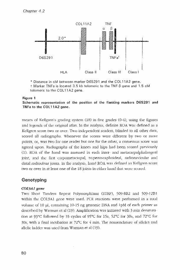
Chapter 4.2
COLllA2
2.0*
D6S291
HLA Class II
TNF
a r
TNFat
Class III Class I
* Distance in eM between marker D6S291 and the COL 11 A2 gene. t Marker TNFa is located 3.5 kb telomerie to the TNF-fl gene and 1.5 eM telomeric to the COL 11 A2 gene.
Figure 1 Schematic representation of the position of the flanking markers 065291 and TNFa to the COL llA2 gene.
means of Kellgren's grading system (18) in five grades (0-4), using the figures
and legends of the original atlas. In the analysis, definite ROA was defined as a
Kellgren-score two or over. Two independent readers, blinded to all other data,
scored all radiographs. Whenever the scores were different by two or more
points, Of, was two for one reader but one for the other, a consensus score was
agreed upon. Radiographs of the knees and hips had been scored previously
(1). ROA of the hand was assessed in each inter- and rnetacarpalphalangeal
joint, and the first carpometacarpal, trapezoscaphoideal, radionavicular and
distal radioulnar joints. In the analysis, hand ROA was defined as Kellgren-score
two or over in at least one of the 18 joints in either hand that were scored.
Genotyping
COL9A 1 ge'!e Two Short Tandem Repeat Polymorphisms (STRP), 509-SB2 and 509-12Bl
within the COL9A1 gene were used. PCR reactions were performed in a total
volume of 10 ~l, containing 10-15 ng genomic DNA and IpM of each primer as
described by Wanl1an et a1 (19). Amplification was initiated with 3 min denatura
tion at 93°C followed by 35 cycles of 95°C for 15s, 52°C for 305, and noc for
30s, with a final incubation at 72°C for 4 min. The nomenclature of alleles and
allelic ladder was used from Warman et a1 (19).
80
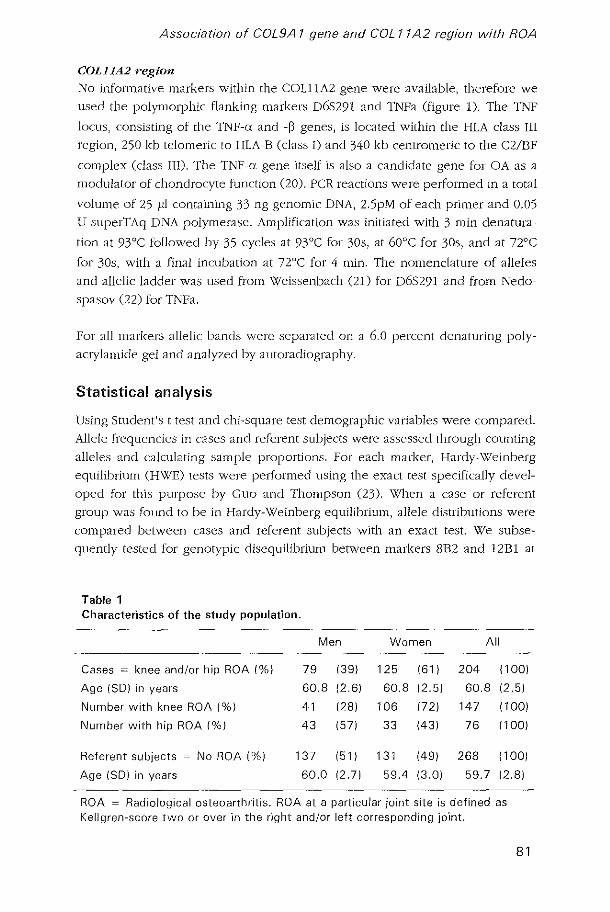
Association of COL9A 1 gene and COL l1A2 region with ROA
COLI1A2 region
No informative markers within the COLI1A2 gene were available, therefore we
used the polymorphic flanking markers D6s291 and TNFa (figure 1). The TNF
locus, consisting of the TNF-a and -p gene.s, is located within the HLA class III
region, 250 kb telomeric to HLA-B (class I) and 340 kb centromeric to the C2/BF
complex (class III). The TNF-a gene itself is also a candidate gene for OA as a
modulator of chondrocyte function (20). peR reactions were performed in a total
volume of 25 pi containing 33 ng genomic DNA, 2.5pM of each primer and 0.05
U superTAq DNA polymerase. Amplification was initiated with 3 min denatura
tion at 93°C followed by 35 cycles at 93°C for 30s, at 60°C for 30s, and at noc for 305, with a final incubation at 72°C for 4 min. The nomenclature of alleles
and allelic ladder was used from Weissenbach (21) for D6s291 and from Nedo
spasov (22) for TNFa.
For all marker::; allelic bands were separated on a 6.0 percent denaturing poly
aClylamicie gel and analyzed by autoradiography.
Statistical analysis
Using Student's t test and chi-square te::;t demographic variables were compared.
Allele frequencies in cases and referent subjects were assessed through counting
alleles and calculating sample proportions. For each marker, Hardy-Weinberg
equilibrium (HWE) tests were performed using the exact test specifically devel
oped for this purpose by Guo and Thompson (23). When a case or referent
group was found to be in Hardy-Weinberg equilibrium, allele distributions were
compared between cases and referent subjects with an exact test. We subse
quently tested for genotypic disequilibrium between markers 8B2 and 12Bl at
Table 1 Characteristics of the study population.
Men Women All
Cases = knee and/or hip ROA (%) 79 (39) 125 161) 204 (100)
Age (SO) in years 60.8 (2.6) 60.8 12.5) 60.8 (2.5)
Number with knee ROA r%) 41 128) 106 (72) 147 (100)
Number with hip ROA (%) 43 157) 33 (43) 76 (100)
Referent subjects ~ No ROA 1%) 137 (51) 131 (49) 268 1100)
Age (SO) in years 60.0 12.7) 59.4 (3.0) 59.7 12.8)
ROA = Radiological osteoarthritis. ROA at a particular joint site is defined as Kellgren-score two or over in the right and/or left corresponding joint.
81
Chapter 4.2
the COL9A1 gene and markers D6s291 and TNFa at the COLllA2 region. HWE
test, test for population differentiation and test for genotypic disequilibrium
were conducted, using the Genepop statistical package (24). These tests are
ba.sed on the Markov chain method, which yields exact p-values. Furthermore,
an expectation maximization (EM) algorithm was used to obtain estimated hap
lotype frequencies and a chi-square test was used to compare haplotype fi'e
quencies between cases and referent slJbjects (25).
Results
The characteristics of our study population are shown in table 1. Out of 204
cases 147 subjects had knee ROA (106 women and 41 men) and 76 subjects had
hip ROA 03 women and 43 men). In the referent group, consisting of subjects
without ROA in the knees, hips, or hands, men and women were equally dis
tributed. On average, cases were 1.1 years older than referent subjects.
COl9A1 gene
Genotype and allele analysts
We detected 12 alleles of the COL9A1 STRl' 8B2 and 10 alleles of the STRP 12B1,
which means in both cases t\Vo additional alleles as compared to Warman et al
(9). In table 2, allele frequencies of 268 referent subjects and 199 cases (geno
typing was unsuccessful in five cases) of STRPs 12B1 and 8B2 are shown. In
table 3, the allele frequencies are shown for men and women separately. Hardy
Weinberg equilibrium testing showed that for both microsatellite marker loci
cases as well as referent subjects were in equilibrium. This did not alter when
stratifying cases and referent subjects by gender (table 3). The frequency distri
bution of alleles of the STRP 12B1 differed statistically significantly between
cases and referent subjects (p ~ 0.002) (see table 2). An increased frequency of
allele AS and decreased frequency of allele A2 in cases as compared to referent
subjects was obselved in both men and women, though this was statistically
Significant (p < 0.05) in women only (see table 3). No differences were observed
between cases with knee ROA and cases with hip ROA (data not shown). The
frequency distribution of alleles of the STRP 8B2 was neither overall (p ~ 0.20)
nor in men or women separately statistically Significantly different in cases and
referent subjects.
LD and haplotype analysis
Linkage disequilibrium (LD) analysis showed that overall in cases and referent
subjects no evidence existed for LD bet\Veen the STRPs 12B1 and 8B2 (referent
subjects p = 0.36 and cases p = 0.29). However, in the gender specific analyses I
82
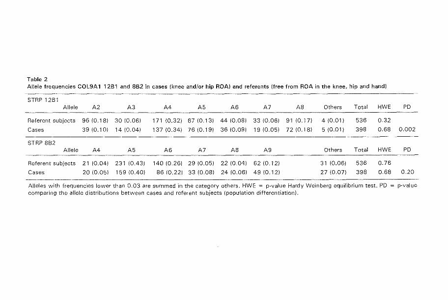
Table 2 Allele frequencies COL9A1 12B1 and 8B2 in cases (knee and/or hip ROA) and referents (free from ROA in the knee, hip and hand)
STRP 12Bl Allele A2 A3 A4 A5 A6 A7 AS Others Total HWE PD
Referent subjects 96 (O.lS) 30 (0.06) 171 (0.32) 67 (0.13) 44 (0.08) 33 (0.06) 9110.17) 4 (0.01) 536 0.32
Cases 39 (0.10) 14 (0.04) 137 (0.34) 76 (0.19) 36 (0.09) 19 (0.05) 72 (0.18) 5 (0.01) 398 0.68 0.002
STRP 882 Allele A4 A5 A6 A7 A8 A9 Others Total HWE PD
Referent subjects 21 (0.04) 231 (OA3) 140 (0.26) 29 (0.05) 22 (0.04) 62 (0.12) 31 (0.06) 536 0.76
Cases 20 (0.05) 159 (OAO) 86 (0.22) 33 (0.08) 24 (0.06) 49 (0.12) 27 (0.07) 398 0.68 0.20
Alleles with frequencies lower than 0.03 are summed in the category others. HWE = p-value Hardy Weinberg equilibrium test. PO p-value
comparing the allele distributions between cases and referent subjects (population differentiation).

Table 3 Allele frequencies COl9A1 12B1 and 8B2 in cases (knee and/or hip ROA) and referents (free from ROA in the knee, hip and hand), stratified by gender.
STRP 12Bl Allele A2 A3 A4 A5 A6 A7 A8 Others Total HWE PO
Referents women 43 (0.16) 16 (0.06) 77 (0.29) 36 (0.14) 21 (O.OS) 17 (0.06) 51 (0.19) 1 (0.01) 262 0.53
Cases women 19 (0.08) 9 (0.04) 83 (0.34) 47(0.19) 20 (0.08) 11 (0.05) 51 (0.21) 4 (002) 244 0.44 0.04
Referents men 53 (0.19) 14 (0.05) 94 (0.34) 31 (0.11) 23 (0.08) 16 (0.06) 40(0.15) 3 (0.01) 274 0.30
Cases men 20 (0.13) 5 (0.03) 54 (0.35) 29 (0.19) 16 (0.1 D) 8 (0.05) 21 (0.14) 1 (0.01) 154 0.S4 0.21
STRP 882 Allele A4 A5 A6 A7 A8 A9 Others Total HWE PO
Referents women 10 (0.04) 110 (0.42) 72 (0.27) 15 (0.06) 11 (0.04) 30 (0.11) 14 (0.05) 262 0.68
Cases women 14 (0.06) 97 (0.39) 56 (0.23) 22 (0.09) 14 (0.06) 27 (0.11) 14 (0.06) 244 0.60 0.67
Referents men 11 (0.04) 121 (0.44) 68 (0.25) 14 (0.05) 11 (0.04) 32 (0.12) 17 (0.06) 274 0.98
Cases men 6 (0.04) 62 (0.40) 30 (0.19) 11 (0.07) 10 (0.06) 22 (0.14) 13 (0.08) 154 0.49 0.44
Alleles with frequencies lower than 0.03 are summed in the category others. HWE = p-value Hardy Weinberg equilibrium test. PO p-value comparing the allele distributions between cases and referent subjects (population differentiation).
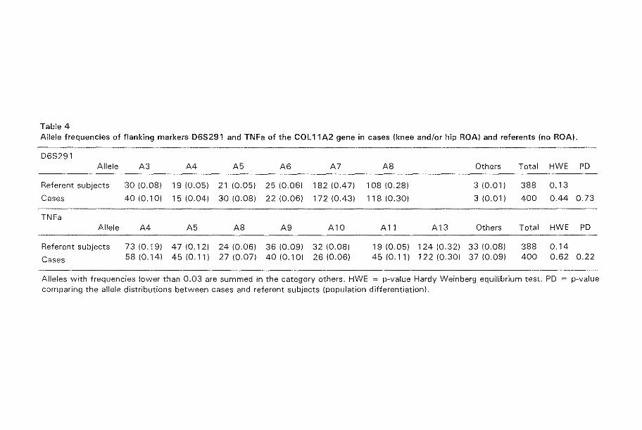
Table 4 Allele frequencies of flanking markers D65291 and TNFa of the COL 11A2 gene in cases (knee and/or hip ROA) and referents (no ROA).
06S291 Allele A3 A4 A5 A6 A7 A8 Others Total HWE PO
Referent subjects 30 (0.08) 19 (0.05) 21 (0.05) 25 (0.06) 182 (0.47) 108 (0.28) 3 (0.01) 388 0.13
Cases 40(0.10) 15 (0.04) 30 (0.08) 22 (0.06) 172 (0.43) 118 (0.30) 3 (0.01) 400 0.44 0.73
TNFa Allele A4 A5 A8 A9 Al0 All A 13 Others Total HWE PO
Referent subjects 73 (0.19) 47(0.12) 24 (0.06) 36 (0.09) 32 (0.08) 19 (0.05) 124 (0.32) 33 (0.08) 388 0.14
Cases 58 (0.14) 45 (0.11) 27 (0.07) 40 (0.10) 26 (0.06) 45 (0.11) 122 (0.30) 37 (0.09) 400 0.62 0.22
Alleles with frequencies lower than 0.03 are summed in the category others. HWE == p-value Hardy Weinberg equilibrium test. PO = p-value comparing the allele distributions between cases and referent subjects (population differentiation).

Chapter 4.2
we observed LD in the female case group (p = 0.02). Neither in male cases nor
in male or female referent subjects LD was observed. \Ve subsequently investi
gated whether in women specific haplotypes were responsible for the associa
tions observed between ROA and alleles of STRP 12Bl. These analyses were
based on the estimated haplotype frequencies derived from an EM algorithm.
The occurrence of haplotypes 12Bl·A2/SB2·A5 and 12Bl·A2/SB2·A6 was reo
spectively 3.2 times (adjusted p·value ~ 0.01) and 7.1 times (adjusted p·value <
0.002) decreased in female cases as compared to female referent subjects. The
frequencies of both BE2 alleles, AS and A6, were decreased in cases as COlll
pared to referent subjects in the overall (table 2) and gender specific (table 3)
analyses, although these differences were not statistically significant. Allele
12Bl·A5 forms haplotypes with all alleles of STRP SB2. No specific haplotype
with the allele 12BI-A5 was observed with a statistically significantly increased
frequency in cases as compared to referent subjects.
COL 11 A2 region
Of the microsatellite marker locus D6s291 8 alleles were detected, while for
TNFa 14 different alleles were observed. In table 4, allele frequencies of 194 ref·
erent subjects and 200 cases (genotyping was unsuccessful in four cases) of mi
crosatellites D6s291 and TNFa are shown. Genotypes of both dinucleotide reo
peats were in Hardy-Weinberg equilibrium in cases and referent subjects (see
table 4), No significant differences were found in allele distributions between
cases and referent subjects for D6s291 or TNFa, neither in the overall analysis
(table 4) nor in the analy.ses stratified according to gender (data not shown),
Also) LD analysb failed to show significant evidence for an association between
the COLllA2 region and ROA.
Discussion
We found a statistically significant dIfference in the allele distribution between
cases with ROA in the knee and/or hip joint and referent subjects without ROA
for the STRP 12Bl of the COL9Al gene. 11,is difference was mainly due to the
lower frequency of allele A2 and higher frequency of allele AS in cases as com~
pared to referent subjects. 'TI1e association between STRP 12B1 and ROA was
strongest in women. Linkage disequilibrium was found in the female case group,
in whom subsequent ha plotype analysis led to a refinement of the association of
ROA with 12Bl·A2/SB2·A5 and 12Bl·A2/SB2·A6, suggesting a protective effect
of these haplotypes. Allelic and haplotype distributions of the microsatellite
marker loci D6s291 and TNFa, flanking the COLllA2 gene, were not different in
cases as compared to referent subjects.
86

Association of COL9A 1 gene and COL I1A2 region with ROA
'I11e allele frequencies in our :-;tudy population are similar to those previ~
Gusly reponed in Caucasian populations (19,21)2). All radiographs were scored
before genotyping and genotyping was performed blind to the disease status.
Therefore, misclassification will most likely be random and is not likely to be the
cause of the associations found. In the present study cases and referent subjects
were derived [rom a homogeneous Caucasian population, making bias due to
population admL'Cture unlikely (26).
STRPs 12B1 and BB2 are both located within the COL9Al gene, but linkage dis
equilibrium was found in the female case group only. This suggest.s that the fre
quency of recombination within the COL9Al gene is likely to be high and may
be different in this region for men and women. Gender specific recombination
rates have previously been found for other region's (27). Interestingly, the asso
ciation reported in this paper between the COL9Al gene and ROA was strongest
for women, in palticular for the lZBl allele A2 and specifically for haplotypes
12BI-AZ/BB2-AS and 12Bl-AZ/8B2-A6. The effect of 12Bl allele AS, which fre
quency is increased in caseS as compared to referent subjects, was not con
firmed by haplotype analysis. In women, STRP 8B2 itself was not signifrcantly
associated with ROA, despite the fact that linkage disequilibrium was found with
STRP 12Bl in the female case group. This can be explained by the relatively
high frequencies of the AS and A6 alleles of STRP BB2. These alleles occur
therefore on many other haplotypes than the ones involved in the linkage dis
equilibrium with allele A2 of STRP 12Bl, with which allele an association with
ROA was detected.
Our findings did not show evidence for a role of the COLIIA2 region in the
occurrence of ROA. Keeping in mind the relatively large distance between the
COLllA2 gene and the two flanking microsatellite marker loci D6s291 and
TNFa, in particular the haplotype analysis decreases the chance that the
COLllA2 gene itself plays an important role in the occurrence of ROA in the
general population. However, this finding needs to be confirmed in another
study. Both TNFa and the COLl J A2 gene are located within the HLA region,
which region has a low frequency of recombination and therefore TNFa can
se1ve as a reliable marker locus for the COLllA2 gene. TNF-cx gene may be con
sidered a candidate gene for OA (20), but was not associated with ROA in our
study. This minimizes the possibility that the TNF-cx gene plays a role in the oc
currence of ROA in the general population.
Although present in small amounts, type IX collagen is an impOltant con
stituent of normal alticular cartilage. It stabilizes and lends cohesion to the colla
gen network (28). Differences in the gene products encoding type IX collagen
may lead to the synthesis of abnormally assembled type IX collagen fibrils, that
are more prone to damage from repetitive and long-term mechanical stress. This
87

Chapter 4.2
has indeed been shown in transgenic mice, in which the synthesis of shortened
al(IX) chains gives rise to osteoanhritic changes in knee joints (8),
The present study shows a significant association between the STRP 12B1
of the COL9Al gene and ROA in the general population. following the studies
in transgenic mice, our population-based findings emphasize the COL9Al gene
as an interesting candidate gene for human ~A.
References
1. Odding E: Locomotor disability in the elderly. An epidemiological study of its occurrence and determinants in a general population of 55 years and over. PhD thesis, Erasmus Universily Rotterdam, The Netherlands, 1994.
2. O'Reilly SC, Nluir KR, Dohc11Y M: Screening for pain in knee osteoaI1hritis: which question? Ann Rheum Dis 1996; 55: 931-933.
3. Bruckner P, Van del" Rest M: Structure and function of ca1tHage collagens. Min Res & Tech 1994; 28: 37H-384.
1. WU JJ, Woods PE, Eyre DR: Identification of cross-linking sites in bovine cartilage type IX collagen reveals an antiparallel type II-type IX molecular relationship and lype IX to type IX bonding . .J Bioi ehem 1992; 267: 23007-23014.
5. Diab M: The role of type IX collagen in osteoai1hritis and rheumatoid arthritis. 011h Review 1993; 26: 165-170.
6. Diab M, \'Vu JJ, Eyre DR: Collagen type IX from human cartilage: a structural profile of intermolecular cross-linking sites. BiochemJ 1996; 311: 327-332.
7. Kimura T, Mattei MG, Stevens JW, Goldring MB, Ninomiya Y, Olsen BR: Molecular cloning or rat and human type IX collagen eDNA and localization of the alphal(IX) gene on the human chromosome 6. Eur J Biochem 1989; 179: 71-78.
8. Nakata K, Ono K, j'vfiyazaki JI, Olsen BR, Muragaki Y, Adachi E, Yamamura KI, Kimura T: Osteoal1hritis associated with mild chondrodysplasia in transgenic mice expressing a1(IX) collagen chains with a central deletion. Proc Natl Acad Sci USA 1993; 90: 2870-2874.
9. Eissler R, Schnegelsberg PNJ, Dausman j, Shinya T, Muragaki Y, IvIcCal1by MT, Olsen BR, Jaenisch R: Mice lacking al(IX) collagen develop noninfiammatolY degenerative joint disease. Proc Natl Acacl Sci USA 1994; 91: 5070-5074.
10. Muragaki Y, Mariman ECM, van Beersurn SEC, Perala M, van MourikJI3, Warman ML, Olson I3R, Hamel BCJ: A lnutation in the gene encoding the a2 chain of the fibril associated collagen IX, COL9A2, causes multiple epiphyseal dysplasia (EDM2). Nature Genet 1996; 8: 103-105.
11. Thom JR, Morris NP: Biosynthesis and proteolytic processing of type Xl collagen in embtyonic chick sterna. J BioI Chem 1991; 266: 7262-7269.
12. Vikkula M, Mariman EC1\-I, Lui VC, Zhidkova N, Tiller G, Goldring 1'1'1, van Beersum S, de \'Vaal Maldijt M, van den Hoogen F, Ropers HH, Mayne R, Cheah KS, Olsen BR, Warman wIL, Brunner HG: Autosomal dominant and recessive osteochondrodysplasias associated with the COLllA210cus. Ce!11995; 80: 431-137.
1.3. mith GN, \'ijilliams JM, Brandt KD: Interaction of proteoglycans with the pericellular (la, 2a, 30'.) collagens of cal1ilage. J Bioi Chern 1985; 260: 10761-10767.
ltl. Lui VC, Ng LJ, Sat EWY, Cheah KS: The human a2(XI) collagen gene (COLlIA2): completion of coding information, identification of the promoter sequence, and pre-
88

Association of COL9A 1 gene and COL 11 A2 region with ROA
cisc localization 'within the major histocompatibility complex reveal overlap with the KE5 gene. Genomic." 1996; 32: 401-112.
15. U Y, Lacerda DA, Warman NIL, Beier DR, Yoshioka H, Ninomiya Y, Oxfonl ]T, i'vlorris NF, Andrikopou!os K, Ramirel F, Wardell BB, Liffel1h GD, Teuscher C, Woodward SR, Taylor I3A, Seegmiller RE, Olsen BR: A fibrillar collagen gene, Call1al, is essential for skeletal morphogenesis. Cell 1995; 80: 423-430.
16. Richards AJ, Yates JRW, \'Villiams R, Payne Sj, Pope FM, Scott JD, Snead MP: A family with Stickler syndrome type 2 has a mutation in the COL llAl gene resulting in the substitution or glycine 97 by valine in al(XI) collagen, Hum Mol Genet 1996; 5: 13.39-13'U.
17. Hofman A, Grobbee DE, De Jong PTVM, Van den Ouweland FA: Determinants of disease and disability in the elderly. Eur] Epidemiol1991; 7: 403-422.
18. Kellgren JH, Jeffrey MR, Ball] (cds): The epidemiology of chronic rheumatism. Volume 11: Atlas of standard radiographs of aI1hritis. Oxford: Blackwell Scientific Publications, 1963.
19. \X1arman !vIi, Tiller GE, Polumuo PA, Seldin 1\iIF, Rochelle JM, Knoll JHM, Cheng SD, Olsen BR: Physical and linkage mapping of the human and murine genes for the cd chain of type IX coHagen (COL9A1). Genomics 1993; 17: 691.-698.
20. Saklatvala J: Tumor necrosis factor u stimulates resorption and inhibits synthesis of proteoglycan in cartilage. Nature 1986; 322: 547-519.
21. \Veissenbach J, Gyapay G, Dib C, Vignal A, Morisette J, Millasseau P, Vaysseix G, Lathrop M: A second generation linkage map of the human genome. Nature 1992; 359, 791-801.
22. Nedospasov SA, Udalova lA, Kuprash DV, Turetskaya H.I: DNA sequence polymorphism at the human tumor necrosis factor (TNF) locus. J Immunol 1991; lti7: 1053-1059.
23. Guo S\V, Thompson EA: Performing the exact test of Hardy-Weinberg propol1ion for multiple alleles. Biometrics 1992; 48: .361-372.
21. Raymond J\:I, Rousser F: Genepop (version 1.2): population genetics software for exact tests and ecumenicism. J Heredity 1995; 86: 248-249.
25. Schneider S, Kueffer JM, Roessli D, Excoffier L: Arlequin ver 1.1: a softw"are for population genetic data analysis. Genetics and Biometly LaboratOlY, University of Geneva, Switzerland, 1997.
26. Lander ES, Schork NJ: Genetic dissection of complex traits. Science 1994; 265: 20.37-2048.
27. Paldi A, Gyapay G, Jami.J: Imprinted chromosomal regions of the human genome display sex-specific meiotic recombination frequencies. Current Biology 1995; 5: 1000-1035.
28. Eyre DR: Structure and function of the caI1ilage collagens: role or type IX collagen in al1lcular canilage. In: Brandt KD (cd) Canilage changes in osteoal1hrltis, Indiana University School of lvledicine, Indianapolis, pp 12-16, 1990.
89

A sibling pair study on the role
of the COL2Al and COL9Al genes
in radiological osteoarthritis
Abstract
4.3
Objective To investigate the role of the COL2A 1 and COL9A 1 genes in
the occurrence of radiological osteoarthritis (ROA) and/or disk degenera
tion through a sibling pair approach.
Methods Probands (n = 118) were derived from a random sample of a
population-based cohort study (the Rotterdam Study) and were affected
with ROA and/or disk degeneration at multiple joint sites. All available sib
lings that were willing to participate were recruited (n = 257). ROA in the
knees, hips, and hands and disk degeneration of the spine was scored
according to the Kellgren grading system. ROA was considered a quanti
tative trait locus, using a sum score of ROA and disk degeneration a sum
score of hand ROA, and a sum score of disk degeneration. Analyses were
performed using MAPMAKER/SIBS.
Results For the COL2A 1 locus, a reduction was observed in the variance
for the sum score of ROA and disk degeneration and for the sum score of
hand ROA (quantitative trait loci), with an increase in the number of al
leles shared between female siblings (not statistically significant). Effects
were stronger for women with ROA and Heberden's nodes and for
91

Chapter 4.3
women with generalized ROA. For the COL9A 1 locus, no evidence for
linkage was found between the three phenotypes studied and this locus.
Conclusion Findings of the present study are compatible with a role of
the COL2A 1 locus in the occurrence of ROA in the peripheral joints.
Introduction
Osteoatthritis eOA) is a disease characterized by degeneration of 3Iticuiar carti
lage and formation of osteophytes at the joint margins. Recent population-ba::;ed
studies have demonstrated a considerable genetic component in the etiology of
common forms of OA 0-3). High sib-sib correlations were obtained in these
studies when OA was regarded a quantitative trait, by summing all joints in the
hands and knees affecteel with radiological OA (ROA). Which genes are respon
sible for this genetic influence in OA remains largely to be determined, although
several candidate genes have been proposed that could explain part of this ge
netic susceptibility (4-8). Among others these are genes encoding cartilage spe
cific collagens, growth factors, rnetalloproteases and non-collagenous cal1ilage
proteins.
For the COL2Al gene, encoding the major cartilage protein, collagen type
II, findings have been controversiaL Two British hospital based studies showed
an association between rare alleles of the COL2Al gene and radiological OA
(ROA) at multiple sites (4,5), Association studies in the Finnish population and
Belgian population (postmenopausal women) and a sibling pair study of limited
size in the British population failed to show an association with the COL2Al lo
cus (9-11). We found an association between the COL2Al locus and ROA in
women, in a population-based study (see Chapter 4.1). Strongest effects were
found for women with generalized OA and for women with Heberden's nodes.
111e COL9Al gene encodes the al chain of the heterotrimeric protein colla
gen type IX, which is covalently cross linked to collagen type II fibrils and stabi
lizes the articular collagen network (12). We recently performed the first asso
ciation study on ROA and the COL9Al gene and found evidence for a protective
effect of this locus on the occurrence of ROA, especially in women (see Chapter
4.2)
Our findings in both association studies need to be interpreted with cau
tion, as these studies can easily give rise to a false positive result (13). We have
conducted a sibling pair study in 257 siblings of 118 probands with ROA and
disk degeneration at multiple joint sites. We examined the genes, i.e. the
COL2A! and COL9A! genes, of which we previously found an association with
ROA. ROA was considered a quantitative trait based on the number of joints in
the knees, hips) hands, and spine affected with ROA or disk degeneration.
92
Sibling pair study and COL2A 7 and COL9A 1 genes
Materials and Methods
Subjects
Probands were derived from a prospective population-based follow-up study of
determinant.s and prognosis of chronic diseases in the elderly) the Rotterdam
study (14). In thio study all persons living in Ommoord, a suburb of Rotterdam,
aged 55 years and over were invited to participate. In total 7983 participants (re
sponse rate of 78 percent) were examined. The medical ethics conmlittee of
Erasmus University Medical School has approved the study and written informed
consent was obtained from all participant.s.
A random sample of 944 non-institutionalized individuals aged between 55
and 65 years was drawn from the Rotterdam Study. In all individuals, radio
graphs of the peripheral joints, Le. knees, hips and hands, and of the thoraco
lumbar spine were scored fOf respectively ROA and disk degeneration. Probands
for the sibling pair study had to have tvvo or more of these four joint sites af
fected in order to be selected. In case individuals had hand ROA and disk de
generation of the spine, which i.s the most common combination present, they
had to have additional Heberden's nodes to be included as probands. The 221
probands that were willing to participate (response rate 88 percent), had a total
of 708 siblings born alive, of which 368 siblings were eligible to our study. We
were able to recruit 257 siblings (70 percent), whom were all examined at the
research center.
Measurements
For all individuals the following radiographs were obtained: weightbearing ante
rior-posterior pelvic radiographs with both feet in 10" endorotation, weightbear
ing knee radiographs with the patellae in central position, anteroposterior radio
graphs of the hands and wrists and lateral radiographs of the spine CTh4-SlJ.
TIl(~ use of radiologically defined changes, e.g. osteophyte formation and
joint space narrowing, is widely accepted in epidemiological research concern
ing OA (15). ROA of the spine could not be assessed on the lateral radiographs
that were made. Instead, disk degeneration of the spine was scored, which may
share a common genetic etiology with ROA of the peripheral joints (16). ROA of
the peripheral joints and disk degeneration of the spine were assessed by means
of the Kellgren grading system in five grades (0-4), using the figures and legends
of the original atlas (7). Two independent readers, who had no knowledge of
the other data of the participant, scored all radiographs. After each set of about
150 radiographs the scores of the two readers were evaluated. Whenever the
scores were two or more points different, Of, was two for one reader but one for
93
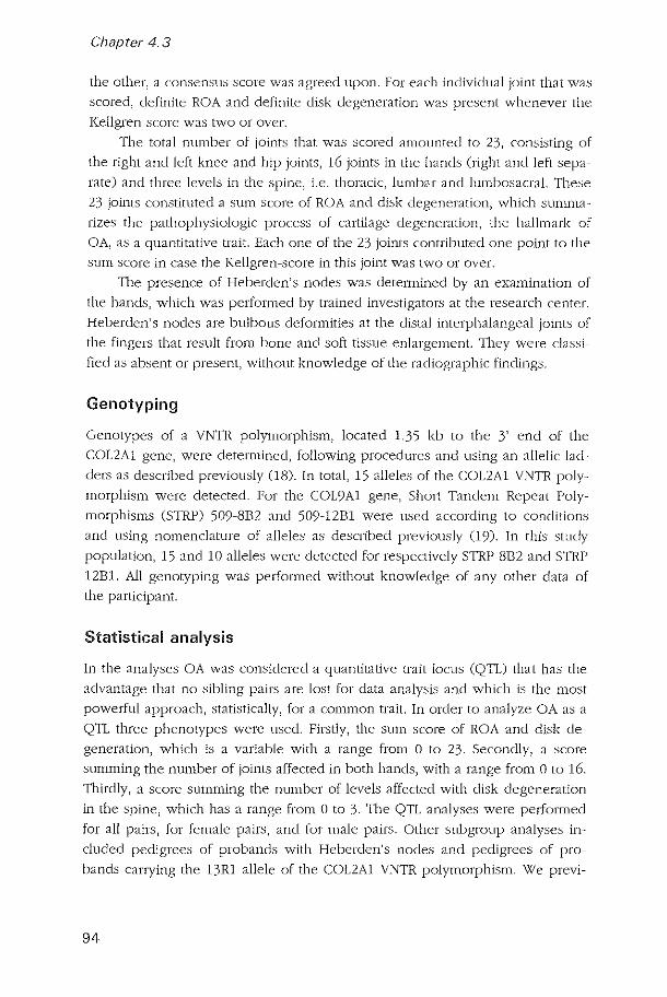
Chapter 4.3
the other, a consensus score was agreed upon. For each individual joint rh8t was
scored, definite ROA and definite disk degeneration was present whenever the
Kellgren-score was two or over.
rTI1e total number of joints that was scored amounted to 23, consi.sting of
the right and left knee and hip joints, 16 joints in the hands (right and left sepa
rate) and three levels in the spine, i.e. thoracic, lumbar and lumbosacral. The.<;e
23 joints constituted a surn score of ROA and dbk degeneration, which SUl1l11lJ
rizes the pathophysiologic process of cartilage degeneration, the hallmark of
OA, as a quantitative trait. Each one of the 23 joints contributed one pOint to the
sum score in case the Kellgren-score in this joint was two or over.
111e presence of Heberclen's nodes was determined by an exarnination of
the hands, which was performed by trained investigators at the research center.
Heberden's nodes are bulbous defonnities at the distal interphalangeal joints of
the fingers that result from bone and soft tissue enlargement. They were classi
fied as absent or present, without knowledge of the radiographic findings.
Genotyping
Genotypes of a VNTR polymorphism, located 1.35 kb to the 3' end of the
COL2Al gene, were determined, following procedures and using an allelic lad
ders as described previously (IS). In total, 15 alleles of the COL2Al VNTR poly
morphism were detected. For the COL9Al gene, Short Tandem Repeat Poly
morphisms (STRP) 509-SIl2 and 509-12B1 were used according to conditions
and using nomenclature of alleles as descIibed previously (19). In this study
population, 15 and 10 alleles were detected for respectively STRl' BB2 and STRl'
12Bl. All genotyping was performed without knowledge of any other data of
the pal1icipant.
Statistical analysis
In the analyses OA was considered a quantitative trait 10CllS (QTI) that has the
advantage that no sibling pairs are lost for data analysis and which is the most
powerful approach, statistically, for a common trait. In order to analyze OA as a
QTI three phenotypes were used. Firstly, the sum score of ROA and disk de
generation, which is a variable with a range from 0 to 23. Secondly, a score
summing the number of joints affected in both hands, with a range from 0 to 16.
Thirdly, a score summing the number of levels affected with disk degeneration
in the spine, which has a range from 0 to 3. The QTL analyses were performed
for all pairs, for female pairs, and for male pairs. Other subgroup analyses in
cluded pedigrees of probands with Heberden's nodes and pedigrees of pro
bands canying the 13Rl allele of the COL2Al VNTR polymorphism. We previ-
94
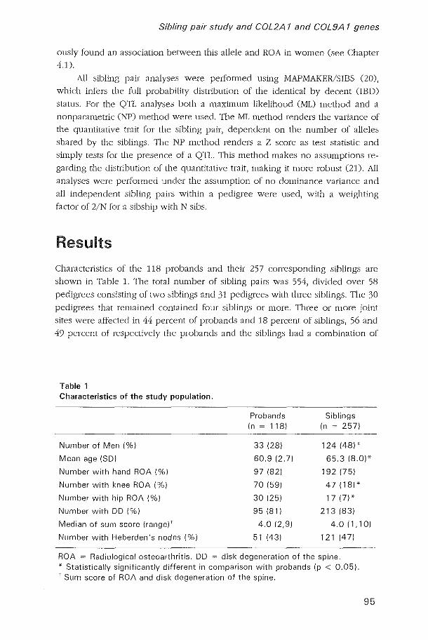
Sibling pair study and COL2A 1 and COL9A 1 genes
ously found an association between this allele and ROA in women (see Chapter
4.1).
All sibling pair analyses were performed using MAPMAKER/SIBS (20),
which infers the full probability distribution of the identical by decent (IBD)
status. For the QTL analyses both a maximum likelihood (ML) method and a
nonparametric (NP) method were used. 'I11e .ML method renders the variance of
the quantitative trait for the sibling pair, dependent on the number of alleles
shared by the siblings. The NP method renders a Z score as test statistic anel
simply tests for the presence of a QTI. This method makes no assumptions re
garding the dbtrlbution of the quantitative trait, making it more robust (21). All
analyses were performed uncler the assumption of no dominance variance and
all independent sibling pairs within a pedigree were used, with a weighting
factor of 2/N for a sibship with N sibs.
Results
Characteristics of the 118 probands and their 257 corresponding siblings are
shown in Table 1. The total number of sibling pairs was 554, divided over 58
pedigrees consisting of two siblings and 31 pedigrees with three siblings. The 30
pedigrees that remained contained four siblings or more. Three or more joint
sites were affected in 44 percent of probands and 18 percent of siblings, 56 and
49 percent of respectively the probands and the siblings had a combination of
Table 1 Characteristics of the study population.
Probands Siblings (n ~ llB) (n ~ 257)
Number of Men (%) 33 (2B) 124 (4B) *
Mean age (SD) 60.9 (2.71 65.3 (B.O)*
Number with hand ROA (%) 97 (B2) 192 (75)
Number with knee ROA (%) 70 (59) 47 (lB)*
Number with hip ROA (%) 30 (25) 17 (7) *
Number with DO (%) 95 (Bl) 213(B3)
Median of sum score (range) t 4.0 (2,9) 4.0 (1,10)
Number with Heberden's nodes (%) 51 (43) 121 147)
ROA = Radiological osteoarthritis. DD disk degeneration of the spine, * Statistically significantly different in comparison with probands (p < 0.05). t Sum score of ROA and disk degeneration of the spine.
95

Chapter 4.3
tvlo joint sites affected. Finally, 33 percent of the ::;lbling.':l had no or just one joint
site affected with ROA or disk degeneration.
COl2A1 gene
For the .sum score of ROA anel disk degeneration, the variance between .siblings
in a sibling pair was lower when they .shared one or two allele.s of the COL2Al
VNTR polymorphism as compared to siblings that shared no alleles Crable Z).
This effect was strongest in female pairs, whereas in male pairs almost no effect
was obsclved. for the score summing the Dumber of joints affected with ROA in
the hands, the variance was three times lower in female sibling pairs sharing tvlo
alleles as compared to female sibling pairs sharing no alleles (LOD ~ LO) (Table
2). For disk degeneration of the spine, analyzed separately, no evidence for
linkage with the COL2Al locus was founel (data not shown). When considering
the pedigrees of pro bands with Heberden's nodes, a reduction in variance with
an increase in number of alleles shared was observed for the sum score of ROA
and disk degeneration and for the sum score of hand ROA. These variances
were respectively 020 = 21.6,02
] = 9.9, and a22 "" 9.9, with LOD score 0.6 and a 2
0
= 17.9, a21 = 7.6, and a 22 = 7.6, with LOD score 0.7. Earlier we repOlied an asso
ciation between allele 13RI (frequency O.4Z) of the COLZA! VNTR polymor
phism and ROA in the peripheral joints, I.e. the knees, hips and hands. In an
analysis uoing only the sibling pairs of probands canying a 13RI allele with their
Table 2 Quantitative trait locus (UTU analysis in 554 sibling pairs for the COL2A 1 gene.
Maximum likelihood analysis Nonparametric
analysis
,,'0 ,,', ,,', LaD Z score
LaD score score
Sum score of ROA and DO
All pairs 16.3 12.5 12.5 0.2 -0.2 0.0
Female pairs 18.0 13.3 8.2 0.5 1.3 0.3
Male pairs 14.5 13.5 13.5 0.0 -1 .5 0.0
Sum score of hand ROA
All pairs 12.5 8.6 8.6 0.3 0.1 0.2
Female pairs 14.3 9.8 4.1 1.0 1.6 0.6
Male pairs 10.3 9.3 9.3 0.0 -1.1 0.0
ROA = Radiological osteoarthritis. DO = Disk degeneration. 020, 0
21, 0
22 ~ The
variance of the QTL in sibling pairs sharing respectively zero, one or two alleles. Z score = The ratio of the test statistic Xw(s) and square root of the variance V.
96

Sibling pair study and COL2A 1 and COL9A 1 genes
respective siblings, a marked decrease in the variance for the sum score of ROA
anel disk degeneration was observed when siblings shared alleles (a2o = 16.2, a2l
~ 10.0, and 0-2
, ~ 6.7, with LOD score 0.2).
COL9A1 gene
For the COL9Al gene, the variance was equal in all analyses for sibling pairs
sharing zero alleles or one allele of the two marker loci STRP-8B2 and !2BI (Ta
ble 3). Sibling pairs sharing t\Vo alleles had lower variances as compared with
pairs sharing zero or one allele. However, the reduction in variance was mini
mal.
Discussion
Earlier, we performed population-based association studies to examine the role
of the COLZAI and COL9AI loci in the occurrence of ROA. The present sibling
pair study was performed with probands derived from these population-based
studies and their corresponding siblings. For the COL2Al locus) we earlier ob
selved an association in women between the most frequent allele of the COL2Al
rable 3 Quantitative trait locus (OTl) analysis in 554 sibling pairs for the COL9A 1 gene. *
Maximum likelihood analysis Nonparametric
analysis
,,'0 ,,', ,,', LOD Z score
LOD score score
Sum score of ROA and DD
All pairs 13.8 13.8 13.1 0.1 -0.4 0.0
Female pairs 13.2 13.2 11.2 0.0 -0.1 0.0
Male pairs 15.7 15.7 10.8 0.1 0.1 0.0
Sum score of hand ROA
All pairs 9.6 9.6 9.6 0.0 -1.2 0.0
Female pairs 9.1 9.1 9.1 0.0 -0.7 0.0
Male pairs 11 .1 11. 1 6.8 0.2 -0.1 0.1
ROA = Radiological osteoarthritis. DO = Disk degeneration. 0'20, 0'21, 0'22 = The
variance of the QTL in sibling pairs sharing respectively zero, one or two alleles. Z score = The ratio of the test statistic Xw{s) and square root of the variance V. ~. Results shown are derived from multipoint analyses using marker loci STRPs 1281 and 882 for the COL9A 1 gene.
97

Chapter 4.3
VNTR polymorphism, allele 13R1 (frequency 0.42), and ROA (Chapter 4.1). The
findings on the COL2Al locus in the present study, which show a reduction in
variance for OA as quantitative trait with an increase in the nurnber of alleles
shared between female siblings, are in line with the detected association bc
tv\reen the COL2Al locus and ROA. for the COL9Al locus, an association was
earlier observed between STRp·12B1 (a marker locm for the COL9A1 gene) and
ROA that was stronger in women as compared to men (Chapter 4.2). In the pres
ent study, no evidence for linkage was found between the sum score of ROA
and disk degeneration, hand ROA, or di::ik degeneration of the spine and the
COL9A1 locus.
The most important limitation of our study concerns the statistical power.
Although, 554 sibling pairs were available for analyses, no significant result.')
could be reported. The maximum LOD score that was reached amounted to 1.0
with 80 percent ability to e:h'1ract the full IBD .')tatu.') in case of the COL9Al 10c11.'),
where two closely linked marker loci were used. In case of the COL2Al VNTR
polymorpbism full lED status could be calculated for 68 percent of all sibling
pairs.
Previou.')ly, one .')ibling pair study of limited size concerning the COL2Al
locus has been publisbed (10). Tbis affected sibling pair study did not show evi·
dence for linkage of OA in three or more joint sites before the age of 60 years to
the COL2Al locu.'). The .')tronge.')t indications for linkage in the pre.')ent sibling
pair study were observed in two out of three subgroups that earlier showed the
strongest association between this locus and ROA, i.e. women, in particular
those with Heberden' s nodes. 111cse findings funher support the hypothesi.')
that the COL2Al locus plays a role in the development of ROA in peripberal
joints. However, given the limited size of the present study the.')e finding.') are
not conclusive.
In case of the COL9Al locus no earlier association or sibling pair study has
been performed. In our previous association study, we found no allele at the
COL9Al locus predisposing for the occurrence of ROA or disk degeneration, but
instead obselved an association indicating a possibly protective effect of this
locus. nle present sibling pair .')tudy mainly consisted of siblings that were af
fected with ROA and/or disk degeneration (only 12 siblings were free from ROA
and disk degeneration). Therefore, it was unlikely that in this sample it would be
possible to confirm this protective effect, because of a lack of statistical power.
Alternatively, the pre.')ent .')tudy supports the finding of our previous association
study that the COL9Al locus does not have a predisposing effect on the occur
rence of ROA or disk degeneration.
In conclusion, findings of the present study are compatible with a role of
the COL2Al locus in the occurrence of ROA in peripheral joints, although in the
Cllfl'ent setting no statistically significant results were obselved. We could not
98
Sibling pair study and COL2A 1 and COL9A 1 genes
find evidence that the COL9Al locus increases the risk of developing ROA or
disk degeneration.
References
1. Spector TD, Cicuttini F, Baker J, Loughlin], Hellt D. 1996. Genetic innuences on 05-teoaI1hritis in women: a twin study. BMJ 312:9IJO-3.
2. Fclson DT, C:ouropmitree NN, Chaisson eE, Hannan IV1T, Zhang Y, McAlindon 'fE, LaValley lvI, Levy D, lVIycrs RI-L 1998. Evidence for a mendclian gene in a segregation analysis of generalized radiographic osteoarthritis. Arthritis Rheum 41: 1064-7l.
3. Hirsch R, LClhbridge-Cejku lvI, Hanson R, Scott \Yf\V, Reichle R, Plato ee, Tobin ]D, [Iochberg IvJC. 1998. Familial aggregation of osteoalthritis. Al1hritis Rheum 11:1227-32.
4. Hull R and Pope FM. 1989. Osteoal1hritis and cal1ilage collagen genes. Lancet i:1337-8.
5. Loughlin], lrven C, AthanasoLl N, Carr A, Sykes B. 1995. Differential 'allelic expression of the type II collagen gene (COL2Al) in osteoarthritic cartilage. Am ] Hum Genet 56,1186~9.).
6. Loughlin], liven C, AthanasOll N, et a1. Differential allelic expression of the type II collagen gene (COL2Al) in osteoalihritic callilage. Am] Hum Genet 1995;56:1186-93.
7. IVleulenuelt I, Bijkerk C, lVliedema HS, Breedvcld FC, Hotinan A, Valkenburg HA, Pols HAP, Slagboom PE, van Duijn eM. A genetic association study of the IGF-l gene and radiological osteoarthriLis in a population-based cohOI1 study (the Rotterdam Study). Ann Rheum Dis 1998;57:371-4.
8. Meu!enbelt I, I3ljkerk C, de \X7ildt SC, Miedema US, Valkenburg HA, Breedveld FC, Pols HAP, te Koppcle JM, Sloas VF, Hofman A, Slagboom PE, van Duijn CM. Investigation of the association of the CRTM and CR'J'Ll genes with radiographically evident osteoal1hriLis in subjects from the Rotterdam Study. A1thritis Rheum 1997;40:1760-5.
9. Vikkula lvI, Nissila M, Hirvensalo E, NuoHo P, Palotie A, Aho K, Pcltonen L. 1993. lVfultiallelic polymorphism of the cal1i1age collagen gene: no association with osteoarthritis. Ann Rheum Dis 52:762-4.
10. Loughlin .1, Irven C, Fergusson C, Sykes B. 1994. Sibling pair analysis shows no linkage of generalized osteoarthritis to the loci encoding type II collagen, cattilage link protein, or clllilage matrb:: protein. Br J Rheumatol 33:1103-6.
11. Aerssens J, Dequeker J, Peeters J, Breemans S, Boonen S. ]998. Lack of association between osteoallhritis of the hip and gene polymorphisms of VDR, COllA], COL2Al in postmenopausal women. Al1hritis Rheum 41:1946-50.
12. Diab NI, \X7u JJ, Eyre DR. 1996. Collagen type IX from human cal1ilage: a structural profile of intcrmolentlar cross-linking sites. Biochem J 3] 4:327-32.
1.3. Weeks DE, Lathrop GM. 1995. Polygenic disease: methods for mapping complex disease traits. 'I'rends Genet 11:513-9.
11. Hollnan A, Grobbee DE, De .rong PVT.lVI, van den Ouwcland FA. 1991. Determinants of disease and disability in the elderly. Em J Epidemiol 7:403-422.
15. Silman AJ, I-Iochuerg .MC 1993. The epidemiology of the rheumatic diseases. Oxford: Oxford University Press 257-88.
16. Kimura T, Nakata K, Tsumaki N, j\'Uyamoto 5, Matsui Y, Ebara S, Ochi T 1996. Progressive degeneration of articular cartilage and intervellebral discs. An experimental study in transgenic mice bearing a type IX collagen mutation. Int Orthopaed 20:177-81.
99
Chapter 4.3
17. Kcllgrcn jH, Jeffrey lVlR, Ball J (editors). 1963. The epidemiology of chronic rheumatism. Volume II: Atlas of standard radiographs of 'H1hritis. Oxford: Blackwell Scientific Publications.
18. Berg ES, Olaisen B. 199,3, Characterization of the COL2Al VNl'H polymorphism. Genomics 16:350-4.
19. \'(Tarman 1\·11, Tiller GE, Pohllnbo PA, Seldin MF, Rochelle .11\11, Knoll .11 [.I'd) Cheng SD, Olsen BR. 1993. Physical and linkage mapping of the human and murine genes for the cd chain of type IX collagen (COL9Al). Genomics 17:G9'i-8.
20. Kruglyak L, Lander E. 1995. Complete multipoint sib pair analysis of qualitative and quantitative traits. Am J I Ium Genet 57;1i39-54.
21. Kruglyak L, Lander E. 1995. A non parametric approach for mapping quanLiLative trait loci. Genetics 139:1421-8.
100

5
Gene Interaction in Osteoarthritis

The IGF-l gene and
radiological osteoarthritis in a population-based study
Abstract
5. 1
Objective A genetic association study was performed to investigate
whether radiological osteoarthritis (ROA) was associated with specific
genotypes of the insulin-like growth factor I (IGF-I) gene.
Methods Subjects aged 55-65 years were selected from a population
based study of which ROA at the knee, hip, and hand and disk degenera
tion of the spine was assessed. Genotypes were determined of a poly
morphism in the promoter region of the IGF-1 gene.
Results The IGF-1 locus was significantly associated with the presence
of ROA (overall adjusted OR for heterozygous subjects = 1.9, 95% CI
1.2-3.1 and for homozygous subjects 3.6, 95% CI0.8-16.2).
Conclusion Our results suggest that variation at the IGF-1 locus is asso
ciated with ROA and/or disk degeneration and may playa role in OA
pathogenesis. To confirm these findings replication in another population
based sample is needed.
103

Chapter 5. 1
Introduction
Osteo3nhritis eOA) is a disease characterized by the degradation of articular car
tilage and formation of new bone Costeophytes and sclerosis), Several reports
suggest that genetic influences contribute considerably to the development of
OA (1)). The relevance of the genetic component, however, varies among
subgroups of patients and as yet it is not clear which genes are involved 0).
Insulin-like growth factor 1 (lGF-I) stimulates chondrocytes to synthe.:.;ize
extracellular matrix (ECM) components in cartilage (4,5). Its action is mediated
through the type 1 [GF receptor. The function of IGF-l and its receptor in carti
lage formation both during developmental stages and remodeling of adult carti
lage may be relevant to the etiology of ~A. IGF-l may also influence OA by os
teophyte formation (6). Although osteophyte growth during OA progression and
bone mineral density (BMD) b positively correlated to high serum IGF-l levels
(6,7), no consbtent relationship betvveen serum IGF-l levels and OA has been
found (8,9). FUl1hermore, Serum IGF-l is inversely correlated to age (10) and
body mass index (BMIl (11). Since individual IGF-l levels are liable to temporal
variations, assessing the role of IGF-l in OA by serum levels is complex. We
have examined the relation betvveen the IGF-l gene and the presence of radio
logical OA eROA) in subjects aged 55-65 years.
Materials and methods
Subjects
Individuals were derived from a prospective population-based cohort study of
determinant.s and prognosis of chronic diseases in the elderly, the Rotterdam
study (12). Weight beaIing anterior-posterior radiographs of the hips and knees,
anterior-posterior radiographs of the hands and wrists and lateral radiographs of
the spine (Th4-S1) were obtained fro111 a random population of 944 unrelated
individuals aged 55-65 years. ROA and disk degeneration was assessed by tvvo
independent readers and by mean.:.; of the grading system proposed by Kellgren
(13). By applying these criteria a hip joint with Kellgren score 2 requires the
presence of both definite joint space narrowing and definite o.:.;teophytes. A knee
or hand joint with Kellgren score 2 requires the presence of definite osteophytes
and possible narrowing of jOint space. A spine jOint with Kellgren score 2 de
notes definite lateral osteophytes. For both knees and hips radiographs had pre
viously been scored (14). ROA of the hand was assessed in each inter- and
Inetacarpalphalangeal joint individually, and the first carpometacarpal and tra
pezoscaphoideal joint. ROA of the wrist was assessed at the radiocarpal and
104
Association of IGF-l gene with ROA
distal radioulnar joints. For the spine three levels, from Th4 to 51, were scored
with regard to osteophytes and disk space narrowing, e.g. thoracic, lumbar and
lumbosacral. In the analysis definite ROA was defined as Kellgren-score 2 or
over in the left and/or right corresponding joint. Hand ROA was defined as Kell
gren-score 2 or over in at least one of the 36 joints that were scored. For this
purpose the joints of the wrist were included in the categolY hand ROA. Disk
degeneration of the spine was defined as Kellgren-score 2 or over in at least one
out of three levels scored. 111C presence or absence of osteophytes of the knee
was scored separately.
Information on age (in years), BMI (measured as weight in kg divided by
height2 in metres), and EMD (measured as gram mineral divided by area in cm2)
of the neck of the femur were used in the study. In view of the hypothesis tha t
the genetic contribution to ROA differs in men and women and may depend on
the joint site, stratified analysis was performed with respect to gender and joint
site.
Genotyping and Statistical analysis
Genotypes of the dinucleotide repeat polymorphism of the IGF-l gene were
detennined as previously described (15) of 786 individuals for whom cells were
available. Demographic variables (sex, age, EMI and BMD) were compared
bet\Veen subjects with and without ROA by using t-tests for independent sam
ples. Counting alleles and calculating sample proportions assessed allele fre
quencies. Chi square (X') for Hardy Weinberg equilibrium (HWE) were calcu
lated using the HWE-program (LINKUTIL package) (16). Alleles with an allele
frequency < 0.05 were pooled. A likelihood ratio test was used to test for asso
ciation of !GF-I alleles with the occurrence of ROA (J 7). This method is specifi
cally suitable to perform association studies with polymorphic markers with
multiple alleles. To measure the strength of association bet\Veen ROA and IGF-l
genotypes, a logistic regression model was used to estimate the odds ratio (OR).
ORs were adjusted for risk factors of OA i.e., sex, age, EMI and BMD. ORs are
presented with 95% confidence intervals (95% CI). The statistical package SPSS
was used and P-values < 0.05 (2-sided) were considered significant.
Results
Table 1 shows the number of genotyped individuals with ROA and/or disk de
generation of tbe spine (ROA +) and with none of the four joint sites affected
(ROA-) and tbe mean age, body mass index (EMI), and bone mineral density
(EMD). The mean age and EM! differed Significantly between individuals with
and without ROA and/or disk degeneration (p < 0.001). Only 17% of individuals
105

Chapter 5.7
were free of ROA and disk ciegeneration of the spine in evcIY joint investigated
in this relatively young age group.
In total 9 different alleles were identified (AI-A9) with allele frequencies
ranging from 0.002-0.66. IGF-l allele frequencieo of the 4 moor frequent alleleo
are ohown in Table 1. Except for the relatively rare allele A2, the IGF-l allele
frequencies observed in the random population, and the subjects with and with
out ROA and/or disk ciegeneration were si.milar as described by Weber and May
(15) (oee Table J). The low frequency alleles Al (frequency 0.02), A6 (frequency
0.02), A7 (frequency 0.003), and A9 (frequency 0.002) (not shown in Table 1)
were not previously described in a population-based study (15). The distribution
of genotype frequencies was not Significantly different from that expected for a
population in H\VE neither overall (P ~ 0.76) nor for the ROA+ (p ~ 0.78) or the
ROA- group (p ~ 0.47).
Table 1 shows association of the IGf-l polymorphism in ROA+ as com
pared to ROA- subjects (p ~ 0.02). Differences in allele frequency A3 and A4
between ROA+ and ROA- subjects caused this association. The strength of the
association of the IGF-l locus with ROA was estimated using a logistic regres-
Table 1 Characteristics of random sample of 786 individuals aged 55 to 65 years with allele frequencies of the dinucleotide repeat polymorphism in the promotor region of the IGF-1 gene.
Overall ROA+ ROA-
Total number of individuals 786 651 135
Characteristics of subjects Number of men (%) 317(40) 254 (39) 63 (47) Age in years (SO) 60.5 (2.7)' 59.5 (2.7) BMI in kg/m2 (SO) 26.6 (3.7)' 25.1 (3.1) BMO in glcm' (SO) 0.87 10.13) 0.86 10.12)
Allele frequencies (number of alleles)
Alleles * * Weber t Overall ROA+ ROA-(88) (1572) (1302) (270)
A2 0.18 0.07 0.07 0.07 A3 0.16 0.19 0.20 0.13 A4 0.60 0.66 0.65 0.72 A5 0.06 0.05 0.05 0.05 other 0.00 0.04 0.04 0.04
* significantly different from the control group with p < 0.01 i(. i(. IGF-1 a!!eles with nomenclature as in Weber and May (15) t allele frequencies as in Weber and May (15) t likelihood-ratio test statistic ~ -2Inll(Ho)/L(HdJ
106
LRT' p-
value
4.9 0.02
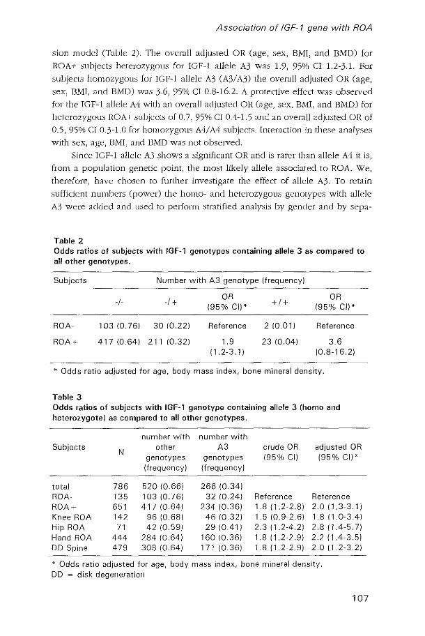
Association of IGF-l gene with ROA
sion model (Table 2). The overall adjusted OR (age, sex, EMI, and EMD) for
ROA+ subjects heterozygous for IGF-l allele A3 was 1.9, 95% CI 1.2-3.1. For
subjects homozygous for IGF-l allele A3 (A3! A3) the overall adjusted OR (age,
sex, EM!, and EMD) was 3.6, 95% C! 0.8-16.2. A protective elfect was observed
for the IGF-I allele A4 with an overall adjusted OR (age, sex, EMI, and EMD) for
heterozygous ROA+ subjects of 0.7, 95% CI 0.4-1.5 and an overall adjusted OR of
0.5,95°;6 CI 0.3-1.0 for homozygous A4/A4 subjects. Interaction in these analyses
with sex, age, BMI, and BMD was not obselved.
Since IGF-l allele A3 shows a significant OR anel is rarer than allele A4 it is,
from a population genetic point, the most likely allele associated to ROA. We,
therefore, have chosen to further investigate the effect of allele A3. To retain
sufficient numbers (power) the homo- and heterozygous genotypes with allele
A3 were added and used to perform stratified analysis by gender and by sepa-
Table 2 Odds ratios of subjects with IGF-1 genotypes containing allele 3 as compared to all other genotypes.
Subjects Number with A3 genotype (frequency)
-1- -I + OR
+1+ OR
(95% el)* (95% el) *
ROA- 103 {O.76} 30 (O.22) Reference 210.Q1) Reference
ROA+ 417 (O.64) 211 10.32) 1.9 23 10.04} 3.6 {1.2-3.1 } (O.8-16.2)
* Odds ratio adjusted for age, body mass index, bone mineral dens'lty.
Table 3 Odds ratios of subjects with IGF-1 genotype containing allele 3 (homo and heterozygote) as compared to all other genotypes.
number with number with Subjects
N other A3 crude OR adjusted OR
genotypes genotypes (95% el) 195% el)* (frequency) (frequency)
total 786 520 (O.66) 266 (O.34) ROA- 135 103 10.76) 32 {O.24} Reference Reference ROA+ 651 417 (O.64) 234 10.36) 1.8 (1.2-2.8) 2.0 (1.3-3.1) Knee ROA 142 96 (O.68) 46 (O.32) 1.510.9-2.6} 1.8 {1.0-3.4} Hip ROA 71 42 10.59) 29 {O.41} 2.311.2-4.2} 2.811.4-5.7) Hand ROA 444 284 (O.64) 160 10.36) 1.8 (1.2-2.9) 2.2 (1.4-3.5) DO Spine 479 30810.64) 171 (O.36) 1.811.2-2.9) 2.0 11.2-3.2}
* Odds ratio adjusted for age, body mass index, bone mineral density, DO = disk degeneration
107

Chapter 5, 1
rate jOint sites in ROA+ subjects. The overall adjusted OR calculated for homo
and heterozygous carriers of the A3 allele together using the remaining geno
types as reference was 2.0, 95% CI 1.3-3.1. This association with IGF-l A3
genotypes was found in both men (adjusted OR ~ 2.4, 95% CI 1.2-4,8) and
women (adjusted OR = 1.8, 95(YlJ CI 1.0-3.3) being strongest in men. \"'\Ihen sub
jects were selected on joint site specific ROA, a signific;:mt effect of the IGF-l
genotype with allele A3 was observed for each individual joint (Table 3), The
strongest effect as measured by the OR was observed in subjects with hip ROA
with an adjusted OR of 2,8, 95% CI 1.4-5,7, When subjects were stratified by
number of joint groups affected (knee, hip, hand, and spine) the strength of the
association measured by the OR of A3 genotypes was not higher for individuals
with ROA in 3 joint sites (generalized ROA).
Discussion
We investigated whether a polymorphic marker of the IGF-l gene is associated
with the presence of ROA at the knee, hip, or hand or with disk degeneration of
the spine. In a population-based study of 786 subjects, the frequency of the
subjects heterozygous for the IGF-1 allele A3 was found to be approximately
two times increased in ROA+ subjects as compared to ROA- subjects (adjusted
OR 1.9,95% CI 1.2-3,J) A 3,5 times increased ti'equency of the A3/A3 homozy
gous IGF-1 genotype was observed among subjects with ROA+ (adjusted OR
3.6, 95% CI 0.8-16.2\ The latter association was not significant, most likely be
cause of the small number of subjects homozygous for IGF-1 allele A3 (for
ROA+ n""'23, for ROA- n"'" 3). The associations obselved were not explained by
age, sex, BMD, or BMI 0,10,11). 11,e deviation of IGF-1 allele 4 may be a con
sequence of compensating allele frequencies. Moreover, no association was
found between IGF-1 alleles and BMD or EMI nor did we observe an association
of IGF-1 locus with the presence of osteophytes in the knee as has been sug
gested for serum IGF-1 levels (6) (results not .':ihown). Since association .':itudies
are subject to false positive results the observed associations require cautious
interpretation and replication in a second population based .':itudy.
In view of the function of IGF-1 in articular cartilage metabolism and t.he
intragenic location of the dinucleotide repeat polymorphism, the association
found may be due to a role of the IGF-l gene in the onset of ROA. We, how
ever, cannot exclude the possibility that a gene closely linked to the IGF-1 locus
influences the association. Two other genes on chromosome 12 which may play
a role in the onset of OA are the procollagen type II (COLZAl) (18) and the vi
tamin D receptor (VDR) genes (19). The location of these genes, however, is on
l2ql2-l4, which is at least 80 cM of tbe IGF-1 gene, Since in unrelated subjects
108

Association of IGF-l gene with ROA
linkJge disequilibrium extend.s only over short genetic distances 0-2 eM), it is
not likely that mutations in these genes caUSE the currently described associa
tions.
In addition to rGF-I, genotypes of a polymorphism in the gene encoding
the receptor of the IGF-l gene (20) were studied but did not show any associa
tion in subjects with ROA in the knee and/of the hip as compared to subjects
without ROA (results not shown).
Although the IGF-l polymorphism is located in the promotor region, and
may affect the expression of the gene, its relation to IGF-l level.s is not yet
known. The association observed may be explained by differences in carriers
versus non-carriers in the response of the chondrocyte, via lGF-I, to cartilage
damage and degradation during the OA process (4,5). The observation that IGF-
1 allele A3 is associated with ROA at any joint site (knee, hip, hand, and spine)
and does not increase specifically for subjects with generalized ROA may incli
cate a mild genetic predisposing effect, which phenotypic outcome may depend
on other factors e.g. mechanical stresS. Carriers of the IGF-l allele A3 may thus
be predisposed to ROA at any possible joint.
The observed association was strongest in subjects with hip ROA, a site for
which the role of genetic factors was not previously assessed. It has, however,
been rep011ed tbat ROA of tbe bip bas a specific sex and geographically preva
lence pattern, which may suggest the involvement of systemic factors to the on
set of hip ROA. Furthermore, hip ROA is often considered to arise due to an
anatomical abnormality (21). Since IGF-l is expressed during developmental
stages and plays (In important role in cartilage formation (22), the effect of the
IGF-1 locus may be exerted via this way.
In our population-based stlldy we were able to study specifically the
pathophy.siology of ROA of the hip, knee, hand, Jnd spine. Our selection criteria
for ROA negative subjects were strict in that we included only subjects without
ROA at any of the 4 joints studied. Due to the high prevalence of hand and spine
ROA only 135 (J 7%) ROA negative subjects fulfilled these criteria. This may have
contributed to the detection of association since the observed association was
mainly due to reduced frequency of allele A3 in subjects without ROA, suggest
ing that especially absence of this allele in the genotype lower::; the risk for ROA.
It is also possible that another IGF-1 allele protects subjects without ROA. The
frequency of IGF-l allele 4 in this re::;pect is higher among subjects without ROA
and may possibly protect. The relative high zlliele frequency of this allele, how
eveJ, may have decreased power to signiftcantly prove such a protective effect.
The independent effect of allele A3 and A4 could not be tested by exclusion of
individuals with either IGF-l allele since the remaining number of individuals
was not sufficienL
109

Chapter 5. 1
Our study shows an association of IGF-l genotype with the prevalence of
ROA in knee, hip, hand, or disk degeneration of the spine irrespective of BMO,
BMI, and age. These findings suggest tbat IGF-l plays a role in OA patbogene
sis.
References
1. Spector 1D, Cicuttini F, Baker j, Loughlin./, Halt Pl Genetic influences on osteoarthritis in women: a twins study. Bl'vIJ 1996;312:940-4.
2. Kellgren ]H, Lawrence S, Bier F: Genetic factors in generalized ostcoal1hrosis. Ann Rheum Dis 1963;22:237-253.
3. Cicllttinc FM and Spector TD \\lhat is the evidence that osteoal1hritis is genetically determined? Bail! Clin Rheum 1997;11:657-669.
1. i\r[cQuillan DJ, Handley CJ, Campbell fvlA, Bolis S, .J\tlilway VE, Herington AC Stimulation of protcoglycan biosynthesis by serum and insulin-like growth factor-1 in cultured bovine al1icular cal1ilage. BiochemJ 1986;240:423-1\.10.
5. Schoen Ie E, Zapf .1, Humber RE, Fmesch ER. Insulin-like gmwth factor I stimulates growth in hypophysectomized rats. Nature 1982;296:252-253.
6. SChOll ten .1SAG, Van den Ouweland FA, Valkenburg HA, Lambel1s SWJ. Insulin-like grov.,rth factor-l: a prognostic J~lctor of knee osteoalthritis. Br .1 Rheumatol 199.0;02;274-280.
7. Dequeker .1, Mohan S, Finkelman D, Aerssens .1, and Baylink DJ (199.1) Generalized osteoaI1hritis associated 'with increased insulin-like gmwth factor types II and transforming growth factor r~ in cOl1ical bone from thee iliac crest. Al1hritis and Rheumatism 1993;36;1702-8.
8. Lloyd ME, I tal1 DJ, Nandra D, l\iIcAlindon TE, \V'heeler lvI, Doyle DV, Spector TD. Relation between insulin-like growth factor-I concentrations, osteoarthritis, bone denSity, and fractures in the general population: the Chingford study. Ann Rheum Dis 1996;55;870-4.
9. Denko C\X1, Baja B, Moskowitz R\X1. Gwwth promoting peptides in osteoalthritis: insulin, insulin-like grovvth factor-1, growth hormone . .1 Rheumatol 1990; 17: 1217-21.
10. Bennet AE, Wahner HW, Riggs 13L, IIintz RL. Insulin-like growth factors I and II: Aging and bone density in ""vornen. J Clin Endocrinol Metab 1984;59:701-4.
11. Rudman D, Feller AG, Hoskote SN, Gergans GA, Lalitha PY, Goldberg AF. Effects of gro\vth hormone in men aged over 60 ye;us old. N Engl J J\:led 1990;323:1-6.
12. Hofman A, Grobbee DE, De Jong PTVW, Van den Ouweland FA: Determinants of disease and disability in the elderly: the Rotterdam Elderly Study. Eur J Epldem 1991;7;403-22.
1.3. Kellgren JIl, Lawrence .IS. Atlas of Standard Radiographs; The Epidemiology of Chronic Rheumatism. Vol 2 Oxford: Blackwell Scientific Publications, 1963.
14. Odding E, Valkenburg HA, Algra D, van den Ouweland FA, Grobbee DE, Hofman A. Associations of radiological osteoal1hritis of tile hip and knee with locomotor disability in the H.ottcrdam Study. Ann Rheum Dis 1998;57:203-8.
15. \"X1eber .IL, lI-'lay PE. Abundant class of human DNA polymorphisms which can be typed using the polymerase chain reaction. Am] Hum Genet 1989;1j;J:388-96.
16. Ott J Analysis of human genetic linkage, revised ed. Baltimore: John Hopkins University Press, 1991
17. Telwilliger .ID. A powerf-ul likelihood method for the analysis of linkage disequilibriUIn between trait loci and one or more polymorphic marker loci. Am J Hum Genet 1995;56;777-87.
110

Association of IGF-l gene with ROA
18. \X'illiams e), Jimenez SA. Heredity, genes and osteoa11hritis. Rheum Dis Clin North Am 1993; 19;523-43.
19. Keen R\V, Hart DJ, Lanchbury, and Spector TD. Association of carly ostcoal1hritis of the knee with a Taq I polymorphism of the vitamin D receptor gene. Arthritis RJleum 1997;40; 1441-9.
20. Meloni R, Fougeroussc F, Rouciaut C, Beckmann ]S. Trinucleotide repeat polymorphism at the human insulin-like grO\v1h factor I receptor gene (lGF-IR). Nucleic Acids Res 1992;20:1127.
21. liarris \Y!. Etiology of osteoat1hritis of the hip. Clin 011hop 1980;213:20-33. 22. Humbel RE. Insulin-like growth factors I and II. Em] Biochem 1990;190:145-62.
111


5.2
Interaction between the IGF-l and
COL2Al genes in the association with
radiological osteoarthritis
Abstract
Objective We performed a study to investigate a possible interaction be
tween the IGF-l and COL2A 1 genes on the occurrence of radiological os
teoarthritis (ROA) and of disk degeneration of the spine.
Methods In 783 subjects, aged 55 to 65 years, derived from a popula
tion-based study ROA of the knees, hips and hands and disk degeneration
of the spine was assessed. Genotvpes were determined of a polvmor
phism in the promotor region of the IGF-l gene and a VNTR polvmor
phism located 1.35 kb to the 3' end of the COL2A 1 gene.
Results The prevalence of ROA and/or disk degeneration was signifi
cantlv increased in individuals who carry both the A3 allele of the IGF-7
gene and the 13R 1 allele of the COL2A 1 gene as compared to individuals
carrying none of these alleles (odds ratio (OR) = 2.2, 95% CI 1.1-4.3).
Stratification according to affected joint site showed for all four joint sites
a similar interactive effect. The effects were strongest for individuals ho
mozvgous for allele 13Rl (OR = 4.7,95% CI 1.0-21.0) and for individu
als with Heberden's nodes (OR = 4.0, 95% CI 0.7-22.5). There was no
evidence for an effect of COL2A 1 allele 13R 1 in the absence of the IGF-l
allele A3 or for an effect of the IGF-l allele A3 in the absence of COL2A 7
allele 13R 7.
113

Chapter 5.2
Conclusion The present studv suggests that the IGF-7 and COL2A 7
genes are involved in the occurrence of ROA and/or disk degeneration of
the spine through a mechanism in which both genes interact.
Introduction
Radiological osteoal1hritis (ROA) of the peripheral joints is to a large extent de
termined by genetic factors (1-3), The influence of the genetic component varies
between different subgroups of osteoarthritis (OA) phenotypes, e.g. hand ROA
or knee ROA (see Chapter 3.2), Several candidate genes that could playa role in
the occurrence of OA have been investigated) of which some have shown an
association with C0l111110n forms of OA (4). However, interaction between genes,
through which the genetic influence on OA is probably exerted, has not been
investigated.
Previously, we have reported an association between a polymorphism in
the promotor region of the insulin-like growth factor 1 (IGF-I) gene and ROA in
a population-based study (5). This polymorphism is associated witb serUlll IGF-l
level (6). IGF-l mediates the effects of growtb hormone at tbe local tissue level
and is important for proteoglycan synthesis by chondrocytes and for bone re
modeling, i.e. bone cell proliferation, differentiation, and collagen synthesis 0-9). The relationship bernreen serum IGF-l levels and OA is unclear, since high,
normal and low levels of IGF-l bave been reponed in individuals witb OA (10-
13).
Several mutations in the COL2AI gene, encoding the predominant cartilage
collagen type II, have been reported to lead to severe early-onset OA often with
chondrodysplasia (14). Findings on the role of the COL2A1 gene in the occur
rence of con11110n forms of OA) with onset later in life) remain controversial.
Previously) we found an association between the most common allele of the
COL2Al VNTR polymorphislll, the 13Rl allele (frequency 0.42), and ROA in
WOlnen (see Chapter 4.1).
The aim of the present study was to test for interaction bernreen the IGF-I
gene and the COL2AI gene on the occurrence of ROA in the knees, hips) and
hands and of disk degeneration of the spine.
Methods
Subjects
All individuals were derived froIn a prospective population-based cohort study
of determinants and prognosis of chronic diseases in the elderly (15). In order to
114
Interaction between the IGF-1 and COL2A 1 genes
ascertain ROA of relatively early-onset and herewith to distinguish genetic pre
disposition from environmental determinants of OA, the study was restricted to
non-institutionalized individuals aged 55 to 65 years. Weight-bearing radio
graphs of the hips and knees, anteroposterior radiographs of the hands and lat
eral radiographs of the spine were obtained in a random sample of 944 indi
viduals. ROA was assessed by means of the Kellgren grading system (16) in five
grades (0-4). Two independent readers, who were blinded to all other data of
the participant, scored all radiographs. Radiographs of the knees and hips had
previously been scored (J 7). ROA of the hand was assessed in each inter- and
metacarpal phalangeal joint, the first carpometacarpal, the trapezoscaphoideal,
the radionavicular and the distal radioulnar joints. Definite knee or hip ROA was
defined as a Kellgren-score two or over in the left and/or right corresponding
joint. Definite hand ROA was defined as a Kellgren-score two or over in at least
one of the 36 joints that were scored in both hands. In the Kellgren grading sys
tem mild ROA (grade 2) of the hip requires both narrowing of joint space and
definite osteophytes) whereas mild ROA of the knee or hand only requires defi
nite osteophytes with possible narrowing of joint space.
ROA of the spine could not be assessed on the lateral radiographs of the
spine that were available. Instead) disk degeneration of the spine was assessed
at three levels, i.e. thoracic (Th4 to ThlZ), lumbar (Ll to L4) and lumbosacral
(L5~SI or L5-L6). Disk degeneration was scored using the Kellgren grades (0-4),
in which a grade two denotes vertebral osteophytosis and grades three and four
vertebral osteophytosis accompanied by respectively moderate or severe disk
space narrowing.
Body height Gn em) and weight (in kg) was measured under standardized
conditions. Body mass index (in kg/ml) was used as a measure of obesity. Bone
mineral density (In g/cm2) was measured at the femoral neck by dual energy x
ray absorptiometlY as described previously (18).
Genotyping and statistical analysis
Genotypes of a VNTR polymorphism located 1.35 kb to the 3' end of the
COL2Al gene and of a dinucleotide polymorphism in the promotor region of the
IGF-} gene were determined as described previously (19) 20). DNA was avail
able and genotyping was successful in 78,) individuals (83 percent). In total, 16
alleles of the COLZAI VNTR polymorphism and 9 alleles of the IGF-l gene
polymorphism were detected. All genotyping was performed without know
ledge of any other data of the participant.
Demographic variables were compared using the Student's t test and the
chi-square test. Allele frequencies were assessed by counting alleles and calcu
lating sample proportions. To measure the strength of the association between
ROA and/or disk degeneration and C:OL2Al- or IGF-l genotypes, multiple logis-
115
Chapter 5.2
tic regression analysis was used to estimate the odds ratio (OR). ORs are pre
sented with 95 percent confidence intervals (CD and were adjusted for age, sex,
body mass index, anel bone mineral density. In the analyses, individuals with
ROA and/or disk degeneration were compared with individuals frcc from both
ROA and disk degeneration.
Results
The baseline characteristics of the study population are shown in Table 1. ROA
anel/or disk degeneration was present in at least one of four joint sites, i.e.
kneeo, hips, hands, and opine, in 648 individuals (83 percent). Only 133 indio
viduals (17 percent) were free of ROA and disk degeneration at all sites. Rela
tively more men were free of radiographic abnormalities than women, although
this was not statistically Significant. Both mean age and body mass index were
statistically significantly higher in affected individuals as compared to not af
fected individuals.
Previously, we found alleles of both loci associated with ROA and/or disk
degeneration, i.e. allele A3 of a polymorphism in the promotor region of the
IGF-1 gene and allele 13R1 in case of the COL2A1 VNTR polymorphism (oee
Chapters 4.1 and 5.1). To examine a possible interactive effect between these
two loci, individuals carrying either the IGF-1 allele A3 or the COL2Al allele
13Rl or canying both risk alleles were compared with individuals without the
IGF-1 and COL2A1 risk alleleo.
Table 2 shows that the occurrence of ROA and/or disk degeneration is
statistically significantly higher in individuals canying risk alleles at both loci as
compared to individuaL canying none of these alleles (OR = 2.2, 95% CI 1.1-
4.3). There was no statistically significant evidence for an association between
Table 1 Characteristics study population
Number of individuals
Number of men (%) Age in years (SO) BMI in kg/m' ISOI BMO in g/cm' ISO)
ROA and/or disk degeneration present
648
253 139) 60.512.71 26.5 (3.71
0.87 10.13)
ROA and disk degeneration absent
133
62 (47) 59.5 12.7)' 25.1 (3.1)'
0.86 {0.121
* * p < 0.05 in comparison with individuals with ROA and/or disk degeneration. ROA = radiological osteoarthritis. BMI = body mass index. BMD = bone mineral density.
116
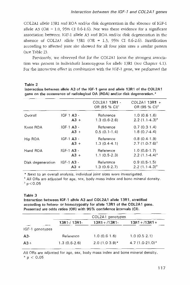
Interaction between the IGF-7 and COL2A 1 genes
COL2AI allele URI and ROA and/or disk degeneration in the absence of IGF-I
allele A3 (OR ~ 1.0, 95% CI 0.6-1.6). Nor was there evidence for a significant
association between IGF-I allele A3 and ROA and/or disk degeneration in the
absence of COL2AI allele I3RI (OR ~ 1.3, 950/() CI 0.6-2.6). Stratification
according to affected joint site showed for all four joint sites a similar pattern
(see Table 2).
Previously, we observed that for the COL2AI locus the strongest associa
tion was present in individuals homozygous for allele URI (see Chapter 4.1).
For the interactive effect in combination with the IGF-I gene, we performed the
Table Z Interaction between allele A3 of the IGF-l gene and allele 13R1 of the COLZA1 gene on the occurrence of radiological OA (ROA) and/or disk degeneration. *
COL2A 1 13R1 - COL2Al 13Rl + OR 195 % CII' OR 195 % CII'
Overall IGF-l A3- Reference 1.0 10.6-1.61 A3 + 1.3 10.6-2.61 2.211.1-4.31'
Knee ROA IGF-l A3- Reference 0.710.3-1.41 A3 + 0.510.1-1.41 1.8 10.7-4.41
Hip ROA IGF-IA3- Reference 0.8 10.4-1.91 A3 + 1.3 10.4-4.1 I 2.711.0-7.61'
Hand ROA IGF-l A3- Reference 1.010.6-1.71 A3 + 1.1 10.5-2.31 2.211.1-4.41'
Disk degeneration IGF-l A3- Reference 0.910.5-1.51 A3 + 1.3 10.6-2.71 2.211.1-4.31'
* Next to an overall analysis, individual joint sites were investigated. tAli ORs are adjusted for age, sex, body mass index and bone mineral density. , p<0.05
Table 3 Interaction between IGF-l allele A3 and COLZA 1 allele 13Rl, stratified according to hetero- or homozygosity for allele 13R1 of the COl2A 1 gene. Presented are odds ratios (OR) with 95% confidence intervals (CI).
COL2A 1 genotypes
13R1-/13R1- 13Rl +/13R1- 13R1 +/13R1 +
IGF-l genotypes
A3- Reference 1.010.6-1.61 1.010.5-2.11
A3+ 1.3 10.6-2.61 2.011.0-3.91' 4.7 11.0-21.01'
All ORs are adjusted for age, sex, body mass index and bone mineral density. , p < 0.05
117

Chapter 5.2
analyses for COL2A! allele 13R! heterozygotes and l101ll0zygotes separately.
Table 3 shows that the interactive effect of both genes was strongest in individu
als homozygous for allele 13R! (OR = 4.7, 95% CI 1.0-22.0\ Also, individuals
with Heberden's nodes showed a stronger association between ROA and/or disk
degeneration and carriership of both risk alleles than individuals without Heber
den's nodes (Heberden's (+) OR = 4.0,95% CI 0.7-22.5 versus Heberden's (-) OR
= 1.7, 95% CI 0.8-3.5\
Discussion
Earlier we found an association between a frequent allele of a polymorphislll in
the promotor region of the IGF-! gene and ROA and/or disk degeneration (5).
For the COL2Al gene, another major candidate gene for OA, we found evidence
for a role of the most frequent allele of a VNTR polymorphism in the occurrence
of ROA (Chapter 4.1). In the present study we observed that the association of
allele 13R! of the COLZA! gene with ROA and/or disk degeneration was only
present in carriers of allele A3 of the IGF-l gene, suggesting an interactive effect
between both loci. Also, the effect of the IGF-l A3 risk allele was conditional on
the presence of the COL2Al risk allele l3Rl. The interaction was a generalized
effect that was not observed joint site specific.
The observed interactive effect between IGF-! allele A3 and COLZA! allele
l3Rl was independent of the effects of age, sex, body mass index, and bone
mineral density. The Vitamin D receptor (VDR) gene is located at a distance of
750 kb from the COLZA! gene and was found to be associated with knee ROA
and bone mineral density (Z!, 22). We can not exclude the possibility that the
VDR gene is contributing to the obselved interaction between the IGF-l and
COL2Al loci. However, the fact that this interactive effect was independent of
bone mineral density suggests that a role of the VDR gene is unlikely. When
testing for interaction, the interaction term was not statistically significant. How
ever, the statistical power of this analysis was low. We did not investigate for
interactive effects between other alleles of both loci, because in our earlier asso
ciation studies no other alleles were statistically Significantly associated with the
occurrence of ROA and/or disk degeneration.
In a recent study the currently investigated polymorphism in the promotor
region of the IGF-l gene was associated with the serum IGF-l level (6). Carriers
of allele A3 of this polymorphism had a higher level of serum IGF-L The finding
of an increased frequency of the IGF-! allele A3 in individuals with ROA and/or
disk degeneration is in line with higher serum IGF-l levels that were observed
in subjects with osteophytic progression in the knee joints (10) and in women
with knee OA (11). Possibly, the interactive effect between the IGF-! and
118

Interaction between the IGF-1 and COL2A 1 genes
COL2Al loci obselved in the present study could be cau.sed by a predisposing
effect of the COL2A1 gene that is only exhibited in the co-existence of allele A3
of the IGF-l gene. One previous report suggested that the expression of the
COL2A1 gene in cultllred chondrocytes was stimulated by IGF-1 (23). High se
rum IGF-l levels may parallel high tissue IGF-l leveb, which may stimulate
chondrocytes, for example in respon.se to cal1ilage damage, to overexpression in
case COL2Al allele 13Rl is present. This lllay lead to an inappropriate produc
tion of coHagen type II fibers that can not be incorporated in the extracellular
ca11ilage network (24). This may remit eventually in a more rapid progression of
OA.
In conclusion the present study suggests an interactive effect between the
IGF-l and COL2Al genes, through a mechanism in which the prevalence of
ROA and/or disk degeneration was only increased in individuals canying the
risk alleles at both loci. The allele of the IGF-1 gene responsible for this interac
tion has previously been associated with a higher serum IGF-l level, suggesting
that high level.s of IGF-l are associated with the pathogenesis of OA.
References
L Spector TD, Cicuttini F, Baker J, Loughlin J, Hart D. Genetic influences on osteoarthritis in women: a t\vin study. BIVU 1996;312:940-3.
2. Felson DT, Couropmitree NN, Chaisson CE, }-£annan II/IT, Zhang Y, McAlindon TE, et aL Evidence for a mendelian gene in a segregation analysis of generalized radiographiC osteoaltilritis. Al1hritis Rheum 1998;41:1061-71.
3. Hirsch R, Lethbridge-Cejku M, I ranson R, Scott \Y/W, Reichle R, Plato CC, Tobin JD, Hochberg MC. Familial aggregation of osteoaI1hritis. Anhritis Rheum 1998;41 :1227-32.
4. Ritvaniemi P, Korkko.J, Bonaventure J, Vikkula M, IIyland J, Paassilta P, el al. Identifica lion of COL2Al gene mutations in patients with chondrodysplasias and familial osteoalthritis. AJlhritis Rheum 1995;38:999-]004.
5. Meulenbelt I, Bijkerk C, Miedema HS, Breedveld FC, Hofman A, Valkenburg HA, Pols HAP, Slagboom PE, van Duijn CM. A genetic association study of the IGF-1 gene and radiological osleoal1hritis (ROA) in a population-based cohort study (the Rotterdam Study). Ann Rheum Dis 1998;57:371-4.
6. Rosen Cj, Kurland ES, Vereault D, Adler RA, RackoffPJ, Craig \YfY, Witte 5, H.ogersJ, BilezikianJP. Association between serum insulin growlh factor-1 ClGF-l) and a simple sequence repeat in IGF-1 gene: implications [or genetic studies of bone mineral density. j Clin Endocrinol J'vIetab 1998;83:2286-90.
7. LeRoith D. Insulin-like gro'\\'1h factors in health and disease. Ann Intern Med 1992; 116:851-862.
8. )'vfcQuillan Dj, Handley Cj, Campbell [VIA, Bolis S, LVIihvay VE, Herington AC. Stimulation of proteoglycan biosynthesis by serum and insulin-like grmvlh factor-l in cultured bovine Jl1icular cat1ilage. Biochem j 1986;240:123-430.
9. Rosen CJ, Donahue LR, Hunter S]. Insulin-like grovvth factors and bone: the osteoporosis connection. Proc Soc Exp BioI Med 1994;206:83-102.
119
Chapter 5.2
10. Schouten JSAG, Van den Ouweland FA, Valkenburg IIA, Lamberts S\X1]. Insulin-like growth [actor-1; a prognostic factor of knee ostcoal1hritis. Br J Rheumatol 1993;32;274-280.
11. Lloyd ME, Hell1 DJ, Nandra D, IVkAlindon TE, \'Vheclcr lvI, Doyle DV, Spector 'I'D. Relation between insulin-like grO\vth factor-I concentrations, osteoaJ1hritis, bone density, and fractures in the general population: the Chingforcl study. Ann Rheum Dis 1996;55;870-4.
12. Hochberg ['dC, Lethbridgc-Cejku M, Scott \Y/W/, Reichle R, Plato ee, Tobin .If). Serum concentrations of insulin-like growth [actor 1 in subjects with osteoarthritis of the knee. Al1hritls Rheum 1994;.17:1177-80.
n. Dcnko C\X1, Baja B, iVloskowitz RW. Growth promoting peptides in osteoat1hritis: insulin, insulin-like growth factor-I, grO\vth hormone. J Rheumatol 1990;17:1217-21.
11. Palotie A, Ott J, Elim K, Cheah K, Vaisanen P, Ryhanen L, et al. Predisposition to familial osteoa11hrosis linked to type II coHagen gene. Lancet 1989;i:924-7.
15. Hofman A, Grobbee DE, De .long PVTM, van den Ouwcland FA. Determinants of disease and disability in the elderly. Eur.J EpidemioI1991;7:403-22.
16. KellgrenJH, Jeffrey MR, Ball J. (editors). 1963. The epidemiology of chronic rheumatism. Volume II: Atlas oj' standard radiographs of arthritis. Oxford: I31a('kwell Scientific Publications.
17. Odding E, Valkenburg HA, A!gra D, van den Ouwe!and FA, Grobbee DE, Hofman A. Associations of radiological osteoaI1hritis or the hip and knee with locomotor disability in the Rotterdam Study. Ann Rheum Dis 1998;57:203-8.
18. Burger I-I, van Dade PLA, adding E, Valkenburg HA, Hofman A, Grobbee DE, et a1. OsteoaJthritis is associated with higher bone mineral density and increased bone loss with age: The Rotterdam Study. Arthritis Rheum 1996;39:81-6.
19. Berg ES, Olaisen B. Characterization of the COL2Al VNTR polymorphism. Genomics 199.0; 16;.050-4.
20. Weber )L, May PE. Abundant class of human DNA polymorphisms which can be typed using the polymerase chain reaction. AmJ HUm Genet 1989;1j1j:388-396.
21. Uitterlinden AG, Burger H, Huang Q, Odding E, van Duiin eM, Hofman A, Birkenhager JC, van Leeuwen JPTM, Pols IJAP. Vitamin D receptor genotype is associated with radiographic osteoa11hritis at the knee. J Clin Invest 1997;100:259-63.
22. Eisman jA. Vitamin D receptor gene alleles and osteoporosis: an alIirmative view. J Bone Miner Res 1995;10:1289-9.3.
23. Sandell LJ, Dudek E. IGF-1 stimulates type II collagen gene expression in cultured chondrocytes (abstract). 011hop Trans 1988;13:300.
24. Matyas jR, Adams ME, Huang D, Sandell LJ. Discoordinate gene expression of aggrecan and type II collagen in experimental osteoal1hritis. Arthritis Rheum 1995;38:120-5
120

6
General Discussion


Genetic epidemiological approach to
studying osteoarthritis
I n this chapter, the main findings of the present thesis will be discussed in
light of previous findings reported in the literature. As this thesis primarily
concerns radiological osteo;uthritis (ROA) in the general population, the general
discussion will focus on the as.':.iociation between genetic factors and radiological
osteoarthritis. Funhennore, special attention is addressed to methodological is
sues in genetic epidemiological studies. Recommenciations for future research
Jnd developments in osteoarthritis eOA) research will be discussed.
Findings and methodological considerations
Determinants of generalized radiological osteoarthritis
TI1e findings of the present thesis support the existence of a subset of polyar
ticular ROA in women, meaning that clu~tering wa~ found of ROA and disk de
generation at different site~ (see Chapter 3.1), In women, age, body ma~~ index,
bone mineral den~ity, and Heberden's nodes were all statbticaily ~ignificant ri~k
factors of polyarticular ROA. For women the findings of the present study are
consbtent with previou~ studies (1, 2). To our knowledge this thesis b the first
~tudy to sugge~t that disk degeneration can be included in the concept of poly
articular ROA in women. The a~sociations observed in women are in keeping
123

Chapter 6
with a genetically determined generalized susceptibility for cartilage degrada
tion, which may be modified by the effects of systemic factors like body mass
index and bone mineral density 0).
In men, only hand ROA at multiple sites was associated with disk degen
eration of the spine. Only age and body mass index were statistically signifi
cantly associated with poJyal1icular ROA in men (see Chapter 3.1), One earlier
study found evidence for polyarticular disease in men, based on the statislically
significant association of hand and knee ROA (1),
'I11C finding in both men and women that disk degeneration of the spine is
associated with hand ROA confirms the findings of an experimental study (4)
and suggest.s a .':ilured, possibly genetic, pathogenesis of ROA and disk degen
eration (5). An important implication for genetic studies of this finding is that
disk degeneration of the spine should be included in the concept of polyarticu
lar ROA. Disk degeneration may be regarded the progressive deterioration of
hyaline cartilage of the intervertebral disk. A shared pathogenesis in ROA and
disk degeneration could [or example be explained by changes in the collagen
network (e.g. collagen types II, IX, X or XI), the extracellular proteoglycan me
tabolism, the chondrocyte function or function of proteolytic enzymes or cytoki
nes (6).
Heritability of OA
The findings of this thesis show that hand ROA and disk degeneration of the
spine in the general population are largely determined by genetic factors, with
heritahility estimates of respectively 0.56 (95 % CI 0.34-0.76) and 0.75 (95 % CI
0.30-1.00) (see Chapter 3.2). These results were detected independent of the
effects of age, sex, body mass index and bone mineral density. That OA is a
heritahle disorder was already recognized by Stecher in the 1940's (7), and con
finned in the 1950's, in studies on generalized OA, by Kellgren and Lawrence
(8). Recent population-based studies have shown results for sib-sib correlations
in hand ROA, knee ROA anel the combination of hand and knee ROA, that are
largely similar to the results presented in this thesis, as is outlined in Table 1 (9-
11). The presented correlations were in all studies at least adjusted for age and
weight. In our study, no statistically significant genetic influence was observed
on the variance of knee ROA in the general population. Moreover, in this thesis
no genetic effect was obselved on the occurrence of hip ROA in the general
population (see Chapter 3.2). However, a genetic influence on severe hip OA
leading to joint replacement has been reported (12). No earlier data have been
published concerning the heritability of disk degeneration of the spine, although
one recent report suggests a genetic susceptibility to this disorder based on an
observed association with the Vitamin D receptor gene (13). In view of possible
linkage disequilibrium between the VDR and COL2A1 genes, it is not dear
124
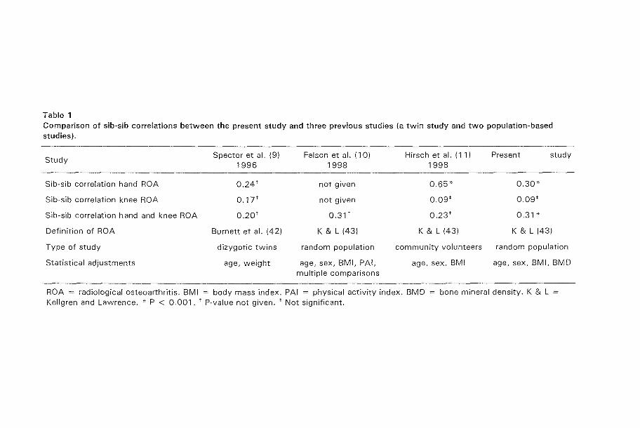
Table 1 Comparison of sib-sib correlations between the present study and three previous studies (a twin study and two population-based studies).
Study Spector et al. 19) Felson et al. [10) Hirsch et al. [11) Present study
1996 1998 1998
Sib-sib correlation hand ROA 0.24' not given 0.65* 0.30*
Sib-sib correlation knee ROA 0.17' not given 0.09' 0.09'
Sib-sib correlaflon hand and knee ROA 0.20' 0.31 ' 0.23' 0.31 *
Definition of ROA Burnett et al. [42) K & L [43) K & L [43) K & L [43)
Type of study dizygotic twins random population community volunteers random population
Statistical adjustments age, weight age, sex, BMI, PAl, age, sex, 8MI age, sex, BMI, BMD multiple comparisons
ROA = radiological osteoarthritis. 8MI = body mass index. PAl = physical activity index. SMD Keligren and Lawrence. * P < 0.001. i P-value not given. t Not significant.
bone mineral density. K & L
Chapter 6
which gene actu<llly underlies the obselved association. In OUf study, we ob
sClved the highest heritability estimate for a score slIIllming the total number of
joints affected with ROA and/or disk degeneration (0.78 with 95 % CI 0.52-0.98),
funher supporting the evidence for a shared genetic etiology of both conditions
(4, 5)
Radiological osteoarthritis and collagen genes
The collagen network is essential for the dynamic mechanical and structural in
tegrity of alticular Gutilage. The lnain constituents are collagen type IT, about 90
percent of the collagen content of cartilage, collagen type IX, forming the cross
links between the type II fibrils, and collagen type XI, regulating the diameter of
the type 1I fibrils. Collagen type II consists of three similar alpha chains encoded
by the COL2Al gene on chromosome 12. Collagen type IX and Xl, both consist
of three separate alpha chains, encoded respectively by the COL9Al, COL9A2,
and COL9A3 genes and the COL2Al, COLll Al, and COLllA2 genes. Except for
the COL9A3 gene, mutations have been identified for all above-mentioned
genes leading to some form of severe, mostly early-onset, familial OA (14-18).
COL2A 1 gene
~ Association study This thesi.s describes the association of the most frequent
allele (allele frequency 0.42) of a VNTR polymorphism 1.35 kb to the 3' end of
the COL2Al gene with ROA (see Chapter 4.1). The strongest effects were ob
selved in women homozygous for this allele 13Rl (OR ~ 1.86, 95 % CI 1.0-3.6).
The ORs for women with polyalticular OA were respectively 3.87, 95 % CI 1.1-
13.3 for homozygotes and 1.59 with 95 ')" CI 0.6-4.1 for heterozygotes. Also the
effect was stronger in women with Heberden's nodes as compared to women
without Heberden's nodes. In two previous association studies concerning rela
tively severe OA cases, rare alleles of the COL2Al gene were statistically signifi
cantly associated with the occurrence of ROA (l9, 20). Two other association
studies, respectively carried out in Finnish cases with generalized OA or hand
OA and in Belgian hip OA cases, elid not find evidence for an association of OA
with the COL2/d gene (21, 22). This thesis provides the first report of an asso
ciation betvveen common forms of ROA and the COL2A1 gene in a population
based study. Given the cross-sectional design of the present study, the observed
association with the COL2A1 VNTR locus gives no dues about the biological
unportance of this finding in terms of the etiology or prognosis of ~A.
- Sibling pair study We performed a sibling pair study with probands, derived
from the above-mentioned population-based association study (Chapter 4.1),
and their corresponding siblings. For the COL2A1 gene, the findings of the sib
ling pair study were in agreement with the association reponed in Chapter 4.1,
126
General discussion
although no stJtisticaliy significant results were found (Chapter 4.3). For sibling
pairs of probands canying the 13H.1 allele, a marked decrease of t.he variance in
a score summing the total number of joints affected with ROA and disk degen
eration was observed when siblings shared one or tvlo alleles (rr2o = 16.2, rr\ =
10.0 and a' , = 6.7, with LOD score 0.2). One earlier sibling pair study of limited
size (38 sibling pairs), examining siblings with three or more joint sites affected,
failed to show linkage to the COL2Al locus (23). In a recent unpublished repOlt,
concerning a sibling pair study, derived from 52 ext.ended families, in the
Framingham Study, no evidence for linkage WJS found between the COL2A1
locus and ROA (24). This study was carried out with five microsZltellite markers
flanking the COL2A1 locus. Each of these studies, including our study, lacked
statistical power for showing a Significant result.
- Interaction with the insulin-like growth factor-l (IGF-l) gene The IGF-I
gene is also a candidate gene for OA and was found to be associated with ROA
(Chapter 5.1). Allele A3 of a polymorphism in the promotor region of the IGF-I
gene was associated with ROA (OR for heterozygous subjects was 1.9, 95 O;() CI
1.2-3.1 and for homozygous subjects 3.6, 95 % CI 0.8-16.2), independent of the
influences of bone mineral density and body mass index (25).
\X1e observed an interactive effect. on the presence of ROA betvleen the
IGF-1 gene and the COL2A1 gene (Chapter 5.2). This study showed that COL2A1
VNTR allele l3RI is only associated with ROA in the presence of allele A3 of the
IGF-I gene (OR = 2.0, 95 % CI 1.0-3.9 for individuals heterozygous for allele
13Rl, and OR = 4.7, 95 % CI 1.0-21.0 for individuals homozygous [or allele
13R1). Alternatively, no association was obselved between IGF-l allele A3 and
ROA in the absence o[ COL2AI VNTR allele l3R1. 111is clearly suggests the ex
istence of an epistatic effect of both loci on the occurrence of ROA.
- COL2A 1/VDR locus TI,e Vitamin D receptor (VDR) gene, located about 750
kb distant from the COL2A1 gene, was found to be associated with disk degen
eration (13), knee ROA (26, 27), and bone mineral density (28). Given the dis
t.ance betvleen both genes linkage disequilibrium I113Y exist, raising the question
whether the COL2Al gene itself contributes to the etiology of ROA or the VDR
gene instead. The obselved Jssociation between the COL2A1 VNTR polymor
phism and ROA (Chapter 4.]) and the interactive effect of this locus with the
IGF-l gene on the occurrence of ROA (Chapter 5.2) was independent of bone
minerZil density (BMD). This suggests that it is unlikely that the VDR gene un
derlies the observed association through its effect on BMD.
~ Genomic screens Recent reports, concerning two genomic screens in re
spectively finnish sibships from a genetiC isolate, and affected sibling pairs who
127

Chapter 6
had under'\vcnt joint replacement, showed no evidence for linkage to chromo
some 12 (the localization of tbe COL2Al/VDR locus) (29, 30). In the genetically
isolated Finnish population genetic drift could explain the finding that the
COL2Al gene does nol JJlJy a role in the etiology of ~A. The second genomic
screen was carried out in Brltbh palients who Ul1e!clwent joint replacements
which represents severe cases of principally hip OA, that could well be a OA
phenotype with an etiology in which the type II collagen is not involved. In our
heritability studies hip ROA was not highly genetic. Therefore, in the present
study of ROA, in contrast to studies of hip replacement.s, a different phenotype
was studied.
- Conclusion Thb thesis suggests a role of the COL2Al locus in the etiology of
common forms of OA, by mcans of an association study. The findings of the
sibling pair stuny subsequently carried out in the same study poptJiation were in
line with these findings. \XTc observed that this association of the COL2Al locus
with ROA was fully determined by the influence of the IGF-1 gene, Such an
epistatic model will reduce the power of past and future sibling pair studies
concerning the COL2Al genc, unless the effect of both genes is studied simulta
neously. Although, we can not exclude the possibility that the adjacent VDn.
gene is contributing to the detected association, we can conclude that it will not
be through an effect of the VDR gene on bone mineral density.
COL9Al gene We observed an association between a Short Tandem Repeat Polymorphism
(STRP) within the COL9Al gene and ROA in women from a population-based
sample (Chapter 4.2). Two distinct haplotypes were detected that showed re
spectively a three (p = 0.01) and a seven (p < 0.002) times decreased frequency
in women with ROA. This obselved association could not be confirmed in the
sibling pair st.udy with probands who were affected with ROA at two or more
joint sites (Chapter 4.3). Ilowever, the present sibling pair study mainly included
affected siblings (only 12 siblings were free from ROA and disk degeneration),
therefore it seems likely that in this sample it was not possible to confirm the
obselved protective efTect, because of a lack of statistical power. For this gene
no previous association or sibling pair stllC.lies have been published. Given t.he
absence of consistency bet\veen our association study and sibling pair study, the
role of the COL9Al gene in the etiology of OA remains to be confirmed.
COL l1A2 gene
Since no intra genic markers were available, this gene was studied indirectly
through the use of two flanking markers; tumor necrosis factor a (TNFa) within
the TNF-jS gene (1.5 eM from the COLllA2 gene) and a dinucleotide repeat
128

General discussion
marker (D6S291) at a 2 cM distance from the C0L11A2 gene. The COLllA2 gene
is located within the major histocompatibility complex (MHC) on chromosome
6) just as the TNF-S gene. The MHC region exhibits a very low recombination
fraction and therefore TNFa and D6s291 can be used in a haplotype analysis of
the C0L11A2 gene. Funhermore, the TNF-a gene, directly adjacent to the 1NF-fS
gene was considered a candidate gene for OA itself, given the role of TNF-a in
the pathophysiology of cartilage degradation (31). Both genes have not been
studied before in OA in humans. No association was found between the
C0L11A2 gene or the TNF-a gene and the presence of ROA in the general
population (Chapter 4.2).
Strategies for future research
Family-based studies
The role of classical linkage studies in detecting disease-susceptibility loci in
common forms of OA is expected to be limited, given the multifactorial nature of
the disease and the subsequent difficulty of detecting extended families with
affected pedigree members. Classical linkage studies remain imp011ant for de
tecting causative mutations in early-onset, mostly more severe, OA phenotypes.
OA is a multifactorial disease that is caused by mUltiple genes interacting
with each other and with environmental factors, which creates a gradient of ge
netic susceptibility to disease. The degree and type of interaction between these
genes influences the chances of detecting genes through a sibling pair analysis
(32). Another factor determining the successfulness of sibling pair studies is the
definition of the phenotype under investigation. Sibling pair methods can be
applied in both a candidate gene approach and random genome scans, although
it has been suggested that in a candidate gene approach association studies are
to be preferred (33). A sibling pair approach considering OA a quantitative trait
may render a high power for detecting disease-susceptibility loci as the herita
bility of this trait is high (see Chapter 3.2) and both affected and unaffected rela
tives can be included in the study (34). The sum score of the total number of
joint sites affected with ROA in peripheral joints and disk degeneration of the
spine was used in this thesis and was shown to be determined largely by genetic
influences (Chapter 3.2). Concerning the statistical power of a study, it has been
shown that, for a common trait, the use of the population distribution of a phe
notype is to be preferred over an affected sibling pair approach (35). Recmit
ment of sibling pairs from both extremes of a phenotype distribution) e.g. high
est versus lowest decile) rnay be an alternative powerful approach (36). How-
129
Chapter 6
ever, the usefulness of the extreme discordant sibling pair approach in practice
needs to be established.
Recently, two unpublished reports concerning genomic screens in affected
sibling pairs with relatively rare OA phenotypes showed excess sharing in OA
patients of chromosomal regions on chromosome 2q and l1q (29, 3D), Candi
date genes located in these regions are the interleukin-l cluster and the cluster
of genes encoding matrix metalloproteases. The relevance of these findings
needs to be established with lise of a dense set of marker loci in these chromo
somal regions. Further genomic screens in sibling pairs with more common
fonns of OA are needed in order to obtain a more complete view on suscepti
bility loci for ~A. The availability of a genome-Wide high-resolution map (0.5-
1.0 Mb) will increase the possibilities for fine mapping and eventually positional
cloning of a disease-susceptibility locus 07, 38).
Association studies
At present, only a candidate gene approach is feasible in unrelated individuals,
except for genomic screens that are carried out in genetically isolated popula
tions. Population stratification is the ma jor confounding factor in association
studies and can be due either to recent admixture of different populations or to
inappropriate matching of patients and controls 09, 40). Given this possibility of
confounding, findings need to be confirmed using a transmission/disequilibrium
test (TDT) or using haplotype analysis (41). However, the TDT is not a powerful
approach and is practically difficult in late-onset diseases. An impol1ant issue for
future genetic epidemiological studies will be to quantify the presence of pOplJ
Jation stratification. Association studies are powerful as a follow-up strategy for
examining disease-susceptibility loci that have been identified in genomic
screens or linkage studies as long as these studies are carried out in well-defined
large homogeneous populations to reduce confounding and information bias.
Population relevance
OA is considered a genetically heterogeneous disorder in which several genes
act in interaction with each other, either in a multiplicative or in an additive
model. In this way genetically susceptible individuals are characterized by a dis
tinct set of genes that give rise to articular caltilage more prone to progressive
degeneration. Different individuals have different sets of genes leading to the
same phenotype. In future, large-scale population-based studies are needed to
establish which genes demonstrate an interactive effect on the development or
progression of ~A.
Finally, it is impOltant to realize that data derived from association and sib
ling pair studies and from genomic screens only provide indirect evidence for
130

General discussion
the role of a gene or a locus in the etiology of OA. In case of non-coding se
quences that are found to be a.:;.:;ocialed with OA in genomic ':;('1"een.:; or a candi
date gene .:;tudy, po.:;itional cloning and mutation analy.:;is should indicate which
mutJ tion i.:; responsible for the observed a.:;.:;ociatiol1. Abo, ftuther molecular
genetic studies in affected subjects, cell and animal model.:; are needed to deter
mine through what mechani.:;m a mutation, e.g. leading to a causative protein
isofo1'1n or an altered gene expression, is involved in causing OA. The final test
for the relevance of a mutation in the occurrence of OA is to determine the asso
ciated risk of disease in a prospective follow-up .:;tudy. Accordingly, the studies
presented in this the.:;is are to be interpreted as the first step in unraveling the
genetically determined pathogenesis of OA.
References
1. fIirsch H, Lethbridge-Cejku M, Scott Jr \XIWi, Reichle R, Plato CC, Tobin ], Hochberg Me. Association of hand and knee osleoal1hritis: evidence for a polyarlicular disease subset. Ann l{heuJ11 Dis 1996;55:25-9.
2. Cooper C, Egger P, Coggon D, Halt D], IvIasud T, Cicuttini F, Doyle DV, Spector TD. Generalized osteoarthritis in women: pattern of joint involvement and approaches to definition for epidemiological studies. J Rhcumatol 1996;2;$:1938-42.
3. Cicuttini FM, Speclor 'I'D. Genetics of osteoal1hritLs. Ann Rheum Dis 1996;55:665-7. 4. Kimura T, Nakata K, Tsumaki N, Miyamoto S, Matsui Y, Ebara S, Ochi T. Progressive
degeneration of articular cartilage and interver1ebral discs. An experimental study in transgenic mice bearing a type IX collagen mutation. Int Onhop 1996;20:177-8l.
5. Il,tfatsui H, Kuramori lvI, Ishihara H, Yudoh K, Naruse Y, Tsuji H. Familial predisposition for lumbar degenerative disc disease. A case-control study. Spine 1998;23:1029-31.
6. Bleasel .IF, Ilolderbaulll D, I3rancolini V, Moskowitz RW, Considine EL, Prockop Dj, Devoto 11'1, \\Tilliams C). Five families with arginine 519-cystcine mutation in COL2A"!: evidence for lhree distinct founders. I hun .lVIulat 1998; 12: 172-6.
7. Stecher RM. Hcberden's nodes. Heredity in hypenrophic al111ritis of the finger joints. Am} IvIed Science 1911;201:80l.
8. Kellgrcn JI-l, Lawrence ]S, Bier P. Gcnetic factors in generalized osteoar1hrosis. Ann Rheum Dis 1963;22:237-55.
9. Spector TD, Ckuttini P, Baker ], Loughlin J, Iran D. Genetic influences on osteoarthritis in women: a twin study. 11M] 1996;312:940-3.
10. Felson DT, Couropmitree NN, Chaisson CE, Hannan IVIT, Zhang Y, IV1cAJindon TE, LaValley 1'1'1, Levy D, .Myers RH. Evidence for a ll1endeli:l1l gene in a segregation analysis or gencralized radiographic osteoanhritis. Anhritis Hheum 1998;41:1064-71.
II. I Iirsch R, Lethbridge-Cejku ivl, I ranson Fl., Sco!t \V\Y/, Reichle R, Plato ec, Tobin JD, Hochberg [viC. [<';.1111i1ial aggrcgation of osteoalthritis. A11hritis Rhculll 1998;41:1227-32.
12. Cbitnavis J, Slnshcimer JS, Clipsham K, Loughlin J, Sykes B, Burge PD, Carr AJ Gcnetic influenccs in end-stage osteoarthritis. Siblings risks of hip and knee replacements for idiopathic osteoarthritis.] Bone Joint Surg 131' 1997;79:660-4.
131
Chapter 6
13. Vidernan T, Leppavllori j, Kaprio j, Battie ;,'!C, Gibbons LE, Peltoncn L, Koskenvuo {>d, Intragenic polymorph isms of the Vitamin D receptor gene asociated with intervertebral disc degeneration. Spine 1998;23:2477-85.
ILi. Spranger J, Winterpacht A, Zabel B. The type II collagenopathies: a spcct1llm of chondrocly!;plasias. Eur J Pcdiatr 1994; 153:56-65.
15. Nakata K, 0110 K, tv1iyazaki .II, Olsen BR, IVIuragaki Y, At-belli E, Yamamura K1, Kimura 1'. OsteoanhritLs associated with mild chondrodyspbsia in transgenic mice
expressing a 1 (IX) collagen chains with a central deletion. Proc Natl Acad Sci USA 1993;2870-:'.
16. IVluragaki Y, Mariman ECM, van Ueersum sr..:c, Perala M, van lvIourik.JB, Warman 1\-1L, Olsen BR, Hamel Be]. A mutation in the gene encoding the a2 chain of the fibril-associated collagen IX, C:OL9A2, causes multiple epiphyseal dyspl:1sia (EO)\112). Nat Genet 1996;1:103-5.
17. Brunner He, van Reersu111 SEC, \\'1arman IVIL, Olsen BR, Ropers 1 Ill, Mariman ECl'v1. A Stickler syndrome gene is linked to chromosome 6 near the COLl1A2 gene. Hum Idol Genet 1994;3::l561-Ij.
18. Richard AJ, Yates .1RW, Williams R, Payne SJ, Pope FM, Scott .1D, Snead .MP. A family with Stickler syndrome type 2 has a mutation in the COLl1A1 gene resulting in the
substitution of glycine 97 by valine in uJ (Xl) collagen. Hum Mol Genet 1996;5:Li39-13.
19. 1 lull R, Pope FM. Ostcoalthritis and calti!Jge coHagen genes. Lancet 1989;i:1337-8. 20. Loughlin .1, Irven C, Athanasou N, Carr A, Sykes B. Differential allelic expression of
the type II collagen gene (CUL2A1) in os!eoanhritic cartilage. Am .1 Hum Genet 1995;56,1186-90.
21. Vikkula .lVI, Nissila lvI, Hilvensalo E, Nuotio P, Palotie A, Aho K, Pdtonen L. Muitiallelie polymorphism of the cat1ilage collagen gene: no association with osteoat1hritis. Ann Rheum Dis 1993;52:762-4.
22. Aerssens .1, Dequeker .1, Peeters .1, Breemans S, Boonen S. Lack of association between osteoal1hritis of the hip and gene polymorphisms of VDR, COLlA1, COL2A1 in postmenopausal women. Anhritis Rheum 1998;IJ1:1946-50.
2."3. Loughlin .1, lrven C, Fergusson C, Sykes B. Sibling pair analysis shows no linkage of generalized osteoanhritis to the loci encoding type II collagen, cal1ilage link protein or cartilage matrix protein. Sr J Rheumatol1994;3}1103-0.
2-1. Baldwin CT, Joost 0, Chaisson C, IVfcAlindon T, Farrer L, Ordovas J, Schaefer E, Levy D, Myers R, Fe!.son D. The type II collagen/Vitamin D receptor loclls and osteoarthritis: The Framingham Osteoarthritis Study. Arthritis Rheum 1998;'11:S352.
25. Meulcnbelt I, Bijkerk C, .lVliedema HS, I3reedveld FC, Hofman A, Valkenburg HA, Pols HAP, Slagboom PE, van Duijn C1v1. A genetic association study or the IGF-1 gene and radiological osteoalthritis in a populalion-based COhOll study (the Rotterdam Study). Ann Rheum Dis 1998;57:371-1.
26. Uitterlinden AG, Burger I-I, Huang Q, Odding E, van Duijn CM, Hofinan A, mrkenager ]C, van Leemven .1PT.I\IJ, Pols HAP. Vitamin D receptor genotype is associated with radiographic osteoallhritis at the knee . .J Clin Invest 1997;100:259-0."3.
27. Keen R\'V, Hart DJ, Lanchbury JS, Spector TD. Association of early osteo:111hritis of the knee with a Taq 1 polymorphism of the Vitamin D receptor gene. Al1hritis Rheum 1997;40:1444-IJ9.
28. Eisman JA. Vitamin D receptor gene alleles and osteoporosis: an affirmative view. J Bone Ivliner Res 1995; 1 0: 1289-93.
29. LeppaHlori .1K, Kujala U.lVf, Kap1'io J, Nissila lvf, JIelibvaara .lVI, Kinnunen .1, Koskenvuo M. Genome scan for predisposing loci of dital interphalangeal joint osteoalthritis. Am.1 Hum Genet 1998;G3:A297.
30. Chapman K, Mustafa Z, lrven C, Smith A, Carr A, Clipsham K, Chitnavis .1, Sinsheimer .1, Sykes B, Loughlin J. A comprehensive genome-wide screen for susceptibility lod in osteoarthritis: linkage to chromosomes 2q and l1CJ. Am] HUm Genet 1998;03:A16.
132

General discussion
31. Saklatvala J. Tumour necrosis factor 'if stimulates resorption and inhibits synthesis of proteoglycan in ca!1iiage. Nature 1986;322:547-59.
32. Eaves LJ. Elfect of genetic architecture on the power of human linkage studies to resolve the contribution of quantitative trait loci. Heredity 1991;72:175-92.
33. Risch N, tvlerikangas K. The future of genetic studies of complex human diseases. Science 1996;2nI516-7.
YL \Veeks DE, Lathrop Clv1. Polygenic disease: methods for mapping complex disease traits. Trends Genet 1995;11:51.3-9.
35. Wijsman El\tI, Amos Cl. Genetic analysis of simulated oligogenic traits in nuclear and extended pedigrees: Summary of GA\VlQ contributions. Genet Epidemiol 1997;14: 719-35.
36. Risch N, Zhang l-I. Extreme discordant sib-pairs for mapping quantitative trait loci in humans. Science 1995;268: 1584-9.
37. Collins FS. Positional doning moves from perditional to traditional. Nat Genet 1995;9;317-50.
38. \Valter .J'vIA, Spillet DJ, Thomas P, \Vdssenbach J, Goodfellow PN. A method for constructing radiation hybrid lnaps of whole genomes. Nat Genet 1991j;7:22-8.
39. Knowler \VC, Williams RC, Pettitt DJ, Steinberg AG. Gm3;5,1.J,"l4 and type 2 diabetes mellitus: an association in American Indians with genetic admixture. Am J Hum Genet 1988:43:520-6.
40. Lander ES, Schork I\~. Genetic dissection of complex traits. Science 1994;265:2037-48. IiI. Spielman RS, IvlcGinnis RE, Ewens \Xl). The transmission/disequilibrium test detects
segregation and linkage. Am J I-lum Genet 1994;54:559-60. 42. l3urnett S, Harl Dj, Cooper C, Spector 'I'D. A radiographic atlas of osteoarthritis.
Springer-Verlag, London. 1994. 4.3. Kellgren jB, Jeffrey I'vIR, Ball J. (editors). The epidemiology of chronic rheumatism.
Volume Jj: Atlas of standard radiographs of al1hritis. Oxford: Blackwell Scientific Publications. 196.1.
133


7
Summary

Summary
T his thesis describes an approach of population-based genetic association
studies and sibling pair studies to study COIllmon forms of osteoanhritis
eOA) occurring in the general population. OA is characterized by a progressive
degeneration of al1icuiar c31tilage of diarthrodial joints and has a multifactorial
etiology. The genetic influence on the etiology of OA has been recognized for
familial forms of .':icvere early-onset OA I which is often associated with osteo
chondrodysplasia. There is growing evidence from population-based studies
that COIDIDon late-onset forms of OA are also heritable. However, at present it is
unclear which genes are involved in causing these common forms of OA in the
elderly.
First, some methodological considerations concerning genetic epidemiol
ogical studies on OA arc presented (chapte.- 2.1). A candidate gene approach is
powerful in case a genetic association sutdy is applied that was conducted in an
unselected population. However, findings of these association studies need
confirmation in either a sibling pair study or in a haplotype analysis. Genomic
screens can be conducted based on the findings of previous studies or can scan
the entire genome for susceptibility loci. Bias resulting from mUltiple testing is
an impOltant issue of consideration in evelY genetic study. Population
stratification or aclmL"TIure is the main source of bias in association studies.
Sibling pair studies in late-onset complex traits are usually hampered by a lack
of statistical power, because of difficulties in ascertaining sufficient numbers of
siblings.
For the results presented in this thesis, data were used from a random sam
ple derived from the Rotterdam Study (chaptet" 2.2) and from a sample of
sibling pairs (chapter 2.3). The Rotterdam Study is a population-based cohort
study among all inhabitants, aged 55 years and over, of the suburb Ommoord in
Rotterdam. In total, 7983 individuals participated in the Rotterdam Study
(response 78%). For this thesis a random sample was drawn of 1583 individuals
aged 55 to 70 years. All individuals visited the research center where interviews
were held and extensive measurements were performed, including radiographs
of the knees, hips, hands, and thoracolumbar spine and dual energy X-ray
absorptiometly (DEXA) scans. Two independent readers scored all radiographs
for radiological OA (ROA) or disk degeneration according to the Kellgren
scoring system. Individuals with at least two or more joint sites affected were
selected as probands. In case only hand ROA and disk degeneration of the spine
was present, individuals had to have additional Heberden's nodes to be
included as probands. All available sibling.s, who agreed to palticipate, were
recruited for sibling pair studies.
In chapter 3.1 the determinants for ROA and disk degeneration at multiple
sites and the clustering of ROA at all four joint sites are investigated. In women,
body mass index (BMI), bone mineral density (BMD), and IIeberden's nodes
137
Chapter 7
were besides age all statbtically significant risk factors of polyarticular disease
and knee ROA was significantly associated with radiological abnormalities all
other sites studied. In men, only EMI Was in addition to age significantly associ
ated with polyal1icu\Jr disease and only polyarticular hand ROA was associated
with disk degeneration of the spine. These findings support the existence of a
subset of polyarticular disease in women.
Chapter 3.2 describes the genetic influence on the occurrence of ROA in
the knees, hips, and hands and disk degeneration of the spine in the general
population. Heritability estimates for hand ROA and disk degeneration were
statistically Significant, respectively 0.56 (95% CI 0.34-0.76) and 0.75 (95% CI
0.30-1.00), For knee and hip ROA no evidence for a genetic effect in the general
population was found. The heritability estimate for a score summing the total
number of joints affected at four different sites was 0.78 (95% CI 0.52-0.98), sug
gesting a genetic susceptibility to generalized OA.
The association study of the procollagen type II (COL2Al) gene with ROA
is presented in chapter 4.1. The frequency distribution of alleles of the COL2Al
VNTR polymorphism, which is located 1.35 kb to the COL2Al gene, in female
ROA cases differed statistically significantly from that in female referents (p =
0.03). 111is was explained by an increased frequency of the most COIllIllon allele
l3Rl (adjusted OR for female cases heterozygous for allele l3Rl was 1.71, 95%
CI 1.06-2.76 and for female cases homozygous 1.86, 95% CI 0.95-3.64). Strongest
effects were fmmd in women with ROA and Heberden's nodes and in women
with polyal1icular ROA.
In chapter 4.2 the association study concerning the genes encoding the
a-I chain of collagen type IX (COL9AI) and the a-2 chain of collagen type XI
(C0L1IA2) and ROA is described. For the COL9Al gene, one of two ShOlt Tan
dem Repeat Polymorphisms (STRF 509-12BJ), within the COL9AI gene, was
found Significantly associated with ROA in women (p ~ 0.002). Haplotype analy
sis suggested a protective effect of allele 12BI-A2) with a seven times decreased
frequency in female ROA cases. No evidence was observed for a role of the
COLIIA2 gene in the occurrence of HOA.
In chapter 4.3 data is presented on a sibling pair study that was performed
in 257 siblings of 118 probands, derived from the Rotterdam Study, with ROA
and disk degeneration at mlJltiple sites. For the COL2Al locus) a reduction was
observed in the variance for the sum score of ROA and disk degeneration and
for the sum score of hand ROA (quantitative trait loci\ with an increase in the
number of alleles shared between female siblings (not statistically significant).
Effects were stronger for women with ROA and Heberden's nodes and for
women with generalized ROA. We could not find evidence that the COL9AI
locus increases the risk of developing ROA or disk degeneration.
138

Summary
r111e third association study in this thesis concerns the insulin-like growth
factor 1 (lGF-l) gene (chapter 5.1). The IGF-l locus was statistically signifi
cantly associated with the presence of ROA and lor disk degeneration. This
association was explained by an increased frequency of IGF-l allele A3 in
affected subjects (adjusted OR for heterozygous subjects was 1.9, 95% CI 1.2-3.1
and for homozygous subjects 3.6, 95% CI 0.8-16.2).
In chapter 5.2 the findings are reported of a study investigating the possi
ble interaction between the IGF-l and COL2A! genes on the occurrence of ROA
and disk degeneration. The prevalence of ROA and/or disk degeneration was
significantly increased in individuals who cany both the IGF-l allele A3 and
COL2A! allele 13Rl as comp,ued to individuals canying none of these alleles
(adjusted OR = 2.2, 95% CI 1.1-4.3). There was no evidence for an effect of
COL2Al allele 13R1 in the absence of the IGF-l allele A3 or for an effect of the
IGF-l allele A3 in the absence of COL2Al allele 13R1. The findings suggests that
the IGF-l and COL2A1 genes are involved in the occurrence of ROA and/or disk
degeneration of the spine through a mechanism in which both genes interact.
Chapter 6 summarizes our findings together with observations from other
studies and methodological considerations. It is shown that the evidence of a
genetic influence on the occurrence of conunon forms of OA in the general
population is conclusive. The discussion focuses on the contribution of the
COL2A1 gene in this genetic influence. In light of previous and present findings,
a role of the COL2A! locus is velY likely. Furthermore, this influence of the
COL2A1 10c\1O is fully determined by the influence of the IGF-l locus, possibly
through an effect on the expression of the COL2Al gene. Finally, recommenda
tions for future research are given.
139

I
I
I
I
I
I

8
Samenvatting


Samenvatting
D it proc[schrift beschrijft een benadering) gebaseerd op genetische asso
ciatiestudies en broer-zuster studies, tot het bestuderen van gewone
vormen van artrose zoals die optreden in de algemene bevolking. Altrose wordt
gekarakteriseerd door een progressieve degeneratie van gewrkhtskraakbeen
van diarthrodale gewrichten en heeft een multifactoriele etiologie. Een 1'01 van
genetische factoren in de etiologie van altrose is aangetoond voor familia ire
vormen van ernstige vroegtijdige artrose, die vaak gepaard gaat met chondro
c!ysplasie. Er bestaJt toenemend bewijs afkomstig van bevolkingsonderzoeken
dat ook gewone vormen van laat optredende artrose erfeJijk bepaald zijn. Tot op
heden is het niet c!llidelijk welke genen betrokken zijn bij het ontstaan van deze
gewone vormen van altrose bij ouderen.
Tel' introductie worden enkele methodologische aspecten met be trekking
tot genetisch epidemiologische studies naar artrose besproken (hoofdstuk 2.1),
Een kandidaat gen benadering kan sllccesvol zijn indien c!it wordt uitgevoerd
binnen een genetische associatie studie in een niet geselecteerde populatie, De
resllltaten van zo een associatie studie dienen bevestigd te worden in ofwel een
broer-zuster studie ofwel een haplotype analyse. Een genoom scan kan worden
venlcht gebaseerd op de bevlndlngen afk:omstig van vroegere studies of het
gehele genoom kan worden gescand op zoek naar regio's die het fisko op
ziekte be'invloeden. Het optreden van vertekening van de resultaten door het
uitvoeren van herhaalde statistische testen ls een belangrijk punt van aandacht
in elke genetische studie. Populatie stratificatie of vermenging is een belangrijke
bron van veltekening in een genetische associatie studie. Daarentegen worden
broer-zuster studies gekenmerkt door een gebrek aan statistische power doordat
in het algemeen het aanta1 paren dat verzameld kan worden onvoldoencle is.
Voor de resultaten die gepresenteerd worden in dit proefschrift werd
gebruik gemaakt van gegcvens afkornstig van een steekproef van het Erasmus
Rotterdam Gezondheid en Ouderen (ERGO) onderzoek (hoofdstuk 2.2) en van
een verzameling broers en zusters (hoofdstuk 2.3). Het ERGO-onderzoek is een
longituclinaal bevolkingsonderzoek oncler inwoners van 55 jaar en ouder
woonachtig in de Rotterdamse wijk Ommoord. In totaal deden 7983 personen
mee aan het ERGO-onderzock (res pons 78%). In het kader van elit proefschrift
werd hieruit een steekproef genol1len van 1583 personen russen de 55 en 70 jaar
oud, Alle deelnemende personen bezochten het onc!erzoekscentrum alwaar een
anamnese werd afgenomen en uitgebreide metingen werden verricht, inclusief
rontgenfoto's van knieen, heupen, handen en de thoracolumbale wetve1kolol1l
en botdichtheicll1letingen (zogenaamde DEXA-scans). De rontgenfoto's werden
beoordeeld op radiologische artrose en discus degeneratie door twee, onathan
kelijke, beom'delaars met behulp van de gradcringen volgens Kellgren. Perso
nen met tentninste twee aangedane gewrichtsgroepen werden gese1ecteerd als
143

Hoofdstuk 8
pro-bandeo. In het geval dar ceo persoon zowel radiologische handartrose als
discus clegeneratie van de wervelkolom had, dienden additioneel noduli van
Heberden aanwezig te zijn am gei'ncludeerd te kunnen worden als pro-band.
ABe beschikbare broers en zusters van deze pro-bandeD, die bereici waren aan
deze studie deel te nemeo, werden gei'ncludeerd in het onderzoek.
In hoofdstuk 3.1 worden de detenninanten van de aanwezigheid van
radiologische altrose en discus degeneratle in meerdere gewrichtsgroepen en de
aggregatie van radiologische artrose in de perifere gewrichten en disclis de
genera tie van de wervelkolom onderzocht. Bij vrouwen zijn zowe! "body mass
index" (BMIl, botdichtheid, noduli van Heberden als leeftijd statistisch sig
nificant geassodeerd met polYaItfose en was knieartrose geassocieerd met
radiologische afwijkingen in aIle drie andere gewrichtsgroepen. Bij mannen
waren aIleen BMI en leeftijd significant geassocieerd met polyartrose en was
aIleen handanrose geassocieerd met discus degeneratie van de welvelkolom.
Deze bevindingen bevestigen het bestaan bij vrouwen van een subgroep van
personen met polyartrose.
Hoofdstuli 3.2 beschrijft de genetische invloed op het voorkomen van
radiologische artrose in de knieen, heupen en handen en het voorkomen van
discus degeneratie van de welvelkolom in de algemene bevolking. Schattingen
van de erfelijkheid van handaltrose en discus degeneratie van de welvelkolOIn
waren respectievelijk 0.56 (95 % BI 0.34-0.76) en 0.75 (95% BI 0.30-1.00),
hetgeen statistisch significant was. In het geval van knieartrose en heupartrose
werd geen bewijs gevonden voor een genetische invloed op het voorkomen van
deze aandoeningen in de algemene bevolking. De schatting van de erfelijkheid
van een score, die het totale aantal aangedane gewrichten in vier verschillende
gewrichtsgroepen optelt, bec\roeg 0.78 (95% BI 0.52-0.98), hetgeen een gene
tische susceptibiliteit voor gegeneraliseerde artroSe suggereert.
De associatie studie tussen het procoIlageen type II (COL2AI) gen en
radiologische altrose wordt gepresenteerd in Hoofdstuk 4.1. De distributie van
de aIlelfrequenlies van hel COL2AI VNTR (variable number of tandem repeats)
polymorfisme, gelokaliseerd op een afstand van 1,35 kb van het COL2AI gen, in
vrouwen met radiologische artrose verschilde statistisch significant van de
distributie in de vrouwelijke controlegroep (p = 0.03). Dit werd veroorzaakt
door een verhoogde frequentie van het meest voorkomende allel 13Rl (gead
justeerde OR VOOI vrouwen met radiologische altrose heterozygoot v~~r allel
l3RI was 1.71, 95% BI 1.06-2.76 en voor aangedane vrouwen homozygoot voer
aIlel l3RI was de OR 1.86, 95% BI 0.95-3.64). De sterkste effecten werden
gevonden voor vrouwen met zowel radiologische altrose als noduli van
Heberden en voor vrouwen met polyartrose.
In hoofdstuk 4.2 wordt de associatie studie tussen de genen coderend
voor de a-I keten van coIlageen type IX (COL9AJ) en de a-2 kelen van
144

Samenvatting
collageen type Xl (COLlIA2) en radiologische altrose beschreven. Voor het
COL9AI gen was een van de twee Short Tandem Repeat Polymorphisms (STRP),
namelijk 509-12B1, geassocieerd met radiologische altrose bij vrouwen (p =
0.002). Aanvullende haplotype analyse ,')uggereerde een beschermend effect van
aBel 12BI-A2 met een zeven keer verlaagde frequentie in vrouwen met
radiologische artrose ten opzichte van de vrouwelijke contrale groep. Er werden
geen aanwijzingen gevonclen Vaal' een invloed van het COLlI A2 gen op het
voorkomen van radiologische artrose.
In hoofdstuk 4.3 worden de resultaten gepresenteerd van een broer-zuster
stuclie bij 257 braers en zusters van 118 pro-banden met radiologische artrose en
discus degeneratie afi.;:omstig 1.1it de ERGO-stuelie. Een reductie in de variantie
van de sam score van radiologische altrose en di::;cus clegeneratie en van de som
score van radiologische handartrose (kwantitatieve uitkomsten) werd geobser
veerd met een toename van het aantal allelen van het COL2Al locu:::; dat gedeeld
werd door zusters (niet statistisch signHkant). Deze bevindingen waren sterker
voor vrouwen met radiologische altrose en noduli van Heberden en voor
vrouwen met gegeneralbeerde altrose. De resultaten duidclen niet op een in
vloed van het COL9AI locus op het optreden van radiologische altrose enlof
discus degeneratie.
De clercle associatie stuJie die in dit proef:::;chrift beschreven wordt, betreft
het insulin-like growth factor 1 (IGF-l) gen (hoofdstuk 5.1). Het IGF-I locus
was :::;tatistisch significant geassocieercl met de aanwezigheid van racliologische
altrose en/of discu:::; degeneratie van de welvelkolom. Deze associatie werd
veroorzaakt Joor een toegenomen frequentie van het IGF-l allel A3 in
aangedane personen (geadjusteerde OR voor personen heterozygoot voor allel
A3 was 1.9, 95% BI 1 2-3.1 en voor personen homozygoot voor aile A3 3.6, 95%
BIO.8-16.2).
Hoofdstuk 5.2 toont de resultaten van een stu die naar de mogelijke
interactie tus:::;en het IGF-l gen en het COL2Al gen in het optreden van radio
logische altrose en discus degeneratie. De prevalentie van radiologische altrose
en/of disClIS degeneratie was :::;ignificant verhoogd in dragers van zowel het IGF-
1 allel A3 ais het COL2AI allel 13RI in vergelijking met personen die geen van
beicle allelen c1ragen (geadjusteerde OR = 2.2, 95% BI 1.1-4.3). Er was geen
aanwijzing voor een effect van het IGF-I allel A3 in de afwezigheid van COL2AI
aile! 13RI of voor een effect van het COL2AI allel 13Rl in de afwezigheid van
IGF-I aile! A3. Deze resultalen suggereren dat zowel het IGF-I als het COL2AI
gen betrokken zijn bij het optreclen van radiologische artrose en discus
degeneratie van de welvelkolorn door middel van een mechanisme waarbij
beide genen in interactie opereren.
Hoofdstuk 6 vat de bevindingen van het huiclige proefschrift :::;amen in het
licht van eerdere bevindingen en be:::;teedt aandacht aan enkele methodologi-
145

Hoofdstuk 8
sehe aspecten. Er kan worden geconcludeercl dat eeo belangrijke genetische
invloed be staat in het optreden van gewone vannen van altroSe in de algemene
bevolking. De discussie concentreel1 zich velvolgens rond de 1'01 die het
COL2Al gen speelt in deze genetische invloed. Gezien de huidige bevindingen
en mecle in ogenschouw nemend vorige bevindingen is het zeer waarschijnlijk
dat elit gen inderdaad eeo ro1 speelt. Echter, deze 1'01 is in sterke mate af
hankelijk van de invloed van eeo ander gen, te weten het IGF-l gen, wellicht
door een effect op de expressie van het COL2Al gen. Ten slotte worden enkele
aanbevelingen gedaan voar toekomstig onderzoek.
146
Epiloog
Het schrijven van een proefschrift is veel meef een gezamenlijke inspanning dan
blijkt ult de uiteindelijke gedrukte vorm. In eerste instantie ben lk alle ERGO
deelnemers, hun braers en zusters die aan elit onderzoek hebben meegedaan,
evenals aIle medewerkers van ERGO zeer erkentelijk voor hun bijdrage.
VelvoIgens wil lk op deze plaats stilstaan bij de personen die de totstandkoming
van deze disseltatie mede mogelijk hebben gemaakt.
AIlereerst ZOU lk mijn promotor, prof. dL A. Hofman, willen clanken. Besle
BeI1, ik dank je voor de gelegenheid die je me hebt geboden om op de afdeling
Epidemiologie & Biostatistiek mijn promotie onderzoek te kunnen verrichten . .Ie
ninllner aflatencle positieve instelling werkte zeer aanstekelijk.
Mijn co-promotor bij TNO Preventie & Gezondheid was dr. P.E. Slagboom.
Beste Eline, ik dank jouw voor de initierende rol die jij hebt gespeeld bij hel
opzetten van dit promotie onderzoek. Je nauwgezetheid en opbouwende kritiek
heb ik als zeer leerzaam ervaren. Je enthousiasme en betrokkenheid, ook op
niet wetenschappelijk terrein, kon ik erg op prijs stell en.
Mijn co-promotor in Rotterdam, tevens hoafd van de genetisch epide
miologische onderzoeksgroep, was elr. C.M. van Duijn. Beste Cock, je hebt
steeds gewaakt over de inhoudelijke consistentie en plausibiliteit van het
onderzoek. Vaor het overige was je Immer bereid tot een discussie over wijd
uiteenlopende ondelwerpen, hetgeen lk gewaardeerd heb.
Een speciale positie in de voltooiing van elit proefschrift werd ingenomen
door prof. ell'. H.A. Valkenburg. Beste Hans, ik ben je erkentelijk voor je
ol1uitputtelijke inzet, je kritische doch uitermate scherpe blik, je cog voor detail
en " kloppencle getallcn" en je interesse in de menselijke factor.
De onderzoeksgroep bewegingsapparaat staat onder leiding van prof. elr.
HAP. Pols. Beste Huib, bedankt voor het klinische licht dat je liet schijnen over
de onderzoeksresultaten.
Het verzamelen en onderzoeken van de broers en zusters van ERGO~
cleelnemers op het onderzoekscentrum was zeker niet mogelijk geweest zonder
de hulp van Lydia Buist en Anneke Korving.
Het isoleren van DNA lag in vertrouwde l1anden bij Hilda Kornrnan,
Jeanette Vergeer, Angela Jacobs en Bianca de Graaf. Een enorme hoeveelheid
werk, o.a. de genotyperingen, werd verricht op het lab van het Gaubius
Laboratorium van TNO Preventie & Gezondheid te Leiden cloor Saskia de Wildt,
Simone Droog en Ingrid Meulenbelt. Ingrid, jou in het bijzonder wi! ik danken
voor je gedrevenheid in het onderzoek naar de genetica van artroSe.
Het beoorclc1en van de honderden rontgenfoto's was niet altijd een
Jangename bezigheid, echter clit Ieed werd verzacht door de ondersteuning van
147

Epiloog
medebeoordelaars Harald Miedema en Hans Valkenburg. In een eerder stadium
heefl ook Else Odding hieraan een belangrijke bijdrage geleverd.
Dr. J.M te Koppele dank ik voor zijn bijdrage aan hoofstuk 4 van dit
proefschrift. Jeanine Houwing-Duistermaat ben ik dankbaar voor de bijdrage die
ze geleverd heeft aan het onderzoek Daal" de familie aggregatie van artrose.
Anna Bosselaar wil ik clanken voor het verzorgen van de lay-out van elit
proefschrift. liilda Kornman dank ik voar het invoeren van grate hoeveelheclen
data en het maken van afspraken met deelnemers aan het onderzoek.
Vele ERGO collega's hebben eraan bijgedragen dat mijn verblijf op Epib
eeo aangename tijd was. In alfabetische volgorde, zou ik met name Huib
Burger, Ben Cost, Paul van Daele, Sandra Kalmijo, Maalten de Rijk, Leon Testers
en Iris Westendorp willen clanken voor hun" collegialiteit" .
Beide Willies, ook wei bekend als Norbel1 Vaessen en Gerwin Roks, waren
zeer aangename buren op de 22-ste, menigmaal bereid tot een discussie over
helemaal niks. Gerwin, het doet me deugd dat je mijn paranimf wilt zijn, ook al
heb ik het er letterlijk in moeten slaan!
En dan" De Maatschap". Eerst op de 21-ste en later tijdens ons exil op de
22-ste werd ik steevast vergezeld door Arjen Slooter. Ondanks zijn onge
breidelde ambitie en mijn irreversibele eigenwijsheid konden we het uitstekend
met elkaar vinden en ik hoop van harte dat we nog vaak een boompje kunnen
opzetten. ]an-Cees de Groot had altijd een luisterend oar en was immer bereid
tot extra-curriculaire activiteiten; ik reken erop dat we elkaar terugzien in het
Leidse. Met Frank-Erik de Leeuw was het goed praten over de bekommernissen
van het vaderschap; ik hoop dat we hier nag geregeld Hjd voor vrijmaken,
Mijn laatste woorden van dank zijn bestemd voor degene die Vaal' mij
betekenis geeft aan het verstrijken van de tijd, Sarja, je was een onmisbare
steunpilaar in de volbrenging van dit proefschrift; ik hou van je. Ten slotte hoop
ik, dat op een dag zal klinken: "Jammer dat alles inmiddels achterhaald is, papa,
maar je hebt weI je best gedaan" .
148

Curriculum vitae
Casper Bijkerk werd geboren op 21 oktober 1964 te Breda. 1n 1982 behaalde hij
zijn eindex31nen V\X10 aan de Rijksscholengemeenschap te Breda. Na een jaar
geneeskunde gestudeerd te hebben aan het RUCA te Antwerpen, velvolgde hij
zijn medicijnenstudie aan de Vrije Universitelt te Amsterdam. Op 31 mel 1991
behaalde hij hier zijn aI1sexamen, waarna hij gedurende 14 rnaanden zijn mili
taire dienstplicht ven'lllde als elnt-arts in het MiHtair Revalidatie Centrum te
Doorn. Van 1992 tot en met 1994 werkte hij als arts-3ssi.stent op de afcleling In
terne Geneeskunde van het Gnze Lieve Vrouwe Gasthuis te Amsterdam Coplei
der: Dr. B. Silberbuschl. Van januari 1995 tot oktober 1998 was hij aangesteld als
arts-onderzoeker op zowel het Instituut Epidemiologie & Biostatistiek aan de
Erasmus Universiteit Rotterdam (hoofd: Prof. Dr. A. Hofmanl als bij TNO Pre
ventle & Gezondheicl, Gaubius Laboratorium, te Leiden. Tijdens deze aanstelling
voltooide hij de opleiding tot klinisch epidemioloog. Op 1 oktober 1998 is hij
begollnen aan zijn vooropleiding Interne Geneeskunde in het Sint Elisabeth
Ziekenhuis te Tilburg (opleicler: Dr. e. van der Helll) in het kader van zijn oplei
cling tot reumatoloog bij de afdeIing Reumatologie van het Leids Universitair
Medisch Centrum (opleider: Prof. Dr. F.e. Breedveldl.
Sinds 2 september 1994 is hij getrouwd met Sarja de Pijper. Hun dochter heet
Noortje.
149


