38
Genetics and Nephrotic Syndrome Dr Ania Koziell MD PhD HEFC Senior Clinical Lecturer, KCL Consultant Paediatric Nephrologist, Evelina Children’s Hospital

Genetics and Nephrotic Syndrome
Dr Ania Koziell MD PhD
HEFC Senior Clinical Lecturer, KCL
Consultant Paediatric Nephrologist, Evelina Children’s Hospital
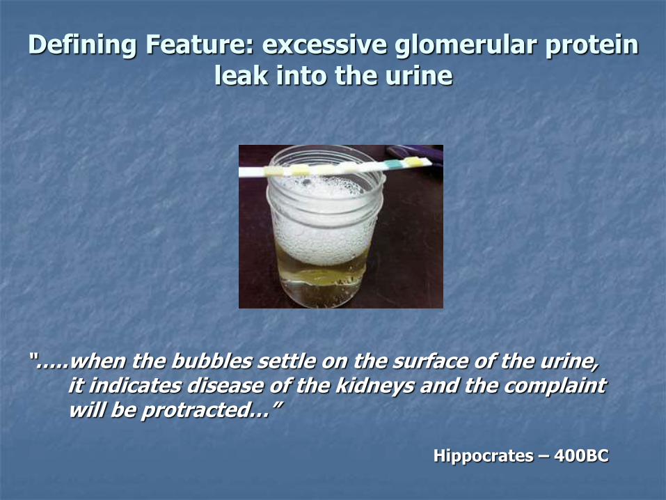
“…..when the bubbles settle on the surface of the urine, it indicates disease of the kidneys and the complaint will be protracted…”
Hippocrates – 400BC
Defining Feature: excessive glomerular protein leak into the urine
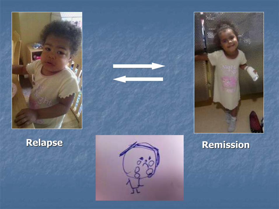
Remission Relapse

many molecules and mechanisms remain a mystery…
Despite 2500 years of clinical description…
PHENOTYPE
GENETICS ENVIRONMENT
IMMUNITY CHANCE

Genetic
Immune dysfunction Reactive
Genetic background
Epigenetic Influences
Immune activation
-innate/acquired
Immunity

Characteristics of childhood NS
heterogeneous:
isolated kidney disease
component of multi-system disorder
hereditary:- improper glomerular development and
subsequent molecular malfunction
idiopathic
acquired - secondary damage
incongruent drug responses: steroid sensitive, steroid resistant, CNI, MMF
malfunction of the glomerular filtration barrier – complex macromolecular sieve and primary ultra-filter for plasma by
the kidney

3D Glomerulus
P

Glomerular basement membrane
1. Endotheium
2.
3. Podocytes
Glomerular Filtration Barrier

DISEASE
?
GFB malfunction: podocyte key culprit
HEALTH

Normal
Nephrotic syndrome
GFB in disease

“Podocentric” view – podocyte is the key culprit
• how dependent is protein filtration simply on the integrity on the slit diaphragms
•role of other GFB components

Clinical clues for a genetic basis
monogenic diseases – AR/AD inheritance
identification of causative genes by standard genetic tools
to date: primarily expressed in podocytes or GBM
positional cloning followed by mutation screening
type of gene and/or mutation can indicate function
in vitro experimental strategies and animal models: how does the mutation cause disease?

Scientific clues for a genetic basis
glomerular cell biology: stereotypic response to injurious stimuli
knockout models: mice, xenopus, zebrafish, C.Elegans, drosophila, chick
monogenic: single podocyte gene defects
genetics of complex disease: rare variant hypothesis
podocytes main target of injury
but, contribution of GBM and endothelium (+/- mesangial cells) may be underestimated

Molecular overview of the slit-diaphragm and podocyte cell–matrix interactions.
Machuca E et al. Hum. Mol. Genet. 2009;18:R185-R194

Candidate genes (n=25) Direct linked
WT1 NPHS1 NPHS2 ACTN4 TRPC6 PLCE1 MYH9 CDAP2 INF2 APOL1
Arhgap24 Cubulin MYOE1 GLEPP1 LAMB2 LMX1B
COL4A3/4A4/4A5 COQ2 COQ6 PDSS2
SMARCAL1
SCARB2/LIMP2 PMM2
Indirect link - mouse models:
NEPH 1,2,3 FAT 1,2 PAX2 PAR4 NOTCH aPKC IL4 Rac
Podocalyxin S-laminin Mpv17 others
P
GBM
M
O

Other Genetic Influences
epigenetic effects
modifier genes: genetic pathways and protein complexes – other genes abnormalities may modulate disease
functional redundancy

Clinical clues – genetic NS
correct description of phenotype isolated kidney disease or syndromic onset < 2 years: >90% chance of genetic mutation onset < 5 years: 50% chance of genetic mutation treatment resistance family history pattern of inheritance: autsomal recessive,
autosomal dominant, (sporadic) ethnic origin renal histology

Phenotype
Isolated kidney disease:
Finnish type (CNF)
diffuse mesangial sclerosis (DMS)
focal segmental glomerulosclerosis (FSGS)
minimal change (MN)
Syndromes:
Denys Drash, Frasier, Galloway-Mowat,
Nail Patella, Pierson, Lowe, Alports, Leigh,
mitocondrial cytopathies, AMRF syndrome,
Schimke

AGE OF ONSET INHERITANCE
CLINICAL PHENOTYPE (DRUG RESPONSE) RENAL HISTOLOGY
Birth to 2 YEARS > 2 – 18 YEARS
Congenital > 3/12
Early onset: >3/12-2years
CNF DMS FSGS
Syndromes
DMS FSGS
Minimal change Syndromes
FSGS DMS
Minimal change Syndromes
Single gene mutations in
>90%
Single gene mutations in
>50%
Single gene mutations less likely unless
family history, predisposition
alleles
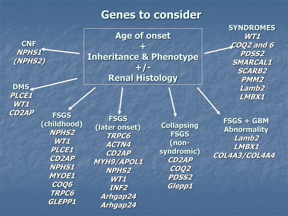
Genes to consider
Age of onset +
Inheritance & Phenotype +/-
Renal Histology
CNF NPHS1
(NPHS2)
FSGS (childhood)
NPHS2 WT1
PLCE1 CD2AP NPHS1 MYOE1 COQ6 TRPC6
GLEPP1
FSGS (later onset)
TRPC6 ACTN4 CD2AP
MYH9/APOL1 NPHS2
WT1 INF2
Arhgap24 Arhgap24
Collapsing FSGS (non-
syndromic) CD2AP COQ2 PDSS2 Glepp1
DMS PLCE1 WT1
CD2AP FSGS + GBM
Abnormality Lamb2 LMBX1
COL4A3/COL4A4
SYNDROMES WT1
COQ2 and 6 PDSS2
SMARCAL1 SCARB2 PMM2 Lamb2 LMBX1

Inheritance pattern
Autosomal dominant
ACTN4
TRPC6 *
INF2*
LIMX1B*
Autosomal recessive
NPHS1 *
NPHS2*
PLEC1*
LAMB2*
Arhgap24
APOL1/MYH9 * - African descent
GLEPP1
many families have “private” gene mutations

? ? ?
? ?
No Fhx renal disease/FSGS
New Zealand
FSGS, transplant
FSGS, transplant x2
FSGS, transplant
FSGS, CKD3
Hereditary FSGS: AD
*

Which genes to test?
Analyse phenotype
CNS: NPHS1, NPSH2, WT1, LAMB2
Later: NPHS1, NPSH2, WT1, LAMB2, PLCE1, APOL1/MYH9, TRCP6, INF2
Use correct genetic tools

NPHS1 mutations
congenital nephrotic syndrome: Finnish and FSGS
Finns: two mutations: Fin major (nt121delCT), Fin minor (R1109X) - truncated, unstable proteins
non-Finns: mutations distributed throughout the gene: - approx 60%: missense or non-frameshift deletions and insertions; approx 40%: nonsense, frameshift, splicing and promotor mutations
steroid/drug resistant
? Predisposition to later onset NS
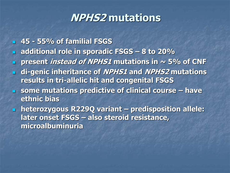
NPHS2 mutations
45 - 55% of familial FSGS
additional role in sporadic FSGS – 8 to 20%
present instead of NPHS1 mutations in ~ 5% of CNF
di-genic inheritance of NPHS1 and NPHS2 mutations results in tri-allelic hit and congenital FSGS
some mutations predictive of clinical course – have ethnic bias
heterozygous R229Q variant – predisposition allele: later onset FSGS – also steroid resistance, microalbuminuria
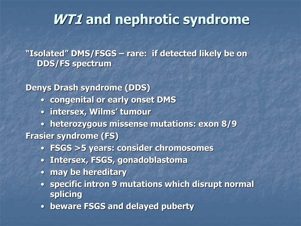
WT1 and nephrotic syndrome
“Isolated” DMS/FSGS – rare: if detected likely be on DDS/FS spectrum
Denys Drash syndrome (DDS)
• congenital or early onset DMS
• intersex, Wilms’ tumour
• heterozygous missense mutations: exon 8/9
Frasier syndrome (FS)
• FSGS >5 years: consider chromosomes
• Intersex, FSGS, gonadoblastoma
• may be hereditary
• specific intron 9 mutations which disrupt normal splicing
• beware FSGS and delayed puberty

PLCE1 Mutations
DMS/FSGS in first two years of life
non-penetrance
15 - 28% incidence idiopathic DMS
not a major cause of familial and later onset FSGS – screening of 69 families (231 individuals) completely negative
may respond to steroids/ciclosporin – cannot be predicted by genotype
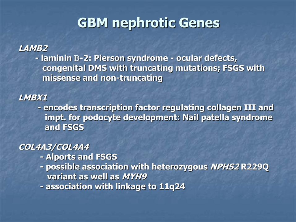
GBM nephrotic Genes
LAMB2 - laminin -2: Pierson syndrome - ocular defects, congenital DMS with truncating mutations; FSGS with missense and non-truncating
LMBX1 - encodes transcription factor regulating collagen III and impt. for podocyte development: Nail patella syndrome and FSGS COL4A3/COL4A4 - Alports and FSGS - possible association with heterozygous NPHS2 R229Q variant as well as MYH9 - association with linkage to 11q24
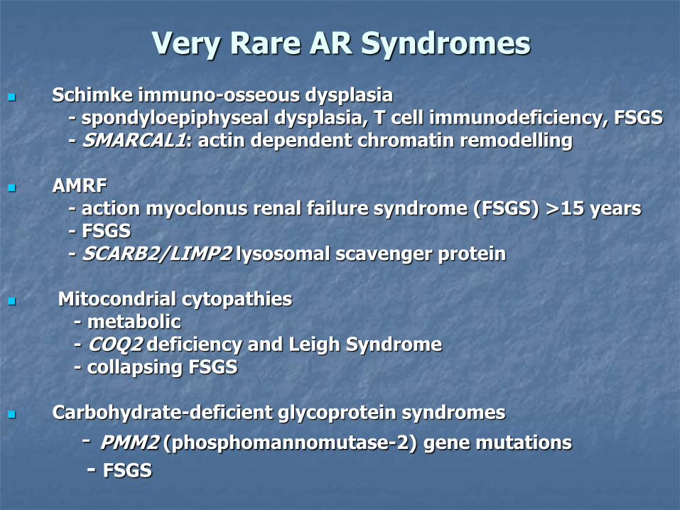
Very Rare AR Syndromes
Schimke immuno-osseous dysplasia - spondyloepiphyseal dysplasia, T cell immunodeficiency, FSGS - SMARCAL1: actin dependent chromatin remodelling AMRF - action myoclonus renal failure syndrome (FSGS) >15 years - FSGS - SCARB2/LIMP2 lysosomal scavenger protein Mitocondrial cytopathies - metabolic - COQ2 deficiency and Leigh Syndrome - collapsing FSGS Carbohydrate-deficient glycoprotein syndromes
- PMM2 (phosphomannomutase-2) gene mutations
- FSGS

Idiopathic steroid sensitive NS
rarely familial
associated with polymorphisms in ACE, VEGF, HLA, NPHS1, MDR1, MIF
exclusion of linkage to NPHS2 - distinct gene loci likely (
homozygosity mapping: locus on chr 2p12-p13.2 (Ruf et al, 2003)
familial and sporadic SSNS in Israeli + Bedouin populations: no linkage to chromosomal loci and no association with mutations in 80 podocyte genes (Landau et al, 2007)
susceptibility genes: rare variant hypothesis

Genetic variants influencing human traits
• Mendelian diseases are caused by extremely rare variants with very large effect size ~1,000-fold) • Complex genetic disease: genetic variants with individually small effect size (typically < 2 fold)
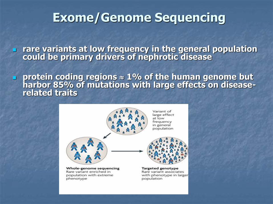
Exome/Genome Sequencing
rare variants at low frequency in the general population could be primary drivers of nephrotic disease
protein coding regions 1% of the human genome but harbor 85% of mutations with large effects on disease-related traits
Aim of Genetic Studies
To understand molecular mechanisms underlying SSNS – design of targeted treatment
Genetic testing to identify “at risk” population
More accurate diagnosis/prognosis
Choosing appropriate treatments
Gene/stem cell therapy
Gene Detection
Linkage - single-gene „Mendelian‟ disorders - inheritance patterns related to several 100‟s to 1000‟s genomic markers Candidate gene approach - small sample sizes - variants assayed limited to a to a select phenotype GWAS - susceptibility loci for complex genetic disease Targeted next generation sequencing - whole exome/whole genome

Molecular Analysis
next generation sequencing population genetics – likely to be complex inheritance detection of potential gene mutation examine effect on protein function in silico functional studies in vitro/in vivo – transgenics biology of relevant cell type in health and disease
Research Approaches
role of innate/acquired immunity complement immune-related genes/mechanisms
Immunological Analysis

Exome Sequencing
Test Population: childhood NS Approaches Targeted Capture: - entire podocyte “genome” 7000 genes - custom designed exon sequence capture hybridization 385K array (NimbleGen) to capture exons of 4000 selected podocyte genes Whole Exome: - entire protein coding region - next generation sequencing - rare disease initiative:
RaDaR https://bridgestudy.medschl.cam.ac.uk/index.shtml
Clinical Applications of Genetic Analysis
Aetiology of disease – targeted therapy Bio-banking - RaDaR/NIHR Bioresource (national rare
disease initiative) Diagnostics – genotype-phenotype to improve diagnosis
and accurate prognosis Pre-transplant: genetic mutation confers minimal risk of
disease recurrence Therapeutics - tailor made medicine in the clinic

Thank you and Questions


