GENETICS IN PEDIATRIC EYE DISEASES CLINICAL IMPLICATIONS Erin M. Salcone, MD MODERATOR Gena Heidary, MD, PhD PROGRAM COMMITTEE COORDINATOR OPTIMIZING OUTCOMES IN CATARACT SURGERY Sherleen Chen, MD, Moderator MODERATORS Larry Piazza, MD, MBA PROGRAM COMMITTEE COORDINATOR 766 th meeting OCTOBER 20, 2017 Hynes Convention Center 900 BOYLSTON STREET | BOSTON, MA 02116 New England Ophthalmological Society
Transcript
GENETICS IN PEDIATRIC EYE DISEASESCLINICAL IMPLICATIONS
Erin M. Salcone, MDMODERATOR
Gena Heidary, MD, PhD PROGRAM COMMITTEE COORDINATOR
OPTIMIZING OUTCOMES IN CATARACT SURGERY
Sherleen Chen, MD, ModeratorMODERATORS
Larry Piazza, MD, MBA PROGRAM COMMITTEE COORDINATOR
766th meeting
OCTOBER 20, 2017 Hynes Convention Center900 BOYLSTON STREET | BOSTON, MA 02116
New England Ophthalmological Society
766th Meeting | 2
GENETICS IN PEDIATRIC EYE DISEASESCLINICAL IMPLICATIONS
Erin M. Salcone, MD, ModeratorGena Heidary, MD, PhD, Program Committee Coordinator
OPTIMIZING OUTCOMES IN CATARACT SURGERY Sherleen Chen, MD, Moderator
Larry Piazza, MD, MBA, Program Committee Coordinator
PO Box 9165 • Boston, MA 02114 | tel: 617.227.6484 | fax: 617.367.4908email: [email protected] | www.neos-eyes.org
Accreditation:The New England Ophthalmological Society designates this live activity for a maximum of
7 AMA PRA Category 1 Credits™. Physicians should claim only the credit commensurate with the extent of their participation in the activity.
The New England Ophthalmological Society is accredited by the Massachusetts Medical Society to provide continuing medical education for physicians.
the 766th meeting of
New England Ophthalmological SocietyA Public Foundation for Education in Ophthalmology
HYNES CONVENTION CENTER900 Boylston StreetBoston, MA 02116
October 20, 2017
3 | New England Ophthalmological Society
766th Meeting | 4
PRESIDENT’S MESSAGE
Welcome to one and all to the 766th meeting of the New England Ophthalmological Society. We have an exciting agenda for everyone this meeting. Please note that during the meeting you will have presentations that will be using the latest in multimedia techniques which including audience participation with both questions and the ARS (audience response system). As always we will have the exhibitors at your disposal for both information and questions. NEOS has actually been a leader in the development of addressing different learning styles and incorporating those individual differences into its programs. During the year we will be bringing you both the Grand Rounds, chaired by Dr. Jeffrey Heier, and the Hal Freeman, MD Video Lecture Series of previous NEOS meetings. Both are intended to reach out to the membership in different ways. Your Board is hopeful that you will continue to find both these programs beneficial and enjoyable.
We are starting off the new NEOS year with our combined meeting of both our membership and the Technician/Administrators’ meeting. Grand Rounds will not be offered at this session secondary to OKAPs and the combined meeting. Both should be informative and transformative for ophthalmic practices. Enjoy and have a great time in Boston.
John J. Dagianis, MD President
5 | New England Ophthalmological Society
ROSA BRAGA-MELE, MED, MD, FRCSCRosa Braga-Mele, MD is a Professor of Ophthalmology, Faculty of Medicine at the University of Toronto, Canada. She graduated magna cum laude from University of Ottawa Medical School. She then completed her residency at the University of Toronto. She went on to complete her Masters Degree in Higher Education with areas of specialty in curriculum development
and patient-physician communication skills.
Dr. Braga-Mele is a cataract specialist and educator who speaks frequently at both the national and international level on advanced surgical techniques and innovations in the area of phacoemulsification surgery and complicated cataract cases and IOL development. She was voted by her peers as one of the top 50 opinion leaders in cataract and refractive surgery. She has over 150 published abstracts and papers. She has also been involved in clinical trials pertaining to wavefront IOL’s and multifocal IOL’s and phacoemulsification surgery.
Dr. Braga-Mele serves as the Chair of the Education Clinical Committee (2015-present) for the American Society of Cataract and Refractive Surgery (ASCRS) and is a member of the Governing Board and Program Committee for ASCRS. She was Chair of the Cataract Clinical Committee for ASCRS from 2011-2015. She was an active member of the AAO Special Programs Committee from 2007-2011, and in 2015-2016 was part of the AAO Cataract Preferred Practice Patterns committee. She was also the chairperson for the cataract section of the Canadian Ophthalmology Society (COS) meeting for 2004, 2005 and 2006.
She is Cataract Section Editor for EyeWorld (an ASCRS publication) and is on the editorial board of other ophthalmic publications.
She was the inaugural Research Director at the Kensington Eye Institute in Toronto from 2007-2012. She was appointed Cataract Director at the Kensington Eye Institute in May 2013-present.
She is involved in the resident surgical teaching curriculum development and implementation at the University of Toronto. She has won multiple teaching awards both at the undergraduate and resident levels for her teaching and mentorship abilities including the Silver Needle award in 2003, 2007, 2012, 2016 and 2017 for best resident surgical teacher, and the University of Toronto, Faculty of Medicine, Community-Based Teaching Award in 2016. She was given the American Academy of Ophthalmology (AAO) Achievement Award in 2007 for distinguished volunteer service and the AAO Secretariat Award in 2012 for special contributions to the Academy and ophthalmology out of proportion to others and making a difference in her efforts. She has recently been awarded the AAO Senior Achievement Award in 2013 for volunteer services.
GUEST OF HONOR
766th Meeting | 6
ARLENE V. DRACK, MDArlene Drack, MD graduated from the University of Scranton in Pennsylvania with a B.S. in Biology and Philosophy. Following a year as an ITT international fellow in Immunology at the University of Oslo, Norway, she attended Pennsylvania State University College of Medicine. Dr. Drack completed her ophthalmology residency at Georgetown University Medical Center and three fellowships: Ophthalmic Genetics at the
Wilmer Institute at Johns Hopkins University; Pediatric Ophthalmology and Strabismus at the University of Iowa; and Molecular Ophthalmic Genetics at the University of Iowa. Dr. Drack held leadership positions at Emory University School of Medicine and the University of Colorado before returning to Iowa City, where she currently is the Ronald V. Keech Associate Professor in Pediatric Genetic Eye Disease Research at the University of Iowa Hospitals and Clinics. As the Director of both the Pediatric and rodent Electroretinogram Services, she maintains a busy clinical practice and research career.
Dr. Drack is well-respected by her colleagues and has been selected the inaugural Chair of the Genetic Eye Disease Task Force of the American Association for Pediatric Ophthalmology and Strabismus (AAPOS). Her professional affiliations include the American Medical Association, Association for Research in Vision and Ophthalmology, AAPOS, and the International Society for Genetic Eye Disease. She has been named “Best Doctors in America” annually since 1996, and has received Senior Achievement Awards from AAPOS and AAO. She was awarded tenure at the University of Iowa in 2015, where she has been integral to the professional development of dozens of research students and residents through her teaching and mentoring roles. She received the Outstanding Translational Research Mentor Award in 2016.
A superb researcher and prolific writer, Dr. Drack has 80 journal publications and over 20 book chapters to her credit. She has written on a wide variety of ophthalmology subjects including pediatric retinal disorders, gene-therapy, genetic mapping of pediatric eye diseases, and congenital and juvenile cataracts. Dr. Drack is an internationally known expert in the diagnosis and treatment of inherited eye diseases. She is actively involved in clinical trials for PEDIG (Pediatric Eye Disease Investigator Group) and Phase III Subretinal gene replacement treatment in RPE65 LCA. Her research has been rewarded with grant support from many entities over the years, and she is currently a co-principle investigator in studies funded by industry, a WIVR Advisory Board Grant, and the NIH. She is on the Editorial Board of the Journal of AAPOS and reviews manuscripts for many major ophthalmology and genetics journals as well as the New England Journal of Medicine. She lectures at conferences and meetings around the United States and the world, most recently at the Canadian Ophthalmologic Society. NEOS will be her 148th lecture or presentation since 1990.
When not working, Dr. Drack enjoys kayaking, hiking with her family, and participating in a women’s book club. She is married to computer software developer, Bill Emerson, and has 2 daughters, a crazy cat, and a genotyped dog.
GUEST OF HONOR
7 | New England Ophthalmological Society
GENETICS IN PEDIATRIC EYE DISEASESCLINICAL IMPLICATIONS
Moderator: Erin M. Salcone, MD Program Committee Coordinator: Gena Heidary, MD, PhD
Educational Gaps:Feedback from NEOS members and Program committee review identified more focus on contemporary pediatric ophthalmology topics has been identified as a need in future educational initiatives. Membership feedback has also indicated that they need better understanding of current diagnostic and therapeutic approaches to pediatric eye disease.
NEOS Program Objectives: 1. Update the membership on new understandings of common pediatric eye diseases from a genetic standpoint. 2. Provide a genetic framework for how to approach eye diseases and how this can guide management. 3. Explain how gene replacement has changed clinical decisions and treatment of pediatric eye diseases.
7:00 am Registration/Exhibits
7:30 Best of the NEOS Hal Freeman Video Library – MAIN HALL
8:30 Introduction ................................................................Erin M. Salcone, MD
8:35 Genetics in Ophthalmology. Practical Relevance and Examples
From inherited Retinal Disorders ...................... Jason Comander, MD, PhD
8:47 Advances in the Genetics of Strabismus –
Will this Change Clinical Care?” .........................Mary Whitman, MD, PhD
8:57 Pediatric Genetic Disorders of the Lens ......................Bharti Gangwani, MD
9:11 Genetics in the Ophthalmology Clinic: The Benefits,
Limitations and Challenges ..................................... Emily Place, MD, CGC
9:23 Introduction of Guest of Honor ..................................Erin M. Salcone, MD
9:28 Results of the RPE65 Gene Therapy Trial .........................Arleen Drack, MD
9:53 Business Meeting
10:03 Refreshment break / Exhibits
10:33 Therapy for Congenital Optic Neuropathies:
Fact vs Fiction ...............................................................Crandall Peeler, MD
10:57 The Workup of Nystagmus in the Molecular Genetic Era .....................................................Arlene Drack, MD
11:20 Panel Discussion ........................................Erin M. Salcone, MD, Moderator
11:45 LUNCHEON SEMINARS:
I. Adventures in Electroretinography
Arlene Drack, MD
ROOM 301
II. Management of the Small Pupil
Rosa Mele-Braga, MD
ROOM 303
Be Sure to Scan in for Afternoon Session Before Going to Room to Receive Credit
Be Sure to Return Your Audience Response Unit Before Leaving the Building!
MORNING SESSION (continued)
Jason Comander, MDArlene Drack, MD
Bharti Gangwani, MDCrandall Peeler, MD
Emily Place, MD, CGCMary Whitman, MD, PhD
Yoshihiro Yonekawa, MD
9 | New England Ophthalmological Society
AFTERNOON SESSION
OPTIMIZING OUTCOMES IN CATARACT SURGERY Moderator: Sherleen Chen, MD Program Committee Coordinator: Larry Piazza, MD, MBA
Educational Gaps: Feedback from NEOS members and Program committee review identified gaps in knowledge and understanding about the management of cataracts in patients with external disease, dry eye, other ocular co-morbidities, and systemic disease, as well as an interest in gaining knowledge about new technologies in the field of cataract surgery.
NEOS Program Objectives: 1. Gain understanding of IOL selection in special situations such as PXF,
keratoconus, post-refractive surgery, premium IOLs. 2. Improve pre-operative evaluation and management of patients with ocular
surface disease or compromise. 3. Review management of cataract surgery in patients with systemic disease or
ocular diseases which may impact the outcomes of cataract surgery.
1:05 Optimizing the Ocular Surface for Cataract Surgery ............Helen Wu, MD
1:15 Cataract Surgery in the Presence of Ocular Surface Disease ..............Lorenzo Cervantes, MD
1:25 Best Outcomes in the Presence of PXF ....... Nicoletta Fynn-Thompson, MD
1:35 Introduction of Guest of Honor .................................... Sherleen Chen, MD
1:40 What to Do When the Capsule Ruptures ..................Rosa Braga-Mele, MD
2:00 Refreshment Break/Exhibits
2:30 Management of Patients with Contact Lenses or Keratoconus .................................. Deborah Jacobs, MD
2:40 Update on Intraocular Lens Calculations......................Roberto Pineda, MD
2:52 Peri-operative Management of Cataracts with Uveitis ................................................. Rebecca Hunter, MD
3:02 Pearls and Pitfalls of Premium Intraocular Lens Cataract Surgery ................................................Rosa Braga-Mele, MD
3:25 Best Cataract Surgery Outcomes in Patients with Retinal Disease ...............................................David Jeng, MD
766th Meeting | 10
3:35 Panel Discussion ..........................................Sherleen Chen, MD, Moderator
4:00 Adjourn
AFTERNOON SESSION (continued)
Rosa Braga-Mele, MDLorenzo Cervantes, MD
Nicoletta Fynn-Thompson, MDRebecca Hunter, MD
Deborah Jacobs, MDDavid Jeng, MD
Roberto Pineda, MD
11 | New England Ophthalmological Society
8:35 AM
GENETICS IN OPHTHALMOLOGY- PRACTICAL RELEVANCE AND EXAMPLES FROM INHERITED RETINAL DISORDERS
Jason Comander, MD, PhD
MASSACHUSETTS EYE AND EAR INFIRMARY | BOSTON, MA
Objective: Understand how the latest developments in gene therapy ares starting to influence clinical practice, such as when to refer patients to a genetics clinic.
Certain ophthalmic genetic disorders have traditionally had no treatments available, limiting the practical need for identification and diagnostic testing. However, advances in gene therapy are beginning to make an appearance in the clinic; for example one form of Leber Congenital Amaurosis, a gene therapy drug is under review for approval by the FDA. Genetic conditions with ongoing human clinical trials include a number of retinal disorders (certain forms of retinitis pigmentosa, achromatopsia, juvenile retinoschisis, Stargardt disease, Usher syndrome, etc.) and Leber Hereditary Optic Neuropathy. Practical tips for diagnosing and referring patients with suspected genetic disorders include: understanding the implications of a positive family history or lack thereof; typical presentations / questions for history taking; and typical exam findings. With some simple knowledge, every ophthalmologist can be equipped to identify patients who might be eligible for the newest generation of genetic treatments.
References: Efficacy and safety of voretigene neparvovec (AAV2-hRPE65v2) in patients with RPE65-mediated inherited retinal dystrophy: a randomised, controlled, open-label, phase 3 trial http://www.thelancet.com/journals/lancet/article/PIIS0140-6736(17)31868-8/abstract
The molecular basis of human retinal and vitreoretinal diseases http://www.sciencedirect.com/science/article/pii/S135094621000025X?via%3Dihub Foundation Fighting Blindness website http://www.blindness.org
766th Meeting | 12
8:47 AM
ADVANCES IN THE GENETICS OF STRABISMUS WILL THIS CHANGE CLINICAL CARE?
Mary Whitman, MD, PhD
BOSTON CHILDREN’S HOSPITAL | BOSTON, MA
Objective: To describe the types of strabismus for which a genetic cause has been identified, discuss how knowing a patient’s genetic diagnosis affects their clinical care, and to discuss advances in understanding the genetics of common forms of strabismus.
Strabismus can be divided into paralytic forms, in which one or both eyes cannot move fully, and non-paralytic forms, in which both eyes have full range of motion. Although less common, the paralytic forms of strabismus often display Mendelian inheritance patterns, and multiple causative genes have been identified. Non-paralytic “common” strabismus represents the vast majority of strabismus cases, and displays complex inheritance. Although there have been some reports linking strabismus to certain genetic regions, causative genes have not yet been identified. Among patients for whom a genetic diagnosis has been found, it is starting to affect clinical care. For ocular care, treatment of both strabismus and ptosis can be tailored based on molecular diagnosis. In addition, certain genetic mutations are associated with additional conditions, and timely referrals and screening can lead to earlier treatments.Strabismus can be divided into paralytic forms, in which one or both eyes cannot move fully, and non-paralytic forms, in which both eyes have full range of motion. Although less common, the paralytic forms of strabismus often display Mendelian inheritance patterns, and multiple causative genes have been identified. Non-paralytic “common” strabismus represents the vast majority of strabismus cases, and displays complex inheritance. Although there have been some reports linking strabismus to certain genetic regions, causative genes have not yet been identified. Among patients for whom a genetic diagnosis has been found, it is starting to affect clinical care. For ocular care, treatment of both strabismus and ptosis can be tailored based on molecular diagnosis. In addition, certain genetic mutations are associated with additional conditions, and timely referrals and screening can lead to earlier treatments.
References: Whitman MC, Engle EC. Ocular congenital cranial dysinnervation disorders (CCDDs): insights into axon growth and guidance. Hum Mol Genet. 2017 Aug 1;26(R1):R37-R44 Ye XC1.
Pegado V, Patel MS, Wasserman WW. Strabismus genetics across a spectrum of eye misalignment disorders. Clin Genet. 2014 Aug;86(2):103-11. doi: 10.1111/cge.12367. Epub 2014 Mar 26.
Khan AO, Shinwari J, Abu Dhaim N, Khalil D, Al Sharif L, Al Tassan N. Potential linkage of different phenotypic forms of childhood strabismus to a recessive susceptibility locus (16p13.12-p12.3).
13 | New England Ophthalmological Society
8:57 AM
PEDIATRIC GENETIC DISORDERS OF THE LENS
Bharti Gangwani, MD
BOSTON CHILDREN’S HOSPITAL | BOSTON, MA
Objective: To provide an update on genetic disorders of lens including congenital and juvenile cataracts in pediatric eyes.
Pediatric genetic disorders of lens include cataractous as well as non-cataractous anomalies. Approximately 50% of childhood cataracts are caused by genetic mutations. The cataracts could be isolated or can be associated with ocular or systemic (and metabolic) anomalies. It is important to obtain a comprehensive history, and perform a detailed ophthalmic and systemic examination in children with bilateral cataracts. The patients with systemic findings should be referred to the geneticists. Non-cataractous anomalies are less common and include lens coloboma, lenticonus, microspherophakia, and dislocation of lens (ectopia lentis). The genetic diagnosis helps the families to better understand the disorder and develop realistic expectations as to the course of their child’s disorder and an understanding regarding future risk in siblings and off springs. Recent advances in genetic testing, including the next generation sequencing, can provide an underlying diagnosis in about 70% isolated congenital cataracts and 63% of those with syndromic congenital cataracts.
Gillespie RL, O’Sullivan J, Ashworth J et al. Personalized diagnosis and management of congenital cataract by next generation sequencing. Ophthalmology 2014;121:2124-37
766th Meeting | 14
9:11 AM
GENETICS TESTING IN THE OPHTHALMOLOGY CLINIC: THE BENEFITS, LIMITATIONS AND CHALLENGES
Emily Place, MS, LCGC
MASSACHUSETTS EYE AND EAR INFIRMARY | BOSTON, MA
Objective: Understand the role of genetic testing in caring for patients with inherited eye disease.
Major advances in the field of genetics and specifically molecular genetic testing is impacting all areas of medicine including ophthalmology. Such advances have improved our abilities to provide an accurate and definitive diagnosis of ocular disease. Given these advances, patients and families have many questions for their eye care clinicians about the availability and utility of genetic testing. There a number of factors for clinicians to consider when exploring the topic of genetic testing with patients including testing method, appropriateness of testing for the patient, implications of the results on clinical care as well as the implications for at risk family members. Discussing the strengths and limitations of genetic testing with patients and families is key to patients making informed decisions about testing.
15 | New England Ophthalmological Society
9:28 AM
RESULTS FOR THE RPE65 GENE THERAPY TRIALArlene Drack, MD
UNIVERSITY OF IOWA | IOWA CITY, IA
Objective: Participants will understand the mechanism of sub retinal gene replacement therapy and the effects it had on patients in the phase III RPE65 gene therapy trial. They will learn how to access information about clinical trials in ophthalmology.Introduction: Leber congenital amaurosis (LCA) is a leading cause of congenital blindness with at least 19 causative genes. A gene therapy (SPK-RPE65) for LCA caused by RPE65 gene mutations recently completed a Phase 3 trial. Many other gene therapy trials for pediatric ocular disorders are in earlier phases. Methods: 31 patients with CLIA laboratory-confirmed RPE65-mediated LCA were enrolled at two centers. 21 participants were randomized to the intervention group; 10 were randomized to 1 year of observation before crossover. Subretinal injection of SPK-RPE65 in a 300 uL volume was delivered to one eye, followed by the contralateral eye within 18 days. Efficacy measures included mobility testing (MT), full-field light sensitivity threshold (FST), and visual acuity (VA). Review of gene therapy trials recruiting patients was obtained from www.clinicaltrials.gov and other sites.Results: The trial met its primary endpoint (p=0.001), demonstrating improvement of functional vision in the intervention group compared to the control group, as measured by bilateral MT change score between baseline and 1 year. FST improved significantly in the intervention group compared to the control group (pSPK-RPE65 or deleterious immune responses. Procedure-related AEs included transient elevated IOP (4), cataract (3), retinal tear (2), and inflammation (2). Patients randomized to no treatment for a year did not improve on testing during this time, but did improve after they were treated following one year of observation. Results have been stable for 2 years in the initial treatment group and for one year in the second group treated. Many other gene therapy trials for pediatric eye disorders are underway. Discussion: SPK-RPE65 resulted in statistically significant improvement in functional vision and visual function measured by MT and FST, respectively. AEs were typical for subretinal surgery and not related to vector or immune response. Parents of children with genetic eye disorders may benefit from knowing about research trials, and how to check on eligibilty of their children.Conclusion: Children with nystagmus and/or poor vision require genetic testing, since subretinal gene therapy improves visual function in RPE65-mediated disease, and other gene therapy clinical trials for different disorders are underway.References: Russell S, Bennett J, Wellman, J, Chung D, et al. Efficacy and safety of voretigene neparvovec (AAV2-hRPE65v2) in subjects with RPE65-mediated inherited retinal dystrophy: a randomised, controlled, open label phase 3 trial. Lancet. 2017 Jul 13. pii: S0140-6736(17)31868-8. doi: 10.1016/S0140-6736(17)31868-8. [Epub ahead of print]
766th Meeting | 16
10:33 AM
THERAPY FOR CONGENITAL OPTIC NEUROPATHIES: FACT VS. FICTION
Crandall Peeler, MD
BOSTON MEDICAL CENTER | CAMBRIDGE, MA
Objective: To discuss the available evidence and safety concerns surrounding stem cell therapy for congenital optic neuropathies.
Congenital optic disc anomalies are responsible for 15% of severe visual impairment in children1. Optic nerve hypoplasia (ONH) is the most common abnormality and is the leading ocular cause of blindness in children in North America and Europe2. While treatment is available for the endocrinopathies often associated with ONH, there is currently no FDA-approved therapy for vision loss stemming from the optic nerve dysfunction. The treatment landscape is similarly stark for other congenital disc anomalies, including coloboma and morning glory discs. Catering to ONH patients seeking visual improvement, medical clinics outside the United States advertise stem cell injections into the cerebrospinal fluid. The aim of this talk is to discuss the available evidence and safety concerns surrounding stem cell therapy for congenital optic neuropathies.
References: Rahi JS, Cable N. Severe visual impairment and blindness in children in the UK. Lancet 2003;362:1359-1365.
Fink C, Garcia-Filion P, Borchert M. Failure of stem cell therapy to improve visual acuity in children with optic nerve hypoplasia. JAAPOS 2013;17(5):490-493.
MASS EYE AND EAR AND BOSTON CHILDREN’S HOSPITAL | BOSTON, MA
Objective: To discuss how genetic testing may help diagnose and optimize the treatment of pediatric vitreoretinopathies.
Pediatric vitreoretinopathies are relatively rare, but can be devastating diagnoses. Examples include retinopathy of prematurity (ROP), familial exudative vitreoretinopathy (FEVR), Norrie disease, persistent fetal vasculature (PFV), Coats’ disease, incontinentia pigmenti, and retinal detachment associated with Stickler syndrome and Marfan syndrome. Children usually do not present with classic textbook findings. Imaging studies coupled with genetic testing are useful in establishing the correct diagnosis, and therefore the optimal treatment plan. Genetic testing also allows the dignosis and surveillance of systemic co-morbidities, and screening of family members.
766th Meeting | 18
10:57 AM
THE WORK UP OF NYSTAGMUS IN THE MOLECULAR GENETIC ERAArlene Drack, MD
UNIVERSITY OF IOWA | IOWA CITY, IA
Objective: Participants will learn about the most common causes of congenital nystagmus and how to structure a workup for individual patients to discover the etiology.
Introduction: Infantile nystagmus has many causes, some life threatening. We determined the most common diagnoses in order to develop a testing algorithm.
Methods: Retrospective chart review. Exclusion criteria were no nystagmus, acquired after 6 months, or lack of examination. Data collected: pediatric eye examination findings, ancillary testing, order of testing, referral, and final diagnoses. Final diagnosis was defined as meeting published clinical criteria and/or confirmed by diagnostic testing. Patients with a diagnosis not meeting the definition were “unknown.” Patients with incomplete testing were “incomplete.” Patients with multiple plausible etiologies were “multifactorial.” Patients with negative complete workup were “motor.”
Results: 284 charts were identified; 202 met inclusion criteria. The 3 most common causes were Albinism(19%), Leber Congenital Amaurosis(LCA)(14%) and Non-LCA retinal dystrophy (13%). Anatomic retinal disorders comprised 10%, motor another 10%. The most common first test was MRI (74/202) with a diagnostic yield of 16%. For 28 MRI-first patients, nystagmus alone was the indication; for 46 MRI-first patients other neurologic signs were present. 0/28 nystagmus-only patients had a diagnostic MRI while 14/46 (30%) with neurologic signs did. Yield of ERG as first test was 56%, OCT 55%, and molecular genetic testing 47%. 90% of patients had an etiology identified.
A testing algorithm is needed.
Conclusion: The most common causes of infantile nystagmus were retinal disorders (56%), however the most common first test was brain MRI. For patients without other neurologic stigmata complete pediatric eye examination, ERG, OCT and molecular genetic testing had a higher yield than MRI scan. If MRI is not diagnostic, a complete ophthalmologic workup should be pursued.
References: Bertsch M, Floyd M, Kehoe T, Pfeifer W, Drack AV. The clinical evaluation of infantile nystagmus: what to do first and why. Ophthalmic Genetics 2017;38(1):22-33.
19 | New England Ophthalmological Society
OPTIMIZING THE OCULAR SURFACE FOR CATARACT SURGERYHelen K. Wu, MD
TUFTS MEDICAL CENTER | BOSTON, MA
Objective: The participant will understand the evaluation and management of patients with ocular surface disease prior to cataract surgery.
Cataract surgery is known to induce dry eye, as well as exacerbate preexisting ocular surface disease. Over half of prospective cataract surgery patients may present with signs of dry eye. After cataract surgery, dry eye may be worsened due to a combination of factors, including damage to the corneal nerves, toxicity from preserved drops, goblet cell loss, meibomian gland dysfunction, and ocular inflammation. While a compromised ocular surface may lead to decreased quality of vision and patient dissatisfaction, it may also predispose to such risk factors as infection and corneal melting. It is thus imperative to recognize and treat preoperatively the signs and symptoms of ocular surface disease, so as to assure optimal outcomes. The diagnosis of ocular surface disease may be made using a variety of tests, often including patient symptom questionnaires, tear break up time, Schirmer testing, conjunctival staining, and tear film osmolarity. Many treatments are available, and the clinical signs should guide the choice of treatment options. A stepwise approach utilizing artificial tears, anti-inflammatory agents, punctal occlusion, treatment of blepharitis and meibomian dysfunction with lid hygiene and antibiotics, oral omega-3 fatty acids and autologous serum tears may be utilized. Systemic immunosuppressant agents may be necessary in patients with severe preexisting ocular surface disease and underlying systemic autoimmune and inflammatory disorders.
References: Movahedan A, Djalilian AR. Cataract surgery in the face of ocular surface disease. Curr Opin Ophthalmol. 2012; 23(1): 68-72.
Sutu C, Fukuoka H, Afshari NA. Mechanisms and management of dry eye in cataract surgery patients. Curr Opin Ophthalmol. 2016 Jan;27(1):24-30.
Trattler WB, et al. The Prospective Health Assessment of Cataract Patients’ Ocular Surface (PHACO) study: the effect of dry eye. Clin Ophthalmol. 2017:11 1423–1430.
1:05 PM
766th Meeting | 20
1:15 PM
CATARACT SURGERY IN THE PRESENCE OF OCULAR SURFACE DISEASE (A.K.A. LEVELING THE PLAYING FIELD)
Lorenzo Cervantes, MD
CONNECTICUT EYE SPECIALISTS | SHELTON, CT
Objective: To help the cataract surgeon recognize and address ocular surface comorbities in order to optimize cataract surgery outcomes.
Patients with eyes with both compromised ocular surfaces and cataracts demand special consideration to optimize their outcomes after cataract surgery. This presentation provides considerations for common conditions that can greatly affect patient expectations, preoperative measurements, and overall quality of vision. Common pathology includes pterygia, corneal scars, epithelial dystrophies, Salzmann nodules, and neoplasias. Strategies for improved cataract surgery outcomes with these comorbidities include patient education and expectation management, and ocular surface optimization techniques such as lesion excision, epithelial debridement, superficial keratectomy, and photherapeutic keratectomy. Photographs and video will be used to demonstrate pathology and these techniques.
References: Kim BZ, Wilson PJ, McGhee CN. Annular Salzmann degeneration: Avoiding perturbations and pitfalls in phacoemulsification surgery. J Cataract Refract Surg. 2015 Nov;41(11):2580-3.
Vanathi M, Goel S, Ganger A, Agarwal T, Dada T, Khokhar S. Corneal tomography and biomechanics in primary pterygium. Int Ophthalmol. 2017 May 13.
Vo RC, Chen JL, Sanchez PJ, Yu F, Aldave AJ. Long-Term Outcomes of Epithelial Debridement and Diamond Burr Polishing for Corneal Epithelial Irregularity and Recurrent Corneal Erosion. Cornea. 2015 Oct;34(10):1259-65.
Kamiya K, Shimizu K, Iijima K, Shoji N, Kobashi H. Predictability of Intraocular Lens Power Calculation after Simultaneous Pterygium Excision and Cataract Surgery. Medicine (Baltimore). 2015 Dec;94(52):e2232.
21 | New England Ophthalmological Society
1:25 PM
BEST OUTCOMES IN THE PRESENCE OF PSEUDOEXFOLIATIONNicoletta A. Fynn-Thompson, MD
OPHTHALMIC CONSULTANTS OF BOSTON | BOSTON, MA
Objective: To highlight the potential complications in eyes with pseudoexfoliation undergoing cataract surgery and provide strategies for optimal intraoperative management.
Pseudoexfoliation (PXF) is an age-related condition causing deposition of fibrillar material on structures in the anterior segment. This material accumulates on the zonular fibers, the anterior lens capsule and the pupillary margin potentially leading to poor pupillary dilation, zonular instability and elevated intraocular pressure. It has been shown that cataracts occur at an increased frequency in eyes with PXF. It is, therefore, prudent to pay special attention to these complex surgical cases.
Cataract surgeons must be cognizant of the increased complexity of cataract surgery in eyes with PXF. We shall highlight the important aspects of preoperative evaluation and the potential intraoperative complications in these eyes. Specifically, management of small pupils, zonular weakness and intraocular lens selection during phacoemulsification will be reviewed. Intraoperative techniques and adjunctive devices to achieve optimal post-operative outcomes will be presented.
References: Shingleton BJ, Crandall AS, Ahmed II. Pseudoexfoliation and the cataract surgeon: preoperative, intraoperative, and postoperative issues related to intraocular pressure, cataract, and intraocular lenses. J Cataract Refract Surg. 2009;35(6):1101-1120.
Turalba A, Cakiner-Egilmez T, Payal AR, et al. Outcomes after cataract surgery in eyes with pseudoexfoliation: Results from the Veterans Affairs Ophthalmic Surgery Outcomes Data Project. Can J Ophthalmol. 2017;52(1):61-68.
Varquez-Ferreiro P, Carrera-Hueso FJ, Poquet Jomet JE, et al. Intraoperative complications of phacoemulsification in pseudoexfoliation: Meta analysis. J Cataract Refract Surg. 2016;42(11):1666-1675.
766th Meeting | 22
1:40 PM
WHAT TO DO WHEN THE CAPSULE RUPTURES?Rosa Braga-Mele, MD, FRCSC
UNIVERSITY OF TORONTO | CANADA
Objective: To help the learner gain understanding of how to recognize, react to and best surgically treat posterior capsular rupture during cataract surgery.
This talk will review pearls on how to recognize when the posterior capsule ruptures during cataract surgery. It will address the best way to respond both in the immediate period and then how best to approach the tear/rupture both in the face of nuclear fragments or without them. It will review best approaches for vitrectomy and/or use of intracameral steroids to aid in surgery to minimize complications and help optimize outcomes.
23 | New England Ophthalmological Society
2:30 PM
MANAGEMENT OF PATIENTS WITH CONTACT LENSES OR KERATOCONUSDeborah S. Jacobs, MD
BOSTON FOUNDATION FOR SIGHT | NEEDHAM, MA
Objective: Attendees will 1. become familiar with guidelines for accurate biometry in contact lens wearers. 2. learn pitfalls of toric IOL for correction of refractive error in patients with keratoconus. 3. understand considerations related to IOL power selection in patient who are habitual RGP contact lens wearers for keratoconus. There are an estimated 41 million contact lens wearers in the United States, 7% of whom wear rigid gas permeable (RGP) lenses. Some of these patients will require cataract surgery each year. Contact lens wear can contribute to corneal warpage. Guidelines for duration of contact lens “holiday” for different types of contact lens prior to biometry for predictable IOL power calculations will be presented.Refractive surgeons of a certain age are aware that patients accustomed to RGP correction of corneal astigmatism may be disappointed by the results of refractive surgery. Likewise, patients with corneal astigmatism who are dissatisfied with toric soft contact lenses may be dissatisfied with toric IOLs. Guidelines for the use of toric IOLs in patients with keratoconus will be presented. Patients with toric IOLs who require RGP correction of residual refractive error will need astigmatism correction in the RGP contact lens (a “bitoric” lens) or spectacle correction over their toric IOL/RGP combo. This is a disappointing outcome for patient who has opted for a toric IOL.In patients who are likely to require RGP correction of astigmatism post-op, one must consider that a corneal or scleral RGP lens will neutralize corneal power. In advanced keratoconus, best results will be obtained when the preop spherical equivalent, AND NOT EMMETROPIA, is selected as the desired post-op refractive error. The habitual contact lens, or one that is only slightly modified, will work post-operatively. Coming up with a satisfactory corneal RGP or scleral contact lens fit can be problematic when high plus power is required for an IOL selected for emmetropia in a patient with a steep cornea. High plus contributes to mechanical instability and/or hypoxic compression over a cone.Remember, patients who are contact lens dependent do best with residual myopia post-op. When in doubt always put more power in the IOL!References: Cope JR, Collier SA, Rao MM, et al. Contact lens wearer demographics and risk behaviors for contact lens-related eye infections--United States, 2014. MMWR Morb Mortal Wkly Rep 2015;64:865-70 Tsai PS, Dowidar A, Naseri A, McLeod SD.Predicting time to refractive stability after discontinuation of rigid contact lens wear before refractive surgery. J Cataract Refract Surg. 2004 Nov;30(11):2290-4. Dell S, Kontos M, Kraff C et al. Abstinence From Contact Lenses Prior to Refractive Surgery https://crstoday.com/articles/2011-oct/abstinence-from-contact-lenses-prior-to-refractive-surgery/ Thebpatiphat N, Hammersmith KM, Rapuano CJ, Ayres BD, Cohen EJ. Cataract surgery in keratoconus. Eye Contact Lens. 2007 Sep;33(5):244-6.
766th Meeting | 24
2:40 PM
UPDATE ON INTRAOCULAR LENS CALCULATIONS
Roberto Pineda II, MD
MASSACHUSETTS EYE AND EAR INFIRMARY | BOSTON, MA
Objective: To review and highlight the newest IOL calculation related material
Over the last decade, IOL power calculations have become a focal point of cataract surgery. In 1977, the state-of-the-art for estimating IOL power for emmetropia was to simply add +19.0 D to the pre-cataractous refraction. A decade later, being within ±1.00 D of the target refraction was still considered a reasonable standard. The standard for accuracy for normal eyes was more recently addressed in the United Kingdom in the 2006 study: “Benchmark standards for refractive outcomes after NHS cataract surgery.” The authors concluded that the “benchmark” standard for refractive outcomes for normal eyes after cataract surgery should be within ±0.50 D for 55% of cases and within ±1.00 D for 85% of cases.1 Today, by carefully optimizing individual component parts of IOL power calculations, combined with advanced surgical techniques it is possible to be within ±0.50 D for better than 70 percent of surgeries and ±1.00 D for better than 90% of surgeries.2 As surgeons have moved to minimally invasive cataract surgery, using premium IOLs and femtosecond lasers, achieving specific postop refraction is as important as cataract surgery itself. Currently, 30% of pts will need glasses or more surgery to address residual refractive error. Cataract surgeons need to maximize accuracy and minimize postoperative refractive misses in order to improve patients’ satisfaction and decrease money spent on additional surgical procedures to refine the postoperative refraction. This talk will review the current trends in IOL calculations and technology to enhance refractive outcomes after cataract surgery.
References: 1. Gale RP, Saldana M, Johnston RL, Zuberbuhler B, McKibbin M. Benchmark standards for refractive outcomes after NHS cataract surgery. Published on-line in Eye, 24 August 2007.
2. Brändle J in Haigis W: IOL calculation in long and short eyes. In Mastering the Techniques of IOL Power Calculations. Hoyos GA, Dementiev JE (eds), Jaypee Brothers Medical Publishers (P) Ltd., New Delhi, 2005
3. Wang L, Hill WE, Koch DD. Evaluation of intraocular lens power prediction methods using the American Society of Cataract and Refractive Surgeons Post-Keratorefractive Power Calculator Intraocular Lens. J Cataract Refract Surg. 2010 Sep;36(9):1466-73. doi: 10.1016/j.jcrs.2010.03.044.
25 | New England Ophthalmological Society
2:52 PM
PERI-OPERATIVE MANAGEMENT OF CATARACTS WITH UVEITISRebecca Hunter, MD
HVMA | BOSTON, MA
Objective: Patients with uveitis pose unique challenges to the cataract surgeon that require special peri-operative attention and management. This talk will review; 1. preoperative considerations and preparations for patients with uveitis who require cataract surgery; 2. special intra-operative procedures that may decrease complications associated with cataract surgery; 3. the postoperative management in this patient population; 4. cataract surgery outcomes in patients with uveitis.
Cataract development is a common complication of patients with uveitis. This can be a result of the primary inflammatory disease itself or as sequelae of treatment with corticosteroids, which remains the most commonly used treatment modality for inflammatory eye disease. Regardless of cause, cataracts remain a major cause of vision loss in these patients and occurs in up to 70% of all patients with uveitis. Cataract surgery in the setting of uveitis adds additional perioperative challenges and risks that are not regularly encountered in patients with age related senile cataracts. Studies have shown that these challenges can be improved with preoperative control of inflammation. Strict preoperative control of inflammation is of significant importance in uveitic eyes undergoing intraocular surgery. However, despite excellent inflammatory control, specific intraoperative challenges are still encountered based on prior inflammatory episodes including anterior and posterior synechiae, inadequate pupil dilation and zonular instability. Even in the event of uncomplicated cataract surgery, there is a considerable increase in postoperative complication rates in these patients given higher risk of recurrent inflammation and cystoid macular edema. Due to these challenges, studies of cataract surgery in uveitic eyes suggest poorer visual outcomes. However with recent studies, various management and treatment strategies have been shown to improve success rates of cataract surgery in patients with uveitis. In this discussion I will review the studies and recommendations for perioperative management that have improved long term outcomes of cataract surgery in this unique patient population.
References: Multicenter Uveitis Steroid Treatment (MUST) Trial Research Group, Kempen JK, Altaweel MM, Holbrook JT, et al. The Multicenter Uveitis Steroid Treatment Trial: Cataract Surgery Outcomes in Uveitis. Ophthalmology 2016; 123: 183-190.
Chu CJ, Dick AD, Johnston RL, et al. Cataract surgery in uveitis: a multicenter database study. Br J Ophthalmol 2017;101: 1132-1137.
Sreekantam S, Denniston AKO, Murray PI. Survey of expert practice and perceptions of the supporting clinical evidence for the management of uveitis-related cataract and cystoid macular edema. Ocul Immunol Inflamm 2011;19:353-357.
766th Meeting | 26
3:02 PM
PEARLS AND PITFALLS OF PREMIUM INTRAOCULAR LENS CATARACT SURGERY
Rosa Braga-Mele, MD, FRCSC
UNIVERSITY OF TORONTO | CANADA
Objective: To help improve knowledge of preoperative tests and diagnoses in premium cataract surgery and a review of IOL’s and picking the best IOL for the patient.
This talk will review the following:
• preoperative tests and assessments for the premium IOL cataract surgery
• looking for corneal surface disease, best practices for treatment and when to proceed with premium surgery
• looking for retinal disease
• which IOL to match with patients
• counselling and chair time
• phaco/femto surgery tips to optimize outcomes
27 | New England Ophthalmological Society
3:25 PM
BEST CATARACT SURGERY OUTCOMES IN PATIENTS WITH RETINAL DISEASE
David Jeng, MD
WEST HARTFORD, CT
Objective: To discuss peri-operative management strategies for patients with retinal disease
Pre-existing retinal disease may affect the visual outcomes for patients undergoing cataract surgery. A detailed retinal exam is essential to rule out undiagnosed retinal disease prior to surgery. Once these retinal conditions have been identified, patients should be made aware of the potential effect on visual prognosis. Proper pre- and post-operative management may prevent visual complications of common retinal diseases, such as diabetic retinopathy, retinal vein occlusions, age-related macular degeneration, and macular puckers. Lastly, specific intra-operative techniques may be helpful in the management of these retinal conditions in the future.
References: Kim SJ, Bressler NM. Optical Coherence Tomography and Cataract Surgery. Curr Opin Ophthalmol. 2009;20(1):46-51.
Loewenstein A, Zur D. Postsurgical Cystoid Macular Edema. Dev Ophthalmol. 2010;47:148-159.
Age-Related Eye Disease Study 2 Research Group. Lutein + zeaxanthin and omega-3 fatty acids for age-related macular degeneration: the Age-Related Eye Disease Study 2 (AREDS2) randomized clinical trial. JAMA. 2013;309:2005-2015.
766th Meeting | 28
As a provider accredited by the Massachusetts Medical Society, NEOS must ensure balance, independence, objectivity, and scientific rigor in all its individually and jointly provided educational activities. All individuals in a position/role to control the content of an activity are expected to disclose to NEOS any relevant financial relationships they and their spouse/partner have with commercial interests.
The ACCME defines a commercial interest as any entity producing, marketing, reselling or distributing health care goods or services consumed by, or used on, patients. Relevant financial relationships are financial relationships in any amount, which occurred in the twelve-month period preceding the time that the individual was asked to assume a role controlling content of the CME activity, and which relate to the content of the educational activity.
Financial relationships are those relationships in which the individual benefits by receiving a salary, royalty, intellectual property rights, consulting fee, honoraria, ownership interest (e.g., stocks, stock options or other ownership interest, excluding diversified mutual funds), or other financial benefit. Financial benefits are usually associated with roles such as independent contractor (including contracted research), consulting, promotional speaking and teaching, membership on advisory committees or review panels, board membership, and other activities for which remuneration is received or expected. The MMS/ACCME considers relationships of the person involved in the CME activity to also include financial relationships of a spouse or partner.
Dagianis, John: Fees for Non-CME Services Received Directly from Commericial Interest or their Agents: Luminus, Speaker Bureau
Drack, Arlene: Contracted Research: Spark Therapeutics, Retrophin and ProQr all supply research grant funding to my institution for research studies on which I am an investigator
NO FINANCIAL INTERESTNone of the other individuals in a position to control the content of this activity, including planners, CME Review Committee members, faculty presenters, moderators, panelists and reviewers have any relevant financial relationship with an ACCME-defined commercial interest to disclose.
766th Meeting | 30
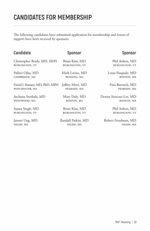
The following candidates have submitted application for membership and letters of support have been received by sponsors:
Candidate Sponsor Sponsor
Christopher Brady, MD, MHS Brian Kim, MD Phil Aitken, MD burlington, vt burlington, vt burlington, vt
Pallavi Ojha, MD Mark Latina, MD Louis Pasquale, MD cambridge, ma reading, ma boston, ma
David J. Ramsey, MD, PhD, MPH Jeffrey Marx, MD Fina Barouch, MD winchester, ma peabody, ma peabody, ma
Archana Seethala, MD Mary Daly, MD Donna Siracuse-Lee, MD westwood, ma boston, ma boston, ma Sujata Singh, MD Brian Kim, MD Phil Aitken, MD burlington, vt burlington, vt burlington, vt
Jannet Ung, MD Randall Patkin, MD Robert Freedman, MD salem, ma salem, ma salem, ma
Thomas Hedges, MD Daniel Lefebvre, MD Nicholas Butler, MD
September 28Cataract (with Pender Lecture) TBA
Ethics and Risk Management TBA
November 30Neuro-ophthalmology and Plastics TBA
Posterior Segment Case Presentations TBA
20 19
March 1
Cornea TBA
Subspeciality Day: Pediatrics Refractive
ImagingTBA
April 12
Anterior Segment Case Presentations (with Chandler-Grant Lecture) TBA
Glaucoma TBA
May 31Uveitis TBA
Retina TBA
FUTURE NEOS MEETINGS
Date Topic Moderator
766th Meeting | 32
THE BOARD AND COMMITTEES 2016-2017
The BoardJohn Dagianis, MD, President Laura Fine, President-elect Mary Daly, MD, Vice-President; Chair Admissions Committee Donna Siracuse-Lee, MD, Secretary Jeffrey Heier, MD, Immediate Past President, Joseph Levy, MD, Treasurer, Chair Finance Committee David Lawlor, MD, Past President, Chair Policies and Nominating Committees Joel Geffin, MD, Past President, Chair Program Committee Michael Price, MD, Chair Educational Endowment Fund Committee Phil Aitken MD, Chair Ophthalmic Services Committee Brendan McCarthy, MD, Chair Public Health and Education Committee Jorge Arroyo, MD, Chair Information Technology Committee Michael Bradbury, MD, Executive Director c omm i t t e e s : Executive CommitteeJohn Dagianis, MD, President Laura Fine, MD, President-elect Joseph Levy, MD, Treasurer Michael Bradbury, MD, Executive Director (ex officio)
Admissions CommitteeMary Daly, MD, Chair Laura Fine, MD John Dagianis, MD
Finance CommitteeJoseph Levy, MD Chair John Dagianis, MD Mary Daly, MD (ex officio) Michael Bradbury, MD (ex officio) Nominations CommitteeJeffrey Heier, MD, Chair Ann Bajart, MD (MA) Mitchell Gilbert, MD (CT) Elliot Perlman, MD (RI) Christopher Soares, MD (VT) David Weinberg, MD (NH) Charles Zacks, MD (ME) ex officio members: Drs. Bradbury, Siracuse-Lee, Fine, Heier, Levy Program CommitteeJoel Geffin, MD, Chair Fina Barouch, MD Geoffrey Emerick, MD Gena Heidary, MD Jeremy Kieval, MD Carolyn Kloek, MD Robert Noecker, MD Lawrence Piazza, MD Shlomit Schaal, MD Michael Yoon, MD John Dagianis, MD (ex officio) Mary Daly, MD (ex officio) Michael Bradbury (ex officio) Public Health and Education CommitteeBrendan McCarthy, MD, Chair Macie Finkelstein, MD Magdalena Krzystolik, MD Joseph Levy, MD
33 | New England Ophthalmological Society
THE BOARD AND COMMITTEES 2016-2017 (continued)
Robert Lytle, MD Vasiliki Poulaki, MD Susannah Rowe, MD Cathryn Welch, MD Michael Wiedman, MD Mary Daly, MD (ex officio) John Dagianis, MD (ex officio) Society Policies CommitteeDavid Lawlor, MD, Chair John Dagianis, MD Michael J. Bradbury, MD Ophthalmic Services CommitteePhil Aitken, MD, Chair Husam Ansari, MD Timothy Blake, MD Kathryn Hatch, MD Edward Jaccoma, MD Marc Leibole, MD Erin Lichtenstein, MD Lauren Shatz, MD Trexler Topping, MD David Vazan, MD Mary Daly, MD (ex officio) John Dagianis, MD (ex officio) Ralph Hinckley (ex officio) Committee for Educational Endowment FundMichael Price, MD, Chair Thomas Coghlin, MD Francis D’Ambrosio, MD Richard Dornfeld, MD Mathew Gardiner, MD Grace Lee, MD Christopher Newton, MD David Lawlor, MD Joseph Levy, MD
Information Technology CommitteeJorge Arroyo, MD, Chair Ankoor Shah, MD Johanna Seddon, MD Nauman Chaudhry, MD Elliot Perlman, MD, (emeritus) Young Ophthalmologists CommitteeJeffrey Heier, MD, Chair Steven Anesi, MD Thomas Berenberg, MD Jennifer Garvey, MD Elizabeth Houle, MD Daniel Lefebvre, MD Michelle Liang, MD Christina Moon, MD Anita Nathan Joshua Ney, MD Archana SeethalaThangappan, MD Ryan Vasan, MD
Jorge Arroyo, MD (ex officio) Michael Bradbury, MD (ex officio) Michael Price, MD (ex officio)
All Donors, please pick up an EEF Ribbon at registration towear at meetings.
Diamond Patrons $100,000 or more
Dr. Michael J. Bradbury In memory of Dr. C. Davis Belcher In honor of Dr. Hal M. Freeman Massachusetts Eye and Ear InfirmaryIn honor of Dr. Joan MillerDr. and Mrs. Paul M. Pender In Memory of Paul D. Pender and Harry V. CareyDr. and Mrs. Richard J. Simmons In memory of Dr. Ruthanne SimmonsOphthalmic Consultants of BostonPhysicians and Patients In honor of Dr. B. Thomas Hutchinson
Platinum Patrons $10,000 to $99,999
Boston Eye Research In memory of Dr. Sanford HechtDr. John Dagianis In honor of Dr. Hal M. Freeman, James and Eleanor Dagianis, and Paul and Verna DobbinsDr. and Mrs. Stuart DuBoff In memory of Dr. Ruthanne Simmons In honor of Samuel and Gloria DuBoff and William and Diane BrownDr. Hal M. FreemanDr. Albert R. Frederick, Jr. In honor of B. Thomas HutchinsonDr. and Mrs. Joseph J. Greco HOYA Optical LaboratoriesDr. B. Thomas HutchinsonNew Hampshire Society of Eye Physicians and Surgeons
Dr. Delia Sang and Dr. Mark Hughes In memory of Dr. Charles L. SchepensDr. Gerald Spindel In honor of Israel and Rose Spindel and Benjamin Burch
Gold Patrons $3,000–$9,999
Drs. A. Robert and Jean Bellows In memory of Dr. W. Morton Grant Dr. and Mrs. Paul P. Dunn In memory of Dr. C. Davis Belcher and in honor of Dr. A. Robert BellowsDr. Joel Geffin Dr. C. Mitchell Gilbert In honor of Drs. Claes Dohlman, Kenneth Kenyon, and Martin WandThe Health Foundation of Central Massachusetts In honor of Dr. Michael J. BradburyJean Keamy for the Keamy Family Foundation In memory of Donald and Yvonne KeamyMaine Society of Eye Physicians and SurgeonsNew England Lens Implant Society In memory of Dr. Sanford HechtDr. and Mrs. Donald Kaplan In memory of Dr. Robert VernlundDr. and Mrs. Elliot Perlman In memory of Drs. C. Davis Belcher and Kathleen MaguireDr. Michael Raizman Dr. Shiyoung Roh and Mrs. Myung Ja RohDrs. Helen and Jack Schinazi In memory of Dr. C. Davis BelcherDr. and Mrs. John Sebestyen In memory of Dr. Taylor R SmithDr. Bradford J. Shingleton In honor of Drs. Albert R. Frederick, B. Thomas Hutchinson,
EDUCATIONAL ENDOWMENT FUND DONORS
766th Meeting | 36
Silvio Von Pirquet and A. Robert BellowsDrs. Richard and Ruthanne Simmons In memory of Dr. W. Morton GrantDr. and Mrs. Richard J. Simmons In memory of Drs. Paul A Chandler, W. Morton Grant, Ruthanne Simmons and C. Davis BelcherDr. and Mrs. Paul Wasson In memory of Dr. Paul Wasson In memory of Dr. Oscar HollanderDr. and Mrs. Hal Woodcome In memory of Dr. Harold Woodcome, Sr.Estate of Dr. Leon Zimmerman
Silver Patrons$1,000–$2,999
Dr. Reid S. Appleby, Jr. In honor of Dr. Harold Woodcome, Jr., and AssociatesDr. and Mrs. Lloyd M. AielloDr. Jorge ArroyoDr. Ann BajartDr. C. Davis Belcher In honor of Dr. Richard SimmonsDr. Harry Braconier In memory of Drs Taylor Smith, Karl Riemer, Carl C. Johnson. In honor of Dr. Hal M. FreemanDr. and Mrs. Sheldon M. BuzneyChildren’s Hospital Ophthalmology FoundationDr. and Mrs. William E. Clark, Jr.Dr. Thomas Coghlin In honor of Dr. Ira Asher and Dr. Kevin O’BrienDr. Joseph L. Dowling, Jr.Dr. Jay S. DukerEye Health Services In memory of Dr. C. Davis Belcher
Dr. Laura FineDr. and Mrs. David GreenseidDr. Bernard HeersinkDr. Jeffrey HeierDr. Ralph HinckleyDr. William S. HoltDr. Robert T. LacyDr. Joseph Levy In honor of Dr. Thomas Hedges IIIDr. Byron S. LingemanDr. Richard LowDr. Kathleen Maguire and Stephen Burke In honor of Dr. Hal M. FreemanDr. Lisa McHamDr. Clifford Michaelson In memory of Dr. Jesse and Mrs. Ruth Lee MichaelsonDr. Stanislaw Milewski In memory of Dr. Taylor R. Smith Dr. Peter B. Mooney In memory of Dr. Henry F. AllenDr. Paul MoultonDr. Stephen J. PhippsDr. and Mrs. Michael PriceDrs. Shiyoung Roh and John WeiterDr. and Mrs. George Santos Dr. Delia Sang In honor of Dr. Lloyd M. AeilloDrs. Jack and Helen Schinazi In memory of Mrs. Mary Santos In honor of Dr. Irving L. PavloDr. Roger F. Steinert In honor of Drs. A. Robert Bellows, S. Arthur Boruchoff, Albert R. Freder-ick, and B. Thomas HutchinsonDr. J. Elliott TaylorDr. Felipe I. Tolentino In honor of Drs. Hal M. Freeman and Roland Houle In memory of Dr. Charles L. SchepensDr. Trexler R. ToppingVermont Ophthalmological Society
EDUCATIONAL ENDOWMENT FUND DONORS (continued)
37 | New England Ophthalmological Society
Dr. Martin Wand In memory of Dr. W. Morton GrantDrs. Peter Wassermann, T. Gordon Hand, Christie Morse and Bradford Hall, In memory of Dr. John Detwiller In honor of Dr. Lewis StieglitzMaster William Weiter In honor of Ann Bajart and Tony Schemmer, and Deborah and Elliot PerlmanDr. Kenneth WolfDr. Allen Zieker Benefactors $500–$999
Dr. Phil Aitken In memory of Drs. Robert Guiduli and Simmons LessellDr. William AtleeDrs. Elliot and Macie FinkelsteinDr. May DalyDr. David Fleishman In memory of Dr. Gary B. Fleishman Dr. George GarciaDr. Robert Guiduli In memory of Dr. Kathleen J. MaguireDr. Lynne KaplinskyDr. Robert LytleMaine Eye CenterDr. and Mrs. Howard MartonDr. Christopher NewtonOphthalmic Consultants of BostonRetina Center of MaineRhode Island Society of Physicians and Surgeons Dr. Joel SchumanDr. Lewis StieglitzDr. Dennis StolerDr. Barry WepmanDr. Charles Wingate
Sponsors $250–$499 Dr. Caroline Baumal In memory of Dr. Jose BerrocalDr. Michael Cooper In memory of Dr. Robert Haimovici and Dr. Behrooz KoleiniDr. Francis Y. Falck, Jr.Dr. Ralph A. Goodwin, Jr.Dr. Timothy GosleeDr. Dana GraichenDr. Payson B. Jacobson In memory of Dr. Abraham Pollen Dr. Glenn P. KimballDr. Peter LouDr. Brendan McCarthy In Memory of Dr. Behrooz KoleiniDr. Carmen PuliafitoDr. Sarkis Soukiasian In Honor of Dr. Roger SteinertDr. Caldwell W. SmithDr. Neal G. SneboldDr. Jonathan TalamoDr. Yvonne Tsai In memory of Helena ToksozDr. Andrew Wong In memory Dr. Charles L. Schepens Worcester Ophthalmology Associates Dr. Charles Zacks
Friends Up to $250
In Memory of C. Davis Belcher Accent Eyewear James Bernson Dr. Charles Beyer-Machule Philip Cacciatore Eye Health Services Milton Feinson Dr. Richard Getnick Evelyn John
EDUCATIONAL ENDOWMENT FUND DONORS (continued)
766th Meeting | 38
Dr. Ernest Kornmehl Don Lesieur Joyce Marshall Rebecca Murphy Therese O’Keefe Dr. Stephen Poor, III Eileen Raffferty Elizabeth Reece Dr. Richard Simmons Marian Spilner Dr. Ann Stromberg Elizabeth Sullivan Andrienne Tashjian The Rivers SchoolIn Memory of Dr. Peter Gudas: Naomi Litrowinik Mercedes Sayler Needham Psychotherapy Associates New England Carpenters Health Fund Norfolk Lodge A.F. and A.M. James and Jean Twyning Jacqueline Pepper Jeanne SmithDr. Peter BatsonDr. Richard BrownDr. David CorbitDr. Paul Cotran In memory of Dr. Mariana Mead Dr. Peter DonshikDr. Stuart Fay In honor of Dr. Michael Bradbury and Dr. TuckMelvyn and Eleanor Galin Foundation In honor of B. Thomas HutchinsonDr. Andrew Gillies In memory of Dr. Moshe LahavDr. Timber GormanDr. Jay Gooze In memory of Kirstyn SmithDr. Amy GregoryDr. Walter GriggsDr. Robert HermDr. Ted Houle
Dr. Glenn P. KimballDr. Howard M. Leibowitz In memory of Dr. Behrooz KoleiniDr. Clifford Michaelson In memory of Dr. Behrooz KoleiniDr. Lawrence PiazzaDr. Theodore RennaMolly-Jane Isaacson Rubinger In honor of Trexler ToppingDr. Domenic M. StrazzullaDr. Carter TallmanDr. Michael Wiedman In honor of Dr. Claes Dohlman