113
Genetics of Cardiovascular Diseases Jacques Genest MD Cardiovascular Genetics Laboratory McGill University Health Center
| Date post: | 03-Jan-2016 |
| Category: |
Documents |
| Upload: | beverly-lawson |
| View: | 218 times |
| Download: | 0 times |
Genetics of Cardiovascular Diseases
Jacques Genest MD
Cardiovascular Genetics Laboratory
McGill University Health Center

Genetics loads the gun, environment pulls the trigger
Elliott Joslin
The older I get, the better I was
A Cowboy
Age is a major cardiovascular risk factor
Framingham Heart Study

Human Biochemical Genetics 2009Genetics of Cardiovascular Diseases
General Principles Historical Aspects Monogenic Disorders Genome-wide Association (GWA)Studies Mendelian Randomization Epigenetics
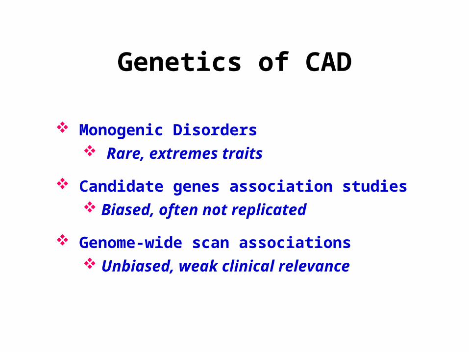
Genetics of CAD
Monogenic Disorders
Candidate genes association studies
Genome-wide scan associations
Rare, extremes traits
Biased, often not replicated
Unbiased, weak clinical relevance

Watkins et al. Nature Reviews Genetics published online 07 February 2006
Genetics of CAD

Genetics and CAD
Genetics of CAD is complex.
Family Hx of premature CAD increases risk > 2.0 fold <55 for father; <65 for mother Corrected for other RF
Association weaker in INTERHEART (case ascertainment of familial CAD weaker than in FHS).
Lloyd-Jones D et al. Lancet 2004;291:2204
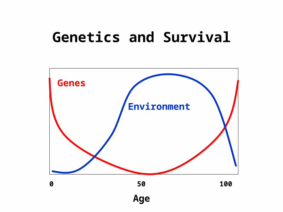
Genetics and Survival
0 50 100
Age
Genes
Environment

General Principles

Chromosomes and DNAChromosomes and DNA
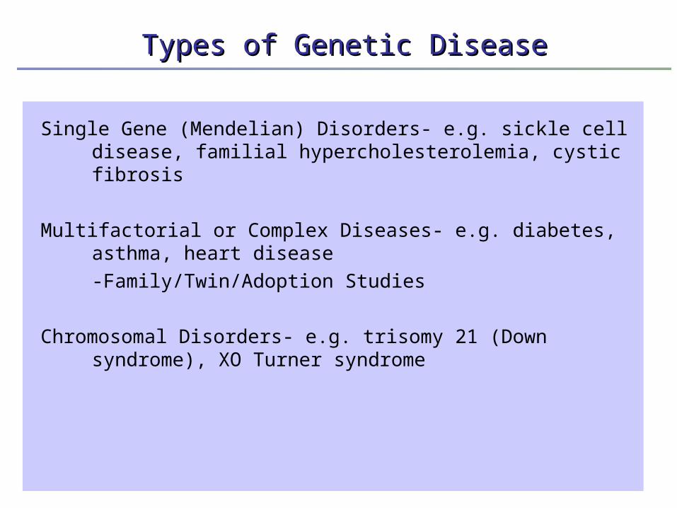
Single Gene (Mendelian) Disorders- e.g. sickle cell disease, familial hypercholesterolemia, cystic fibrosis
Multifactorial or Complex Diseases- e.g. diabetes, asthma, heart disease
-Family/Twin/Adoption Studies
Chromosomal Disorders- e.g. trisomy 21 (Down syndrome), XO Turner syndrome
Types of Genetic DiseaseTypes of Genetic Disease

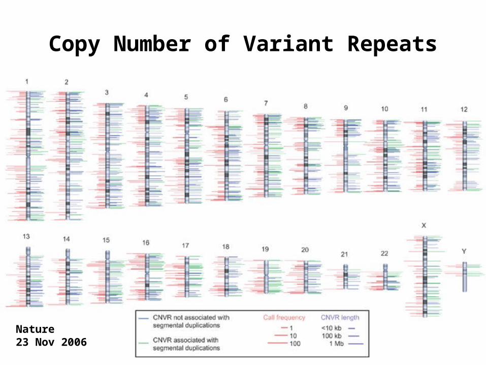
Copy Number of Variant Repeats
Nature 23 Nov 2006

Genetics of Complex Traits

Historical Aspects
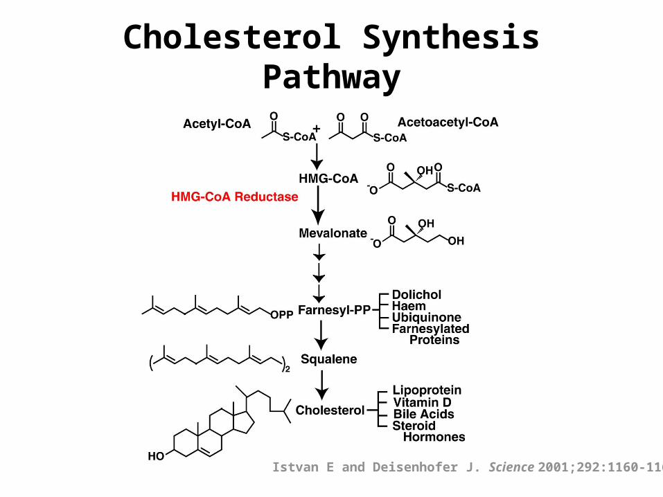
Cholesterol Synthesis Pathway
Istvan E and Deisenhofer J. Science 2001;292:1160-1164.

Risk Factors for CAD
Cigarette Hypertension LDL-cholesterol (apo B) HDL-cholesterol Diabetes Age Atherosclerosis
Circulation 2000;101:111-116
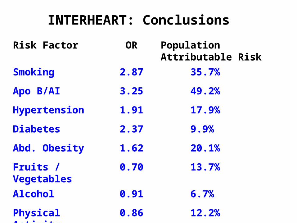
Risk Factor OR Population Attributable Risk
Smoking 2.87 35.7%
Apo B/AI 3.25 49.2%
Hypertension 1.91 17.9%
Diabetes 2.37 9.9%
Abd. Obesity 1.62 20.1%
Fruits / Vegetables 0.70 13.7%
Alcohol 0.91 6.7%
Physical Activity 0.86 12.2%
INTERHEART: Conclusions

Monogenic Disorders

Genetics of Complex Traits
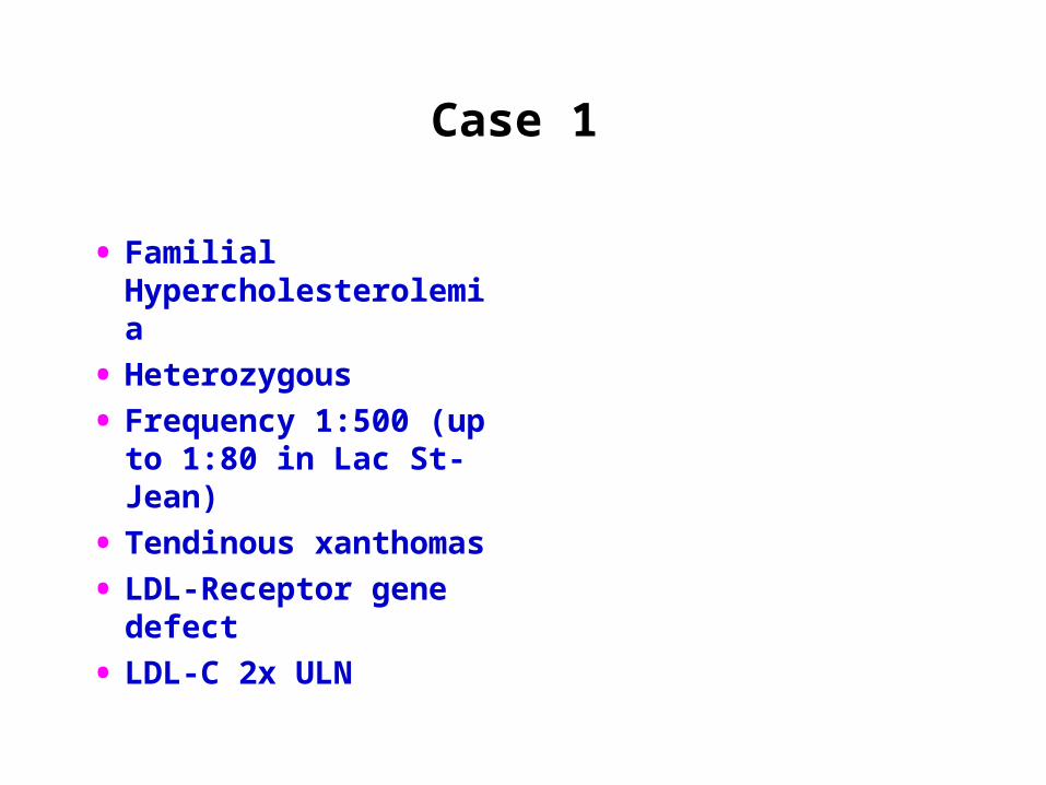
Case 1
• Familial Hypercholesterolemia
• Heterozygous
• Frequency 1:500 (up to 1:80 in Lac St-Jean)
• Tendinous xanthomas
• LDL-Receptor gene defect
• LDL-C 2x ULN

Case 2b
• Familial Hypercholesterolemia
• Homozygous
• Very rare (1:106)
• Premature CAD in childhood
• Extracorporeal LDL removal
Familial Hypercholesterolemia
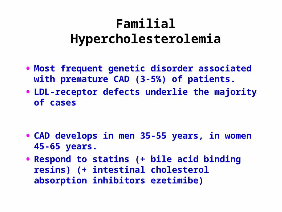
• Most frequent genetic disorder associated with premature CAD (3-5%) of patients.
• LDL-receptor defects underlie the majority of cases
• CAD develops in men 35-55 years, in women 45-65 years.
• Respond to statins (+ bile acid binding resins) (+ intestinal cholesterol absorption inhibitors ezetimibe)
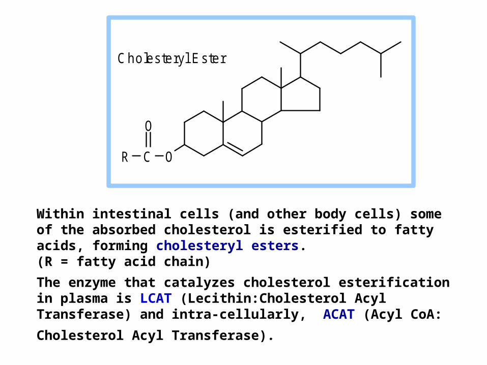
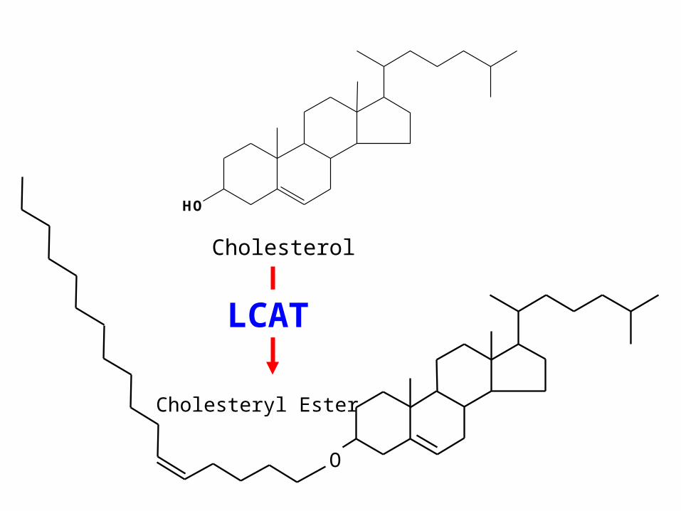
Within intestinal cells (and other body cells) some of the absorbed cholesterol is esterified to fatty acids, forming cholesteryl esters. (R = fatty acid chain)
The enzyme that catalyzes cholesterol esterification in plasma is LCAT (Lecithin:Cholesterol Acyl Transferase) and intra-
cellularly, ACAT (Acyl CoA: Cholesterol Acyl Transferase).
R C O
O
C holesteryl E ster
HO
Cholesterol
O
Cholesteryl Ester
LCAT
H 2C
HC
H 2C
O
O
O
C R 1
O
C
C R 3
OR 2
O
O C R 3
OH 2C
HC
H 2C
O
O
OH
C R 1
O
C R 2
OH 2O
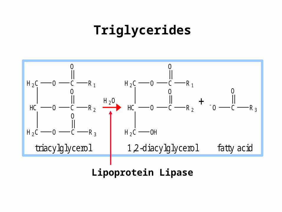
triac ylg lycero l 1 ,2 -d iac ylg lyce ro l fa tty ac id
Lipoprotein Lipase
Triglycerides
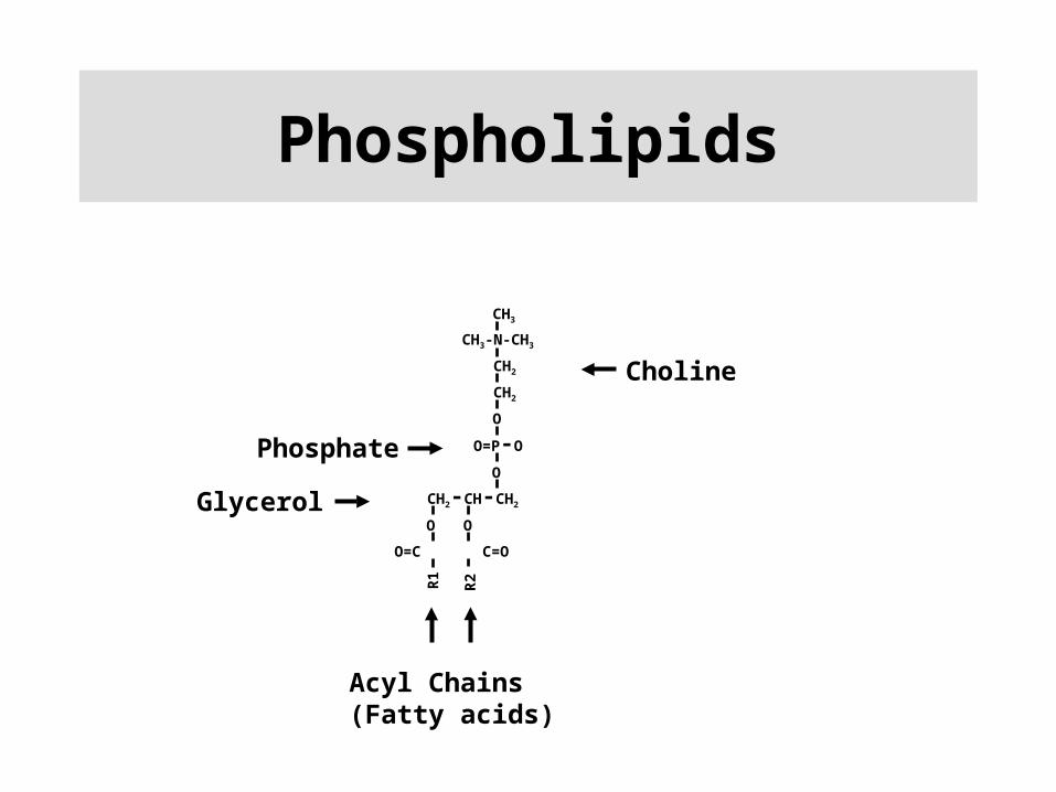
Phospholipids
CH2
O
O=P-O
O
CH2-CH-CH2
O O
O=C C=O
R2
CH3-N-CH3
CH2
CH3
R1
Choline
Phosphate
Glycerol
Acyl Chains(Fatty acids)

Phospholipid
Cholesteryl ester
Apolipoprotein
Triglyceride
Cholesterol

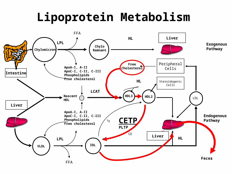
CHYLOMICRONRENNANTS
VLDL
IDL
LDL
HDL2
HDL3
0.95-
1.006-
1.02-
1.06-
1.10-
1.20-
Den
sity
(g
/ml)
Diameter (nm)
5 10 20 40 60 80 1000
Intestine
Liver
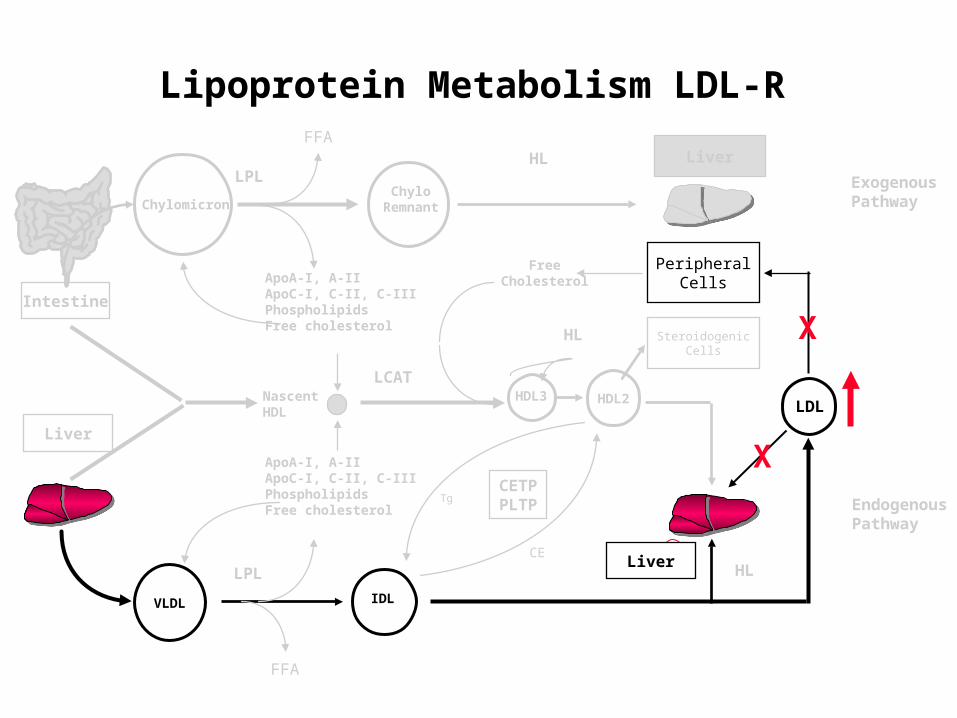
Lipoprotein Metabolism LDL-R
EndogenousPathway
FFA
VLDL
ApoA-I, A-IIApoC-I, C-II, C-IIIPhospholipidsFree cholesterol
ApoA-I, A-IIApoC-I, C-II, C-IIIPhospholipidsFree cholesterol
NascentHDL
PeripheralCells
FFA
FreeCholesterol
HL
Liver
LCAT
HL SteroidogenicCells
ExogenousPathwayChylomicron
ChyloRemnant
HDL2 LDL
IDL
LPL
LPL
CETPPLTP
CE
Tg
HDL3
3Liver
HL
X
X

LDL Receptor
Cells take up LDL by receptor-mediated endocytosis.
The cholesterol in LDL is then used by cells, e.g., for synthesis of cellular membranes.
The LDL receptor was identified by M. Brown & J. Goldstein, who were awarded the Nobel prize for this achievement.

Lipoprotein assembly and secretion
Cholesterol
Fatty acids Cholesteryl esters
VLDL
Bile acids
LDL-R LDL
ApoBVLDL-RLRP
Endosome
sER
HMG CoA Red ACAT
VLDL IDL
Hepatic Cell
ApoB
ApoEApoB
ApoE

LDL-R Pathway Animation
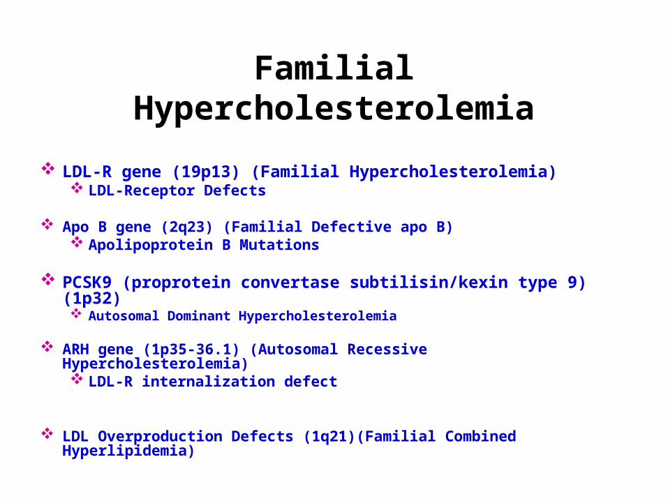
Familial Hypercholesterolemia
LDL-R gene (19p13) (Familial Hypercholesterolemia) LDL-Receptor Defects
Apo B gene (2q23) (Familial Defective apo B) Apolipoprotein B Mutations
PCSK9 (proprotein convertase subtilisin/kexin type 9) (1p32) Autosomal Dominant Hypercholesterolemia
ARH gene (1p35-36.1) (Autosomal Recessive Hypercholesterolemia) LDL-R internalization defect
LDL Overproduction Defects (1q21)(Familial Combined Hyperlipidemia)

Molecular Causes of Familial Hypercholesterolemia (FH)
LDL-R: Primary familial hypercholesterolemia
ARH:Autosomal recessive familial Hypercholesterolemia
PCSK9:Proprotein convertase subtilisin/kexin type 9
ApoB:Familial defective Apo B
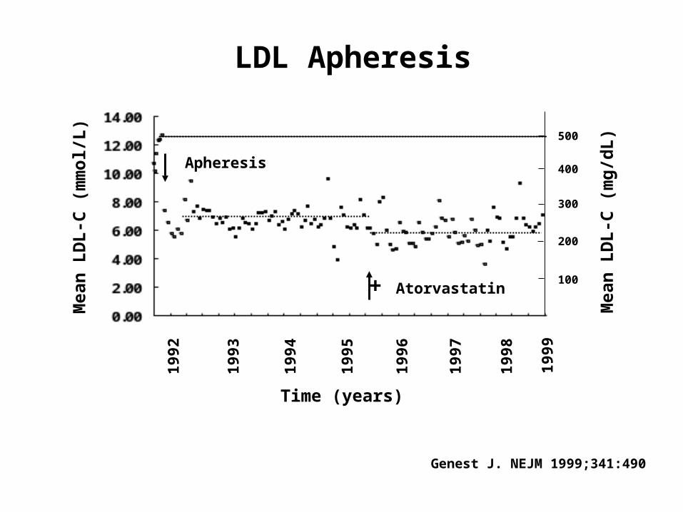
Mea
n L
DL
-C (
mm
ol/
L)
Time (years)
+ Atorvastatin
Apheresis19
92
1993
1994
1995
1996
1997
1998
400
300
200
100
Mea
n L
DL
-C (
mg
/dL
)500
1999
LDL Apheresis
Genest J. NEJM 1999;341:490

PCSK9
Life-long exposure to risk factor:
Principles of Mendelian Randomization

PCSK9 Gene Mutation(African-Americans)
Cohen J. et al. NEJM 2006;354:1264
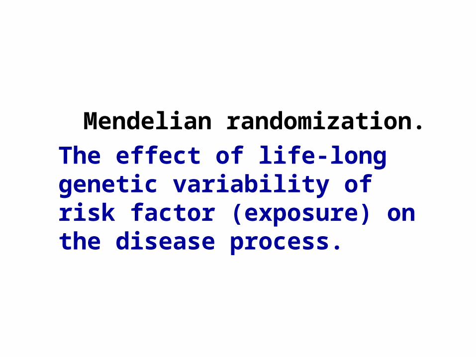
Mendelian randomization.The effect of life-long genetic variability of risk factor (exposure) on the disease process.
HDL
High-Density Lipoproteins
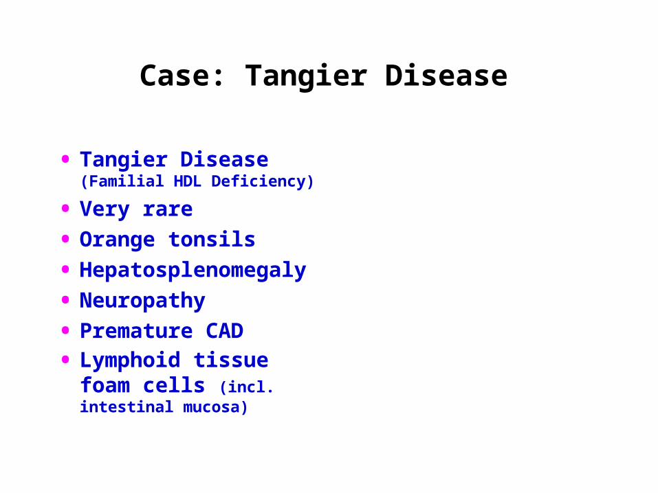
Case: Tangier Disease
• Tangier Disease (Familial HDL Deficiency)
• Very rare
• Orange tonsils
• Hepatosplenomegaly
• Neuropathy
• Premature CAD
• Lymphoid tissue foam cells (incl. intestinal mucosa)

Intestine
VLDL
Liver
ApoA-I, A-IIApoC-I, C-II, C-IIIPhospholipidsFree cholesterol
ApoA-I, A-IIApoC-I, C-II, C-IIIPhospholipidsFree cholesterol
NascentHDL
PeripheralCells
FFA
FFA
FreeCholesterol
HL
Liver
LCAT
HL SteroidogenicCells
ExogenousPathway
EndogenousPathway
ChylomicronChylo
Remnant
HDL2 LDL
IDL
LPL
LPL
CETPPLTP
CE
Tg
HDL3
34
1
Lipoprotein Metabolism: HDL
Liver
HL
X

Cholesterol
Cholesteryl ester Stores
LDL-R
LDL
ApoBEndosome
sER
HMG CoA Red ACAT
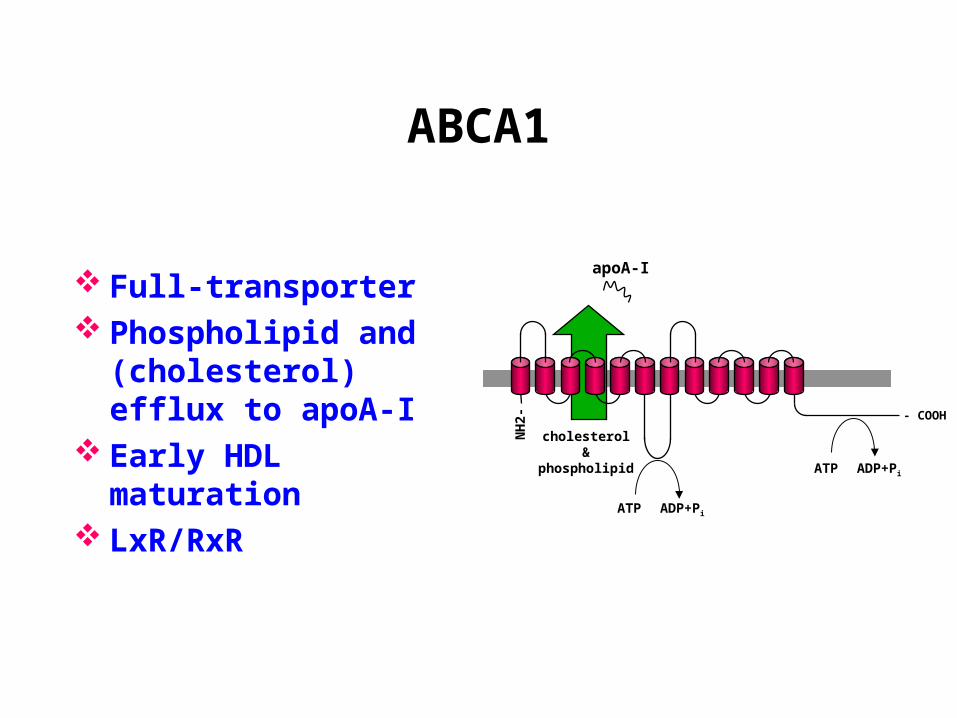
ABCA1
Nascent HDL
HDL3
Lipid-free apo AI
LCAT
- COOH
NH
2-
cholesterol&
phospholipid
ATP ADP+Pi
apoA-I
ABCA1
Full-transporter Phospholipid and
(cholesterol) efflux to apoA-I
Early HDL maturation LxR/RxR
ATP ADP+Pi


HDL Biogenesis
Macrophage <5%
Intestine 20%
Liver 80%
HDL-C Mass
ABCA1
ApoAI
ABCA1
ApoAI
ABCA1
ABCG1
ApoE
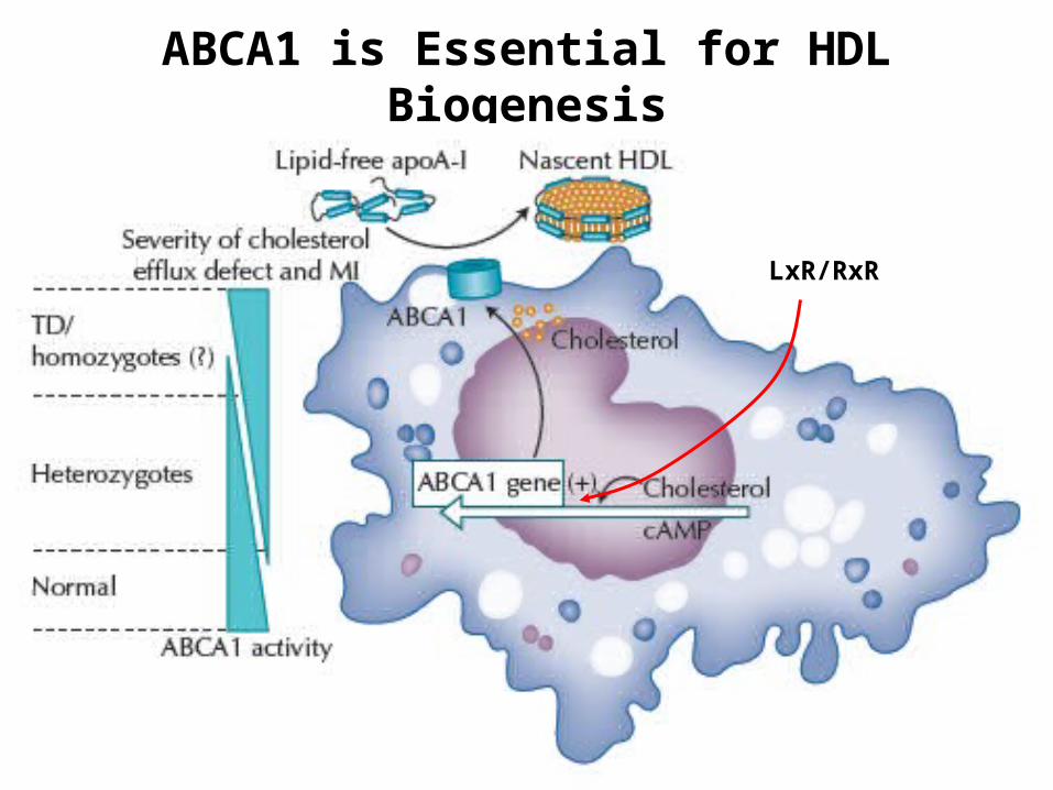
ABCA1 is Essential for HDL Biogenesis
LxR/RxR
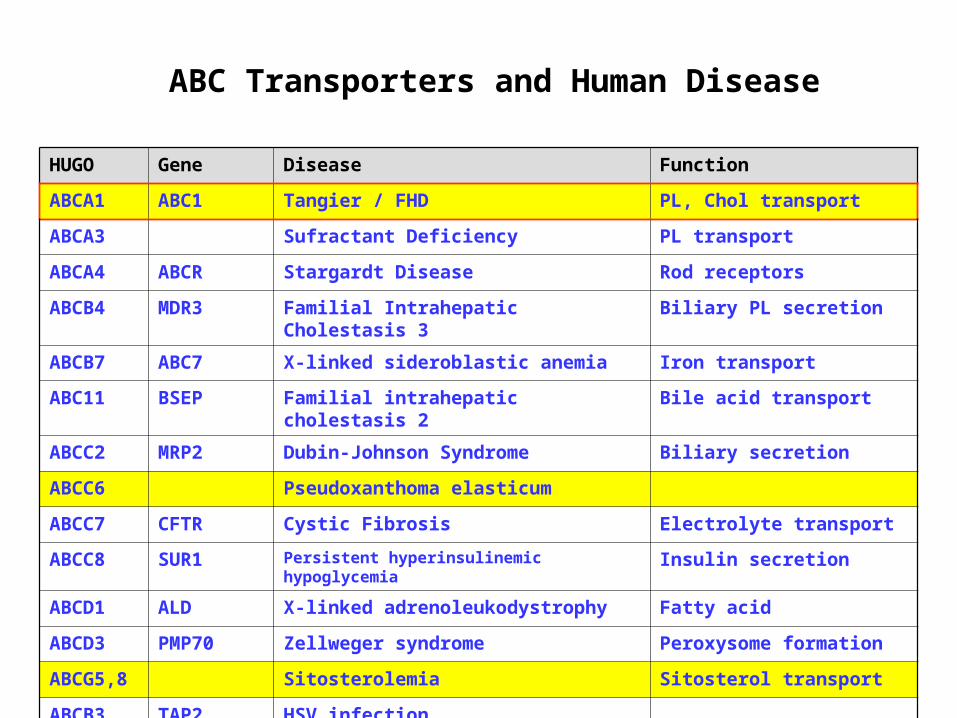
ABC Transporters and Human Disease
HUGO Gene Disease Function
ABCA1 ABC1 Tangier / FHD PL, Chol transport
ABCA3 Sufractant Deficiency PL transport
ABCA4 ABCR Stargardt Disease Rod receptors
ABCB4 MDR3 Familial Intrahepatic Cholestasis 3 Biliary PL secretion
ABCB7 ABC7 X-linked sideroblastic anemia Iron transport
ABC11 BSEP Familial intrahepatic cholestasis 2 Bile acid transport
ABCC2 MRP2 Dubin-Johnson Syndrome Biliary secretion
ABCC6 Pseudoxanthoma elasticum
ABCC7 CFTR Cystic Fibrosis Electrolyte transport
ABCC8 SUR1 Persistent hyperinsulinemic hypoglycemia Insulin secretion
ABCD1 ALD X-linked adrenoleukodystrophy Fatty acid
ABCD3 PMP70 Zellweger syndrome Peroxysome formation
ABCG5,8 Sitosterolemia Sitosterol transport
ABCB3 TAP2 HSV infection
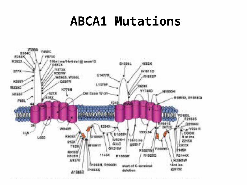
ABCA1 Mutations

HDL-C is Highly Heritable
Studies in twins (n=9) Family studies (n=14) Heritability of HDL-C 0.24 – 0.83 Canadian data: 0.58
Peackock JM ATVB 2001;21:1823

Candidate Gene Sequence Variants in Low HDL subjects
Cohen JC et al. Science 2004;305:869• ABCA1 variants seen in ~10% of low HDL-C (p<0.001)
Frikke-Schmidt R et al. J Clin Invest. 2004;114:1343 • Genetic variation in ABCA1 contributes to HDL-C (~10%)
Alrasadi M et al. Atherosclerosis 2006 • ABCA1 mutations found in 20% of French Canadians with
HDL deficiency.
1 2 3 4 5 6 7 8 9 10 11 12 13 14 15 16 17 18 19 20 21 22 23 24 2725 26 28 3029 31 3332 34 35 36 37 38 39 40 41 42 43 44 45 46 4847 49 503’5’ UTR UTR
1 2 3 4 5 6 7 8 9 10 11 12 13 14 15 16 17 18 19 20 21 22 23 24 2725 26 28 3029 31 3332 34 35 36 37 38 39 40 41 42 43 44 45 46 4847 49 503’5’ UTR UTR
1 2 3 4 5 6 7 8 9 10 11 12 13 14 15 16 17 18 19 20 21 22 23 24 2725 26 28 3029 31 3332 34 35 36 37 38 39 40 41 42 43 44 45 46 4847 49 503’5’ UTR UTR
1 2 3 4 5 6 7 8 9 10 11 12 13 14 15 16 17 18 19 20 21 22 23 24 2725 26 28 3029 31 3332 34 35 36 37 38 39 40 41 42 43 44 45 46 4847 49 503’5’ UTR UTR
1 2 3 4 5 6 7 8 9 10 11 12 13 14 15 16 17 18 19 20 21 22 23 24 2725 26 28 3029 31 3332 34 35 36 37 38 39 40 41 42 43 44 45 46 4847 49 503’5’ UTR UTR
1 2 3 4 5 6 7 8 9 10 11 12 13 14 15 16 17 18 19 20 21 22 23 24 2725 26 28 3029 31 3332 34 35 36 37 38 39 40 41 42 43 44 45 46 4847 49 503’5’ UTR UTR
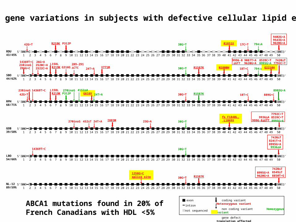
ABCA1 gene variations in subjects with defective cellular lipid efflux
RDU43/45%
SBO44/62%
RPH68/71%
GOB39/58%
LBO54/66%
NL-1289/59%
R1851X42G>T R219K P312P 30G>T 17C>T 794A
14369T>C289-291CTC2522C>A
26G>A2520C>A
R219K G316G 24T>A V771M 30G>T R1587K N1800H 18T>C 794A Q2210H
8539C>T9620G>A
9996-87792C>T
9087T>A 7420T8995G>A
42G>T
238insG 14369T>CR219K P312P
270insGG616V
4152A
24T>A 30G>T R1587K 18T>C 889G>C
8995G>A
270insG 452T I883M 23G>A 30G>T
14369T>C 30G>T
1258G>Csplice site 30G>T R1587K
GTT238insG296G>C
L158L
L158L
24T>A
fs F1840L, L1869X
8995G>A9620G>A
7420T8549T8850T>G
7420T8241T>A8995G>A
9936A
8539C>T9996-8GTT
7792C>T
8995G>A9936A
9402G>A9543G>A9620G>A
coding variant Heterozygous variant
non coding variant Homozygous variant
gene defect translation affected
exon
intron
not sequenced
ABCA1 mutations found in 20% of French Canadians with HDL <5%
Intestine
Liver
EndogenousPathway
FFA
VLDL
ApoA-I, A-IIApoC-I, C-II, C-IIIPhospholipidsFree cholesterol
ApoA-I, A-IIApoC-I, C-II, C-IIIPhospholipidsFree cholesterol
NascentHDL
PeripheralCells
FFA
FreeCholesterol
HL
Liver
LCAT
HL SteroidogenicCells
ExogenousPathwayChylomicron
ChyloRemnant
HDL2 LDL
IDL
LPL
LPL
CETPPLTP
CE
Tg
HDL3
3Liver
HL
Lipoprotein Metabolism
Feces

Modulators of HDL in Humans
SR-BICellularfactors { ABCA1
ELHL
CETPLPL
PLTPs-PLA2
Extracellularfactors {
LCAT
-17.0
-12.2
- 7.1
[nm]
-9.5
ApoA-I-containinglipoproteins
-LpA-Ipre-1-LpA-I
O pre-
-17.0
-12.2
- 7.1
[nm]
-9.5
-17.0
-12.2
- 7.1
[nm]
-9.5
ApoA-I-containinglipoproteins
-LpA-Ipre-1-LpA-I
O pre-
ApoAI
SMase
Dastani Z et al. Figure 1Gene defects identified in man
Lipases

Candidate genes and HDL-C
Structural Receptor/Transport Lipases Exchange
Apo AI ABCA1 Hep Lipase (LIPC) PLTP
Apo AII NPC1 LPL CETP
Endo Lipase LCAT
S-PLA2
SMAse

The Proprotein convertase kexin/subtilisin type 5 gene (PSCK5)
affects HDL-C
PCSK5 inactivates Endothelial Lipase (EL) directly and via ANGPTL3 expression
Iatan I. Circ Cardiovasc Genet 2009 Oct

The PCSK5 gene (302 714 bp)) contains 21 exons and is located on chr 9q21.13

PCSK5 gene SNPs
SNP locations in the PCSK5 gene. Schematic representation of the human PCSK5 gene locus showing the exon structure and the location of the 19 variants (bottom panel) identified through sequencing and the 9 genetic variants associated with HDL-C (upper panel) identified by genotyping. SNPs in bold are associated with HDL-C with P<0.01. Locations are based on RefSeq NM_006200.3.
Quantitative Trait Analysis PCSK5 and HDL-C
Chromosome SNP Trait Beta P-Value
9 rs11144782 HDL -0.07623 0.002021
9 rs11144766 HDL -0.06287 0.005061
9 rs1339246 HDL 0.05575 0.01767
9 rs1331384 HDL 0.03717 0.03825
9 rs11144688 HDL -0.05339 0.03921
9 rs11144690 HDL -0.09334 0.04026
9 rs1338746 HDL -0.03619 0.04393
9 rs4745522 HDL 0.05101 0.04487
9 rs2050833 HDL 0.04527 0.04515
9 rs11144782 TRIG 0.5019 0.04878
9 rs11144782 VLDL 0.1686 0.03858
9 rs11144782 APOB 10.72 0.02234
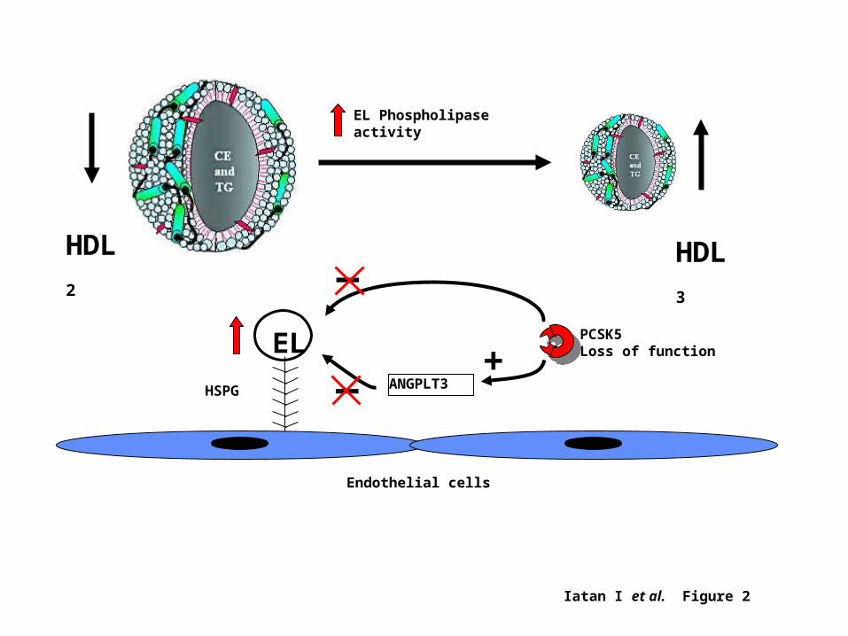
Iatan I et al. Figure 2
HDL2
Endothelial cells
EL Phospholipase activity
HSPG
EL PCSK5Loss of function
ANGPLT3+
_
_
HDL3
The Genetics of HDL
Genome-Wide Scans of FamiliesGenome-Wide Association Studies
Genome-Wide Scans and HDL-C
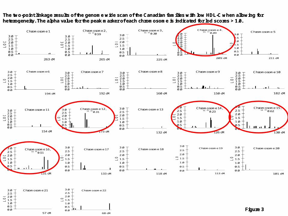
The two-point linkage results of the genome wide scan of the Canadian families with low HDL-C when allowing for heterogeneity. The alpha value for the peak marker of each chromosome is indicated for lod scores > 1.0.
Chromosome 1
0.00.51.01.52.02.53.0
263 cM
LO
D
Chromosome 2,0.59
0.00.51.01.52.02.53.0
265 cML
OD
Chromosome 3,0.30
0.00.51.01.52.02.53.0
225 cM
LO
D
Chromosome 6
0.00.51.01.52.02.53.0
194 cM
LO
D
Chromosome 7
0.00.51.01.52.02.53.0
192 cM
LO
D
Chromosome 8
0.00.51.01.52.02.53.0
160 cM
LO
D
Chromosome 9
0.00.51.01.52.02.53.0
150 cM
LO
D
Chromosome 10
0.00.51.01.52.02.53.0
182 cM
LO
D
Chromosome 11
0.00.51.01.52.02.53.0
154 cM
LO
D
Chromosome 12,0.31
0.00.51.01.52.02.53.0
173 cM
LO
D
Chromosome 13
0.00.51.01.52.02.53.0
132 cM
LO
D
Chromosome 14,0.23
0.00.51.01.52.02.53.0
126 cM
LO
D
Chromosome 15,0.62
0.0
0.51.0
1.5
2.02.5
3.0
130 cM
LO
D
Chromosome 16,0.51
0.00.51.01.52.02.53.0
131 cM
LO
D
Chromosome 17
0.0
0.5
1.0
1.5
2.0
2.5
3.0
133 cM
LO
D
Chromosome 18
0.0
0.5
1.0
1.5
2.0
2.5
3.0
118 cM
LO
D
Chromosome 19
0.0
0.5
1.0
1.5
2.0
2.5
3.0
113 cM
LO
D
Chromosome 21
0.00.51.01.52.02.53.0
57 cM
LO
D
Chromosome 5
0.0
0.5
1.0
1.5
2.0
2.5
3.0
211 cM
LO
D
Chromosome 22
0.0
0.5
1.0
1.5
2.0
2.5
3.0
68 cM
LO
D
Chromosome 20
0.0
0.5
1.0
1.5
2.0
2.5
3.0
101 cM
LO
D
Chromosome 4,0.49
0.00.51.01.52.02.53.03.54.04.55.0
209 cM
LO
D
Figure 3
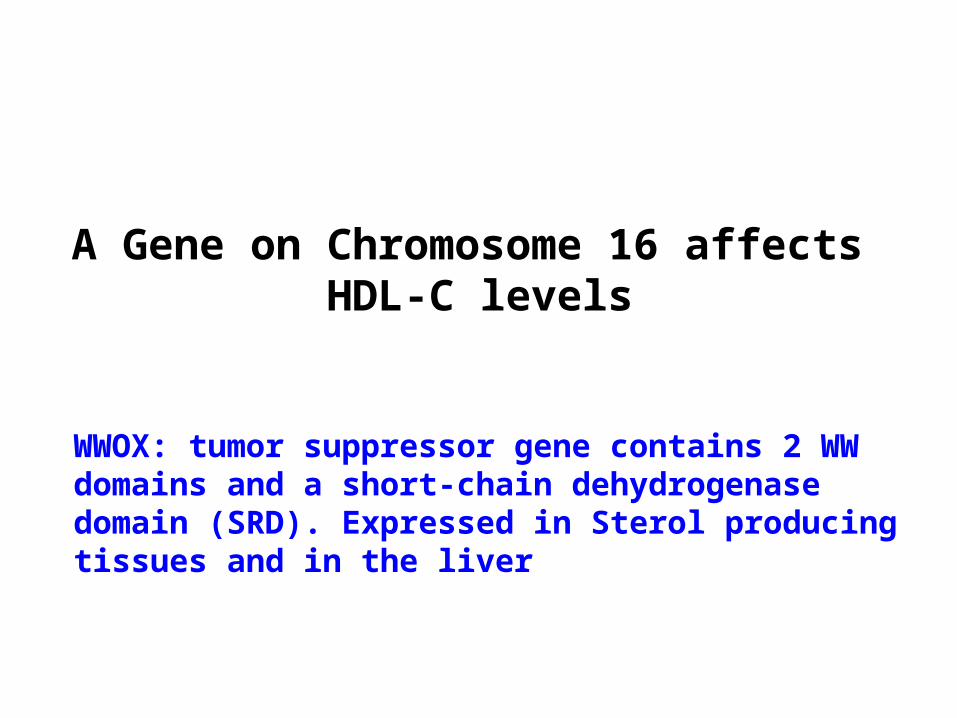
A Gene on Chromosome 16 affects HDL-C levels
WWOX: tumor suppressor gene contains 2 WW domains and a short-chain dehydrogenase domain (SRD). Expressed in Sterol producing tissues and in the liver
D8M
it12
LO
D=
3.5
MouseQTL
PSM
D7
GL
G1
FA
2HR
FW
D3
ML
KL
WD
R59
ZN
RF
1L
DH
DZ
FP
1C
TR
B1
AK
1270
04B
CA
R1
CF
DP
1B
X64
8484
CH
ST
6C
HS
T5
GA
BA
RA
PL
2A
DA
T1
KA
RS
TE
RF
2IP
CA
SP
R4(
CN
TP
)H
SR
G1(
MO
N1B
AD
AM
TS1
8N
UD
T7
KIA
A15
76C
LE
CS
F1
WW
OX
AF
4477
09M
AF
DN
CL
2BC
DY
L2
DC
13K
IAA
0431
BC
0027
01G
CS
HP
KD
1L2
BC
MO
1G
AN
CIM
P
tel
D1 6
S5 1
4
8.1
D1 6
S5 1
6
D1 6
S5 0
5
D1 6
S7 6
3
1.1 .6 10
D1 6
S3 1
07
D1 6
S3 0
95
D1 6
S3 1
06
D1 6
S3 0
66D
1 6S
3 018
D1 6
S5 0
4
D1 6
S3 0
40
D1 6
S5 0
7
D1 6
S3 0
98
2.6 1.1 1.4 0.8 1.4 0.8 3.2 0.8 3.2
18.1cM~7.8Mb
CH
ST6
LC
AT
CE
TP
D1 6
S5 0
3
D1 6
S5 1
5
DiscreteLOD=1.7QUE(D16S505)
LOD(QTL)=2.3QUE(95cM)
RegionforSNPsassociationinSLSJ(23Mb)
LOD(QTL)=2.55SLSJ(85cM)
D16
S31
40
1.87.4
25.5cM~18Mb
C
ABD
cen
1-1430581
7-27413
2-27587.2
8-1440369
5-27587.1
6-1435859
3-1200242
4-1434384
10-24292
9-1384255
54.9
75.1
77.7
80.262
.2
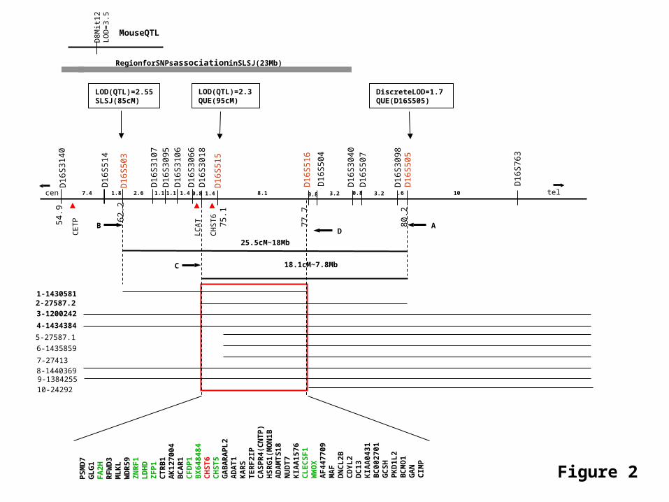
Figure 2


WWOX Protein
WW domains indicates a role in protein-protein interactions. WWOX binds the proline-rich domain PPxY. Highest expression detected in hormonally regulated tissues such as testis, ovary, prostate. Expression pattern and presence of SRD domain suggests a role in steroid metabolism. Wwox-/- mice testis and ovaries display impaired gene expression of key steroidogenesis enzymes.
Interrogation of several online databases show that WWOX is strongly associated with HDL-C (P = 0.0000225). [Willer 08]

HDL Biological Networks
SMPD1
HDL-C
ABCA1
Apo AI
EL
PCSK5
LCAT
LPL
sPLA2
CETPHL
PLTP
SR-B1 ABCG1
?
??
AGPL4
Apo E
WWOX

Genetics of HDL:Family Studies and Candidate Genes
Several genes account for ~25% of severe HDL deficiency (HDL-C <5th percentile) i.e.:
A genetic basis for HDL is identified in 1-2% of subjects
New genes (SMAse, PCSK5, WWOX) provide novel pathways in a complex network

J Clin Invest 2007:117:748
HDL are More Complex than Imagined

Copyright ©2007 American Society for Clinical Investigation
Vaisar, T. et al. J. Clin. Invest. 2007;117:746-756

Questions?
Second session 21 Jan 2010

Genome-Wide Association Studies(GWAS)
Katherisan S, Willer C, Nat Genet 2009

Genetics of Complex Traits

The yet identified genes together explain only a small amount of less than 10% of the HDLC variance, which leaves an enormous room for further yet to be identified genetic variants.
This might be accomplished by large population-based genome-wide meta-analyses and by deep-sequencing approaches on the identified genes.
The resulting findings will probably result in a re-drawing and extension of the involved metabolic pathways of HDLC metabolism.
Exp Gerontol 2008

Genome-Wide Scans and Quantitative traits
Broad and Lund databaseWellcome-Trust Case Control
Consortium (WTCCC)Framingham Study Database Fusion/Sardinia/FHS
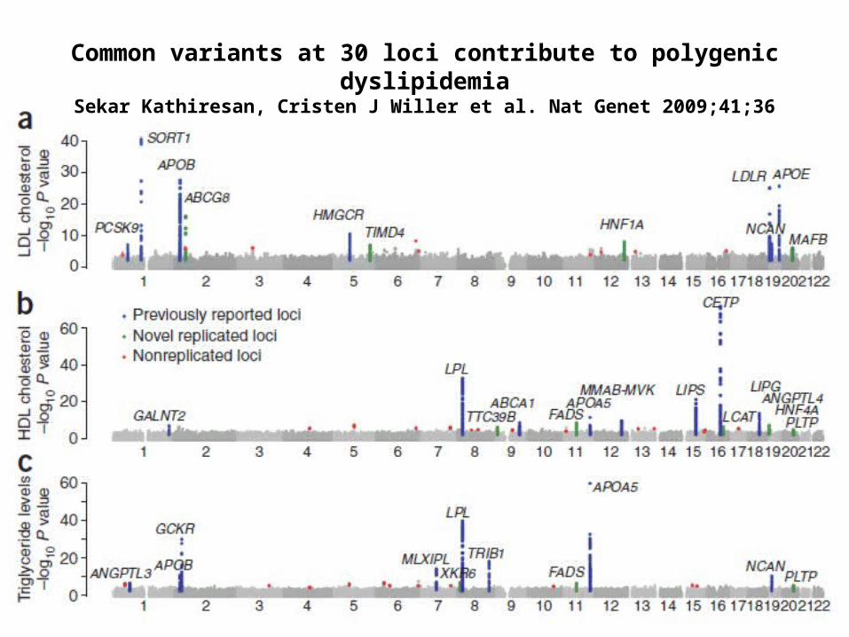
Common variants at 30 loci contribute to polygenic dyslipidemiaSekar Kathiresan, Cristen J Willer et al. Nat Genet 2009;41;36

Common variants at 30 loci contribute to polygenic dyslipidemiaSekar Kathiresan, Cristen J Willer et al. Nat Genet 2009;41;36
Genes you have seen before in candidate gene approach
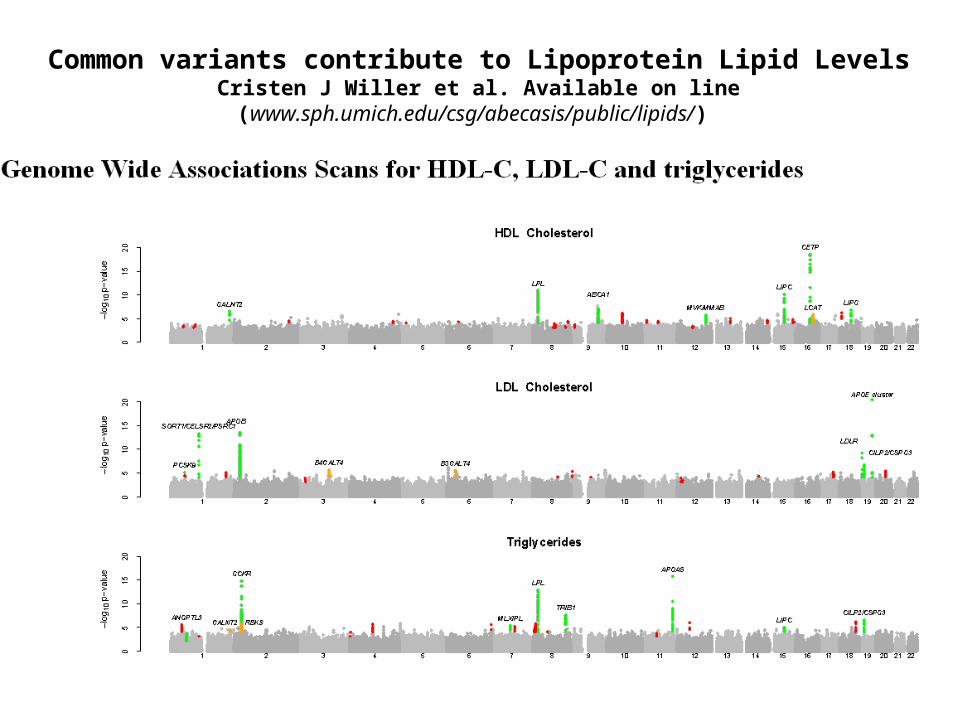
Common variants contribute to Lipoprotein Lipid LevelsCristen J Willer et al. Available on line (www.sph.umich.edu/csg/abecasis/public/lipids/)
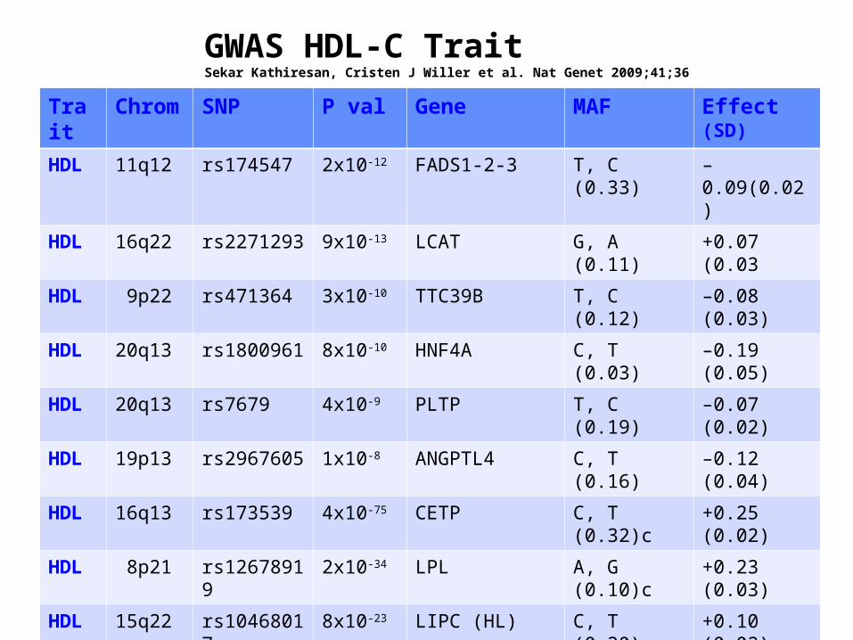
Trait Chrom SNP P val Gene MAF Effect (SD)
HDL 11q12 rs174547 2x10-12 FADS1-2-3 T, C (0.33) –0.09(0.02)
HDL 16q22 rs2271293 9x10-13 LCAT G, A (0.11) +0.07 (0.03
HDL 9p22 rs471364 3x10⁻¹⁰ TTC39B T, C (0.12) –0.08 (0.03)
HDL 20q13 rs1800961 8x10⁻¹⁰ HNF4A C, T (0.03) –0.19 (0.05)
HDL 20q13 rs7679 4x10⁻⁹ PLTP T, C (0.19) –0.07 (0.02)
HDL 19p13 rs2967605 1x10⁻⁸ ANGPTL4 C, T (0.16) –0.12 (0.04)
HDL 16q13 rs173539 4x10-75 CETP C, T (0.32)c +0.25 (0.02)
HDL 8p21 rs12678919 2x10-34 LPL A, G (0.10)c +0.23 (0.03)
HDL 15q22 rs10468017 8x10-23 LIPC (HL) C, T (0.30)c +0.10 (0.02)
HDL 18q21 rs4939883 7x10-15 LIPG C, T (0.17) –0.14 (0.02)
HDL 1q23 rs964184 1x10-12 A1-C3-A4-A5 C, G (0.14)c –0.17 (0.03)
HDL 12q24 rs2338104 1x10⁻¹⁰ MMAB, MVK G, C (0.45) –0.07 (0.02)
HDL 9q31 rs1883025 1x10⁻⁹ ABCA1 C, T (0.26)c –0.08 (0.02)
HDL 1q42 rs4846914 4x10⁻⁸ GALNT2 A, G (0.40) –0.05 (0.02)
GWAS HDL-C TraitSekar Kathiresan, Cristen J Willer et al. Nat Genet 2009;41;36

Common variants at 14 loci contribute to HDL-CSekar Kathiresan, Cristen J Willer et al. Nat Genet 2009;41;36
Based on combined GWAS analysis of >40,000 subjects, 11% of variance of HDL-C levels can be explained.
The Genetics of HDL:Impact on HDL-C or CAD?
Monogenic Disorders Genome-wide Associations Association with CAD

Mendelian randomization is a method of using non-experimental studies to examine the causal effect of a modifiable exposure on disease by making use of measured variation in genes of known function.
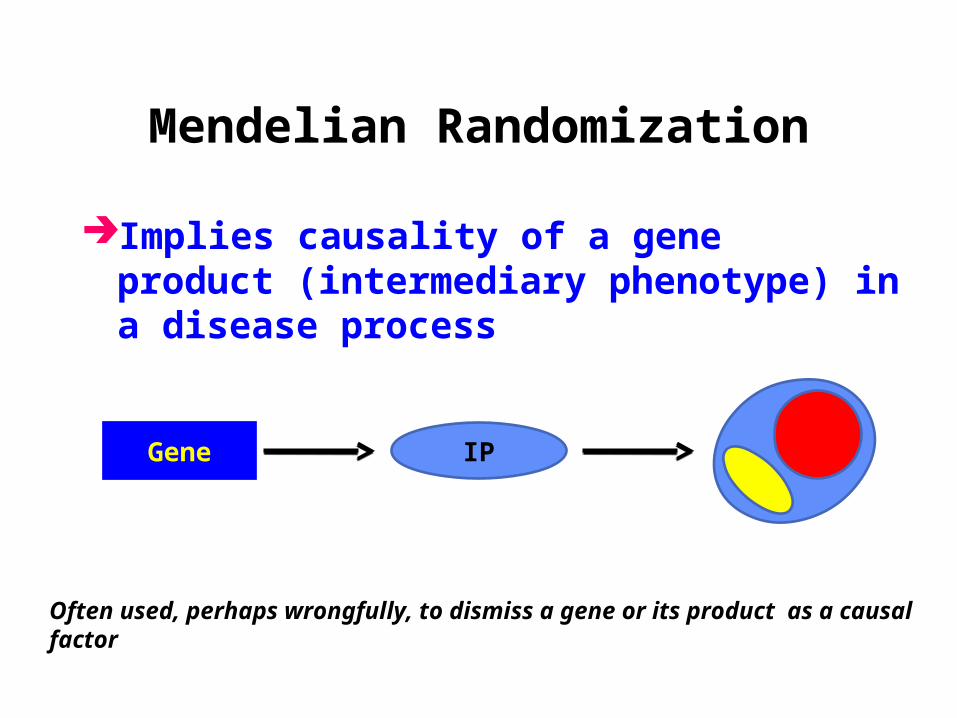
Mendelian Randomization
Implies causality of a gene product (intermediary phenotype) in a disease process
Gene IP
Often used, perhaps wrongfully, to dismiss a gene or its product as a causal factor

Copenhagen Heart Study: HDL-C and CVD Risk
Frikke-Schmidt R JAMA. 2008;299(21):2524-2532.

Copenhagen Heart Study: ABCA1 Mutations and CVD Risk
Frikke-Schmidt R JAMA. 2008;299(21):2524-2532.
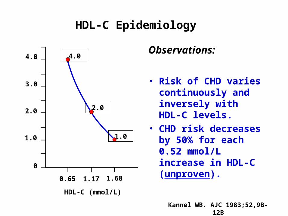
HDL-C Epidemiology
4.0
3.0
2.0
1.0
0.65 1.17 1.68
HDL-C (mmol/L)
CHD risk ratio 2.0
1.0
0
4.0Observations:
• Risk of CHD varies continuously and inversely with HDL-C levels.
• CHD risk decreases by 50% for each 0.52 mmol/L increase in HDL-C (unproven).
Kannel WB. AJC 1983;52,9B-12B

CHD risk ratio
4.0
3.0
2.0
1.0
0.65 1.17 1.68
HDL-C (mmol/L)
2.0
1.0
0
4.0
HDL Epidemiology: Yin and YangC
V R
isk
Apo AI Milano (▼ CV Risk)
CETP Deficiency(▲ CV Risk)
3.16
ABCA1*
*Copenhagen Heart Study** Wellington S, Genest J
ABCA1**
LCAT HL (LIPC)

Genome-Wide Associations(GWA) Studies

The Genetics of Complex traits
Genome-Wide Scans of FamiliesGenome-Wide Association Studies

Genetics of Complex Traits

Genome-Wide Scans for CAD and MI

(Lack of )Validation of Genetic Markers for CAD
Morgan T et al. JAMA 2007;297:1551
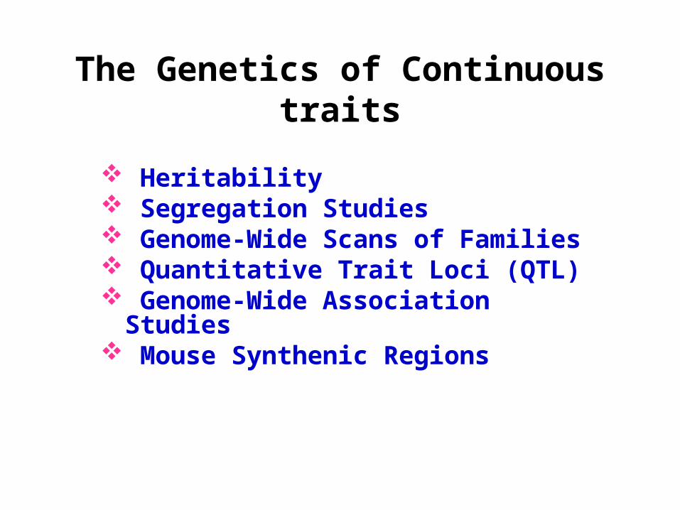
The Genetics of Continuous traits
Heritability Segregation Studies Genome-Wide Scans of Families Quantitative Trait Loci (QTL) Genome-Wide Association Studies Mouse Synthenic Regions

Phenotype Distance
Genetics of complex traits: longer phenotype distance decreases specificity
Risk Factor BiomarkersSurrogate End-Points
Mortality/Morbidity
Phenotype Distance
IMT CHD death, MIBiol. PathwaysLipid Levels
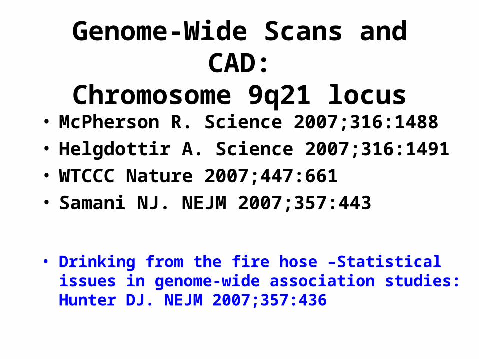
Genome-Wide Scans and CAD:Chromosome 9q21 locus
• McPherson R. Science 2007;316:1488• Helgdottir A. Science 2007;316:1491• WTCCC Nature 2007;447:661• Samani NJ. NEJM 2007;357:443
• Drinking from the fire hose –Statistical issues in genome-wide association studies: Hunter DJ. NEJM 2007;357:436
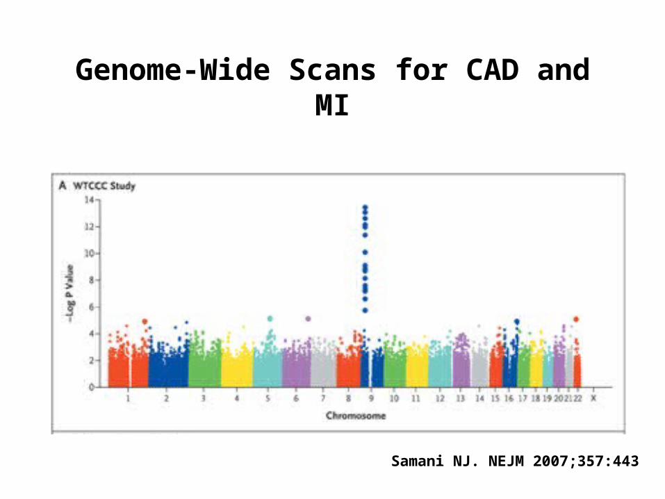
Genome-Wide Scans for CAD and MI
Samani NJ. NEJM 2007;357:443

Chromosome 9q21: which gene?

Schunkert et al. Circulation 2008
Meta-analysis of 9p21 and heart disease

Mendelian Randomization

Women’s Genome Health Study GWAS for Plasma C-Reactive Protein Level
Am J Hum Genet 2008;82:1185-1192

CRP Genetics and Outcome
Zacko et al NEJM 2008;359:1897.
Mendelian Randomization
Epigenetics
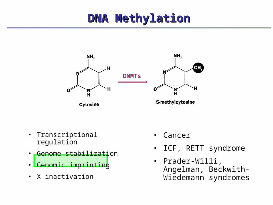
• Transcriptional regulation
• Genome stabilization
• Genomic imprinting
• X-inactivation
DNMTs
DNA MethylationDNA Methylation
• Cancer
• ICF, RETT syndrome
• Prader-Willi, Angelman, Beckwith-Wiedemann syndromes
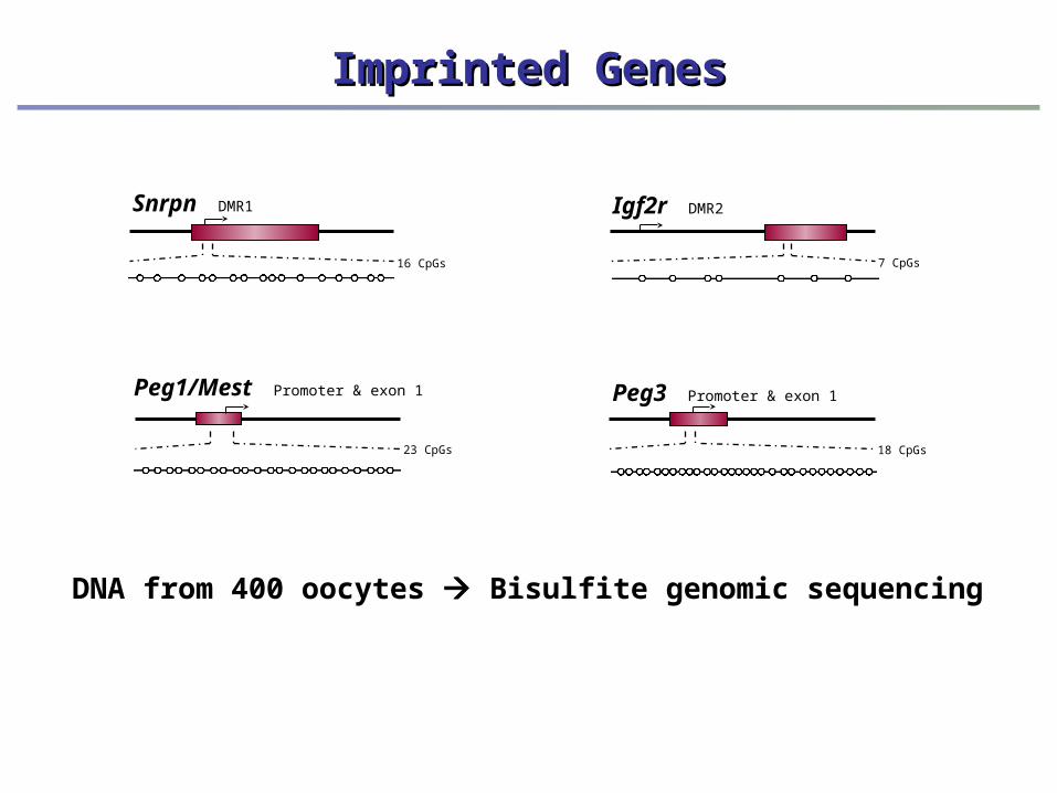
Imprinted GenesImprinted Genes
Igf2r DMR2
7 CpGs
Snrpn DMR1
16 CpGs
Peg1/Mest Promoter & exon 1
23 CpGs
Peg3 Promoter & exon 1
18 CpGs
DNA from 400 oocytes Bisulfite genomic sequencing
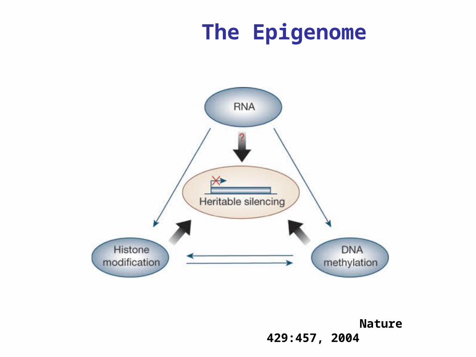
The Epigenome
Nature 429:457, 2004

Imprinting DiseasesImprinting Diseases
- Angelman Syndrome
- Prader-Willi Syndrome
- Beckwith-Wiedemann Syndrome
CancerCancerInactivation of tumor
suppressor genesInactivation of
DNA repair genes
CpG island hypermethylation
Normal DNA methylationNormal DNA methylation
Global hypomethylation
Chromosome instability Retrotransposon
activation
Oncogene activation
?? ?
Adapted from Strathdee et al., Expert Reviews in Molecular Medicine (2002).
ICF SyndromeICF Syndrome
Mutation in DNMT3B
hypomethylation of centromeric
chromatin
IImmunodeficiency, CCentromeric region instability, FFacial
anomalies
Human Diseases Associated with Altered Methylation
Profiles
Human Diseases Associated with Altered Methylation
Profiles


Imprinted Genes
PAR5SNURF-SNRPN
PAR-SN
ZNF217 NDN MAGEL2
IPW
PAR1UBE3A
GABRB3
GABRA5
GABRG3
UBE3A-AS
ICZNF217-AS
Angelman and Prader-Willi syndromes locus 15q11-q13
H19INS IGF2ASCL2
KCNQ1OT1
KCNQ1CDKN1C CD81 TH
IGF2-AS
TSSC5TSSC3NAP1L4 WT1
Beckwith-Wiedemann syndrome locus 11p15.5
Loss of Maternal Methylation at SNRPN and KCNQ1OT1

Female Predominance and Transmission Distortion in the Long-QT Syndrome
Imboden M et al, NEJM 2007;355:2744-2751
Long QT syndrome caused by KCNQ1 and
KCNH2 mutationsAutosomal DominantTransmission Ratio 55% Female; 45% Male
(p=0.005 from normal) Possible genomic imprinting
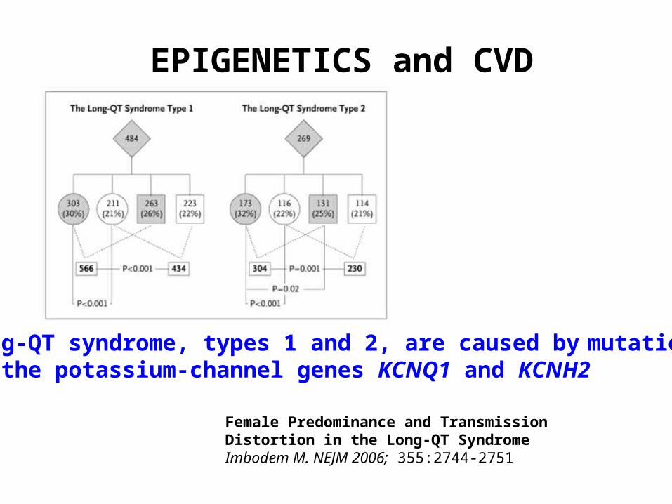
EPIGENETICS and CVD
Female Predominance and Transmission Distortion in the Long-QT SyndromeImbodem M. NEJM 2006; 355:2744-2751
Long-QT syndrome, types 1 and 2, are caused by mutations in the potassium-channel genes KCNQ1 and KCNH2
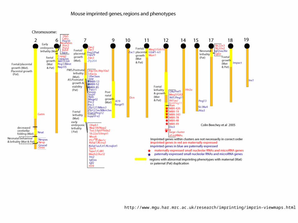
http://www.mgu.har.mrc.ac.uk/research/imprinting/imprin-viewmaps.html

Human Biochemical Genetics 2009Genetics of Cardiovascular Diseases
General Principles Historical Aspects Monogenic Disorders Genome-wide Association Studies (GWA) Mendelian Randomization Epigenetics


