425 Introduction Tuberculosis (TB) is a major public health problem worldwide despite a declining trend in mortality, with effective diagnosis and treatment. An estimated 10.4 million people developed TB in 2015 and more than half of the TB cases (60%) were seen in South-East Asia and Western Pacific Regions 1 . About 60 per cent of TB cases and deaths occur among males, but the disease burden is high among women also 1 . In 2015 nearly 500,000 women died from TB, and among them, 28 per cent had human immunodeficiency virus (HIV) co-infection 1 . Genital TB in females is well recognized as an important aetiological factor for infertility in countries with high prevalence of TB. Genital TB usually occurs secondary to TB in other sites (primarily, the lungs). The spread is generally through haematogenous or lymphatic routes 2 . Tuberculous infection of the female genital organs can result in infertility, dyspareunia, menstrual irregularities and chronic pelvic inflammatory disease (PID) 3 . Drug therapy for female genital TB (FGTB) is similar to the standard treatment regimens used for pulmonary TB. In patients with infertility, conception rate is not very encouraging after anti-TB treatment (ATT) 2 . Here we review the epidemiology, clinical presentations, recent advances in diagnosis and treatment of FGTB. Female genital tuberculosis - epidemiology and pathogenesis Genitourinary TB is a common form of extrapulmonary TB (EPTB) worldwide (27%) with Genital tuberculosis in females G. Angeline Grace, D. Bella Devaleenal & Mohan Natrajan Department of Clinical Research, ICMR-National Institute for Research in Tuberculosis, Chennai, India Received September 29, 2015 The morbidity and mortality due to tuberculosis (TB) is high worldwide, and the burden of disease among women is significant, especially in developing countries. Mycobacterium tuberculosis bacilli reach the genital tract primarily by haematogenous spread and dissemination from foci outside the genitalia with lungs as the common primary focus. Genital TB in females is a chronic disease with low-grade symptoms. The fallopian tubes are affected in almost all cases of genital TB, and along with endometrial involvement, it causes infertility in patients. Many women present with atypical symptoms which mimic other gynaecological conditions. A combination of investigations is needed to establish the diagnosis of female genital TB (FGTB). Multidrug anti-TB treatment is the mainstay of management and surgery may be required in advanced cases. Conception rates are low among infertile women with genital TB even after multidrug therapy for TB, and the risk of complications such as ectopic pregnancy and miscarriage is high. More research is needed on the changing trends in the prevalence and on the appropriate methods for diagnosis of FGTB. Key words Anti-tuberculosis treatment - conception - fallopian tubes - genital tuberculosis - infertility - laparoscopy Quick Response Code: Review Article Indian J Med Res 145, April 2017, pp 425-436 DOI: 10.4103/ijmr.IJMR_1550_15
Transcript
425
Introduction
Tuberculosis (TB) is a major public health problem worldwide despite a declining trend in mortality, with effective diagnosis and treatment. An estimated 10.4 million people developed TB in 2015 and more than half of the TB cases (60%) were seen in South-East Asia and Western Pacific Regions1. About 60 per cent of TB cases and deaths occur among males, but the disease burden is high among women also1. In 2015 nearly 500,000 women died from TB, and among them, 28 per cent had human immunodeficiency virus (HIV) co-infection1. Genital TB in females is well recognized as an important aetiological factor for infertility in countries with high prevalence of TB. Genital TB usually occurs secondary to TB in other sites
(primarily, the lungs). The spread is generally through haematogenous or lymphatic routes2. Tuberculous infection of the female genital organs can result in infertility, dyspareunia, menstrual irregularities and chronic pelvic inflammatory disease (PID)3. Drug therapy for female genital TB (FGTB) is similar to the standard treatment regimens used for pulmonary TB. In patients with infertility, conception rate is not very encouraging after anti-TB treatment (ATT)2. Here we review the epidemiology, clinical presentations, recent advances in diagnosis and treatment of FGTB.
Female genital tuberculosis - epidemiology and pathogenesis
Genitourinary TB is a common form of extrapulmonary TB (EPTB) worldwide (27%) with
Genital tuberculosis in females
G. Angeline Grace, D. Bella Devaleenal & Mohan Natrajan
Department of Clinical Research, ICMR-National Institute for Research in Tuberculosis, Chennai, India
Received September 29, 2015
The morbidity and mortality due to tuberculosis (TB) is high worldwide, and the burden of disease among women is significant, especially in developing countries. Mycobacterium tuberculosis bacilli reach the genital tract primarily by haematogenous spread and dissemination from foci outside the genitalia with lungs as the common primary focus. Genital TB in females is a chronic disease with low-grade symptoms. The fallopian tubes are affected in almost all cases of genital TB, and along with endometrial involvement, it causes infertility in patients. Many women present with atypical symptoms which mimic other gynaecological conditions. A combination of investigations is needed to establish the diagnosis of female genital TB (FGTB). Multidrug anti-TB treatment is the mainstay of management and surgery may be required in advanced cases. Conception rates are low among infertile women with genital TB even after multidrug therapy for TB, and the risk of complications such as ectopic pregnancy and miscarriage is high. More research is needed on the changing trends in the prevalence and on the appropriate methods for diagnosis of FGTB.
Indian J Med Res 145, April 2017, pp 425-436DOI: 10.4103/ijmr.IJMR_1550_15
426 INDIAN J MED RES, APRIL 2017
genital TB alone accounting for 9 per cent of all EPTB cases4. However, the burden of genital TB in females is underestimated as most of the patients are asymptomatic and usually diagnosed during evaluation for infertility. A study on FGTB among patients with infertility from India has shown an incidence of 3-16 per cent5. Higher rates have been reported from tertiary referral hospitals in India probably due to referrals from different parts of the country for the diagnosis and management of difficult and complicated cases6. A study among women with infertility registered for in vitro fertilization in north India reported the prevalence of genital TB in patients with tubal factor infertility as 48.5 per cent7. A survey by the Indian Council of Medical Research (ICMR) reported that prevalence of FGTB in India has increased from 19 per cent in 2011 to 30 per cent in 2015. A multicentric ICMR study team is working on developing a nationally applicable algorithm for diagnosis and management of FGTB8. The existing literature on the prevalence of genital TB among women with infertility and conception rates (spontaneous or assisted) is shown in Table I9-15.
Genital TB is mostly secondary to pulmonary TB or extrapulmonary foci such as kidneys, meninges, skeletal system and gastrointestinal system. TB bacilli infect the genital tract by four routes - haematogenous route (with lungs as the common primary focus), descending direct spread, lymphatic spread and rarely as primary infection of the genitalia through sexual transmission5. The genital organs affected by Mycobacterium tuberculosis (in descending order of frequency) are as follows: fallopian tubes (95-100%),
The morphology of genital organs infected with TB varies widely. The organs appear normal in the early stages. The ampullary region of the fallopian tubes shows the earliest changes and the fimbrial processes become swollen later. TB endometritis is often focal, and pathological changes such as ulceration, caseous necrosis and haemorrhage are seen in advanced endometrial TB. In later stages, adhesions may occur between ovaries and adjacent pelvic organs resulting in adnexal mass. Intrauterine adhesions if occur can result in partial obliteration of the uterine cavity. Cervix, vulva and vagina are rarely affected16,17.
Clinical presentations of female genital tuberculosis
M. tuberculosis affects the female genital organs, especially the fallopian tubes, and thereby causes infertility. It can occur in any age group, but women in the reproductive age group (15-45 yr) are the most affected18. In most cases, the disease is asymptomatic or can present with a few symptoms among which infertility is the most common. Other symptoms reported are menstrual irregularities such as oligomenorrhoea, hypomenorrhoea, amenorrhoea, menorrhagia, dysmenorrhoea, metrorrhagia, pelvic pain and abnormal vaginal discharge. In postmenopausal women, genital TB presents with symptoms resembling endometrial malignancy, such as postmenopausal bleeding, persistent leucorrhoea
Table I. Studies on the prevalence of genital tuberculosis among women with infertility reported in the literatureAuthor (s), country (yr) Study design Number of study
participantsPrevalence of genital TB (%) Conception
rate (%)Tripathy and Tripathy, India (2002)9
Prospective study 91 3 (overall) 41 (in cases with tubal factor
infertility)
19.2
Jindal, India (2006)10 Retrospective record review 150 7.2 13.3Shaheen et al, Pakistan (2006)11 Prospective study 534 2.43 23Singh et al, India (2008)7 Retrospective record review 140 48.5 (in tubal factor infertility) -Nadgouda et al, India (2010)12 Prospective study 170 10 11.8Khanna and Agrawal, India (2011)13
Cross-sectional study 100 26 -
Shahzad, Pakistan (2012)14 Cross-sectional study 150 20 -Abdelrub et al, Yemen (2015)15 Prospective study 682 6.9 (overall)
31.1 (in tubal factor infertility)12.8
Superscript numerals denote reference numbers
GRACE et al: FEMALE GENITAL TB 427
and pyometra5. Genital TB can mimic or coexist with other gynaecological and abdominal pathologies such as genital carcinomas, acute appendicitis, ovarian cysts, PID, or ectopic pregnancy. Varied clinical presentations of FGTB are shown in Table II19-28.
Diagnosis of female genital tuberculosis
The discovery of tubercle bacilli in 1882 and isolation of the bacilli in samples of urine and sputum in 1883 contributed immensely to the diagnosis and management of TB29. Despite availability of various diagnostic techniques, diagnostic dilemma still exists, especially for genital TB. Hence, FGTB needs a thorough systematic clinical examination with high degree of suspicion and use of intensive investigations30. The possibility of FGTB should be considered in patients with chronic PID not responding to standard antibiotic treatment, unexplained infertility or in women with irregular menstrual cycle or postmenopausal bleeding and persistent vaginal discharge (where genital neoplasias have been excluded)31. Risk factors include contact with a smear-positive pulmonary TB patient, past history of TB infection, residence in or recent travel to endemic areas, low socio-economic background, people living with HIV and drug abuse32. There is no single diagnostic test available to confirm the diagnosis of FGTB. High degree of clinical suspicion, elaborate history taking, systemic examination, battery of tests to document M. tuberculosis as well as imaging methodologies for characteristic structural changes are essential for the diagnosis33.
Investigations
As per the WHO definition of EPTB, diagnosis of EPTB should be made on the basis of ‘one culture-positive specimen, or positive histology or strong clinical evidence consistent with active EPTB’1. A general examination to exclude a TB focus elsewhere in the body, X-ray chest, tuberculin skin test (TST), erythrocyte sedimentation rate (ESR) and complete blood count should be done at baseline. It has been reported that 10 to 75 per cent of patients with genital TB may have abnormal X-ray34-36. However, a negative chest X-ray does not rule out the possibility of genital TB. TST has limited utility in populations with high TB burden and where Bacille Calmette–Guérin (BCG) vaccination is followed as a routine. False-positive (non-TB mycobacterial, previous vaccination with BCG) and false-negative reactions (patients on steroid therapy, coexisting HIV infection, recent TB infection, chronic renal failure and people with typhoid fever,
typhus, brucellosis, leprosy, pertussis) can also occur with TST. Abdelrub et al15 showed that TST was positive in 42.6 per cent of patients with genital TB. Raut et al37 reported sensitivity and specificity of TST as 55 and 80 per cent, respectively, in women with laparoscopically diagnosed TB.
Imaging techniques
The two imaging techniques useful in the diagnosis of FGTB are hysterosalpingography (HSG) and ultrasonography (USG)36. HSG evaluates the internal structure of the female genital tract and tubal patency whereas USG allows simultaneous evaluation of ovarian, uterine and extrapelvic involvement38.
Hysterosalpingography (HSG)
Genital TB is associated with characteristic structural changes in the organs involved, and HSG is a useful tool in visualizing the abnormalities. In HSG, presentation of tubal TB varies from non-specific changes such as tubal dilatation, tubal occlusion, irregular contour, diverticular outpouching (salpingitis isthmica nodosa), hydrosalpinx to specific pattern such as ‘cotton wool plug’, ‘pipestem tube’, ‘golf club tube’, ‘cobblestone tube’, ‘beaded tube’, ‘leopard skin tube’, tubal occlusion and adhesions in the peritubal region which may present as straight spill, corkscrew appearance and peritubal halo39. TB should be strongly suspected in the presence of synechiae, tubal obstruction in the transition zone between the isthmus and ampulla40, multiple constrictions, calcified lymph nodes, irregular linear or nodular calcifications in the adnexal area38.
The uterine changes due to TB may be seen as specific features such as ‘collar-stud abscess’, ‘T-shaped’ uterus and ‘pseudounicornuate’ uterus or non-specific features such as synechiae formation, uterine contour distortion, obliteration of the uterine cavity, venous and lymphatic intravasations41,42. Chronic infection may lead to extensive destruction of the endometrium and myometrium resulting in complete narrowing of the uterine cavity called Netter syndrome. It appears in the HSG as a gloved finger consisting of cervical canal and small part of the uterus43. Cervical TB is rare as the stratified epithelium of the ectocervix is naturally resistant to bacterial penetration; hence, cervical TB is mostly secondary to TB of the fallopian tubes and endometrium44. Cervical involvement is visualized in HSG as irregularity in contours and diverticular outpouching with a feathery appearance, cervical distortion and serrated
428 INDIAN J MED RES, APRIL 2017
Tabl
e II
. Var
ied
clin
ical
pre
sent
atio
ns o
f gen
ital t
uber
culo
sis i
n fe
mal
es re
porte
d in
the
liter
atur
eA
utho
r (s)
, cou
ntry
Clin
ical
pre
sent
atio
nSy
mpt
oms
Salie
nt fi
ndin
gsLe
arni
ng p
oint
s
Ara
keri
and
Sink
ar, I
ndia
19A
cas
e of
se
cond
ary
vulv
al
TB m
asqu
erad
ing
as a
tum
our i
n a
40 y
r old
fem
ale
patie
nt
Vul
val g
row
th a
s m
ultip
le n
odul
es
(with
sinu
ses)
, ye
llow
ish-
whi
te
disc
harg
e
Past
his
tory
of i
ncom
plet
e tre
atm
ent t
o pu
lmon
ary
TB.
Pres
ente
d as
hyp
ertro
phie
d ca
ulifl
ower
-like
mas
s cov
erin
g th
e en
tire
labi
a. P
artia
l vul
vect
omy
was
don
e; H
PE o
f the
exc
ised
m
ass s
ugge
sted
TB
. Clin
ical
impr
ovem
ent w
as se
en a
fter
ATT.
Det
aile
d hi
stor
y ta
king
, th
orou
gh c
linic
al e
xam
inat
ion,
gr
anul
omat
ous
chan
ges
in
hist
opat
holo
gy a
ct a
s re
liabl
e to
ols
to d
etec
t vul
val T
B
in p
atie
nts
with
aty
pica
l pr
esen
tatio
ns.
Sach
an e
t al,
Indi
a20C
ase
serie
s: O
ne
case
of c
ervi
cal
TB a
nd tw
o ca
ses
of e
ndom
etria
l TB
in w
omen
of
repr
oduc
tive
age
grou
p
Cas
e 1:
Cer
vica
l TB
-pol
ymen
orrh
agia
, po
st-c
oita
l ble
edin
g C
ases
2 a
nd 3
: En
dom
etria
l TB
-vag
inal
di
scha
rge,
pos
t-coi
tal
blee
ding
, low
er
abdo
min
al p
ain
Cas
e 1:
Spe
culu
m e
xam
inat
ion
show
ed c
onge
sted
, an
gry-
look
ing
cerv
ix. C
ervi
cal c
arci
nom
a w
as s
trong
ly
susp
ecte
d. H
PE o
f cer
vica
l tis
sue
reve
aled
tube
rcul
ar
cerv
iciti
s. R
emar
kabl
e im
prov
emen
t afte
r 12
mon
ths
of A
TT
Cas
es 2
and
3: C
linic
al e
xam
inat
ion
rais
ed a
stro
ng
susp
icio
n of
car
cino
ma.
End
omet
rial t
issu
e sa
mpl
ing
was
po
sitiv
e fo
r TB
PC
R. B
oth
case
s im
prov
ed w
ith 6
-9 m
onth
s of
ATT
. C
ase
3, a
pat
ient
dia
gnos
ed w
ith s
econ
dary
infe
rtilit
y co
ncei
ved
afte
r ATT
.
In e
ndem
ic a
reas
, hig
h de
gree
of
susp
icio
n fo
r TB
is re
quire
d in
end
omet
rial l
esio
ns a
nd
mal
igna
nt-a
ppea
ring
lesi
ons o
f ce
rvix
in fe
mal
es o
f rep
rodu
ctiv
e ag
e gr
oup.
Akb
ulut
et a
l, Tu
rkey
21A
cas
e of
tube
rcul
ar
tubo
-ova
rian
cyst
ic
mas
s pre
sent
ing
as
acut
e ap
pend
iciti
s in
a 17
yr o
ld fe
mal
e
Pain
in th
e rig
ht
low
er q
uadr
ant o
f ab
dom
en, n
ause
a,
vom
iting
Ultr
asou
nd ra
ised
susp
icio
n of
retro
caec
al a
ppen
dici
tis.
Per o
pera
tive
findi
ng w
as a
mas
s aris
ing
from
the
right
tu
bo-o
varia
n co
mpl
ex. C
ystic
mas
s was
exc
ised
and
HPE
fin
ding
s wer
e su
gges
tive
of T
B. P
atie
nt re
cove
red
com
plet
ely
with
six
mon
ths o
f ATT
.
In T
B-e
ndem
ic a
reas
am
ong
wom
en o
f rep
rodu
ctiv
e ag
e gr
oup,
gen
ital T
B is
an
impo
rtant
di
ffere
ntia
l dia
gnos
is o
f acu
te
appe
ndic
itis.
Agr
awal
et a
l, In
dia22
A c
ase
of c
ervi
cal
tube
rcul
osis
m
imic
king
ca
rcin
oma
in a
26
yr
old
fem
ale
Abd
omin
al p
ain,
va
gina
l dis
char
ge,
post
-coi
tal b
leed
ing,
in
term
enst
rual
bl
eedi
ng, l
oss o
f w
eigh
t
Irre
gula
r fri
able
cer
vica
l gro
wth
on
spec
ulum
ex
amin
atio
n. C
ervi
cal s
mea
r was
pos
itive
for A
FB. B
iops
y of
cer
vica
l mas
s sh
owed
gra
nulo
mat
ous
infl
amm
atio
n an
d ca
seou
s ne
cros
is o
n H
PE. A
fter
six
mon
ths
of A
TT, p
atie
nt
was
tota
lly s
ympt
om fr
ee a
nd c
ervi
x w
as a
lmos
t nor
mal
.
Tube
rcul
osis
is a
n im
porta
nt
diffe
rent
ial d
iagn
osis
of a
bnor
mal
ce
rvic
al le
sion
s.
Con
td...
GRACE et al: FEMALE GENITAL TB 429
Aut
hor (
s), c
ount
ryC
linic
al p
rese
ntat
ion
Sym
ptom
sSa
lient
find
ings
Lear
ning
poi
nts
Lobo
and
Won
g,
Indi
a23C
oexi
sten
ce o
f tu
berc
ulos
is a
nd
beni
gn o
varia
n se
rous
cys
tade
nom
a in
a 2
9 yr
old
w
oman
Low
er a
bdom
inal
pa
in, a
bdom
inal
di
sten
sion
, pal
lor,
wei
ght l
oss
Exam
inat
ion
show
ed a
cys
tic, n
on-te
nder
, mob
ile p
elvi
c m
ass.
Ultr
asou
nd s
ugge
sted
left
ovar
ian
cyst
ic n
eopl
asm
. Ex
plor
ator
y la
paro
tom
y w
as d
one.
Due
to s
trong
sus
pici
on
of o
varia
n ca
rcin
oma,
tota
l abd
omin
al h
yste
rect
omy
and
bila
tera
l oop
hore
ctom
y w
ere
perf
orm
ed. H
PE o
f the
cys
tic
porti
on o
f the
left
ovar
y co
nfirm
ed th
e di
agno
sis
of b
enig
n se
rous
cys
tade
nom
a. Z
iehl
-Nee
lsen
sta
in o
f the
ova
ry w
as
posi
tive
for A
FB. D
iagn
osed
with
gen
ital T
B, p
atie
nt w
as
star
ted
on A
TT.
Sync
hron
ous o
ccur
renc
e of
ova
rian
cyst
ic n
eopl
asm
s and
gen
ital T
B
pose
a g
reat
er d
iagn
ostic
cha
lleng
e.
His
topa
thol
ogic
al fi
ndin
gs h
elp
in
defin
itive
dia
gnos
is w
hen
clin
ical
, ra
diog
raph
ic a
nd la
bora
tory
dat
a ar
e in
conc
lusi
ve.
Kok
kayi
l et a
l, In
dia24
Coe
xist
ence
of
Myc
obac
teri
um
tube
rcul
osis
, M
ycop
lasm
a ge
nita
lium
and
C
hlam
ydia
tr
acho
mat
is in
a
34 y
r old
infe
rtile
w
oman
Inab
ility
to c
once
ive
afte
r eig
ht y
ears
of
mar
riage
HSG
show
ed le
ft fa
llopi
an tu
be b
lock
with
per
ituba
l ad
hesi
ons.
Endo
met
rial a
spira
te w
as p
ositi
ve fo
r M.
tube
rcul
osis
DN
A b
y PC
R. E
ndoc
ervi
cal s
wab
s tes
ted
posi
tive
for M
. gen
italiu
m a
nd C
hlam
ydia
trac
hom
atis
. Pa
tient
was
trea
ted
with
ATT
and
sing
le d
ose
(1 g
) of
azith
rom
ycin
. The
follo
w u
p sa
mpl
es a
fter t
reat
men
t wer
e ne
gativ
e.
In c
ases
of i
nfer
tility
, cha
nces
of
mix
ed in
fect
ions
exi
st w
hich
is
qui
te o
ften
igno
red.
Tre
atin
g ph
ysic
ians
shou
ld sc
reen
pat
ient
s w
ith in
ferti
lity
for c
oexi
stin
g in
fect
ions
to g
ive
appr
opria
te
ther
apy
and
prev
ent f
urth
er
com
plic
atio
ns.
Neo
naki
s et a
l, G
reec
e25A
cas
e of
gen
ital
TB in
a 6
1 yr
old
w
oman
trea
ted
for
brea
st c
ance
r
Wei
ght l
oss,
low
er a
bdom
inal
pa
in, b
lood
y va
gina
l dis
char
ge,
leuc
orrh
oea
Past
his
tory
of l
umpe
ctom
y fo
r duc
tal c
arci
nom
a of
bre
ast
and
adju
vant
radi
othe
rapy
and
che
mot
hera
py; H
PE o
f en
dom
etria
l sam
ple
sugg
este
d gr
anul
omat
ous
dise
ase
and
cultu
re c
onfir
med
the
diag
nosi
s of
FG
TB. P
atie
nt
was
trea
ted
with
dai
ly re
gim
en o
f iso
niaz
id, r
ifam
pici
n an
d et
ham
buto
l for
nin
e m
onth
s, a
nd s
he s
how
ed m
arke
d im
prov
emen
t.
FGTB
is a
n im
porta
nt d
iffer
entia
l di
agno
sis o
f vag
inal
ble
edin
g in
po
stm
enop
ausa
l fem
ales
.
Gas
cón
and
Aci
én,
Spai
n26B
ilate
ral t
uber
cula
r py
osal
pinx
in a
18
yr o
ld fe
mal
e w
ith p
elvi
c ki
dney
an
d se
ptat
e ut
erus
(g
enito
urin
ary
mal
form
atio
n)
Hyp
ogas
tric
disc
omfo
rt, p
elvi
c in
flam
mat
ory
dise
ase,
re
curr
ent u
rinar
y tra
ct
infe
ctio
ns
USG
and
tum
our m
arke
rs su
gges
ted
ovar
ian
mal
igna
ncy.
La
paro
tom
y re
veal
ed la
rge
pelv
ic a
bsce
sses
and
bila
tera
l py
osal
pinx
. HPE
and
cul
ture
con
firm
ed tu
berc
ulou
s inf
ectio
n.
Patie
nt im
prov
ed a
fter s
ix m
onth
s of A
TT.
This
repo
rt hi
ghlig
hts t
he
nece
ssity
of s
uspe
ctin
g ge
nita
l TB
, par
ticul
arly
in c
ases
of l
arge
py
osal
ping
es.
Con
td...
430 INDIAN J MED RES, APRIL 2017
endocervical canal41,44. As TB of the cervix will most frequently be misdiagnosed as cervical cancer, the need for ruling out the later immediately is critical in the management45.
Ultrasonogram
The fallopian tubes may appear dilated, thickened and may be filled with clear fluid called hydrosalpinx or thick caseous material called pyosalpinx38. The endometrium is affected in 60-90 per cent of cases with genital TB, and the uterine enlargement may be due to filling by caseous material46. The endometrium may appear heterogeneous with hyperechoic areas representing foci of calcification or fibrosis, intrauterine adhesions and a distorted uterine cavity38. Findings may vary from a normal scan to abnormalities such as thin or thickened endometrium, cornual obliteration, alteration in the endometrial vascularity during midcycle in stimulated menstrual cycles, calcification of the sub endometrium, variation in the uterine artery flow during midcycle, tubal fluid, free and loculated peritoneal fluid, heterogeneous enlargement of ovaries and adnexal fixation. Some findings with greater specificity are oligemic myometrial cysts, follicles with echogenic rims and presence of endometrial fluid along with a hydrosalpinx47. Computed tomography and magnetic resonance imaging are employed in FGTB in the presence of an abdominal or pelvic mass48.
Laparoscopy
Although laparoscopy is an invasive procedure, it aids in visual inspection of the ovaries, fallopian tubes, peritoneal cavity and biopsy of the tuberculous lesions. The advantages of combining hysteroscopy with laparoscopy include not only the exclusion of endometrial involvement but also to do interventions such as lysis of synechiae or endometrial priming with oestrogen49. The laparoscopic findings suggestive of genital TB may vary from normal appearance to tubercles on the surface, fimbrial block, fimbrial phimosis, tubal beading, peritubal adhesions, periovarian adhesions, tubo-ovarian mass, hydrosalpinx and rigid tubes50,51. Baxi et al51 showed that the sensitivity, specificity and negative predictive value of endoscopic evaluations were 85.7, 22.2 and 77 per cent, respectively, when compared with polymerase chain reaction (PCR).
Histopathological examination (HPE)
HPE of the specimens shows typical features of TB infection in the form of granulomatous caseous
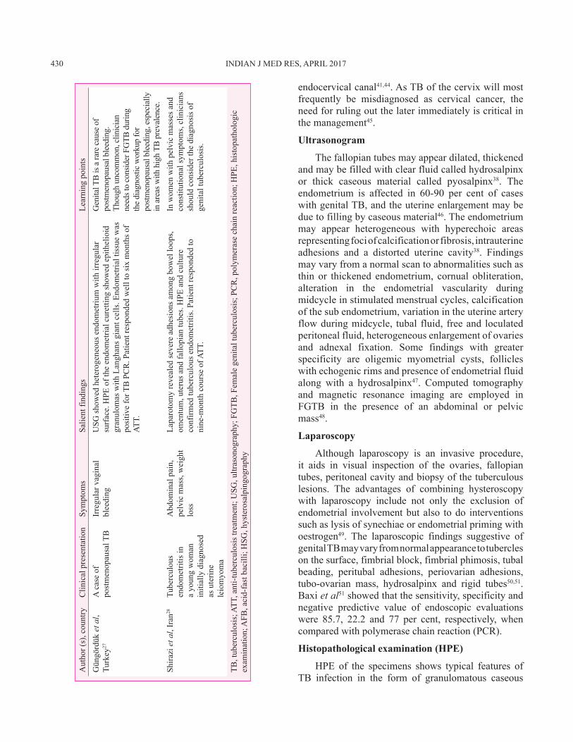
Aut
hor (
s), c
ount
ryC
linic
al p
rese
ntat
ion
Sym
ptom
sSa
lient
find
ings
Lear
ning
poi
nts
Gün
görd
ük e
t al,
Turk
ey27
A c
ase
of
post
men
opau
sal T
BIr
regu
lar v
agin
al
blee
ding
USG
show
ed h
eter
ogen
eous
end
omet
rium
with
irre
gula
r su
rfac
e. H
PE o
f the
end
omet
rial c
uret
ting
show
ed e
pith
elio
id
gran
ulom
as w
ith L
angh
ans g
iant
cel
ls. E
ndom
etria
l tis
sue
was
po
sitiv
e fo
r TB
PC
R. P
atie
nt re
spon
ded
wel
l to
six
mon
ths o
f AT
T.
Gen
ital T
B is
a ra
re c
ause
of
postm
enop
ausa
l ble
edin
g.
Thou
gh u
ncom
mon
, clin
icia
n ne
eds t
o co
nsid
er F
GTB
dur
ing
the
diag
nosti
c w
orku
p fo
r po
stmen
opau
sal b
leed
ing,
esp
ecia
lly
in a
reas
with
hig
h TB
pre
vale
nce.
Shira
zi e
t al,
Iran
28Tu
berc
ulou
s en
dom
etrit
is in
a
youn
g w
oman
in
itial
ly d
iagn
osed
as
ute
rine
leio
myo
ma
Abd
omin
al p
ain,
pe
lvic
mas
s, w
eigh
t lo
ss
Lapa
roto
my
reve
aled
seve
re a
dhes
ions
am
ong
bow
el lo
ops,
omen
tum
, ute
rus a
nd fa
llopi
an tu
bes.
HPE
and
cul
ture
co
nfirm
ed tu
berc
ulou
s end
omet
ritis
. Pat
ient
resp
onde
d to
ni
ne-m
onth
cou
rse
of A
TT.
In w
omen
with
pel
vic
mas
ses a
nd
cons
titut
iona
l sym
ptom
s, cl
inic
ians
sh
ould
con
side
r the
dia
gnos
is o
f ge
nita
l tub
ercu
losi
s.
TB, t
uber
culo
sis;
ATT
, ant
i-tub
ercu
losi
s tre
atm
ent;
USG
, ultr
ason
ogra
phy;
FG
TB, F
emal
e ge
nita
l tub
ercu
losi
s; P
CR
, pol
ymer
ase
chai
n re
actio
n; H
PE, h
isto
path
olog
ic
exam
inat
ion;
AFB
, aci
d-fa
st b
acill
i; H
SG, h
yste
rosa
lpin
gogr
aphy
GRACE et al: FEMALE GENITAL TB 431
lesions. The demonstration of typical caseous granulomas with giant epithelioid cells is suggestive of TB; however, these lesions also appear in fungal infections, syphilis, leprosy, rheumatoid arthritis, systemic lupus erythematous, pneumoconiosis and sarcoidosis51. Mondal52 reported histopathological findings from 110 FGTB patients which included isolated small-to-medium epithelioid cell granulomas in different stages, caseation and rare detection of acid-fast bacilli (AFB). Features of chronic salpingitis include occasional non-caseating granulomas in the early stage and single and/or multiple confluent epithelioid granulomas in the lamina propria in the later stage52. Caseation and AFB may be observed in the tissue sections of Fallopian tubes. In ovarian TB, caseation is rare and granulomas are usually observed in the cortical area of the ovaries53. Epithelioid granulomas may be present in cervical TB and caseation, and AFB is a rare entity in vaginal and vulval TB52. As TB of the cervix is frequently misdiagnosed as carcinoma, it is critical to differentiate both at the earliest45. For maximizing the yield in HPE, specimens should be collected from multiple sites as the infecting organisms are scarce in genital TB54,55, sampling site may not be the infected site and cyclical shedding leads to inadequate granuloma formation in endometrium. Ideal time for endometrial sampling is the late secretory phase of the menstrual cycle16 which is favourable to identify the classic giant cells and tubercles.
Bacteriological evaluation
Acid-fast bacilli (AFB) staining and culture
Definitive diagnosis of TB requires the isolation of TB bacilli. Conventional methods for diagnosis of TB include microscopy and culture. Microscopy for AFB is a rapid test for diagnosis but with variable sensitivity56. Acid-fast [Ziehl–Neelsen (ZN), Kinyon] staining or fluorescent (auramine, rhodamine) staining is generally used. For ZN staining to yield a positive result, a sample should contain 104-106 bacilli/ml. Culture for Mycobacterium is more sensitive and requires 10-100 bacilli/ml of tissue/fluid sample for the diagnostic yield16. Though bacteriologic examination of menstrual blood for smear and culture is recommended by some experts, the sensitivity of these tests is quite low36. For diagnostic tests on menstrual blood, menstrual fluid can be collected from the vagina on the first day of menstruation57. An acid-fast staining of the endometrial curetting is a rapid test and requires 10 organisms per ml for a positive result36.
Culture methods
The diagnosis of TB is confirmed based on the identification of M. tuberculosis in culture. Solid cultures are usually performed on the egg-based Lowenstein–Jensen (LJ) medium or agar-based Middlebrook 7H10 medium, and the liquid culture is performed using automated BACTEC Mycobacterial Growth Indicator Tube 960 (MGIT 960) based on modified Middlebrook 7H9 Broth with an oxygen-sensitive fluorescent detection technology58. The advantages of liquid culture include its sensitivity, identification of Mycobacterium species and ability to perform phenotypic drug susceptibility tests (DSTs) and genotyping for further molecular epidemiology studies. The disadvantage of culture methods is the time needed for the growth of mycobacteria. Liquid cultures require at least 9-10 days for positive results and six weeks for being considered negative and in LJ medium cultures, the minimum time-to-positivity is 4-8 weeks54. Thangappah et al55 showed that, among the 72 infertile women studied, AFB smear positivity and culture positivity were 8.3 and 5.2 per cent, respectively, when endometrial samples were tested. Goel et al59 showed that the positivity in LJ medium and BACTEC for premenstrual samples were 1.83 and 8.8 per cent, respectively.
Molecular methods
Molecular techniques for the detection of TB are increasingly evaluated and used nowadays. The nucleic-acid amplification tests (NAAT) provide results in a few hours. PCR is a rapid molecular method for identification of nucleic acid sequences specific to M. tuberculosis and other mycobacteria in tissue samples of patients with FGTB. PCR assays can detect <10 bacilli/ml including dead bacilli and has a testing time of 8-12 h60. Sensitivity of PCR is higher than culture and histopathology and specificity may be as high as 100 per cent in detecting FGTB30,61-63. Recognition of genes encoding the virulence determinants, targets in genome and expressing factors are currently important biomarkers for the detection of FGTB64. Clinicians should not initiate ATT for patients only on the basis of positive PCR due to high false positivity and should correlate with clinical evidence and laparoscopic findings65.
Serology
The WHO has banned the usage of serological tests in individuals suspected of any form of active TB, regardless of their HIV status66. A retrospective study
432 INDIAN J MED RES, APRIL 2017
by Goel et al59 compared different methods i.e., HPE, smear microscopy, LJ culture, BACTEC culture and PCR-DNA for diagnosing endometrial TB in females with infertility. The study concluded that none of the available tests were sensitive enough to diagnose all cases of genital TB, but conventional methods such as HPE and LJ culture still have an important role in the diagnosis of endometrial TB in resource-limited settings. PCR has higher specificity and sensitivity, faster turnaround time but limited by high false-positive rates. Recently, GeneXpert MTB/RIF assay has been endorsed by the WHO for worldwide application that permits the simultaneous detection of M. tuberculosis and resistance to rifampicin. GeneXpert is a useful diagnostic test for all forms of EPTB and provides results in less than two hours67. Further research is needed in identifying the role of Xpert in the diagnosis of FGTB.
Currently, there are no standard guidelines or algorithm for the diagnosis of FGTB, and extensive research is needed for early diagnosis and appropriate interventions. We suggest an algorithm which can aid the clinicians in the diagnosis of FGTB (Figure).
Differential diagnosis
Differential diagnosis of FGTB varies based on the site of involvement. Tripathy and Sapkal68 have
elaborated a variety of conditions based on the site involved (Table III).
Treatment
Treatment of FGTB is similar to pulmonary TB. The regimen recommendation for many forms of EPTB is mostly not based on evidence from vigorous studies as those for PTB and the duration of treatment for six months though debatable is considered adequate69. In patients with organisms sensitive to first-line drugs, six-month regimen is highly effective69. The WHO treatment guidelines for TB (2010)70 recommend that patients newly diagnosed with TB should receive a regimen containing rifampicin (R) for six months: intensive phase with isoniazid (H), R, ethambutol (E) and pyrazinamide (Z) for a duration of two months followed by continuation phase with HR for four months. Alternative to the daily regimen is that TB patients may receive a daily intensive phase followed by thrice weekly continuation phase [2HRZE/4(HR)3] or thrice weekly dosing throughout therapy [2(HRZE)3/4(HR)3] provided that each dose is directly observed. Retreatment TB patients who default or relapse from their first treatment course may receive 2HRZES/1HRZE/5HRE. According to Standards for TB Care Guidelines for new TB patients, the initial
Suspicion of FGTBUnexplained infertility,Menstrual irregularities,
PID not responding to treatment,Postmenopausal bleeding,Persistent white discharge
phase should consist of two months of HREZ followed by HR for four months71. Studies have reported the usage of ATT for duration of six months consisting of H, R, E, Z for two months, followed by H and R for the subsequent four months for the management of patients with genital TB15,72. There is very limited literature available regarding randomized clinical trials which have investigated the optimal drugs and the duration of treatment for genital TB73.
Patients should be monitored for adverse drug reactions during the course of treatment. Since all the four drugs can cause hepatitis, monitoring of liver function is absolutely necessary70. Good adherence to first-line drugs is essential as irregular drug intake can lead to the development of treatment failure, development of multidrug resistant TB as well as TB recurrence and subsequent complications. The bacteriological confirmation of response to treatment is often not possible due to the difficulty in obtaining follow up samples and lack of follow up guidelines for these patients.
Treatment outcomes in women with genital tuberculosis
Laparoscopic findings such as tubercles, caseous tubercles and encysted ascites may disappear after ATT; however, severe findings such as adhesions may persist74. Post-ATT hysteroscopy findings show significant differences in Grade I adhesions and Grade II-IIa adhesions75. The spontaneous conception rate may vary from 31 to 59 per cent among patients treated with ATT for FGTB with better rates in patients diagnosed and treated earlier. The outcomes of pregnancy may be live birth, spontaneous abortion or ectopic pregnancy72,76.
If the patient fails to conceive spontaneously, assisted reproduction techniques can be considered to increase the conception rate. In a prospective study done in India among patients treated for infertility due to genital TB, the conception rate was reported as 19.2 per cent, with a much lower live birth rate (7.2%)9. In a study by Jindall et al77 among PCR-positive and PCR-negative FGTB patients with infertility, treatment with ATT and additional assisted reproduction techniques showed an overall pregnancy rate of 60 per cent.
Surgery was the treatment of choice before the chemotherapeutic era; however, its role is minimal now except in patients with recurrent pelvic pain, persistent pelvic mass or TB sinus and excessive bleeding78. There may be a role for limited surgical procedures such as drainage from tubo-ovarian abscesses or pyosalpinx followed by ATT for improved treatment outcomes79. Sharma et al80 have reported complications during laparoscopic procedure among patients with genital TB which include an inability to create pneumoperitoneum, inability to visualize the pelvis, excessive bleeding, injury to the bladder and peritonitis. The surgical procedure of choice is total hysterectomy with bilateral salpingo-oophorectomy with proper chemotherapy coverage81. Vaginal hysterectomy in patients with FGTB may be associated with complications such as increased bleeding, peritonitis, bowel injury and flare up in the post-operative period82.
Prevention
Primary prevention of TB includes strategies to minimize the risk of exposure to mycobacteria. It is, therefore, essential to educate patients of pulmonary TB to follow respiratory hygiene at home and in public places and adhere to standard treatment. Specific to genital TB, adopting safe sexual practices may decrease the chances of acquiring genital infection. In countries with high TB burden like India, BCG immunization is used as a preventive strategy. The BCG vaccine is up to 80 per cent effective in preventing the development of severe forms of TB, but its protective effect varies widely in the population83.
Conclusions
Genital TB is a major cause of infertility in women, and prevalence is generally underestimated because of the asymptomatic nature of the infection and diagnostic challenges. Large multicentric studies are needed to estimate the magnitude of FGTB and to identify the most sensitive test for diagnosis. Clinicians
Table III. Differential diagnosis of female genital tuberculosisSite of involvement Differential diagnosisTuberculous salpingitis Pelvic inflammatory disease
Ectopic pregnancyOvarian cystEndometriosisCarcinoma of the colonDiverticulitis
Ovarian tuberculosis Ovarian malignancyCervical tuberculosis Carcinoma of the cervixVulval tuberculosis Elephantiasis vulvaSource: Ref 68
434 INDIAN J MED RES, APRIL 2017
need to be aware of this important cause of infertility and menstrual dysfunction in women. Screening for genital TB needs to be a part of evaluation of infertility and menstrual abnormalities. Most of the patients present in advanced stage with scarring, severe fibrosis and adhesions and treatment outcomes, especially with regard to infertility, are poor. Hence, early diagnosis and correct treatment is vital to avoid complications and to restore fertility.
Conflicts of Interest: None.
References1. WHO. WHO global tuberculosis report 2016. Available
from: http://www.who.int/tb/publications/global_report/en/, accessed on April 27, 2017.
2. Aliyu MH, Aliyu SH, Salihu HM. Female genital tuberculosis: A global review. Int J Fertil Womens Med 2004; 49 : 123-36.
3. Namavar Jahromi B, Parsanezhad ME, Ghane-Shirazi R. Female genital tuberculosis and infertility. Int J Gynaecol Obstet 2001; 75 : 269-72.
4. Golden MP, Vikram HR. Extrapulmonary tuberculosis: An overview. Am Fam Physician 2005; 72 : 1761-8.
5. Sharma JB. Current diagnosis and management of female genital tuberculosis. J Obstet Gynaecol India 2015; 65 : 362-71.
6. Gupta N, Sharma JB, Mittal S, Singh N, Misra R, Kukreja M. Genital tuberculosis in Indian infertility patients. Int J Gynaecol Obstet 2007; 97 : 135-8.
7. Singh N, Sumana G, Mittal S. Genital tuberculosis: A leading cause for infertility in women seeking assisted conception in North India. Arch Gynecol Obstet 2008; 278 : 325-7.
8. Indian Scientists Developing Diagnostic Algorithm for Female Genital TB. Hans India. Available from: http://www.thehansindia.com/posts/index/2014-09-21/Indian-scientists-developing-diagnostic-algorithm-for-female-genital-TB-108475, accessed on March 18, 2016.
9. Tripathy SN, Tripathy SN. Infertility and pregnancy outcome in female genital tuberculosis. Int J Gynaecol Obstet 2002; 76 : 159-63.
10. Jindal UN. An algorithmic approach to female genital tuberculosis causing infertility. Int J Tuberc Lung Dis 2006; 10 : 1045-50.
11. Shaheen R, Subhan F, Tahir F. Epidemiology of genital tuberculosis in infertile population. J Pak Med Assoc 2006; 56 : 306-9.
12. Nadgouda SS, Mukhopadhyaya PN, Acharya A, Nagee A, Kunjadia PD. A study on genital tuberculosis and infertility in Indian population. Clin Pract 2010; 2 : 1.
13. Khanna A, Agrawal A. Markers of genital tuberculosis in infertility. Singapore Med J 2011; 52 : 864-7.
14. Shahzad S. Investigation of the prevalence of female genital tract tuberculosis and its relation to female infertility: An observational analytical study. Iran J Reprod Med 2012; 10 : 581-8.
15. Abdelrub AS, Al Harazi AH, Al-Eryani AA. Genital tuberculosis is common among females with tubal factor infertility: Observational study. Alex J Med 2015; 51 : 321-4.
16. Das P, Ahuja A, Gupta SD. Incidence, etiopathogenesis and pathological aspects of genitourinary tuberculosis in India: A journey revisited. Indian J Urol 2008; 24 : 356-61.
17. Rajamaheswari N. Pelvic tuberculosis. Int J Microbiol 2008; 27 : 361-3.
18. Qureshi RN, Samad S, Hamid R, Lakha SF. Female genital tuberculosis revisited. J Pak Med Assoc 2001; 51 : 16-8.
19. Arakeri SU, Sinkar P. An unusual gross appearance of vulval tuberculosis masquerading as tumor. Case Rep Obstet Gynecol 2014; 2014 : 815401.
20. Sachan R, Patel ML, Gupta P, Verma AK. Genital tuberculosis with variable presentation: A series of three cases. BMJ Case Rep 2012; 2012. pii: Bcr2012006665.
21. Akbulut S, Arikanoglu Z, Basbug M. Tubercular tubo-ovarian cystic mass mimicking acute appendicitis: A case report. J Med Case Rep 2011; 5 : 363.
22. Agrawal S, Madan M, Leekha N, Raghunandan C. A rare case of cervical tuberculosis simulating carcinoma cervix: A case report. Cases J 2009; 2 : 161.
23. Lobo FD, Wong MY. Coexistence of benign ovarian serous cystadenoma and tuberculosis in a young woman. Singapore Med J 2013; 54 : e154-7.
24. Kokkayil P, Rawre J, Malhotra N, Dhawan B. Co-infection of Mycoplasma genitalium and Chlamydia trachomatis in an infertile female patient with genital tuberculosis. Indian J Pathol Microbiol 2013; 56 : 457-9.
25. Neonakis I, Mantadakis E, Gitti Z, Mitrouska I, Manidakis LG, Maraki S, et al. Genital tuberculosis in a tamoxifen-treated postmenopausal woman with breast cancer and bloody vaginal discharge. Ann Clin Microbiol Antimicrob 2006; 5 : 20.
26. Gascón J, Acién P. Large bilateral tubercular pyosalpinx in a young woman with genitourinary malformation: A case report. J Med Case Rep 2014; 8 : 176.
28. Shirazi M, Shahbazi F, Pirzadeh L, Mohammadi SR, Ghaffari P, Eftekhar T. Tuberculosis endometritis presenting as a leiomyoma. Int J Fertil Steril 2015; 8 : 481-4.
29. Zajaczkowski T. Genitourinary tuberculosis: Historical and basic science review: Past and present. Cent European J Urol 2012; 65 : 182-7.
30. Bhanothu V, Theophilus JP, Rozati R. Use of endo-ovarian tissue biopsy and pelvic aspirated fluid for the diagnosis of female genital tuberculosis by conventional versus molecular methods. PLoS One 2014; 9 : e98005.
31. Varma TR. Tuberculosis of the female genital tract. Glob Libr Women’s Med 2008. Available from: http://www.glowm.com/section_view/heading/Tuberculosis%20of%20the%20
GRACE et al: FEMALE GENITAL TB 435
Female%20Genital%20Tract/item/34, accessed on March 18, 2016.
32. Chowdhury NN. Overview of tuberculosis of the female genital tract. J Indian Med Assoc 1996; 94 : 345-6, 361.
33. Bose M. Female genital tract tuberculosis: How long will it elude diagnosis? Indian J Med Res 2011; 134 : 13-4.
34. Hoppe LE, Kettle R, Eisenhut M, Abubakar I; Guideline Development Group. Tuberculosis—diagnosis, management, prevention, and control: Summary of updated NICE guidance BMJ 2016; 352 : h6747.
35. Diagnostic Standards and Classification of Tuberculosis in Adults and Children. This official statement of the American Thoracic Society and the Centers for Disease Control and Prevention was adopted by the ATS Board of Directors, July 1999. This statement was endorsed by the Council of the Infectious Disease Society of America, September 1999. Am J Respir Crit Care Med 2000; 161 (4 Pt 1) : 1376-95.
36. Sengupta VB. Gynaecology for postgraduate and practitioners. India: Elsevier; 2007.
37. Raut VS, Mahashur AA, Sheth SS. The Mantoux test in the diagnosis of genital tuberculosis in women. Int J Gynaecol Obstet 2001; 72 : 165-9.
38. Shah HU, Sannananja B, Baheti AD, Udare AS, Badhe PV. Hysterosalpingography and ultrasonography findings of female genital tuberculosis. Diagn Interv Radiol 2015; 21 : 10-5.
39. Ahmadi F, Zafarani F, Shahrzad G. Hysterosalpingographic appearances of female genital tract tuberculosis: Part I. Fallopian tube. Int J Fertil Steril 2014; 7 : 245-52.
40. Khalaf Y. ABC of subfertility. Tubal subfertility. BMJ 2003; 327 : 610-3.
41. Ahmadi F, Zafarani F, Shahrzad GS. Hysterosalpingographic appearances of female genital tract tuberculosis: Part II: Uterus. Int J Fertil Steril 2014; 8 : 13-20.
42. Merchant SA, Bharati AH, Badhe PB. Female genital tract tuberculosis: A review of hysterosalpingographic appearances part 2-the uterus. J Womens Imaging 2004; 6 : 153.
43. Netter A, Musset R, Lambert A, Salomon Y, Montbazet G. Tuberculous endo-uterine symphysis; an anatomo-clinical and radiologically characteristic syndrome. Gynecol Obstet (Paris) 1955; 54 : 19-36.
44. Farrokh D, Layegh P, Afzalaghaee M, Mohammadi M, Fallah Rastegar Y. Hysterosalpingographic findings in women with genital tuberculosis. Iran J Reprod Med 2015; 13 : 297-304.
45. Danforth WC. Tuberculosis of the cervix. Ann Surg 1937; 106 : 407-12.
46. World Health Organization. Manual of diagnostic ultrasound. Geneva: WHO; 2013.
47. Khurana A, Sahi G. OC14. 04: Ultrasound in female genital tuberculosis: A retrospective series. Ultrasound Obstet Gynecol 2013; 42 : 28.
48. Gatongi DK, Gitau G, Kay V, Ngwenya S, Lafong C, Hasan A. Female genital tuberculosis. Obstet Gynaecol 2005; 7 : 75-9.
49. Schaaf HS, Zumla A. Tuberculosis: A comprehensive clinical reference. St Louis (MO): Elsevier Health Sciences; 2009.
50. Sharma JB, Roy KK, Pushparaj M, Kumar S, Malhotra N, Mittal S. Laparoscopic findings in female genital tuberculosis. Arch Gynecol Obstet 2008; 278 : 359-64.
51. Baxi A, Neema H, Kaushal M, Sahu P, Baxi D. Genital tuberculosis in infertile women: Assessment of endometrial TB PCR results with laparoscopic and hysteroscopic features. J Obstet Gynecol India 2011; 61 : 301-6.
52. Mondal SK. Histopathologic analysis of female genital tuberculosis: A fifteen-year retrospective study of 110 cases in Eastern India. Turk J Pathol 2013; 29 : 41-5.
53. Altchek A, Deligdisch L, Kase N. Diagnosis and management of ovarian disorders. San Diego (CA): Academic Press; 2003.
54. Norbis L, Alagna R, Tortoli E, Codecasa LR, Migliori GB, Cirillo DM. Challenges and perspectives in the diagnosis of extrapulmonary tuberculosis. Expert Rev Anti Infect Ther 2014; 12 : 633-47.
55. Thangappah RBP, Paramasivan CN, Narayanan S. Evaluating PCR, culture & histopathology in the diagnosis of female genital tuberculosis. Indian J Med Res 2011; 134 : 40-6.
56. Kashyap B, Srivastava N, R Kaur I, Jhamb R, K Singh D. Diagnostic dilemma in female genital tuberculosis- staining techniques revisited. Iran J Reprod Med 2013; 11 : 545-50.
57. Simon HB, Weinstein AJ, Pasternak MS, Swartz MN, Kunz LJ. Genitourinary tuberculosis. Clinical features in a general hospital population. Am J Med 1977; 63 : 410-20.
58. Tortoli E, Cichero P, Piersimoni C, Simonetti MT, Gesu G, Nista D. Use of BACTEC MGIT 960 for recovery of mycobacteria from clinical specimens: Multicenter study. J Clin Microbiol 1999; 37 : 3578-82.
59. Goel G, Khatuja R, Radhakrishnan G, Agarwal R, Agarwal S, Kaur I. Role of newer methods of diagnosing genital tuberculosis in infertile women. Indian J Pathol Microbiol 2013; 56 : 155-7.
60. Arora S, Merchant R, Allahbadia GN. Reproductive medicine: Challenges, solutions and breakthroughs. New Delhi: JP Medical Ltd.; 2014.
61. Bhanu NV, Singh UB, Chakraborty M, Suresh N, Arora J, Rana T, et al. Improved diagnostic value of PCR in the diagnosis of female genital tuberculosis leading to infertility. J Med Microbiol 2005; 54 (Pt 10) : 927-31.
62. Shrivastava G, Bajpai T, Bhatambare GS, Patel KB. Genital tuberculosis: Comparative study of the diagnostic modalities. J Hum Reprod Sci 2014; 7 : 30-3.
63. Rana T, Singh UB, Kulshrestha V, Kaushik A, Porwal C, Agarwal N, et al. Utility of reverse transcriptase PCR and DNA-PCR in the diagnosis of female genital tuberculosis. J Med Microbiol 2011; 60 (Pt 4) : 486-91.
64. Bhanothu V, Theophilus J, Lakshmi V, Rozati R, Aiman AV, Rosaline M, et al. Detection of female genital tuberculosis
436 INDIAN J MED RES, APRIL 2017
by using endo-ovarian tissue biopsy. Octa J Biosci 2013; 1 : 98-107.
65. Arora R, Sharma JB. Female genital tuberculosis – A diagnostic and therapeutic challenge. Indian J Tuberc 2014; 61 : 98-102.
66. Steingart KR, Ramsay A, Dowdy DW, Pai M. Serological tests for the diagnosis of active tuberculosis: Relevance for India. Indian J Med Res 2012; 135 : 695-702.
67. Raj A, Singh N, Mehta PK. Gene Xpert MTB/RIF assay: A new hope for extrapulmonary tuberculosis. IOSR J Pharm 2012; 2 : 83-9.
68. Tripathy SN, Sapkal R. Tuberculosis manual for obstetricians & gynecologists. New Delhi: Jaypee Brothers Medical Publisher (P) Ltd.; 2015.
69. Lee JY. Diagnosis and treatment of extrapulmonary tuberculosis. Tuberc Respir Dis (Seoul) 2015; 78 : 47-55.
70. WHO. Guidelines for treatment of tuberculosis. Geneva: WHO; 2010.
71. Standards for TB Care in India : Ministry of Health and Family Welfare. Available from: http://www.tbcindia.nic.in/showfile.php?lid=3061, accessed on March 23, 2016.
72. Kulshrestha V, Kriplani A, Agarwal N, Singh UB, Rana T. Genital tuberculosis among infertile women and fertility outcome after antitubercular therapy. Int J Gynaecol Obstet 2011; 113 : 229-34.
73. Abbara A, Davidson RN; Medscape. Etiology and management of genitourinary tuberculosis. Nat Rev Urol 2011; 8 : 678-88.
74. Sharma JB, Sneha J, Singh UB, Kumar S, Roy KK, Singh N, et al. Comparative study of laparoscopic abdominopelvic and fallopian tube findings before and after antitubercular therapy
in female genital tuberculosis with infertility. J Minim Invasive Gynecol 2016; 23 : 215-22.
75. Bahadur A, Malhotra N, Mittal S, Singh N, Gurunath S. Second-look hysteroscopy after antitubercular treatment in infertile women with genital tuberculosis undergoing in vitro fertilization. Int J Gynaecol Obstet 2010; 108 : 128-31.
76. Naredi N, Talwar P, Narayan N, Rai S, Vardhan S, Panda S. Spontaneous conception following anti-tubercular treatment for sub-fertile women with multiple imaging markers suggesting genital tuberculosis. Fertil Sci Res 2014; 1 : 44-9.
77. Jindal UN, Verma S, Bala Y. Favorable infertility outcomes following anti-tubercular treatment prescribed on the sole basis of a positive polymerase chain reaction test for endometrial tuberculosis. Hum Reprod 2012; 27 : 1368-74.
78. Sutherland AM. Surgical treatment of tuberculosis of the female genital tract. BJOG Int J Obstet Gynaecol 1980; 87 : 610-2.
79. Sharma JB. In vitro fertilization and embryo transfer in female genital tuberculosis. IVF Lite 2015; 2 : 14-25.
80. Sharma JB, Mohanraj P, Roy KK, Jain SK. Increased complication rates associated with laparoscopic surgery among patients with genital tuberculosis. Int J Gynaecol Obstet 2010; 109 : 242-4.
81. Tripathy SN, Tripathy SN. Gynaecological tuberculosis - an update. Indian J Tuberc 1998; 45 : 193-8.
82. Sharma JB, Mohanraj P, Jain SK, Roy KK. Increased complication rates in vaginal hysterectomy in genital tuberculosis. Arch Gynecol Obstet 2011; 283 : 831-5.
83. Botha MH, Van der Merwe FH. Female genital tuberculosis. S Afr Fam Pract 2008; 50 : 12-6.
Reprint requests: Dr Mohan Natrajan, Department of Clinical Research, ICMR- National Institute for Research in Tuberculosis No. 1, Mayor Sathyamoorthy Road, Chetpet, Chennai 600 031, Tamil Nadu, India