7 Genomic Study in β-Thalassemia Saovaros Svasti, Orapan Sripichai, Manit Nuinoon, Pranee Winichagoon and Suthat Fucharoen Thalassemia Research Center, Institute of Molecular Biosciences, Mahidol University, Phutthamonthon, Nakhonpathom Thailand 1. Introduction β-Thalassemia is characterized by the reduced or absent production of β-globin chains in the hemoglobin molecule leading to an excess of α-globin chains. The clinical findings of thalassemia caused by the imbalanced α/non α-globin chains synthesis include many pathological changes of various organs and a lower-than-average life expectancy. Most β- thalassemia major patients who are homozygous or compound heterozygous for β 0 - thalassemia mutations have a severe phenotype of β-thalassemia disease and suffer from chronic anemia and requiring regular blood transfusions. In contrast, β 0 -thalassemia/HbE disease is a very heterogeneous disorder and patients who carry identical β-thalassemia genotypes may present with a remarkable variability in disease severity. Their hemoglobin levels range from 3 to 13 g/dL with an average level of 7.7 g/dL (Fucharoen et al., 1987). Patients with mild to moderate clinical symptoms usually have normal growth development and survive without regular blood transfusions. Whereas severely affected patients have marked anemia, growth retardation, severe bone changes, hepatosplenomegaly and heavy iron overload similar to that of β-thalassemia major patients. However, the reasons for this extraordinary clinical heterogeneity are not fully understood. It has been proposed that there are three levels of genetic control of clinical phenotypes in β- thalassemia: 1) primary modifiers due to heterogeneity of the β-thalassemia alleles, 2) secondary modifiers related to the reduced free α-globin pool or increased γ-globin production in adulthood and 3) tertiary modifiers based on candidate genes that may be involved in several pathological alterations, for example, genes that are related to iron absorption, jaundice, cardiac failure and bone defects. However, the fact that many β- thalassemia/HbE patients who have the same β- and α-globin genotypes still have variable clinical symptoms, suggests that there are still some additional factors which influence the severity of the disease. Recently, genome-wide association studies (GWAS) were performed in order to search for other genetic modifying factors. In the first GWAS, approximately 110,000 gene-based single nucleotide polymorphism (SNPs) were initially screened in pooled DNA and then validated in individual samples (Sherva et al., 2010). The second GWAS was conducted using high density SNP arrays to evaluate approximately 600,000 SNPs in individual samples (Nuinoon et al., 2010). The two SNPs association analyses identified highly significant SNPs located in 3 genes/regions (P-value of 1.00×10 -7 to 1.00×10 - www.intechopen.com
Thalassemia Research Center, Institute of Molecular Biosciences, Mahidol University, Phutthamonthon, Nakhonpathom
Thailand
1. Introduction
β-Thalassemia is characterized by the reduced or absent production of β-globin chains in the
hemoglobin molecule leading to an excess of α-globin chains. The clinical findings of
thalassemia caused by the imbalanced α/non α-globin chains synthesis include many
pathological changes of various organs and a lower-than-average life expectancy. Most β-
thalassemia major patients who are homozygous or compound heterozygous for β0-
thalassemia mutations have a severe phenotype of β-thalassemia disease and suffer from
chronic anemia and requiring regular blood transfusions. In contrast, β0-thalassemia/HbE
disease is a very heterogeneous disorder and patients who carry identical β-thalassemia
genotypes may present with a remarkable variability in disease severity. Their hemoglobin
levels range from 3 to 13 g/dL with an average level of 7.7 g/dL (Fucharoen et al., 1987).
Patients with mild to moderate clinical symptoms usually have normal growth development
and survive without regular blood transfusions. Whereas severely affected patients have
marked anemia, growth retardation, severe bone changes, hepatosplenomegaly and heavy
iron overload similar to that of β-thalassemia major patients.
However, the reasons for this extraordinary clinical heterogeneity are not fully understood.
It has been proposed that there are three levels of genetic control of clinical phenotypes in β-
thalassemia: 1) primary modifiers due to heterogeneity of the β-thalassemia alleles, 2)
secondary modifiers related to the reduced free α-globin pool or increased γ-globin
production in adulthood and 3) tertiary modifiers based on candidate genes that may be
involved in several pathological alterations, for example, genes that are related to iron
absorption, jaundice, cardiac failure and bone defects. However, the fact that many β-
thalassemia/HbE patients who have the same β- and α-globin genotypes still have variable
clinical symptoms, suggests that there are still some additional factors which influence the
severity of the disease. Recently, genome-wide association studies (GWAS) were performed
in order to search for other genetic modifying factors. In the first GWAS, approximately
110,000 gene-based single nucleotide polymorphism (SNPs) were initially screened in
pooled DNA and then validated in individual samples (Sherva et al., 2010). The second
GWAS was conducted using high density SNP arrays to evaluate approximately 600,000
SNPs in individual samples (Nuinoon et al., 2010). The two SNPs association analyses
identified highly significant SNPs located in 3 genes/regions (P-value of 1.00×10⁻⁷ to 1.00×10-
www.intechopen.com
Advances in the Study of Genetic Disorders 150
13): the β-globin gene cluster and olfactory receptor genes upstream of β-globin gene cluster
on chromosome 11p15.5, the HBS1L-MYB intergenic region on chromosome 6q23 and
BCL11A on chromosome 2p15 (Nuinoon et al., 2010; Sherva et al., 2010). The three regions
have been shown to be associated with fetal hemoglobin (HbF) level by genetic linkage and
association studies. Moreover, 101 SNPs on 69 genes also showed association with disease
severity with P-values of 1.00×10⁻⁵ to 1.00×10⁻⁶. These genes have been shown to have less
effect on disease severity as compared to the three mentioned regions. However, their roles
in modification of disease severity need further investigation.
2. Hemoglobinopathies
Hemoglobin (Hb) is the protein of the red blood cells that transports oxygen from the lungs
to tissues, and carbon dioxide from the tissues back to the lungs. It is composed of four
globin chains, two α-like and two β-like globin chains, and each globin contains iron-
coordinated heme moieties. The hemoglobinopathies fall into two distinct groups, the
structural hemoglobin variants and the thalassemias. Hemoglobin variants result from
structural alterations in the globin chain, which are mostly due to single amino acid
substitutions, thereby altering the function of the hemoglobin tetramer. The thalassemias are
characterized by the absence or reduced synthesis of one of the globin chains. Some
mutations also lead to both phenotypes. Hemoglobinopathies are the most common
monogenic disorders and it has been estimated that approximately 5.2% of the world
population are carriers. Around 1.1% of couples worldwide are at risk for having children
with a hemoglobin disorder and 2.7 per 1000 conceptions are affected (Modell & Darlison,
2008).
Thalassemias can be divided according to the globin gene(s) defect into ┙-thalassemia, β-
thalassemia, δβ-thalassemia, γ-thalassemia, δ-thalassemia and εγδβ-thalassemia. However,
the major groups of this inherited disorder are α-thalassemia and β-thalassemia.
2.1 α-Thalassemia
The α-like globin locus is located on the short arm of chromosome 16 in band p13.3. It
includes an embryonic gene (ζ2), two fetal/adult genes (α2 and α1), three pseudogenes
(ψζ1, ψα2, ψα1) and a gene (θ) of unidentified function. α-Thalassemia is classified by a
reduction or absence in synthesis of α-globin chains. Molecular defects of α-thalassemia are
mainly associated with deletions or mutations of one or more of the α-globin genes, of
which now more than 35 known deletional mutations have been discovered (Hardison et al.,
1998). α-Thalassemia 1 (--/αα) occurs from a deletion of the duplicated α-globin genes,
while deletion or mutation of one copy of the duplicated α-globin genes, which produces a
reduced amount of α-globin leads to α-thalassemia 2 (-α/αα). The clinical pathology of the
patients is heterogeneous depending mainly on the number of defective genes.
2.2 β-Thalassemia
The β-globin locus is located on the short arm of chromosome 11 in band p15.5. It includes
an embryonic gene (ε), two fetal genes (Gγ and Aγ), two adult genes (δ and β) and a
pseudogene (ψβ). β-Thalassemia occurs as a consequence of a quantitative reduction of β-
globin chain production. More than 200 point mutations and, rarely deletions, have been
www.intechopen.com
Genomic Study in β-Thalassemia 151
reported (Hardison et al., 1998). These genetic defects lead to a variable reduction in ┚-
globin output ranging from a minimal deficit, mild ┚+-thalassemia alleles, to the complete
absence, β0-thalassemia.
2.3 Hemoglobin E
HbE, the most common Hb variant among Southeast Asian populations, results from a G to
A substitution at codon 26 (GAG to AAG) in exon 1 of the β-globin gene, which substitutes
an amino acid residue from Glu to Lys. This abnormal gene produces a structurally
abnormal hemoglobin which consists of α2βE2-globin chains. Moreover, the abnormal gene
also activates a cryptic 5′ splice site that causes abnormal pre-mRNA splicing. The aberrant
splicing leads to a 16 nucleotide deletion of the 3′ end in exon 1 and creates a new inframe
stop codon (Orkin et al., 1982). This new cryptic splice site competes with the normal doner
splice site, consequently the level of correctly spliced βE-globin mRNA is decreased. As a
result, HbE is synthesized at a reduced rate and thus the βE-globin gene behaves like a mild
form of β+-thalassemia.
3. β-Thalassemia/HbE disease
β-Thalassemia/HbE disease is the most common form of β-thalassemia in many Asian
countries. In Thailand, approximately 3,000 children are born with this condition each year,
and there are some 100,000 patients in the population (Fucharoen & Winichagoon, 2000). It
accounts for over 50% of cases of severe β-thalassemia in Indonesia and Bangladesh and is
also very common in Vietnam, Cambodia, Laos, and Malaysia. It also occurs frequently in
the eastern side of Indian subcontinent, including Sri Lanka and Maldives (Fucharoen &
Winichagoon, 1997).
The major mechanism underlying the pathophysiology of β-thalassemia/HbE is due to the
absence or inadequate β-globin chain production, and can be related to the deleterious
effects of imbalanced globin chain synthesis. The excess α-globin chains in β-thalassemia,
which are highly unstable, precipitate and lead to oxidative damage in developing (causing
dyserythropoiesis) and mature red cells (causing shortened red blood cell survival).
Hemolysis and ineffective erythropoiesis cause anemia in β-thalassemia (Rund &
Rachmilewitz, 2005). Ineffective erythropoiesis leads to the expansion of marrow cavities
and the massive medullar cell proliferation, resulting in skeletal deformities. The erythroid
hyperplasia and ineffective erythropoiesis are responsible for the increased iron absorption,
which together with regular blood transfusions, results in chronic iron overload and death
in these patients.
3.1 Clinical Heterogeneity of β-Thalassemia/HbE disease
β-Thalassemia/HbE is generally classified as thalassemia intermedia. The patients have
inherited a β-thalassemia allele and hemoglobin E, which acts as a mild β+-thalassemia.
However, despite seemingly identical genotypes, β-thalassemia/HbE patients have a
remarkably wide spectrum of clinical phenotypes. Notable are variations in anemia, growth
development, hepatosplenomegaly, and transfusion requirements. The clinical features
range from asymptomatic or mild clinical symptoms with normal growth development and
survival without transfusions, to transfusion-dependent thalassemia major who have
www.intechopen.com
Advances in the Study of Genetic Disorders 152
marked anemia, growth retardation, severe bone changes, hepatosplenomegaly and heavy
iron overload. A study of 802 β-thalassemia/HbE patients showed that hemoglobin levels in
the steady state range from 3 to 13 g/dL with an average level of 7.7 g/dL (Fucharoen et al.,
1987). Interestingly, the discordant severity of anemia ranged from 0 to 8.6 g/dL, while the
distribution of differences of hemoglobin levels in 216 sib pairs from 98 families showed a
remarkable skewness toward the lower values with a mode at 0-0.5 g/dL. This suggests that
multiple genetic factors must be involved in determining the clinical variability (Fucharoen
et al., 1984). The reasons for this extraordinary clinical heterogeneity are not fully
understood.
3.2 β-Thalassemia/HbE disease modifiers
The pathophysiologic change in homozygous β-thalassemia patients is predominantly
determined by the amount of excess α-globin chains. In vitro globin chain synthesis
techniques available in the 1960s revealed that the central mechanism underlying the
pathophysiology of β-thalassemia was related to the excess α-globin chains and the degree
of chain imbalance (Weatherall et al., 1965). Similar to homozygous β-thalassemia, the effect
of imbalanced globin chain synthesis to disease severity of β-thalassemia/HbE patients has
been demonstrated. The in vitro globin chain synthesis showed a significant difference
between α/non α-globin chains ratio of 2.08±0.42 and 2.55±0.34 in mild and severe groups
(P-value<0.005), respectively (Fucharoen et al., 1987). Interestingly, there were overlaps in
the range of α/non α-globin chains ratio of 1.34-2.90 in the mild group and 2.23-3.30 in the
severe group. This suggested that other factors also contributed to the clinical symptoms of
these patients.
The well-characterized or possible genetic modifiers which influence the severity of β-
thalassemia have been classified. As the amount of excess α-globin chains determined the
pathophysiologic change in β-thalassaemia, the factors affecting the excess α-globin pools,
which may lead to variable clinical phenotype of the patients are classified as primary and
secondary modifying factors. While the tertiary modifying factors are genetic factors that do
not affect globin imbalance directly but modify complications of the disease in several
different ways (Thein, 2008).
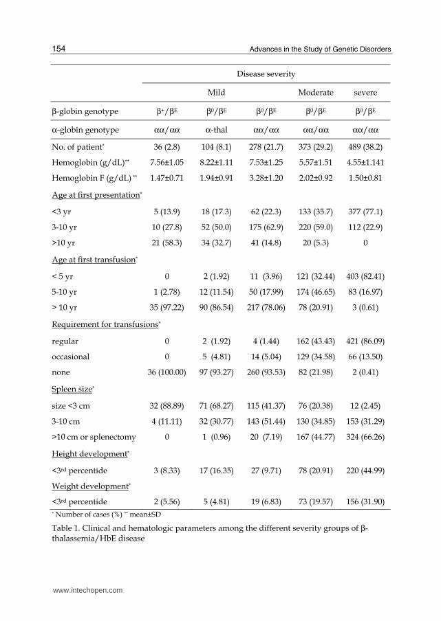
Recently in order to explore disease modifying factors we recruited about 1,300 Thai β0-thalassemia/HbE patients and classified them into three groups, mild, moderate and severe using a scoring system based on criteria of 6 representative parameters; hemoglobin level, age onset, age at the first blood transfusion, requirement of blood transfusion, spleen size or splenectomy, and growth development (Sripichai et al., 2008a). The analysis of known genetic modifiers and genome-wide SNPs association studies (GWAS) were performed using MassARRAY platform (Sequenom, Inc., San Diego, CA, USA) and Illumina Human 610-Quad BeadChips array (Illumina, San Diego, CA, USA) (Nuinoon et al., 2010; Sherva et al., 2010).
3.2.1 Primary modifying factors
The primary modifying factor is described by expression of the β-globin alleles due to the
nature of the underlying β-thalassemia mutation itself. β-Thalassemia mutations range from
null mutations (β0-thalassaemia) that cause a complete absence of β-globin production, to
those that cause a minimal deficit (β+-thalassemia). Generally, interaction of two β+-
www.intechopen.com
Genomic Study in β-Thalassemia 153
thalassemia alleles, in which there is some β-globin production such as the mutations at -28
ATA box (AG), codon 19 (AG), results in a milder disease. A recent study in a Thai β-
thalassemia/HbE cohort showed that all β+-thalassemia/HbE patients had mild clinical
symptoms (Table 1). On the contrary, co-inheritance of a β0-thalassaemia mutation, which
causes a complete absence of β-globin production from the allele, and HbE results in a wide
spectrum of phenotypes.
Since mutation of the βE-globin gene leads to the alternative splicing of βE-globin pre-
mRNA, the amount of alternative spliced βE-globin mRNA may play a role in the variability
of disease severity. Analysis of in vitro globin chain synthesis showed α/βE-globin chains
ratio of 2.69±0.58 and 3.18±0.36 in mild and severe groups respectively (Fucharoen et al.,
1987). A study in the 1990s by the RT-PCR technique showed that aberrantly spliced βE-globin mRNA in patients with severe clinical phenotype was higher than those of mild cases
and the level of aberrantly spliced βE-globin mRNA was correlated with the degree of anemia in the patients (Winichagoon et al., 1995). A recent study using allele specific RT-
qPCR confirmed that there were differences in the alternative splicing of the βE-globin
mRNA among the patients and the correctly/aberrantly spliced βE-globin mRNA ratio in the mild group was higher than that of the severe group (Tubsuwan et al., 2011 ). Excluding
factors correlated to high HbF production, XmnI -158 Gγ-globin, HBS1L–MYB intergenic
region and BCL11A, the alternative splicing of the βE-globin mRNA contributed to at least 7.8% of the mild group.
3.2.2 Secondary modifying factors
The severity of anemia in β-thalassemia reflects the degree of α- to non-α-globin chain
imbalance and the excess of unmatched α-globin chains with all their deleterious effects on the erythroid precursor cells. Therefore secondary modifying factors are described as factors
that affect the degree of globin chain imbalance and the size of the free α-globin chain pool,
either via the co-inheritance of α-globin gene mutations or through genetic determinants which increase the level of HbF production.
┚-Thalassemia patients who co-inherit α-thalassemia will have less excess α-globin chains and tend to have less severe symptoms (Sripichai et al., 2008b; Wainscoat et al., 1983). The
different types of α-thalassemia mutations that predominate in different racial groups display a wide range of severity. The degree of amelioration depends on the number of
functional α-globin genes. None of the patients carrying α-thalassemia were in the severe
group. After exclusion of β+-thalassemia mutations, all patients who co-inherited with α-
thalassemia 1 heterozygote or α-thalassemia 2 homozygote, who have only 2 functional α-globin genes had mild clinical symptoms. About 90% of the patients who have only one
defecteive α-globin gene were in the mild group and 10% were in the moderate group
(Table 2). Co-inheritance of α-thalassemia appears to be a major genetic factor underlying
the mild clinical phenotype of β-thalassemia/HbE. Moreover, a considerable number of
patients, especially those who have co-inheritance of α-thalassemia 1, may have not yet been
diagnosed or not presented to hospitals. The prevalence of 0.5% α-thalassemia 1 and 6.9% α-
thalassemia 2 in the patients was lower as compared to 4% α-thalassemia 1 and 16% α-
thalassemia 2 in Thai population. We suggest that the presence of α-thalassemia genes
should always be considered in apparently mild cases of β0-thalassemia/HbE in Southeast
Asia and probably in other countries where α-thalassemia is also prevalent.
Total 434 (32.6) 389 (29.3) 507 (38.1) 1330 (100.0)
Table 2. The effect of α-globin genotype on β-thalassemia/HbE disease phenotypes
Increased α-globin chains are also synthesized from the α-globin triplicated allele (Higgs et
al., 1980). The excess α-globin chains lead to a greater globin chain imbalance in β-
thalassemia, and hence, the clinical severity of patients is expected to be worsened. The
effect of increased α-globin chains by two extra α-globin genes in homozygous triplicated α-
globin genes (ααα/ααα) or heterozygous quadruplicated α-globin genes (αααα/αα) was
clearly demonstrated in β-thalassemia heterozygotes, which resulted in thalassemia
intermedia (Galanello et al., 1983; Thompson et al., 1989). However, the effect of one extra α-
globin gene on heterozygous triplicated α-globin genes (ααα/αα) in heterozygous β-
thalassemia is more variable and depends on the severity of the β-thalassemia allele
(Camaschella et al., 1997; Traeger-Synodinos et al., 1996). Co-inheritance of triplicated α-
globin genes (ααα/αα) resulting in severe anemia in β-thalassemia patients has been
observed (Camaschella et al., 1995; Galanello et al., 1983; Sripichai et al., 2008a). In this β-
thalassemia/HbE cohort, all patients carrying triplicated α-genes (0.5 % of patients) showed
a severe clinical phenotype (Table 2). However, the triplicated and quadruplicated α-globin
genes occur at a low frequency in most populations. The frequency of triplicated α-globin
genes in Cambodian and Chinese cohorts was about 0.5% of patients (Carnley et al., 2006;
www.intechopen.com
Advances in the Study of Genetic Disorders 156
Ma et al., 2001). A higher frequency (1–3%) was observed in other countries such as
Argentina and Mexico (Bragos et al., 2003; Nava et al., 2006). The quadruplicated α-globin
genes were found at an even lower frequency than the triplicated α-globin genes. In Sri
Lanka the triplicated and quadruplicated α-globin genes frequencies in the patients were
2.0% and 0.2 %, respectively (Fisher et al., 2003).
The HbF level, which is high at birth and decreases thereafter, is an extremely complex
process and still poorly understood. However, the high production of HbF during the
neonatal period protects the newborn from the clinical manifestations of β-thalassemia. The
role of increased HbF as an ameliorating factor was shown in the group of homozygous β-
thalassemia patients who are unable to produce any HbA but showed a mild disease with a
reasonable level of HbF (Thein et al., 1988). Studies in β-thalassemia patients have shown
that increased Hb F production can reduce the ratio of α- to non-α-globins and ameliorate
the severity of the anemia (Rees et al., 1999). The absolute HbF level in mild patients who do
not carry the two known modifying factors (β+-thalassemia or α-thalassemia) was 3.28±1.20
g/dL. This level is significantly higher than that of severe patients, 1.50±0.81 g/dL or mild
patients carrying β+-thalassemia or α-thalassemia, 1.47±0.71 and 1.94±0.91 g/dL,
respectively (Table 1).
The broad distribution of HbF levels in normal adults, in which HbF levels decrease steadily
with age, and in which females have higher levels than males, suggests that more than one
genetic factor may control HbF production. A twin study composed of 264 monozygotic and
511 dizygotic twins showed that approximately 90% of the variations in the level of HbF and
F-cells (HbF containing erythrocytes) production in adult life is genetically controlled by
some factor located near the γ-globin gene, while other factors are present on different
chromosomes (Garner et al., 2000).
It is known that the genetic variant (CT) at position -158 upstream of Gγ-globin gene, XmnI Gγ-globin polymorphism, is associated with high HbF production (Labie et al., 1985a; Thein
et al., 1987). In addition, more than 50% of the genetic variances in levels of F-cells are
caused by factors that are not linked to the β-globin gene cluster (Garner et al., 2000).
Intensive linkage studies have mapped trans-acting quantitative trait loci (QTLs) controlling
F-cell levels to three regions of the genome: chromosome 6q23, Xp22 and 8q11 (Craig et al.,
1996; Dover et al., 1992; Garner et al., 2002). The genetic factors regulating HbF production
are discussed in more detail below.
3.2.3 Tertiary modifying factors
Tertiary modifying factors, while not being involved in hemoglobin synthesis, cause
variation in the progression of the disease. For example, the glucuronosyltransferase 1
(UGT1A) gene was found to be associated with the levels of bilirubin in response to
hemolysis, ineffective erythropoiesis and the incidence of gallstones (Premawardhena et al.,
2001). Similarly, the hemochromatosis (Venter et al.) gene has been identified as influencing
the degree of iron loading (Longo et al., 1999; Rees et al., 1997) and the apolipoprotein E
(APOE) ε4 allele is associated with organ damage and cardiac failure (Economou-Petersen et
al., 1998). Variability of at least three different loci; vitamin D receptor (VDR), type 1
collagen (COLIA1) and transforming growth factor beta 1 (TGFB1), has the potential to
modify the severity of the bone disease, which is particularly common in patients with
severe ┚-thalassemia (Perrotta et al., 2000).
www.intechopen.com
Genomic Study in β-Thalassemia 157
4. General genomic study
Completion of the human genome project (Frazer et al., 2004; Lander et al., 2001; Venter et
al., 2001), dramatically accelerated biomedical research. In parallel, characterization of the
inherited variation in human populations, focusing mainly on DNA variants that are
common in the general population and that confer increases in diseases risk, has led to
exploration of disease susceptibility.
4.1 Genomic variation
Genetic and environmental factors are the two keys that cause human phenotypic variation.
Although more than 99% of human DNA sequences are the same across the population
there is substantial variation in the sequence at many points throughout the genome. DNA
sequence variations are described as mutations and polymorphisms. A mutation is defined
as any change in a DNA sequence to a rare and abnormal variant in comparison to the
normal allele that is prevalent in the population. In contrast, a polymorphism is a DNA
sequence variation that is common in the population. The arbitrary cut-off point between a
mutation and a polymorphism is 1% (Brookes, 1999). Genetic variations are responsible for
individual phenotypic characteristics and impact on how humans respond to diseases,
bacterias, viruses, toxins, chemicals, drugs and other therapies.
Since the early 1980s, humans were known to carry a heterozygous site roughly every 1,300
bases. Genetic maps containing a few thousand markers, which are adequate for
rudimentary linkage mapping of Mendelian diseases, were constructed in the late 1980s and
early 1990s (Lander, 2011; Lander et al., 2001). A map of 1.42 million single nucleotide
polymorphisms (SNPs) distributed throughout the human genome was reported in a
companion to the human genome project (Sachidanandam et al., 2001). Today, the vast
majority of human variants with frequency >5% have been discovered and 95% of
heterozygous SNPs in an individual are represented in current databases. The combination
of marker alleles on a single chromosome is called a haplotype (Haploid Genotype).
Haplotype includes markers that tightly linked with each other and these markers often
display statistical dependence, called linkage disequilibrium (LD). The International
Haplotype Map Project defined the SNPs patterns across the entire genome by genotyping 3
million SNPs (Frazer et al., 2005).
The presence of a specific allele variation can be implicated as a causative factor in human
genetic disorders and a genetic modifying factor in disease phenotypes. Therefore, screening
for such an allele in an individual might enable detection of a genetic predisposition to
disease or particular phenotype. The importance of finding genetic contribution to disease,
therefore, has led to collection of DNA samples from a large number of patients with
particular disorders. Two major approaches have been used to map genetic variants that
influence disease or phenotype: linkage analysis and association studies.
4.2 Linkage studies
Linkage analysis (family-based studies) is based on the principle that a disease locus in a
family will segregate as part of an undisturbed chromosomal region. Genetic markers that
tag a chromosomal region that includes the disease gene will co-segregate with the disease.
In practice, highly variable markers, such as microsatellites that can be distinguished
between most mating couples, have been used to tag regions of the chromosomes (Hauser et
www.intechopen.com
Advances in the Study of Genetic Disorders 158
al., 2004). Families in which sibling pairs are affected with the disorder are typed with
genetic markers to find a polymorphism that is co-inherited with the disease. If the
polymorphic allele is found in a large number of affected sibling pairs, the polymorphism is
probably linked to a gene that confers susceptibility to that disease. The genetic markers and
the disease allele must generally be inherited together within the one or two generations
spanned by the family. To find polymorphisms that are linked to a specific disease requires
the typing of 200–300 families with multiple affected relatives using a set of a few hundred
or a few thousand markers, spaced millions of bases apart along the human genome.
However, the linkages reported by one group have often not been replicated by others.
Failure to replicate the linkages may result from a lack of statistical power or false positive
results in the original study. Furthermore, there might also be different sets of susceptibility
genes operating in different populations. Linkage analysis has been less successful for
polygenic diseases and quantitative traits, perhaps in part because of a limited power to
detect the effect of common alleles with modest effects on disease.
4.3 Association studies
Association studies rely on the retention of adjacent polymorphisms over several
generations across the population. The prevalences of a particular genetic marker or a set of
markers, in affected and unaffected individuals, are compared instead of analysis of large
2005). The “common disease–common variant” hypothesis posited that common genetic
variants could have a role in the etiology of common diseases (Reich & Lander, 2001). The
vast majority of genetic variance in the population is due to common variants. The
susceptibility alleles for a trait will include many common variants unless the alleles have
had a large deleterious effect on reproductive fitness over long periods. For common
diseases or traits, many susceptibility alleles may have been only mildly deleterious, neutral
or even advantageous. By testing all common variants, one could pinpoint key genes and
shed light on underlying mechanisms.
Genome-wide association studies (GWAS) are association studies that survey most of the
genome for causal genetic variants. In GWAS, hundreds of thousand SNPs tested for
association with a disease in hundreds or thousands of persons have revolutionized the
search for genetic influences on complex traits (Manolio, 2010). Such conditions, in contrast
with single-gene disorders, are caused by many genetic and environmental factors working
together, each of which has a relatively small effect and few if any being absolutely required
for the disease to occur. Rapid advances in technology and quality control now permit
affordable, reliable genotyping of up to 1 million SNPs in a single scan of a person’s DNA.
In 2010, nearly 600 genome-wide association studies covering 150 distinct diseases and traits
were published, with nearly 800 SNP–trait associations being reported as significant
(P-value <5×10⁻⁸).
5. Genomic study in thalassemias
Even after exclusion of known genetic modifying factors, the phenotypic severity in the
majority of β-thalassemia/HbE patients still can not be predicted. As mention above, family
studies indicated that multiple genetic factors are involved in determining the clinical
variability and additional genetic modifying factors remain to be discovered. In order to
www.intechopen.com
Genomic Study in β-Thalassemia 159
identify these variants, two GWAS were performed in Thai β-thalassemia/HbE patient
cohorts, from which β- or α-globin mutations that are known to affect disease severity were
excluded. The first GWAS was carried out using a two-stage study design, which allowed cost effective identification of a targeted set of SNPs for individual genotyping. Initially pooled DNA of 197 mild and 198 severe cases was examined (Sherva et al., 2010). Then the SNPs with allele frequency differences between the pooled DNA (P-value < 0.02) were further genotyped in individual samples of 198 mild and 305 severe cases. The assay panel corresponded to 119,811 gene-based SNPs with a median spacing of 10.4 kilobases, covering approximately 99% of all known and predicted human genes. The pooled genotyping was conducted using the MassARRAY platform (Sequenom, Inc., San Diego, CA, USA). The results showed that 50 SNPs were significantly associated with disease severity (P-value <
0.05). Forty-one SNPs in a large linkage disequilibrium (LD) block within the β-globin gene cluster were associated with severe disease, of which the most significant was bthal_bg200 (odds ratio (OR) = 5.56, P-value = 2.6 × 10⁻¹³). The second GWAS was conducted using the Illumina Human 610-Quad BeadChips array
(Illumina, San Diego, CA, USA) with DNAs from 235 mild and 383 severe cases of Thai β0-thalassemia/HbE patients (Nuinoon et al., 2010). The result identified 27 SNPs located in three genes/regions, having P-value of 1.00 x 10⁻⁷ or lower to be associated with the disease severity. The quantile–quantile plot of the observed P-value for association to the disease severity and Manhattan plot are shown in Figure 1. The strongest SNPs associated with the
disease severity were located in the β-globin gene cluster on chromosome 11p15.5 and olfactory receptor genes upstream of ┚-globin gene cluster. The 9 most significant disease severity associated SNPs (P-value <1.00 x 10⁻¹⁰) were located in a large LD spanning over 62 kb containing the locus control region (LCR) of the ┚-globin gene cluster, 4 functional ┚-
globin-like genes (ε-, Gγ-, Aγ- and δ-globin) and 1 pseudogene (ψβ-globin). The most
significantly-associated SNP, rs2071348, was in the ψβ-globin gene (P-value = 2.96 × 10⁻¹³, OR = 4.33 with 95% CI; 2.74-6.84). The second most significant region was mapped to the HBS1L-MYB intergenic region on chromosome 6q23. Five SNPs of this region showed strong
association with disease severity (P-value < 1.00 × 10⁻⁷) and rs9376092 revealed the most
significant association with P-value = 2.36 × 10⁻¹⁰ (OR = 3.07 with 95% CI; 2.16-4.38). The third significant gene is the BCL11A on chromosome 2p15. Four SNPs were significantly associated with the disease severity, with the most significant SNP rs766432 located in
intron 2 (P-value = 5.87 × 10⁻¹⁰, OR = 3.06 with 95% CI; 2.15-4.37). In addition, 101 SNPs on 69
genes also showed association with disease severity with P-values of 1.00 × 10⁻⁵ to 1.00 × 10⁻⁶. These SNPs are located on several genes/regions and can be classified into various biological functions such as cell cycle, cell growth, structural proteins, enzymes, hormones, signaling molecules, genes involved in gene expression, protein degradation, inflammatory response and hypothetical proteins with unknown functions. These genes may have less effect on disease severity compared to the three regions. However, they may play some roles in modification of disease severity and need further investigation. No SNPs in the BCL11A region were typed in the first GWAS, suggested that the pooled genotyping was done on a marker panel that was less dense than the second GWAS and did not have the same level of genome coverage as the high density SNP arrays. However, the low marker density can identify two of the three SNPs highly associated with disease severity identified by the second GWAS, the ┚-globin gene cluster and HBS1L-MYB region.
www.intechopen.com
Advances in the Study of Genetic Disorders 160
Fig. 1. Genome-wide association study scatter plots of P-values (analyzed with disease severity) with chromosome location. The red line denoted the significance threshold (P-value = 1.00 x 10⁻⁷), and the blue line denoted the suggestive P-value (P = 1.00 x 10⁻⁴). Inset shows the quantile–quantile plot of the P-values (allelic association) (Nuinoon et al., 2010). With kind permission from Springer Science+Business Media
Interestingly, the absolute HbF level among mild β0-thalassemia/HbE patients without α-thalassemia co-inheritance was significantly higher than those of the mild cases who were
β+-thalassemia/HbE or having co-inheritance of α-thalassemia and the severe cases (Table 1). The three most significant regions associated with disease severity were also reported to be quantitative trait loci (QTLs) associated with HbF level in the previous genetic linkage and association studies.
6. HbF quantitative trait loci
It is clear from isolated family, linkage and GWAS studies that there are several QTLs for HbF and F-cells. Moreover, in many family studies, the high HbF phenotype segregates
independently of the β-globin gene cluster, implicating the presence of trans-acting factors. The major QTLs for HbF and F-cells that have been extensively studied are located in three
loci, one is in-cis with the β-globin gene cluster and two are in-trans with the β-globin gene cluster, the HBS1L-MYB intergenic region and BCL11A gene.
6.1 β-globin cluster
It has been recognized since the 1980s that the C→T single base substitution at position -158
in the promoter of the Gγ-globin gene is associated with high HbF levels (Gilman &
www.intechopen.com
Genomic Study in β-Thalassemia 161
Huisman, 1985; Labie et al., 1985a). Evidence for the influence of a genetic variant within the
β-globin gene cluster came from families with β-thalassemia or sickle cell anemia which showed a tendency for co-segregation of higher or lower HbF levels with the disease
mutation. The HbS mutation is found on four major β-globin cluster haplotypes (HbS-
Senegal, -Benin, -Bantu, and –Arab/Indian). Carriers for the βS gene on the HbS-Senegal or
HbS-Arab/Indian βS haplotype (including the T allele of XmnI -158 Gγ-globin) have high
HbF levels, and a mild clinical course, whereas carriers of the βS gene on a HbS-Bantu
haplotype (including the C allele of XmnI -158 Gγ-globin) have low HbF levels (Labie et al., 1985b). Despite the long time since the identification of this locus as a major QTL for HbF expression, functional studies have been inconclusive. Identification of the pinpoint causative variant is still unknown, in part because the QTL is located in a large LD block
spanning over 62 kb. A plausible explanation of the genetic variant in β-globin cluster and olfactory receptor genes upstream function is the regulation of globin genes expression and
hemoglobin switching. In erythroblasts, the β-globin locus forms an erythroid-specific spatial structure called the active chromatin hub (ACH). The ACH composed of the HS sites of the LCR, active ┚-globin genes, remote 5’ HS sites (HS-110 in human) and 3’ HS-1 (Fang et al., 2007). A chromatin hub is formed by looping 3’ HS-1, 5’ HS-110, and the 5’ part of the LCR together. The ┚-globin gene locus is surrounded by the genes encoding olfactory receptors that are expressed in olfactory epithelium, but not in erythroid cells (Bulger et al., 1999). The HS-110 and 3’ HS-1 site are located in the olfactory receptor genes (ORGs) cluster.
An RNA FISH study in erythroid cells indicated that intergenic transcription of the β-globin locus occurs over a region of more than 250 kb including several genes in the nearby olfactory receptor gene cluster (Miles et al., 2007).
6.2 HBS1L-MYB intergenic region
Extensive studies over the years of Thein and colleages showed that a quantitative trait locus (QTL) for HbF expression in adults is located in the HBS1L-MYB intergenic region, the so called HBS1L-MYB intergenic polymorphisms (HMIP). A genome-wide linkage analysis study covered seven generations of 210 family members of an Asian-Indian kindred with
heterocellular HPFH, β-thalassemia and α-thalassemia identified QTLs for HbF expression on chromosome 6q23-q24 (Lod score = 12.4) (Craig et al., 1996; Garner et al., 1998). Fine mapping of the 1.5 Mb region of human chromosome 6q23 encompassing the area showed five genes, ALDH8A1, HBS1L, MYB, AHI1 and PDE7B (Close et al., 2004). To narrow down the candidate region, a high-resolution association study was performed in twin pairs of North European origin. Three LD blocks within the 79 kb long HBS1L-MYB intergenic region showed very strong associations with F-cell levels (P-value= 10-75), with the strongest effect in the second block (24 kb) (Thein et al., 2007). The HMIP accounts for about 19% of
the North European population trait variance. In Thai β-thalassemia/HbE, a GWAS study also showed that the most significantly-associated SNP with the HbF level in the HBS1L-MYB region, rs9399137, is also located within the HMIP second block. The HbF expression QTL at HMIP is also reported in healthy individuals of African descent (Creary et al., 2009), sickle cell patients from Tanzania, Brazil, African American (Creary et al., 2009; Lettre et al.,
2008; Makani et al., 2011), β-thalassemia heterozygotes (So et al., 2008) and β-thalassemia patients (Galanello et al., 2009; Nuinoon et al., 2010). The HMIP has been reported to contain distal regulatory elements that generate a key part of the overall control of the MYB expression (Wahlberg et al., 2009). In MYB expressing
www.intechopen.com
Advances in the Study of Genetic Disorders 162
primary human erythroid cells, the core HBS1L-MYB intergenic region harbors several potential cis-regulatory elements for GATA-1 signals that coincided with DNase I hypersensitive sites. HbF expression is linked to the kinetics of erythrocyte maturation and differentiation. MYB plays role in erythroid proliferation and differentiation, and in turn, the control of HbF levels (Jiang et al., 2006). The HMIP was associated not only with the HbF levels but also with the cell numbers of platelets and monocytes in the peripheral blood (Menzel et al., 2007). Recently, a 3-bp deletion, between 135,460,326 and 135,460,328 bp on chromosome 6q23 in HMIP was identified as the functional motif (Farrell et al., 2011).
6.3 BCL11A gene
The association of BCL11A and HbF expression was first identified by two GWAS studies (Menzel et al., 2007; Uda et al., 2008). The first GWAS with the F-cell trait was performed in a European twin cohort, targeting 179 individuals with contrasting extreme F-cell values.
Association analysis identified not only the XmnI -158 Gγ-globin and the chromosome 6 locus but also a new F-cell locus in intron 2 of the oncogene BCL11A on chromosome 2p15 (Menzel et al., 2007). The second GWAS based on HbF of 4,000 individuals from Sardinia
also showed association to the same three loci, the β-globin locus, HMIP and BCL11A, which
was the first replication of the BCL11A locus in patients with SCA and β-thalassemia (Uda et al., 2008). This locus was subsequently replicated in additional SCA patients from the USA
and Brazil (Lettre et al., 2008; Sedgewick et al., 2008), and β-thalassemia heterozygotes from
Hong Kong and the parents of β-thalassemia/HbE patients from Thailand (Nuinoon et al., 2010; Sedgewick et al., 2008). The BCL11A gene encodes several isoforms of a zinc finger transcription factor, the shorter isoforms appeared to be restricted to primitive erythroblasts, and the full-length isoforms to adult-stage erythroblasts (Sankaran et al., 2008). Down-regulation of BCL11A expression in adult human erythroid precursors results in induction of HbF (Sankaran et al., 2008; Wilber et al., 2011). High-resolution chromatin immunoprecipitation (ChIP)–chip analysis and chromosome conformation capture (3C) assay showed that BCL11A binds the upstream
locus control region (LCR), ε-globin and the intergenic regions between γ-globin and δ-
globin genes cooperating with SOX6 that reconfigures the β-globin cluster by modulating
chromosomal loop formation, which finally leads to transcriptional silencing of the γ-globin genes (Xu et al., 2010).
7. Conclusion
Thalassemias are the most common genetic disease in the world resulting from the defective
globin chain synthesis. The main pathophysiologic feature of thalassemia is the
accumulation of unpaired globin chains in erythrocyte precursors and red blood cells (β-
globin in α-thalassemia and α-globin in β-thalassemia). The unmatched globin chains
precipitated in the erythroid precursors in the bone marrow as well as in peripheral
erythrocyte membranes contribute to ineffective erythropoiesis and shortened peripheral
RBC survival resulting in chronic anemia in these patients. As a monogenic disorder,
thalassemia is a very heterogeneous disorder. Patients with identical β-thalassemia
genotypes show a remarkable variability in disease severity, ranging from nearly
asymptomatic (mild disease) to transfusion-dependent anemia with additional
complications (severe disease). Thus, thalassemias are good example of the Mendelian
www.intechopen.com
Genomic Study in β-Thalassemia 163
genetic disease that demonstrate phenotypic variations due to the result of multigene
interactions. Understanding the roles played by genetic factors in diseases will revolutionize
diagnosis, treatment, and prevention, in addition to increase understanding of the
environmental contributions. Three levels of genetic modifiers which influence the severity
of β-thalassemia have been classified into primary, secondary and tertiary modifiers. The
primary modifying factor is described by expression of the β-globin alleles due to the
heterogeneity of the molecular lesions of the underlying the β-thalassemia mutation itself.
Secondary modifying factors are factors that can affect the degree of globin chain imbalance,
which are α-globin genotype and variation in fetal hemoglobin production. Tertiary
modifiers are candidate genes which may be involved in several pathological alterations in
patients. Our recent GWAS in Thai β-thalassemia/HbE patient cohorts suggests that a
number of additional genetic modifier genes may account for the variability in clinical
expression. The three genes/regions that are most significant in β-thalassemia/HbE disease
severity are also associated with HbF expression, namely the β-globin gene cluster and
olfactory receptor genes upstream of β-globin gene cluster on chromosome 11p15.5, the
HBS1L-MYB intergenic region on chromosome 6q23 and the BCL11A gene on chromosome
2p15. It is noteworthy that the HbF expression QTL in the β-globin cluster was discovered
by family and population studies in the 1980s and subsequently validated by genetic
studies. The second QTL, the HBS1L-MYB intergenic region, was discovered by linkage
analysis in extensive kindred in 1990s, and subsequently validated by genetic association
studies. The third QTL, the BCL11A, was discovered in 2000s by GWAS. This shows the
significant development of genomic technologies and knowledge of the human genome. The
rapid development of next-generation sequencing technologies seems likely to accelerate
further genomic studies. The many less significant variants may be identified in near future.
There are several less significant genes compared to the three above mentioned regions,
which may play roles in modification of disease severity. More studies are also needed to
address the effect of these modifying factors.
8. Acknowledgment
This study was supported in part by the Office of the Higher Education Commission and Mahidol University under the National Research University Initiative, Thailand Research Fund and National Science and Technology Development Agency, Thailand.
9. References
Bragos, I. M.; Noguera, N. I., et al. (2003). Triplication (/αααanti3.7) or deletion (-α3.7/)
association in Argentinian β-thalassemic carriers. Ann Hematol, Vol. 82, No. 11, (Nov), pp. 696-698, ISSN 0939-5555
Brookes, A. J. (1999). The essence of SNPs. Gene, Vol. 234, No. 2, (Jul 8), pp. 177-186, ISSN 0378-1119
Bulger, M.; van Doorninck, J. H., et al. (1999). Conservation of sequence and structure
flanking the mouse and human β-globin loci: the β-globin genes are embedded within an array of odorant receptor genes. Proc Natl Acad Sci USA, Vol. 96, No. 9, (Apr 27), pp. 5129-5134, ISSN 0027-8424
www.intechopen.com
Advances in the Study of Genetic Disorders 164
Camaschella, C.; Kattamis, A. C., et al. (1997). Different hematological phenotypes caused by
the interaction of triplicated α-globin genes and heterozygous β-thalassemia. Am J Hematol, Vol. 55, No. 2, (Jun), pp. 83-88, ISSN 0361-8609
Camaschella, C.; Mazza, U., et al. (1995). Genetic interactions in thalassemia intermedia:
analysis of β-mutations, α-genotype, gamma-promoters, and β-LCR hypersensitive sites 2 and 4 in Italian patients. Am J Hematol, Vol. 48, No. 2, (Feb), pp. 82-87, ISSN 0361-8609
Cardon, L. R.&Bell, J. I. (2001). Association study designs for complex diseases. Nat Rev Genet, Vol. 2, No. 2, (Feb), pp. 91-99, ISSN 1061-4036
Carnley, B. P.; Prior, J. F., et al. (2006). The prevalence and molecular basis of hemoglobinopathies in Cambodia. Hemoglobin, Vol. 30, No. 4, pp. 463-470, ISSN 0363-0269
Close, J.; Game, L., et al. (2004). Genome annotation of a 1.5 Mb region of human chromosome 6q23 encompassing a quantitative trait locus for fetal hemoglobin expression in adults. BMC Genomics, Vol. 5, No. 1, (May 31), pp. 33, ISSN 1471-2164
Craig, J. E.; Rochette, J., et al. (1996). Dissecting the loci controlling fetal haemoglobin production on chromosomes 11p and 6q by the regressive approach. Nat Genet, Vol. 12, No. 1, (Jan), pp. 58-64, ISSN 1061-4036
Creary, L. E.; Ulug, P., et al. (2009). Genetic variation on chromosome 6 influences F cell levels in healthy individuals of African descent and HbF levels in sickle cell patients. PLoS One, Vol. 4, No. 1, pp. e4218, ISSN 1932-6203
Dover, G. J.; Smith, K. D., et al. (1992). Fetal hemoglobin levels in sickle cell disease and normal individuals are partially controlled by an X-linked gene located at Xp22.2. Blood, Vol. 80, No. 3, (Aug 1), pp. 816-824, ISSN 0006-4971
Economou-Petersen, E.; Aessopos, A., et al. (1998). Apolipoprotein E epsilon4 allele as a
genetic risk factor for left ventricular failure in homozygous β-thalassemia. Blood, Vol. 92, No. 9, (Nov 1), pp. 3455-3459, ISSN 0006-4971
Fang, X.; Xiang, P., et al. (2007). Cooperativeness of the higher chromatin structure of the β-globin locus revealed by the deletion mutations of DNase I hypersensitive site 3 of the LCR. J Mol Biol, Vol. 365, No. 1, (Jan 5), pp. 31-37, ISSN 0946-2716
Farrell, J. J.; Sherva, R. M., et al. (2011). A 3-bp deletion in the HBS1L-MYB intergenic region on chromosome 6q23 is associated with HbF expression. Blood, Vol 117, No 18, (May 5), pp. 4935-4945, ISSN 0006-4971
Fisher, C. A.; Premawardhena, A., et al. (2003). The molecular basis for the thalassaemias in Sri Lanka. Br J Haematol, Vol. 121, No. 4, (May), pp. 662-671, ISSN 0007-1048
Frazer, K. A.; Ballinger, D. G., et al. (2004). Finishing the euchromatic sequence of the human genome, Nature, Vol 431, No 7011, (Oct 21), pp. 931-945, ISSN 0028-0836.
Frazer, K. A.; Ballinger, D. G., et al. (2005). A haplotype map of the human genome. Nature, Vol. 437, No. 7063, (Oct 27), pp. 1299-1320, ISSN 0028-0836
Fucharoen, S.&Winichagoon, P. (1997). Hemoglobinopathies in Southeast Asia: molecular biology and clinical medicine. Hemoglobin, Vol. 21, No. 4, (Jul), pp. 299-319, ISSN 0363-0269
Fucharoen, S.&Winichagoon, P. (2000). Clinical and hematologic aspects of hemoglobin E β-thalassemia. Curr Opin Hematol, Vol. 7, No. 2, (Mar), pp. 106-112, ISSN 1065-6251
www.intechopen.com
Genomic Study in β-Thalassemia 165
Fucharoen, S.; Winichagoon, P., et al. (1984). Determination for different severity of anemia in thalassemia: concordance and discordance among sib pairs. Am J Med Genet, Vol. 19, No. 1, (Sep), pp. 39-44, ISSN 1552-4825
Fucharoen, S.; Winichagoon, P., et al. (1987). Variable severity of Southeast Asian β0-thalassemia/Hb E disease. Birth Defects Orig Artic Ser, Vol. 23, No. 5A, pp. 241-248, ISSN 1542-0752
Galanello, R.; Ruggeri, R., et al. (1983). A family with segregating triplicated α globin loci
and β-thalassemia. Blood, Vol. 62, No. 5, (Nov), pp. 1035-1040, ISSN 0006-4971
Galanello, R.; Sanna, S., et al. (2009). Amelioration of Sardinian β0-thalassemia by genetic modifiers. Blood, Vol. 114, No. 18, (Oct 29), pp. 3935-3937, ISSN 0006-4971
Garner, C. P.; Tatu, T., et al. (2002). Evidence of genetic interaction between the β-globin complex and chromosome 8q in the expression of fetal hemoglobin. Am J Hum Genet, Vol. 70, No. 3, (Mar), pp. 793-799, ISSN 0002-9297
Garner, C.; Mitchell, J., et al. (1998). Haplotype mapping of a major quantitative-trait locus for fetal hemoglobin production, on chromosome 6q23. Am J Hum Genet, Vol. 62, No. 6, (Jun), pp. 1468-1474, ISSN 0002-9297
Garner, C.; Tatu, T., et al. (2000). Genetic influences on F cells and other hematologic variables: a twin heritability study. Blood, Vol. 95, No. 1, (Jan 1), pp. 342-346, ISSN 0006-4971
Gilman, J. G.&Huisman, T. H. (1985). DNA sequence variation associated with elevated fetal Gγ globin production. Blood, Vol. 66, No. 4, (Oct), pp. 783-787, ISSN 0006-4971
Hardison, R. C.; Chui, D. H., et al. (1998). Access to a syllabus of human hemoglobin variants (1996) via the World Wide Web. Hemoglobin, Vol. 22, No. 2, (Mar), pp. 113-127, ISSN 0363-0269
Hauser, E. R.; Watanabe, R. M., et al. (2004). Ordered subset analysis in genetic linkage mapping of complex traits. Genet Epidemiol, Vol. 27, No. 1, (Jul), pp. 53-63, ISSN 0741-0395
Higgs, D. R.; Old, J. M., et al. (1980). A novel α-globin gene arrangement in man. Nature, Vol. 284, No. 5757, (Apr 17), pp. 632-635, ISSN 0028-0836
Hirschhorn, J. N.&Daly, M. J. (2005). Genome-wide association studies for common diseases and complex traits. Nat Rev Genet, Vol. 6, No. 2, (Feb), pp. 95-108, ISSN 1061-4036
Jiang, J.; Best, S., et al. (2006). cMYB is involved in the regulation of fetal hemoglobin production in adults. Blood, Vol. 108, No. 3, (Aug 1), pp. 1077-1083, ISSN 0006-4971
Labie, D.; Dunda-Belkhodja, O., et al. (1985a). The -158 site 5' to the Gγ gene and Gγ expression. Blood, Vol. 66, No. 6, (Dec), pp. 1463-1465, ISSN 0006-4971
Labie, D.; Pagnier, J., et al. (1985b). Common haplotype dependency of high Gγ-globin gene
expression and high Hb F levels in β-thalassemia and sickle cell anemia patients. Proc Natl Acad Sci USA, Vol. 82, No. 7, (Apr), pp. 2111-2114, ISSN 0027-8424
Lander, E. S. (2011). Initial impact of the sequencing of the human genome. Nature, Vol. 470, No. 7333, (Feb 10), pp. 187-197, ISSN 0028-0836
Lander, E. S.; Linton, L. M., et al. (2001). Initial sequencing and analysis of the human genome. Nature, Vol. 409, No. 6822, (Feb 15), pp. 860-921, ISSN 0028-0836
Lettre, G.; Sankaran, V. G., et al. (2008). DNA polymorphisms at the BCL11A, HBS1L-MYB,
and β-globin loci associate with fetal hemoglobin levels and pain crises in sickle cell disease. Proc Natl Acad Sci U S A, Vol. 105, No. 33, (Aug 19), pp. 11869-11874, ISSN 0027-8424
www.intechopen.com
Advances in the Study of Genetic Disorders 166
Longo, F.; Zecchina, G., et al. (1999). The influence of hemochromatosis mutations on iron overload of thalassemia major. Haematologica, Vol. 84, No. 9, (Sep), pp. 799-803, ISSN 0390-6078
Ma, S. K.; Au, W. Y., et al. (2001). Clinical phenotype of triplicated α-globin genes and
heterozygosity for β0-thalassemia in Chinese subjects. Int J Mol Med, Vol. 8, No. 2, (Aug), pp. 171-175, ISSN 1107-3756
Makani, J.; Menzel, S., et al. (2011). Genetics of fetal hemoglobin in Tanzanian and British patients with sickle cell anemia. Blood, Vol. 117, No. 4, (Jan 27), pp. 1390-1392, ISSN 0006-4971
Manolio, T. A. (2010). Genomewide association studies and assessment of the risk of disease. N Engl J Med, Vol. 363, No. 2, (Jul 8), pp. 166-176, ISSN 0028-4793
Menzel, S.; Jiang, J., et al. (2007). The HBS1L-MYB intergenic region on chromosome 6q23.3 influences erythrocyte, platelet, and monocyte counts in humans. Blood, Vol. 110, No. 10, (Nov 15), pp. 3624-3626, ISSN 0006-4971
Miles, J.; Mitchell, J. A., et al. (2007). Intergenic transcription, cell-cycle and the
developmentally regulated epigenetic profile of the human β-globin locus. PLoS One, Vol. 2, No. 7, pp. e630, ISSN 1932-6203
Modell, B.&Darlison, M. (2008). Global epidemiology of haemoglobin disorders and derived service indicators. Bull World Health Organ, Vol. 86, No. 6, (Jun), pp. 480-487,
Nava, M. P.; Ibarra, B., et al. (2006). Prevalence of -α(3.7) and ααα(anti3.7) alleles in sickle cell
trait and β-thalassemia patients in Mexico. Blood Cells Mol Dis, Vol. 36, No. 2, (Mar-Apr), pp. 255-258, ISSN 1079-9796
Nuinoon, M.; Makarasara, W., et al. (2010). A genome-wide association identified the
common genetic variants influence disease severity in β0-thalassemia/hemoglobin E. Hum Genet, Vol. 127, No. 3, (Mar), pp. 303-314, ISSN 0340-6717
Orkin, S. H.; Kazazian, H. H., Jr., et al. (1982). Abnormal RNA processing due to the exon
mutation of βE-globin gene. Nature, Vol. 300, No. 5894, (Dec 23), pp. 768-769, ISSN 0028-0836
Perrotta, S.; Cappellini, M. D., et al. (2000). Osteoporosis in β-thalassaemia major patients: analysis of the genetic background. Br J Haematol, Vol. 111, No. 2, (Nov), pp. 461-466, ISSN 0007-1048
Premawardhena, A.; Fisher, C. A., et al. (2001). Genetic determinants of jaundice and
gallstones in haemoglobin E β-thalassaemia. Lancet, Vol. 357, No. 9272, (Jun 16), pp. 1945-1946, ISSN 0140-6736
Rees, D. C.; Luo, L. Y., et al. (1997). Nontransfusional iron overload in thalassemia: association with hereditary hemochromatosis. Blood, Vol. 90, No. 8, (Oct 15), pp. 3234-3236, ISSN 0006-4971
Rees, D. C.; Porter, J. B., et al. (1999). Why are hemoglobin F levels increased in HbE/β thalassemia? Blood, Vol. 94, No. 9, (Nov 1), pp. 3199-3204, ISSN 0006-4971
Reich, D. E.&Lander, E. S. (2001). On the allelic spectrum of human disease. Trends Genet, Vol. 17, No. 9, (Sep), pp. 502-510, ISSN 0168-9525
Rund, D.&Rachmilewitz, E. (2005). β-Thalassemia. N Engl J Med, Vol. 353, No. 11, (Sep 15), pp. 1135-1146, ISSN 0028-4793
Sachidanandam, R.; Weissman, D., et al. (2001). A map of human genome sequence variation containing 1.42 million single nucleotide polymorphisms. Nature, Vol. 409, No. 6822, (Feb 15), pp. 928-933, ISSN 0028-0836
www.intechopen.com
Genomic Study in β-Thalassemia 167
Sankaran, V. G.; Menne, T. F., et al. (2008). Human fetal hemoglobin expression is regulated by the developmental stage-specific repressor BCL11A. Science, Vol. 322, No. 5909, (Dec 19), pp. 1839-1842, ISSN 0036-8075
Sedgewick, A. E.; Timofeev, N., et al. (2008). BCL11A is a major HbF quantitative trait locus
in three different populations with β-hemoglobinopathies. Blood Cells Mol Dis, Vol. 41, No. 3, (Nov-Dec), pp. 255-258, ISSN 1079-9796
Sherva, R.; Sripichai, O., et al. (2010). Genetic modifiers of Hb E/β0-thalassemia identified by a two-stage genome-wide association study. BMC Med Genet, Vol. 11, No. pp. 51, ISSN 1471-2350
So, C. C.; Song, Y. Q., et al. (2008). The HBS1L-MYB intergenic region on chromosome 6q23
is a quantitative trait locus controlling fetal haemoglobin level in carriers of β-thalassaemia. J Med Genet, Vol. 45, No. 11, (Nov), pp. 745-751, ISSN 0022-2593
Sripichai, O.; Makarasara, W., et al. (2008a). A scoring system for the classification of β-thalassemia/Hb E disease severity. Am J Hematol, Vol. 83, No. 6, (Jun), pp. 482-484, ISSN 0361-8609
Sripichai, O.; Munkongdee, T., et al. (2008b). Coinheritance of the different copy numbers of
α-globin gene modifies severity of β-thalassemia/HbE disease. Ann Hematol, Vol. 87, No. 5, (May), pp. 375-379, ISSN 0939-5555
Thein, S. L. (2008). Genetic modifiers of the β-haemoglobinopathies. Br J Haematol, Vol. 141, No. 3, (May), pp. 357-366, ISSN 0007-1048
Thein, S. L.; Hesketh, C., et al. (1988). The molecular basis of thalassaemia major and thalassaemia intermedia in Asian Indians: application to prenatal diagnosis. Br J Haematol, Vol. 70, No. 2, (Oct), pp. 225-231, ISSN 0007-1048
Thein, S. L.; Menzel, S., et al. (2007). Intergenic variants of HBS1L-MYB are responsible for a major quantitative trait locus on chromosome 6q23 influencing fetal hemoglobin levels in adults. Proc Natl Acad Sci U S A, Vol. 104, No. 27, (Jul 3), pp. 11346-11351, ISSN 0027-8424
Thein, S. L.; Wainscoat, J. S., et al. (1987). Association of thalassaemia intermedia with a β-globin gene haplotype. Br J Haematol, Vol. 65, No. 3, (Mar), pp. 367-373, ISSN 0007-1048
Thompson, C. C.; Ali, M. A., et al. (1989). The interaction of anti 3.7 type quadruplicated α-
globin genes and heterozygous β-thalassemia. Hemoglobin, Vol. 13, No. 2, pp. 125-135, ISSN 0363-0269
Traeger-Synodinos, J.; Kanavakis, E., et al. (1996). The triplicated α-globin gene locus in β-thalassaemia heterozygotes: clinical, haematological, biosynthetic and molecular studies. Br J Haematol, Vol. 95, No. 3, (Dec), pp. 467-471, ISSN 0007-1048
Tubsuwan, A.; Munkongdee, T., et al. (2011) Molecular analysis of globin genes expression
in different thalassaemia disorders individual variation of βE pre-mRNA splicing determine disease severity. Br J Haematol. Vol. 154, No. 5, (Sep), pp. 635-643, ISSN 0007-1048
Uda, M.; Galanello, R., et al. (2008). Genome-wide association study shows BCL11A associated with persistent fetal hemoglobin and amelioration of the phenotype of
β-thalassemia. Proc Natl Acad Sci U S A, Vol. 105, No. 5, (Feb 5), pp. 1620-1625, ISSN 0027-8424
Venter, J. C.; Adams, M. D., et al. (2001). The sequence of the human genome. Science, Vol. 291, No. 5507, (Feb 16), pp. 1304-1351, ISSN 0036-8075
www.intechopen.com
Advances in the Study of Genetic Disorders 168
Wahlberg, K.; Jiang, J., et al. (2009). The HBS1L-MYB intergenic interval associated with elevated HbF levels shows characteristics of a distal regulatory region in erythroid cells. Blood, Vol. 114, No. 6, (Aug 6), pp. 1254-1262, ISSN 0006-4971
Wainscoat, J. S.; Kanavakis, E., et al. (1983). Thalassaemia intermedia in Cyprus: the
interaction of α and β thalassaemia. Br J Haematol, Vol. 53, No. 3, (Mar), pp. 411-416, ISSN 0007-1048
Weatherall, D. J.; Clegg, J. B., et al. (1965). Globin synthesis in thalassaemia: an in vitro study. Nature, Vol. 208, No. 5015, (Dec 11), pp. 1061-1065, ISSN 0028-0836
Wilber, A.; Hargrove, P. W., et al. (2011). Therapeutic levels of fetal hemoglobin in erythroid
progeny of β-thalassemic CD34+ cells after lentiviral vector-mediated gene transfer. Blood, Vol. 117, No. 10, (Mar 10), pp. 2817-2826, ISSN 0006-4971
Winichagoon, P.; Fucharoen, S., et al. (1995). Role of alternatively spliced βE-globin mRNA
on clinical severity of β-thalassemia/hemoglobin E disease. Southeast Asian J Trop Med Public Health, Vol. 26 Suppl 1, No. pp. 241-245,
Xu, J.; Sankaran, V. G., et al. (2010). Transcriptional silencing of γ-globin by BCL11A involves long-range interactions and cooperation with SOX6. Genes Dev, Vol. 24, No. 8, (Apr 15), pp. 783-798, ISSN 0890-9369
www.intechopen.com
Advances in the Study of Genetic DisordersEdited by Dr. Kenji Ikehara
ISBN 978-953-307-305-7Hard cover, 472 pagesPublisher InTechPublished online 21, November, 2011Published in print edition November, 2011
InTech ChinaUnit 405, Office Block, Hotel Equatorial Shanghai No.65, Yan An Road (West), Shanghai, 200040, China
Phone: +86-21-62489820 Fax: +86-21-62489821
The studies on genetic disorders have been rapidly advancing in recent years as to be able to understand thereasons why genetic disorders are caused. The first Section of this volume provides readers with backgroundand several methodologies for understanding genetic disorders. Genetic defects, diagnoses and treatments ofthe respective unifactorial and multifactorial genetic disorders are reviewed in the second and third Sections.Certainly, it is quite difficult or almost impossible to cure a genetic disorder fundamentally at the present time.However, our knowledge of genetic functions has rapidly accumulated since the double-stranded structure ofDNA was discovered by Watson and Crick in 1956. Therefore, nowadays it is possible to understand thereasons why genetic disorders are caused. It is probable that the knowledge of genetic disorders described inthis book will lead to the discovery of an epoch of new medical treatment and relieve human beings from thegenetic disorders of the future.
How to referenceIn order to correctly reference this scholarly work, feel free to copy and paste the following:
Saovaros Svasti, Orapan Sripichai, Manit Nuinoon, Pranee Winichagoon and Suthat Fucharoen (2011).Genomic Study in β-Thalassemia, Advances in the Study of Genetic Disorders, Dr. Kenji Ikehara (Ed.), ISBN:978-953-307-305-7, InTech, Available from: http://www.intechopen.com/books/advances-in-the-study-of-genetic-disorders/genomic-study-in-thalassemia