2001 7: 499-512 RNA N B Leontis and E Westhof Geometric nomenclature and classification of RNA base pairs. Service Email Alerting click here. right corner of the article or Receive free email alerts when new articles cite this article - sign up in the box at the top http://rnajournal.cshlp.org/subscriptions go to: RNA To subscribe to Cold Spring Harbor Laboratory Press on January 8, 2014 - Published by rnajournal.cshlp.org Downloaded from Cold Spring Harbor Laboratory Press on January 8, 2014 - Published by rnajournal.cshlp.org Downloaded from
Transcript
2001 7: 499-512 RNA N B Leontis and E Westhof Geometric nomenclature and classification of RNA base pairs.
ServiceEmail Alerting
click here.right corner of the article or
Receive free email alerts when new articles cite this article - sign up in the box at the top
http://rnajournal.cshlp.org/subscriptions go to: RNATo subscribe to
Cold Spring Harbor Laboratory Press on January 8, 2014 - Published by rnajournal.cshlp.orgDownloaded from Cold Spring Harbor Laboratory Press on January 8, 2014 - Published by rnajournal.cshlp.orgDownloaded from
Geometric nomenclature and classificationof RNA base pairs
NEOCLES B. LEONTIS 1 and ERIC WESTHOF 2
1Chemistry Department and Center for Biomolecular Sciences, Bowling Green State University,Bowling Green, Ohio 43403, USA
2Institut de Biologie Moléculaire et Cellulaire du Centre National de la Recherche Scientifique,Modélisation et Simulations des Acides Nucléiques, Unité Propre de Recherche 9002,F-67084 Strasbourg Cedex, France
ABSTRACT
Non-Watson–Crick base pairs mediate specific interactions responsible for RNA–RNA self-assembly and RNA–protein recognition. An unambiguous and descriptive nomenclature with well-defined and nonoverlapping param-eters is needed to communicate concisely structural information about RNA base pairs. The definitions should reflectunderlying molecular structures and interactions and, thus, facilitate automated annotation, classification, and com-parison of new RNA structures. We propose a classification based on the observation that the planar edge-to-edge,hydrogen-bonding interactions between RNA bases involve one of three distinct edges: the Watson–Crick edge, theHoogsteen edge, and the Sugar edge (which includes the 2 9-OH and which has also been referred to as the Shallow-groove edge). Bases can interact in either of two orientations with respect to the glycosidic bonds, cis or trans relativeto the hydrogen bonds. This gives rise to 12 basic geometric types with at least two H bonds connecting the bases.For each geometric type, the relative orientations of the strands can be easily deduced. High-resolution examples of11 of the 12 geometries are presently available. Bifurcated pairs, in which a single exocyclic carbonyl or amino groupof one base directly contacts the edge of a second base, and water-inserted pairs, in which single functional groupson each base interact directly, are intermediate between two of the standard geometries. The nomenclature facilitatesthe recognition of isosteric relationships among base pairs within each geometry, and thus facilitates the recognitionof recurrent three-dimensional motifs from comparison of homologous sequences. Graphical conventions are pro-posed for displaying non-Watson–Crick interactions on a secondary structure diagram. The utility of the classificationin homology modeling of RNA tertiary motifs is illustrated.
Nucleic acid bases interact by stacking or by abuttingedge-to-edge+ Whereas stacking interactions providemost of the driving force for folding, the edge-to-edgeinteractions, mediated by hydrogen bonding betweencomplementary arrays of electrically polarized atoms,provide directionality and specificity+ The standard orcanonical Watson–Crick pairs are characterized by theirremarkable isostericity, which gives rise to the regularA-form double helix, and allows each of the four com-
binations to substitute for any of the others withoutdistorting the three-dimensional helical structure+ Thecanonical Watson–Crick pairs, however, represent onlyone of many possible edge-to-edge interactions (Leon-tis & Westhof, 1998c)+ The rapid progress of RNA crys-tallography has revealed a rich variety of base-pairinggeometries (Batey et al+, 1999;Westhof & Fritsch, 2000)+This variety gives rise, in turn, to a multitude of com-plex tertiary structural motifs, as revealed by recentprogress in RNA structural biology (Ferré-D’Amaré &Doudna, 1999; Hermann & Patel, 1999)+
We feel that the growing literature on RNA structuralbiology is hampered by the lack of a systematic no-menclature for base pairing interactions+ Historical, am-biguous, and, sometimes, confusing terms are used(e+g+, “reverse” and “flipped”)+ Frequently, recourse is
Reprint requests to: Neocles B+ Leontis, Chemistry Depart-ment and Center for Biomolecular Sciences, Overman Hall, Bowl-ing Green State University, Bowling Green, Ohio 43403, USA;e-mail: Leontis@bgnet+bgsu+edu, or to Eric Westhof, e-mail:E+Westhof@ibmc+u-strasbg+fr+
made to stating the functional groups involved in theH-bonding interactions, which impedes ready visual-ization of the interactions+ Further, the relationships withthe relative strand orientations are obscured+ We sug-gest that the utility of the recently compiled and ex-haustive database of noncanonical base pairs observedin X-ray and NMR structures, http://prion+bchs+uh+edu/bp_type/, could be significantly enhanced by organizingthe base pairs along geometric principles (Nagaswamyet al+, 2000)+ Indeed, pairwise analysis of hydrogen-bonded, edge-to-edge interactions reveals recurrentgeometric patterns that provide a natural (i+e+, struc-tural) means of classification+ Such a classification canserve to organize the observed pairs into isosteric fam-ilies (Leontis & Westhof, 1998c, 1999) and thus pro-vides for systematic and descriptive nomenclature thatfacilitates prediction of isosteric pairs, necessary stepsin motif recognition in RNA sequences+
The classification that we propose is based on theobservation that, while only about 60% of bases instructured RNAs participate in canonical Watson–Crickbase pairs, the great majority of the remainder partici-pate in some other kind of edge-to-edge interactionswith one or more other bases+ This is borne out in theatomic-resolution structures of the large and small ri-bosomal subunits, the solution of which has expandedour database of RNA structure several-fold (Ban et al+,2000; Schluenzen et al+, 2000; Wimberly et al+, 2000)+The non-Watson–Crick pairs define, in large part, thetertiary structure of an RNA+ Thus, the tertiary struc-ture can be decomposed into a collection of three-dimensional motifs held together by pairwise interactionsthat can be specified simply by indicating the interact-ing edges and the relative orientations of the glycosidicbonds of the two bases+
First it will be shown that there are 12 basic familiesof base pairs+ Examples from each family will be pre-sented and the correspondences between the pro-posed nomenclature and some current usage will bepresented+ Next the default strand orientations for eachbase pair type will be presented with simple rules fortheir visualization, extending previous work (Laveryet al+, 1992; Westhof, 1992)+ Finally, the utility of thenomenclature in summarizing RNA tertiary structure ina two-dimensional format will be illustrated+
Because a nomenclature is fundamentally a workingand networking tool, the adoption of a nomenclature,regardless of its merits, must be the result, in the end,of a consensus agreement between the members of agiven scientific community+ Therefore,we wish to arousediscussions and not controversies+ Informal groups,working on the establishment of conventions useful forRNA research, gather regularly at the annual RNA So-ciety meetings with the coordination of Russ B+ Altman(russ+altman@stanford+edu)+ The proposed nomencla-ture has been presented and discussed at the RNA2000 meeting+
RESULTS AND DISCUSSION
Twelve basic geometric families
RNA purine and pyrimidine bases present three edgesfor H-bonding interactions, as shown in Figure 1 (leftpanel)+ These are the Watson–Crick edge, the Hoogs-teen edge (for purines) or the equivalent “CH” edge (forpyrimidines), and the Sugar edge, so-named becauseit includes the 29-hydroxyl group+ Although “Hoogsteenedge” applies only to purines, it will be used to referalso to the CH edge of pyrimidines, as the atoms in-volved are normally found in the Deep groove of theA-type helix, which corresponds to the Major groove ofB-DNA+ In previous works, the third edge was named“Shallow-groove edge” (Leontis & Westhof, 1998c), be-cause the bases interacting with that edge are locatedin the RNA helix Shallow groove, which is equivalent tothe B-DNA Minor groove+ We thought it was importantto emphasize the distinct and characteristic geometri-cal differences between the two major helices, theB-DNA type and the A-RNA type+ However, with time,the word “minor” as applied to nucleic acid helices hasbeen decoupled from its geometrical meaning+Althoughnames should help memory, they should not conveymistaken meanings (the A-RNA shallow groove is any-thing but “minor,” either regarding function or shape)+The designation “sugar-edge” has the advantage that itmay be applied to B-DNA as well as to A-RNA+
A given edge of one base can potentially interact in aplane with any one of the three edges of a secondbase, and can do so in either the cis or trans orientationof the glycosidic bonds (this nomenclature was usedbefore, see, e+g+, Sundaralingam, 1977)+ The cis andtrans orientations, which follow the usual stereochem-ical meanings, are illustrated in the right panel of Fig-ure 1 for two bases interacting with their Watson–Crickedges+ Thus, 12 distinct edge-to-edge interactions arepossible+ Each pairing geometry is designated by stat-ing the interacting edges of each of the two bases(Watson–Crick, Hoogsteen, or Sugar edge) and therelative glycosidic bond orientation, cis or trans+ Theorder in which the base pairs are listed in Table 1 isdetermined by a historically based priority rule:Watson–Crick edge . Hoogsteen edge . Sugar edge+ The 12base pair geometries are listed in Table 1, with the localstrand orientations in the default anti configurations ofthe bases with respect to the sugars+ Examples takenfrom high-resolution X-ray structures of 11 of the 12basic types are shown in Figures 2 and 3+
When one of the interacting bases occurs in the raresyn configuration of the glycosidic bond, the local strandorientations given in Table 1 are reversed+ Thus, inZ-DNA, the G5C Watson–Crick pair with the G in synpresents a locally parallel orientation of the strands(the O49-atoms of the sugars of the paired bases pointin the same directions), despite the globally antiparallel
500 N.B. Leontis and E. Westhof
Cold Spring Harbor Laboratory Press on January 8, 2014 - Published by rnajournal.cshlp.orgDownloaded from
orientation of the strands+ In the very rare case thatboth bases are syn, the strand orientations revert tothose given in Table 1+ Thus, the proposed system elim-inates the need to speak of “flipped” bases, “reverse”orientations, or to explicitly state the donor and accep-tor atoms+With a mental image of the edges that eachbase of an RNA nucleotide presents for interaction,one can easily visualize and memorize the essentialgeometry of each interaction+ To facilitate the adoptionof the proposed nomenclature, we present in Table 2the correspondence between our nomenclature and thebase-pair designations given in the web-accessible data-base cited above, grouped according to geometric type+
The canonical A-U and G5C pairs belong to the cisWatson–Crick/Watson–Crick (W+C+/W+C+) geometry+Theso-called wobble pairs also belong to this group+ Orig-inally, the term “wobble” designated the pairing be-tween the noncomplementary bases G and U and pairs
FIGURE 1. Left panel: Purine (A or G, indicated by “R”) and pyrimidine (C or U, indicated by “Y”) bases provide three edgesfor interaction, as shown for adenosine and cytosine+ The Watson–Crick edge comprises A(N6)/G(O6), R(N1), A(C2)/G(N2), U(O4)/C(N4), Y(N3), and Y(O2)+ The Hoogsteen edge comprises A(N6)/G(O6), R(N7), U(O4)/C(N4), and Y(C5)+The Sugar-edge comprises A(C2)/G(N2), R(N3), Y(O2), and the ribose hydroxyl group, O29+ Right panel: The cis and transorientations are defined relative to a line drawn parallel to and between the base-to-base hydrogen bonds in the case of twohydrogen bonds or, in the case of three hydrogen bonds, along the middle hydrogen bond+
TABLE 1+ The 12 main families of base pairs between nucleic acidbases together with the local strand orientation (which assumes thatall bases are in the default anti conformation; a syn orientation wouldimply a reversal of orientation; for the global orientation, the stereo-chemistry at the phosphate groups has to be considered)+
involving the modified residue inosine (Crick, 1966)+Such pairs are characterized geometrically by a shift ofone base relative to the other+ We feel the term “wob-ble” should be restricted to those pairs in cis W+C+/W+C+with a shift of the pyrimidine base and should not beextended to trans W+C+/W+C+ pairs even in those caseswhere a shift occurs+ Although wobble pairs often cansubstitute for canonical pairs or constitute intermedi-ates between them, they are not strictly isosteric withthem (nor do they share the property of being self-isosteric)+ That is, a wobble GoU is not isosteric to itsswitched occurrence, UoG+ Likewise, although N1-protonated adenosine forms a pair with cytosine that isisosteric to wobble GoU, the wobble type A(1)oC pairis not isosteric to CoA(1) nor is it isosteric to UoG+Recent reviews of wobble pairs are available (Mas-quida & Westhof, 2000; Varani & McClain, 2000)+
Strand orientations
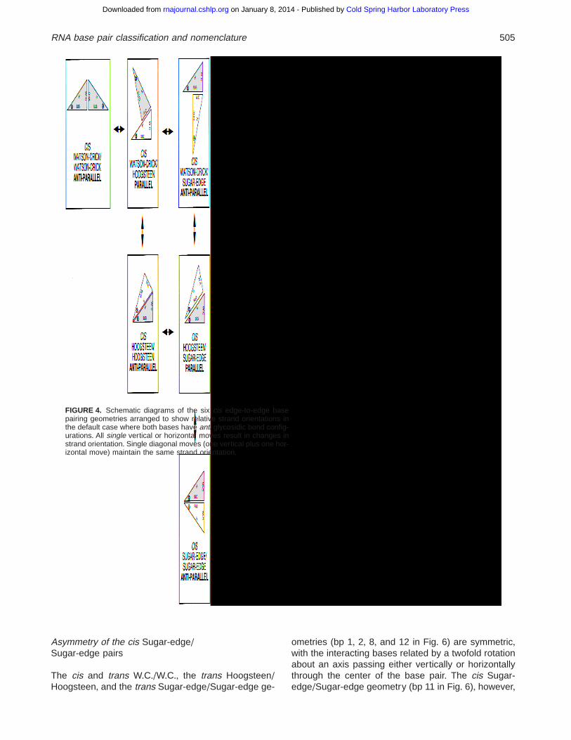
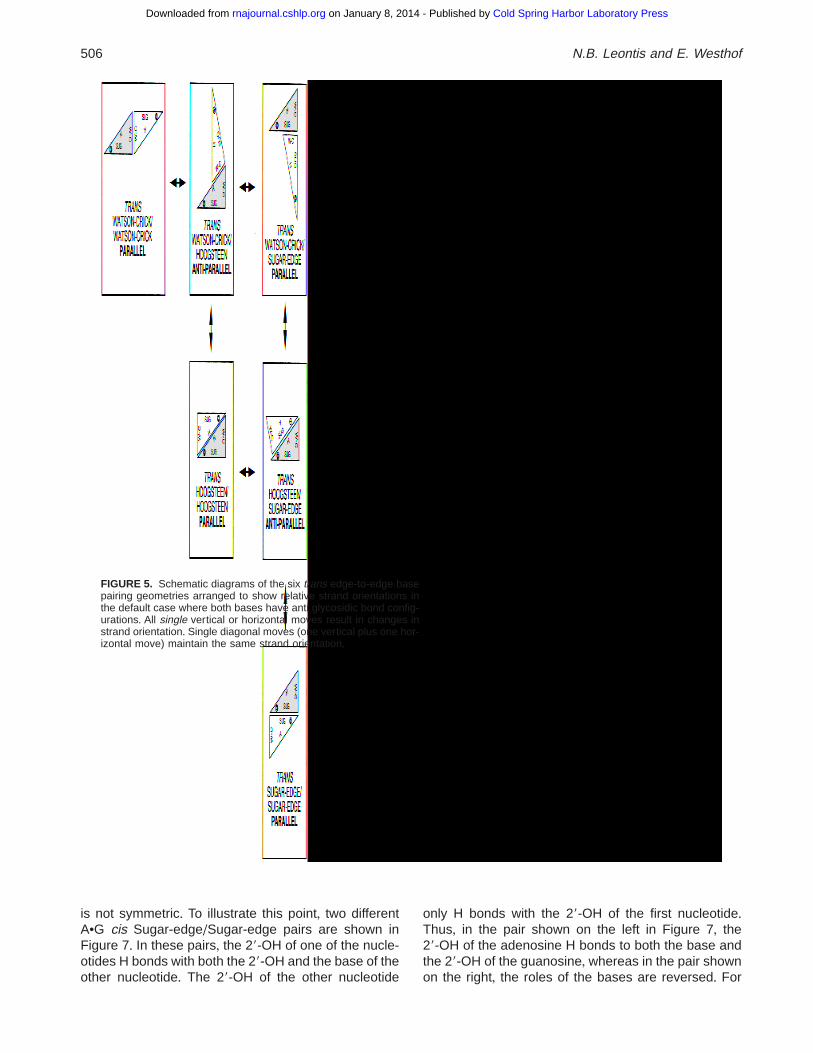
The understanding of RNA folding and architecture, aswell as interactive three-dimensional modeling, re-quires keeping track of the relative orientations of thestrands to which the interacting bases belong+ In Fig-ures 4 and 5, each base-pairing geometry is displayedschematically using two right triangles abutting edge-to-edge+ In each triangle, the sides adjacent to the rightangle represent the Watson–Crick and Sugar edges ofeach base+ The hypotenuse of the triangle representsthe Hoogsteen edge+ A cross or circle in the cornerwhere the Hoogsteen and Sugar edges meet indicatesthe orientation of the sugar-phosphate backbone rela-tive to the plane of the page (59 to 39 or 39 to 59)+ Thesix cis and the six trans edge-to-edge pairing geom-etries are displayed in separate, symmetric 3 3 3 ma-
FIGURE 2. Six possible edge-to-edge base pairing geometries involving Watson–Crick and Hoogsteen edges in all com-binations+ Upper left: Cis Watson–Crick/Watson–Crick A•G NDB file URX053 (Cate et al+, 1996)+ Lower left: Trans Watson–Crick/Watson–Crick G•C (Westhof et al+, 1988)+ Upper center: Cis Watson–Crick/Hoogsteen C(1)•G, NDB file URX053(Cate et al+, 1996)+ Lower center: Trans Watson–Crick/Hoogsteen C(1)•G, UR0004 (Su et al+, 1999)+ Lower left: TransHoogsteen/Hoogsteen A•A, TRNA09 (Westhof et al+, 1988)+ No high resolution example of cis Hoogsteen/Hoogsteen wasidentified (upper right panel)+ Arrows designate Watson–Crick edges available for further interactions with other RNA units,proteins, or small molecules+ The designation of each base pair using the symbols proposed in Figure 6 is also shown+
502 N.B. Leontis and E. Westhof
Cold Spring Harbor Laboratory Press on January 8, 2014 - Published by rnajournal.cshlp.orgDownloaded from
trices+ The elements of each matrix are arranged in theorder Watson–Crick,Hoogsteen, and Sugar edge+ Thus,the W+C+/W+C+ pair is placed in the first row, first columnand the Hoogsteen/Sugar-edge pair in the second row,third column+ In these diagrams, the position and strandorientation of the base on the left is fixed in space+
When arranged in this manner, the base pairs on themain diagonal of each matrix have the same strand ori-entation, antiparallel for cis pairs and parallel for transpairs+ Those in the first diagonal next to the main diag-onal have opposite strand orientations, parallel for cispairs and antiparallel for trans pairs+ The strand orien-tation of the corner element (first row, third column) re-verts to that of the main diagonal+ Thus, any purelyhorizontal or vertical move in the table, correspondingto the change of one edge while retaining the cis or transgeometry, changes the strand orientation, whereas anydiagonal move retains the strand orientation+
Annotation of two-dimensional diagrams
It is desirable to present the non-Watson–Crick pairs ofan RNA molecule on a standard two-dimensional draw-ing+ This helps to recognize and to communicate suc-cinctly in a visually accessible manner the essentialfeatures of a motif+ This, in turn, facilitates recognitionof shared three-dimensional tertiary motifs and fold-ings+ Such diagrams should show, in addition to theclassical secondary structure (contiguous canonicalpairs forming A-form double-stranded helices main-tained by Watson–Crick and wobble pairs), all non-Watson–Crick pairs, all points in the covalent chain atwhich the strand polarity reverses direction, and keybase-stacking interactions, to the degree possible with-out overly cluttering the picture+ As is usually done,nucleotides should be numbered sequentially (59 to 39)to aid in tracing the covalent chain+ Nucleotides are
FIGURE 3. Six possible edge-to-edge base pairing geometries involving the Sugar edge in all combinations+ Upper left: CisWatson–Crick/Sugar Edge A•A, NDB file TRN007 (Westhof et al+, 1988)+ Lower left: Trans Watson–Crick/Sugar Edge A•G,NDB file UR0004 (Su et al+, 1999)+ Upper center: Cis Hoogsteen/Sugar Edge A•A, URX053 (Cate et al+, 1996)+ Lower center:Trans Hoogsteen/Sugar Edge A•G, URL064 (Correll et al+, 1997)+ Upper right: Cis Sugar Edge/Sugar Edge A•G,URX053(Cate et al+, 1996)+ Lower right: Trans Sugar Edge/Sugar Edge A•G UR0004 (Su et al+, 1999)+ As in Figure 2, arrowsdesignate Watson–Crick edges available for further interactions with other RNA units, proteins, or small molecules and thesymbolic designation of each base pair according to Figure 6 is also shown+
RNA base pair classification and nomenclature 503
Cold Spring Harbor Laboratory Press on January 8, 2014 - Published by rnajournal.cshlp.orgDownloaded from
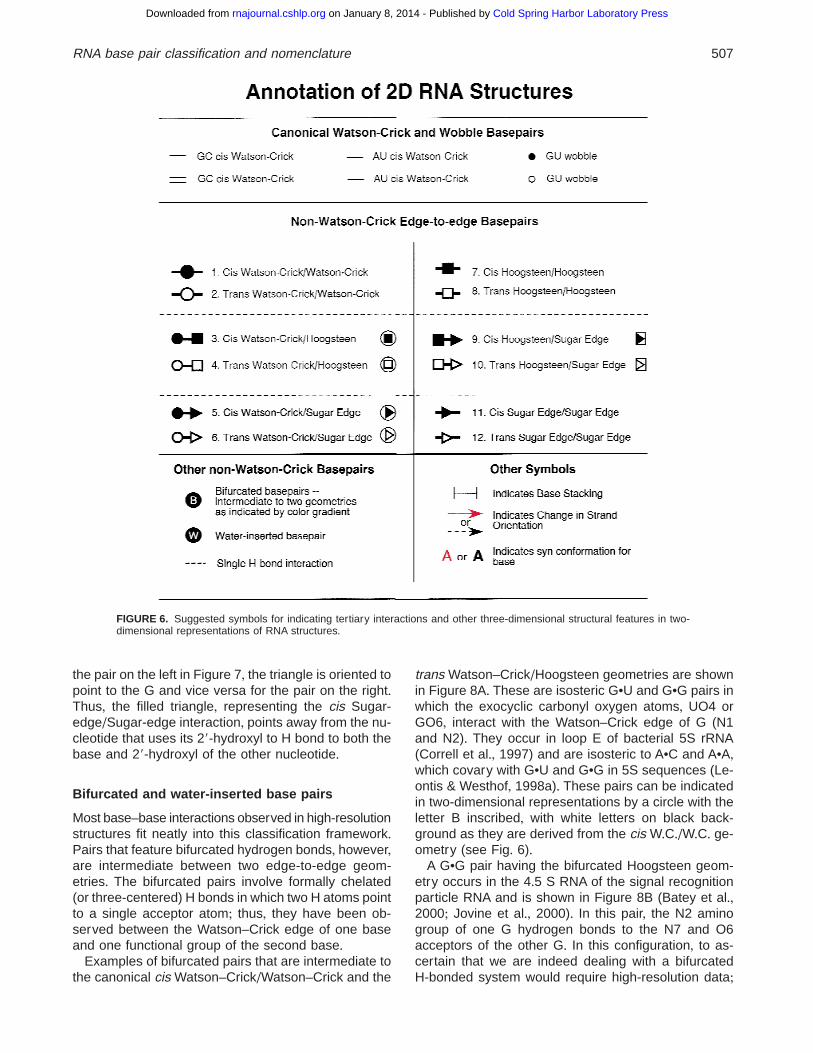
indicated by single, black, capital letters (A, G, C, or U)as usual, except when the base adopts a syn confor-mation about the glycosidic bond, in which case theletter could be printed either bold or colored red+ A redor dotted arrow may be drawn to indicate that a changein strand polarity occurs between two nucleotides+ Todesignate canonical Watson–Crick and wobble pairs,one could use the symbols “–” for both AU and GCpairs and “d” for the wobble GU pair (Damberger &Gutell, 1994), but the convention “–” for AU pairs, “5”for GC pairs, and “C” for GU wobble pairs is moreexplicit (Michel et al+, 1982) and allows the use of “d”as a generic designation for non-Watson–Crick pairs intext+ Both conventions are noted in Figure 6+
Finally, we suggest a set of black-and-white symbolsto accurately specify each kind of non-Watson–Crickedge-to-edge pairing interaction on a secondary struc-ture diagram+ We propose three symbols: circles forWatson–Crick edges, squares for Hoogsteen edges,and triangles for Sugar edges+ The cis and trans ori-entations can be distinguished by filled and open sym-
bols, respectively+When the same edge is used by thetwo bases, only one symbol is necessary (bp 1, 2, 7, 8,11, and 12 in Fig+ 6)+
When an interaction involves two different edges, it isnecessary to designate which edge corresponds towhich base+ For example, “AG cis Watson–Crick/Hoogsteen” designates a pair in which the Watson–Crick edge of the A interacts with the Hoogsteen edgeof the G+ To distinguish the XY and YX pairs in suchcases in two-dimensional diagrams, we suggest usinga horizontal line connecting the two symbols corre-sponding to the two interacting edges, as shown inFigure 6, for bp 3, 4, 5, 6, 9, and 10+ In some situationsit may be desirable to use a more compact symbol todesignate an interaction+ Thus, for each non-Watson–Crick pair we also propose compact symbols consist-ing of the symbol for one edge inside of the symbol forthe other+ The inner symbol is filled or open to desig-nate cis and trans+ A vertical line may be placed adja-cent to the base interacting with the higher priority edge,following the convention discussed above+
TABLE 2+ Correspondence of proposed names to the numbering of Saenger (1984) and the nomenclature used in a recentcompilation (Nagaswamy et al+, 2000)+
Proposed nomenclature Saenger Recent designation
1+ Cis Watson–Crick/Watson–Crick G•A cis W+C+/W+C+ VIII GA IminoC•C cis W+C+/W+C+ (wobble) CC N3(1)-carbonyl, amino-N3G•U cis W+C+/W+C+ (wobble) XXVIIIU•C cis W+C+/W+C+ XVIII UC 4-carbonyl-aminoU•U cis (wobble) W+C+/W+C+ XVI UU imino-carbonyl
2+ Trans Watson–Crick/Watson–Crick A•U trans W+C+/W+C+ XXI AU Reverse Watson–CrickA•A trans W+C+/W+C+ I AA N1-amino, symmetricG•G trans W+C+/W+C+ III GG N1-carbonyl, symmetricG•C trans W+C+/W+C+ XXII GC Reverse Watson–CrickA•C trans W+C+/W+C+ XXVI AC Reverse WobbleG•U trans W+C+/W+C+ XXVII GU Reverse WobbleU•C trans W+C+/W+C+ XVIIC•C trans W+C+/W+C+ XIV, XVU•U trans W+C+/W+C+ XII, XIII UU 4(2)-carbonyl-imino, symmetric
3+ Cis Watson–Crick/Hoogsteen G•G cis W+C+/Hoogsteen VI GG N1-carbonyl, N7-aminoU•A cis W+C+/Hoogsteen XXIII AU HoogsteenG•A cis W+C+/Hoogsteen IX GA N1-N7, carbonyl-aminoA1•G cis W+C+/Hoogsteen GA1 carbonyl-amino, N7-N1
4+ Trans Watson–Crick/Hoogsteen A•A trans W+C+/Hoogsteen V AA N7-aminoG•G trans W+C+/Hoogsteen VII GG N7-iminoU•A trans W+C+/Hoogsteen XXIV AU Reverse HoogsteenC•A trans W+C+/Hoogsteen XXV AC Reverse Hoogsteen
5+ Cis Watson–Crick/Sugar-edge A•G cis W+C+/Sugar-edge GA N3-amino (1 bond)A•U cis W+C+/Sugar-edge AU amino-2-carbonyl
6+ Trans Watson–Crick/Sugar-edge A•G trans W+C+/Sugar-edge X GA N3-amino, amino-N1C•G trans W+C+/Sugar-edge GC N3-amino, amino-N3
7+ Cis Hoogsteen/Hoogsteen
8+ Trans Hoogsteen/Hoogsteen A•A trans Hoogsteen/Hoogsteen II AA N7-amino, symmetric
10+ Trans Hoogsteen/Sugar-edge A•G trans Hoogsteen/Sugar-edge XI GA ShearedA•A trans Hoogsteen/Sugar-edge AA N3-aminoC•U trans Hoogsteen/Sugar-edge UC 2-carbonyl-amino (1 bond)
12+ Trans Sugar-edge/Sugar-edge G•G trans Sugar-edge/Sugar-edge IV GG N3-amino, symmetric
504 N.B. Leontis and E. Westhof
Cold Spring Harbor Laboratory Press on January 8, 2014 - Published by rnajournal.cshlp.orgDownloaded from
The cis and trans W+C+/W+C+, the trans Hoogsteen/Hoogsteen, and the trans Sugar-edge/Sugar-edge ge-
ometries (bp 1, 2, 8, and 12 in Fig+ 6) are symmetric,with the interacting bases related by a twofold rotationabout an axis passing either vertically or horizontallythrough the center of the base pair+ The cis Sugar-edge/Sugar-edge geometry (bp 11 in Fig+ 6), however,
FIGURE 4. Schematic diagrams of the six cis edge-to-edge basepairing geometries arranged to show relative strand orientations inthe default case where both bases have anti glycosidic bond config-urations+ All single vertical or horizontal moves result in changes instrand orientation+ Single diagonal moves (one vertical plus one hor-izontal move) maintain the same strand orientation+
RNA base pair classification and nomenclature 505
Cold Spring Harbor Laboratory Press on January 8, 2014 - Published by rnajournal.cshlp.orgDownloaded from
is not symmetric+ To illustrate this point, two differentA•G cis Sugar-edge/Sugar-edge pairs are shown inFigure 7+ In these pairs, the 29-OH of one of the nucle-otides H bonds with both the 29-OH and the base of theother nucleotide+ The 29-OH of the other nucleotide
only H bonds with the 29-OH of the first nucleotide+Thus, in the pair shown on the left in Figure 7, the29-OH of the adenosine H bonds to both the base andthe 29-OH of the guanosine, whereas in the pair shownon the right, the roles of the bases are reversed+ For
FIGURE 5. Schematic diagrams of the six trans edge-to-edge basepairing geometries arranged to show relative strand orientations inthe default case where both bases have anti glycosidic bond config-urations+ All single vertical or horizontal moves result in changes instrand orientation+ Single diagonal moves (one vertical plus one hor-izontal move) maintain the same strand orientation+
506 N.B. Leontis and E. Westhof
Cold Spring Harbor Laboratory Press on January 8, 2014 - Published by rnajournal.cshlp.orgDownloaded from
the pair on the left in Figure 7, the triangle is oriented topoint to the G and vice versa for the pair on the right+Thus, the filled triangle, representing the cis Sugar-edge/Sugar-edge interaction, points away from the nu-cleotide that uses its 29-hydroxyl to H bond to both thebase and 29-hydroxyl of the other nucleotide+
Bifurcated and water-inserted base pairs
Most base–base interactions observed in high-resolutionstructures fit neatly into this classification framework+Pairs that feature bifurcated hydrogen bonds, however,are intermediate between two edge-to-edge geom-etries+ The bifurcated pairs involve formally chelated(or three-centered) H bonds in which two H atoms pointto a single acceptor atom; thus, they have been ob-served between the Watson–Crick edge of one baseand one functional group of the second base+
Examples of bifurcated pairs that are intermediate tothe canonical cis Watson–Crick/Watson–Crick and the
trans Watson–Crick/Hoogsteen geometries are shownin Figure 8A+ These are isosteric G•U and G•G pairs inwhich the exocyclic carbonyl oxygen atoms, UO4 orGO6, interact with the Watson–Crick edge of G (N1and N2)+ They occur in loop E of bacterial 5S rRNA(Correll et al+, 1997) and are isosteric to A•C and A•A,which covary with G•U and G•G in 5S sequences (Le-ontis & Westhof, 1998a)+ These pairs can be indicatedin two-dimensional representations by a circle with theletter B inscribed, with white letters on black back-ground as they are derived from the cis W+C+/W+C+ ge-ometry (see Fig+ 6)+
A G•G pair having the bifurcated Hoogsteen geom-etry occurs in the 4+5 S RNA of the signal recognitionparticle RNA and is shown in Figure 8B (Batey et al+,2000; Jovine et al+, 2000)+ In this pair, the N2 aminogroup of one G hydrogen bonds to the N7 and O6acceptors of the other G+ In this configuration, to as-certain that we are indeed dealing with a bifurcatedH-bonded system would require high-resolution data;
FIGURE 6. Suggested symbols for indicating tertiary interactions and other three-dimensional structural features in two-dimensional representations of RNA structures+
RNA base pair classification and nomenclature 507
Cold Spring Harbor Laboratory Press on January 8, 2014 - Published by rnajournal.cshlp.orgDownloaded from
therefore, by analogy and to indicate that the pair in-volves unusual geometries we suggest extending theuse of “bifurcated+” This pair is intermediate between
the trans W+C+/Hoogsteen and the trans Sugar-edge/Hoogsteen geometries and is therefore designated byadding a B to the symbol for trans W+C+/Hoogsteen+
FIGURE 7. Two different A•G cis Sugar-Edge/Sugar-edge pairs+ The triangle points from the nucleotide having the 29-OHthat H bonds to both the base and 29-OH of the other nucleotide+ This nucleotide is A23 in the pair from 1F27+PDB (left panel)and G110 in the pair from URX053 (right panel)+
FIGURE 8. Examples of bifurcated pairs+ A: Bifurcated G•G andG•U pairs (G76•G100 and G102•U74 from URL064) intermediate tocis W+C+/W+C+ and trans W+C+/Hoogsteen geometry (Correll et al+,1997)+ B: Bifurcated G•G pair (G162•G149 from PR0021) intermedi-ate to the trans W+C+/Hoogsteen and the trans Sugar-edge/Hoogsteengeometries (Batey et al+, 2000)+
508 N.B. Leontis and E. Westhof
Cold Spring Harbor Laboratory Press on January 8, 2014 - Published by rnajournal.cshlp.orgDownloaded from
Water-inserted pairs have been observed in severalhigh-resolution structures, as recently reviewed (Leon-tis & Westhof, 1998c)+ They often result from an open-ing of a regular type geometry by a rotation of one basewith respect to the other and insertion of one watermolecule (see, e+g+, Fig+ 3 of Westhof & Fritsch, 2000)+We propose that these be designated using the letterW inscribed white on black or black on white depend-ing on whether the interaction is cis or trans (see Fig+ 6)+Pairs in which the inserted water molecule replaces ahydrogen bond in a cis pair are designated cis andlikewise for trans+
Examples of two-dimensionalrepresentations of RNA tertiary structure
To illustrate these conventions, we present in Figure 9examples of two-dimensional representations of RNAmotifs with tertiary interactions added+ The left panelshows the loop E of bacterial 5S rRNA from NDB fileURL064 (Correll et al+, 1997)+ All bases of this sym-metric “internal loop,” in fact, are paired+ A104•G72comprise a trans Hoogsteen/Sugar-edge pair+ This isdesignated using an open symbol (indicating the transgeometry) comprising a square, placed next to A104(for the Hoogsteen edge), connected to a triangle, placednext to G72 (for the Sugar-edge)+ The same interactionoccurs between A78 and G98, but the orientation isreversed, with the Hoogsteen base, A78, on the right+The symbols we propose make these relationships im-mediately clear+U103•A73 and U77•A99 are trans W+C+/Hoogsteen pairs, and are indicated by open symbolscomprising circles (placed next to the Us) connected tosquares (placed next to the As)+ In the U103•A73 pair,the Watson–Crick base (U103) occurs on the left,
whereas the situation is reversed for the U77•A99 pair+G102•U74 and G76•G100 are isosteric cis bifurcatedpairs, intermediate between the cis W+C+/W+C+ and thetrans W+C+/Hoogsteen geometry+ These interactions areindicated by black circles with white B inscribed+A101•G75 is a water-inserted cis W+C+/W+C+ pair+ Thus,it is designated by a black circle with a W super-imposed+ This representation reveals that the bacterialloop E motif in fact comprises two isosteric submotifsoriented in opposite (palindromic) directions+
Sarcin/ricin motif from large ribosomal subunit
The next example (middle panel, Fig+ 9) is the highlyconserved sarcin/ricin motif (Leontis & Westhof, 1998b)+This motif also occurs in loop E of eukaryal 5S rRNAand should not be confused with bacterial loop E+ Thesequence shown is that of rat 28S rRNA, NDB fileUR0002 (Correll et al+, 1998)+ The structure comprisesa GAGA hairpin loop (not shown) and an asymmetric“internal loop+” The dotted arrows between C8 and A9and between A9 and G10 indicate the local strand re-versal that occurs at A9+ The positioning of A9 beneathU11 indicates the stacking between these two resi-dues+ The “bulged” base, G10, is actually hydrogenbonded to U11 and lies in the same plane as theU11•A20 trans W+C+/Hoogsteen pair+ This is indicatedby placing all three bases at the same horizontal levelon the page+ The G10•U11 pair is cis Sugar-edge/Hoogsteen whereas the G19•A12 and U7•C23 pairsare trans Sugar-edge/Hoogsteen+
Domain IV of SRP 4.5S RNA
The SRP motif has been observed as the RNA alone(Jovine et al+, 2000) and in complex to SRP protein 54
FIGURE 9. Left panel: two-dimensional representation of the tertiary structure of loop E of bacterial 5S rRNA (NDB fileURL064)+ Center: two-dimensional representation of the tertiary structure of the sarcin/ricin (S-turn) motif of bacterial 23SrRNA (NDB file UR0002)+ Right panel: two-dimensional representation of the tertiary structure of the internal loop ofDomain IV of the SRP 4+5 S RNA (NDB files PR0021 and UR0009)+
RNA base pair classification and nomenclature 509
Cold Spring Harbor Laboratory Press on January 8, 2014 - Published by rnajournal.cshlp.orgDownloaded from
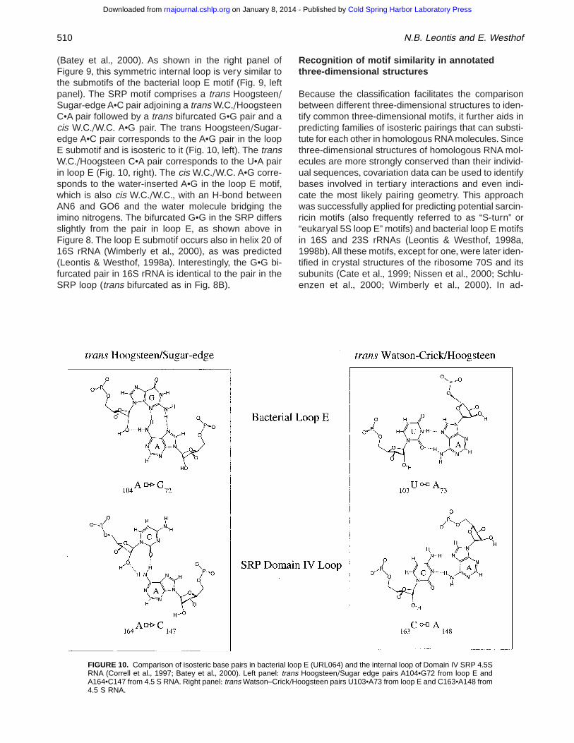
(Batey et al+, 2000)+ As shown in the right panel ofFigure 9, this symmetric internal loop is very similar tothe submotifs of the bacterial loop E motif (Fig+ 9, leftpanel)+ The SRP motif comprises a trans Hoogsteen/Sugar-edge A•C pair adjoining a trans W+C+/HoogsteenC•A pair followed by a trans bifurcated G•G pair and acis W+C+/W+C+ A•G pair+ The trans Hoogsteen/Sugar-edge A•C pair corresponds to the A•G pair in the loopE submotif and is isosteric to it (Fig+ 10, left)+ The transW+C+/Hoogsteen C•A pair corresponds to the U•A pairin loop E (Fig+ 10, right)+ The cis W+C+/W+C+ A•G corre-sponds to the water-inserted A•G in the loop E motif,which is also cis W+C+/W+C+, with an H-bond betweenAN6 and GO6 and the water molecule bridging theimino nitrogens+ The bifurcated G•G in the SRP differsslightly from the pair in loop E, as shown above inFigure 8+ The loop E submotif occurs also in helix 20 of16S rRNA (Wimberly et al+, 2000), as was predicted(Leontis & Westhof, 1998a)+ Interestingly, the G•G bi-furcated pair in 16S rRNA is identical to the pair in theSRP loop (trans bifurcated as in Fig+ 8B)+
Recognition of motif similarity in annotatedthree-dimensional structures
Because the classification facilitates the comparisonbetween different three-dimensional structures to iden-tify common three-dimensional motifs, it further aids inpredicting families of isosteric pairings that can substi-tute for each other in homologous RNA molecules+Sincethree-dimensional structures of homologous RNA mol-ecules are more strongly conserved than their individ-ual sequences, covariation data can be used to identifybases involved in tertiary interactions and even indi-cate the most likely pairing geometry+ This approachwas successfully applied for predicting potential sarcin-ricin motifs (also frequently referred to as “S-turn” or“eukaryal 5S loop E” motifs) and bacterial loop E motifsin 16S and 23S rRNAs (Leontis & Westhof, 1998a,1998b)+All these motifs, except for one,were later iden-tified in crystal structures of the ribosome 70S and itssubunits (Cate et al+, 1999; Nissen et al+, 2000; Schlu-enzen et al+, 2000; Wimberly et al+, 2000)+ In ad-
FIGURE 10. Comparison of isosteric base pairs in bacterial loop E (URL064) and the internal loop of Domain IV SRP 4+5SRNA (Correll et al+, 1997; Batey et al+, 2000)+ Left panel: trans Hoogsteen/Sugar edge pairs A104•G72 from loop E andA164•C147 from 4+5 S RNA+ Right panel: trans Watson–Crick/Hoogsteen pairs U103•A73 from loop E and C163•A148 from4+5 S RNA+
510 N.B. Leontis and E. Westhof
Cold Spring Harbor Laboratory Press on January 8, 2014 - Published by rnajournal.cshlp.orgDownloaded from
dition, a bacterial loop E motif, predicted to occur indomain IV of the 4+5S RNA in the signal recognitionparticle (Leontis & Westhof, 1998a), was later ob-served by X-ray crystallography (Batey et al+, 2000;Jovine et al+, 2000)+ Further, used in conjunction withexperimental evidence, motif prediction, based on phy-logeny and sequence-specific criteria, can be appliedto structure prediction of RNA domains+ Recently, sucha method combining motif recognition with the NMRsignature attached to the three-dimensional structure,led to the rapid identification of a sarcin/ricin (i+e+, eu-karyal 5S loop E) motif in a domain of the IRES ele-ment in the hepatitic C virus (Klinck et al+, 2000)+
CONCLUSIONS
The proposed nomenclature and classification pro-vides a succinct and coherent way to communicateRNA structural information in oral and written presen-tations+Moreover, it facilitates the two-dimensional rep-resentation of complex three-dimensional structures+Thus, we also propose conventions that present theessential three-dimensional features of RNA structuresin a visually accessible and appealing two-dimensionalformat, including: (1) all canonical and non-Watson–Crick pairs, (2) changes in strand polarity in the foldingof the RNA, (3) the occurrence of syn bases, and (4)essential stacking interactions+ The added informationincorporated in two-dimensional representations of RNAmolecules helps in recognizing and memorizing simi-larities between motifs+
MATERIALS AND METHODS
This work relied on visual examination of high-resolution X-raycrystal structures to determine hydrogen-bonding patterns+Structures were obtained from the Nucleic Acid Database,http://ndbserver+rutgers+edu/NDB, and the Protein Data Bank,http://www+rcsb+org/pdb/, and were manipulated with theSwiss PDB Viewer program, available from http://www+expasy+ch/spdbv/ (Guex & Peitsch, 1997)+ Hydrogen-bonding dia-grams were prepared using the Chem3D and ChemDraw Proprograms (CambridgeSoft Corporation)+ Diagrams were pre-pared using Appelworks and Canvas+
ACKNOWLEDGMENTS
We are indebted to Pascal Auffinger, Luc Jaeger, and FrançoisMichel for numerous discussions, and Carl Correll, FrançoisMajor, Steve Harvey, Wilma Olson, Muttaya Sundaralingam,and Brian Wimberly for suggestions regarding motif and base-pair nomenclature+ We thank Jesse Stombaugh for assis-tance with figure preparation (JS was supported by NationalScience Foundation-Research Experiences for Undergradu-ates grant CHE-9732463)+ This work was supported by Na-tional Institutes of Health Grant 2R15 GM55898 to NBL+ EWwishes to thank the Institut Universitaire de France for support+
Received December 13, 2000; returned for revisionDecember 28, 2000; revised manuscript receivedJanuary 24, 2001
REFERENCES
Ban N, Nissen P, Hansen J, Moore PB, Steitz TA+ 2000+ The com-plete atomic structure of the large ribosomal subunit at 2+4 Aresolution [see comments]+ Science 289:905–920+
Batey RT, Rambo RP, Doudna JA+ 1999+ Tertiary motifs in RNAstructure and folding+ Angew Chem Int Ed Engl 38:2326–2343+
Batey RT, Rambo RP, Lucast L, Rha B, Doudna JA+ 2000+ Crystalstructure of the ribonucleoprotein core of the signal recognitionparticle+ Science 287:1232–1239+
Cate JH, Gooding AR, Podell E, Zhou K, Golden BL, Kundrot CE,Cech TR, Doudna JA+ 1996+ Crystal structure of a group I ribo-zyme domain:Principles of RNA packing+ Science 273:1678–1684+
Guex N,Peitsch MC+ 1997+SWISS-MODEL and the Swiss-PdbViewer:An environment for comparative protein modeling+ Electropho-resis 18:2714–2723+
Hermann T, Patel DJ+ 1999+ Stitching together RNA tertiary architec-tures+ J Mol Biol 294:829–849+
Jovine L, Hainzl T, Oubridge C, Scott WG, Li J, Sixma TK, WonacottA, Skarzynski T, Nagai K+ 2000+ Crystal structure of the Ffh andEF-G binding sites in the conserved domain IV of Escherichia coli4+5S RNA+ Struct Fold Des 8:527–540+
Klinck R,Westhof E,Walker S,Afshar M, Collier A,Aboul-Ela F+ 2000+A potential RNA drug target in the hepatitis C virus internal ribo-somal entry site+ RNA 6:1423–1431+
Lavery R, Zakrzewska K, Sun JS, Harvey SC+ 1992+ A comprehen-sive classification of nucleic acid structural families based onstrand direction and base pairing+ Nucleic Acids Res 20:5011–5016+
Leontis NB,Westhof E+ 1998a+ The 5S rRNA loop E: Chemical prob-ing and phylogenetic data versus crystal structure+ RNA 4:1134–1153+
Leontis NB,Westhof E+ 1998b+ A common motif organizes the struc-ture of multi-helix loops in 16S and 23S ribosomal RNAs+ J MolBiol 283:571–583+
Leontis NB,Westhof E+ 1999+ Recurrent RNA motifs: Analysis at thebase pair level+ In: Barciszewki J, Clark BFC, eds+ RNA biochem-istry and biotechnology+ Boston: Kluwer Academic Publishers+pp 45–61+
Masquida B,Westhof E+ 2000+ On the wobble GoU and related pairs+RNA 6:9–15+
Michel F, Jacquier A, Dujon B+ 1982+ Comparison of fungal mitochon-drial introns reveals extensive homologies in RNA secondary struc-ture+ Biochimie 64:867–881+
Nagaswamy U, Voss N, Zhang Z, Fox GE+ 2000+ Database of non-canonical base pairs found in known RNA structures+ NucleicAcids Res 28:375–376+
Nissen P, Hansen J, Ban N, Moore PB, Steitz TA+ 2000+ The struc-tural basis of ribosome activity in peptide bond synthesis [seecomments]+ Science 289:920–930+
Saenger W+ 1984+ Principles of nucleic acid structure. New York:Springer Verlag+
RNA base pair classification and nomenclature 511
Cold Spring Harbor Laboratory Press on January 8, 2014 - Published by rnajournal.cshlp.orgDownloaded from
Schluenzen F, Tocilj A, Zarivach R, Harms J, Gluehmann M, Janell D,Bashan A, Bartels H, Agmon I, Franceschi F, Yonath A+ 2000+Structure of functionally activated small ribosomal subunit at 3+3 Åresolution+ Cell 102:615–623+
Su L, Chen L, Egli M, Berger JM, Rich A+ 1999+ Minor groove RNAtriplex in the crystal structure of a ribosomal frameshifting viralpseudoknot+ Nat Struct Biol 6:285–292+
Sundaralingam M+ 1977+ Non-Watson-Crick base pairs in ribonucleicacids+ Int J Quant Chem: Quant Biol Symp 4:11–23+
Varani G, McClain WH+ 2000+ The G•U wobble pair: A fundamentalbuilding block of RNA structure crucial to RNA function in diversebiological systems+ EMBO Reports 1:18–23+
Westhof E+ 1992+ Westhof’s rule [letter]+ Nature 358:459–460+Westhof E, Dumas P, Moras D+ 1988+ Restrained refinement of two
crystalline forms of yeast aspartic acid and phenylalanine trans-fer RNA crystals+ Acta Crystallogr A 44:112–123+
Westhof E, Fritsch V+ 2000+ RNA folding: Beyond Watson–Crick pairs+Struct Fold Des 8:R55–R65+
Wimberly BT, Brodersen DE, Clemons WM Jr, Morgan-Warren RJ,Carter AP, Vonrhein C, Hartsch T, Ramakrishnan V+ 2000+ Struc-ture of the 30S ribosomal subunit [In Process Citation]+ Nature407:327–339+
512 N.B. Leontis and E. Westhof
Cold Spring Harbor Laboratory Press on January 8, 2014 - Published by rnajournal.cshlp.orgDownloaded from