14
7AD-AISV 479 GEORGIA UNIV ATHENS DEPT OF BIOCHEMISTRY F/6 14/2 CALIBRATED CHEMILUMINESCENCE STANDARDS.(U) AUG 82 J LEE, I B MATHESON. J R LOSEE N0001482K-0440 UNCLASSIFIED NL mhhmmmhmhhhh.

7AD-AISV 479 GEORGIA UNIV ATHENS DEPT OF BIOCHEMISTRY F/6 14/2CALIBRATED CHEMILUMINESCENCE STANDARDS.(U)AUG 82 J LEE, I B MATHESON. J R LOSEE N0001482K-0440
UNCLASSIFIED NLmhhmmmhmhhhh.

Calibrated Chemiluminescence Standards
uo~nrD. NC.4CZO23OC1O ,') 5 02
May - Aug. 1982
John Lee, Professor, Principal Investigatorlain B. C. Matheson, Associate Biochemist
Department of BiochemistryUniversity of Georgia, Athens, GA 30602
(404) 542-1334
in collaboration with
J. R. Losee, Naval Oceans Systems CenterEdward F. Zalewski, National Bureau of Standards
SEP 2 -
C-)-
hlis dOuM-1mnt has been approved
[ for p-'b1ic release and sale; its* - di t-ib oticn is unlimited.
C01:80 w! 1n 01 4
i

Sm y The aim of this project was to provide an absolute photometric
calibration for an underwater photometer in use at the Naval Oceans Systems
Center in San Diego. This was made by using as a reference light standard,
the chemiluminescence reaction of luminol. The quantum yield of this chemi-
luminescence is known. Since it was not possible to conveniently initiate
the chemiluminescence reaction within the underwater photometer, it was
first necessary to develop a liquid light standard which could be used for
substitution. A recipe was developed for making a suspension of luminous
bacteria that would emit light at a constant rate for about 30 minutes. Its
photon emission rate could be easily determined using a second photometer
calibrated against luminol.
In the second part of the project an attempt was made to verify the
published quantum yield of the luminol chemiluminescence using the new
silicon photodiode absolute photometer now available at the National Bureau
of Standards. The results show satisfactory agreement with the previous
measurements but the measurements are still preliminary and significant optical
problems still need to be resolved.
D
\5
D .
1AL

Report on Calibration of Photomultiplier Photometers at the
Naval Ocean Systems Center - San Diego.
Technique: A dilution of marine bacteria, Photobacterium phosphoreum strain A-13
into filtered sea water gave stable emission over a period of >30 minutes.
The emission rate of these bacteria, a 1 ml sample was measured absolutely
using a bioluminescence field photometer, property of the Bioluminescence Group,
University of Georgia. This photometer hereafter known as YB (yellow box) was
field calibrated using a radioactive standard of C-14 POPOP. POPOP has a somewhat
different emission spectrum from the A-13 bacteria and previous experiments on YB
had shown that for a 1 ml sample.
1 photon count of bacteria
= (1.26 ± 0.07) photon/count relative to the POPOP standard.
Thus measured counts/ml. were measured as bacterial counts = YB counts X 1.26.
Three photometer systems were calibrated. These were A, a vial photometer
with a 5.6 ml (measured volume) vial, B a flow photometer with a 25 ml chamber,
and C a submersible flow photometer with a 25 ml chamber.
Photometer A
Photometer A had a vial of 5.6 ml measured volume and a ND 2 filter in front
of one photomultiplier and a UV filter in front of the other. No significant counts
were observed for the UV PM.
Calibration factor C = 1.26 X 5.6 = 7.056.
I counts/100 sec. YB hv F - CXYB/I
dark counts negligible
9.4 X 10⁶ 9.5 X 10⁸ 713
9.17X 10⁶ 9.2 X 10⁸ 722
8.87X 1O6 9.0 X 10 716
8.53x 1O6 8.8 X 10 728
_ _IO X dilu. dark counts 3.6 X i04 - D
9 -
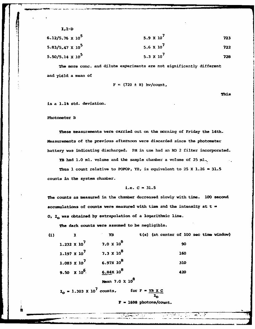
ZI,I-D
6.12/5.76 X 105 5.9 X 10⁷ 723
5.83/5.47 X 105 5.6 X 10⁷ 722
5.50/5.14 X 105 5.3 X 10 728
The more conc. and dilute experiments are not significantly different
and yield a mean of
F (720 8 8) hv/count,
This
is a 1.1% std. deviation.
Photometer B
These measurements were carried out on the morning of Friday the 14th.
Measurements of the previous afternoon were discarded since the photometer
battery was indicating discharged. PM in use had an ND 2 filter incorporated.
YB had 1.0 ml. volume and the sample chamber a volume of 25 ml.
Thus 1 count relative to POPOP, YB, is equivalent to 25 X 1.26 = 31.5
counts in the system chamber.
i.e. C - 31.5
The counts as measured in the chamber decreased slowly with time. 100 second
accumulations of counts were measured with time and the intensity at t -
0, I0 was obtained by extrapolation of a logarithmic line.
The dark counts were assumed to be negligible.
i) I YB t(s) (at center of 100 sec time window)
1.232 X 10 7 7.0 X 10⁸ 90
1.197 X 10⁷ 7.3 X 10⁸ 160
1.083 X 10⁷ 6.97X 10⁸ 310
9.50 X 10⁶. 6.84X 10⁸ 420
Mean 7.0 X 10⁸
10 1.303 X 10⁷ counts, for F = YB X C
F - 1698 photons/count.

(ii) I YB t
1.056 X 10⁷ 4.76 X 10⁸ 88
9.78 X 10⁶ 5.0 x 10⁸ 214
9.12 X 10⁶ 5.04 X 10⁸ 326
8.33 X 10⁶ 4.81 X 168 442
I = 1.116 X 10⁷ B = 5.0 X 10
8
0
i.e. F = 1411
(iii) System B.
I YB t
1.306 X 10⁷ 5.65 X 10⁸ 82
1.171 X 10⁷ 5.68 X le 191
1.0895X 10⁷ 5.71 X 10⁸ 303
1.0516X 10⁷ 5.77 X 10⁸ 415
9.40 x 10⁶ 5.70 X i08 " 537
I = 1.37 X 7 -B = -.7 X 108
and F = 1311
(iv) I YB t
1.0803 X 10⁷ 5.2 X 10⁸ 74
9.935 X 10⁶ 5.2 X le 188
9.026 X 10⁶ 5.02X 10⁸ 301
8.317 X 10⁶ 4.92X 10⁸ 415
I - 1.139 X 107 =5.2 X 1080
and F = 1438
*

(v) YBt
78i.275 X 10 5.14 X 10 66
1.1395X 10⁷ 5.20 X 10⁸ 177
1.067 X 10⁷ 5.26 X 10⁶ 290
9.254 X 10⁶ 5.26 X 10⁸ 409
1 = 1.35 X 10 7 YB=5.26 X 10 8
0
and F 1227
collecting F values,
(i) 1698 disregarding (1) F = (1347 ± 97) hv/count.
(ii) 1411
(iii) 1311
(iv) 1438, this is a 7% std. deviation
(v) 1227
Photometer C
Submersible photometer "Black hole". As with B, sample volume was 25 ml
and C = 31.5. The decay of the bacteria light standard in this chamber, while less
serious than that of B, was allowed for by the same back extrapolation method.
Dark counts 2.4 X 10⁵
(i) I/I-D YB t(sec)
8.22/8.205 X 106 3.83 X 10⁸ 68
7.487/7.463X 106 4.08 X 10⁸ 178
7.264/7.240X 106 3.98 X 10⁸ 288
5.3916/5.368X106 3.94 X 10⁸ 395
5.658/5.634X 106 3.96 506
0 = 8.67 X 10⁶ YB = 4 X 10⁸
F = C X YB = 1453. NB10
Black
hole power supply unsteady, replaced with another power supply for later experi-
~ments.
. .I .... . r
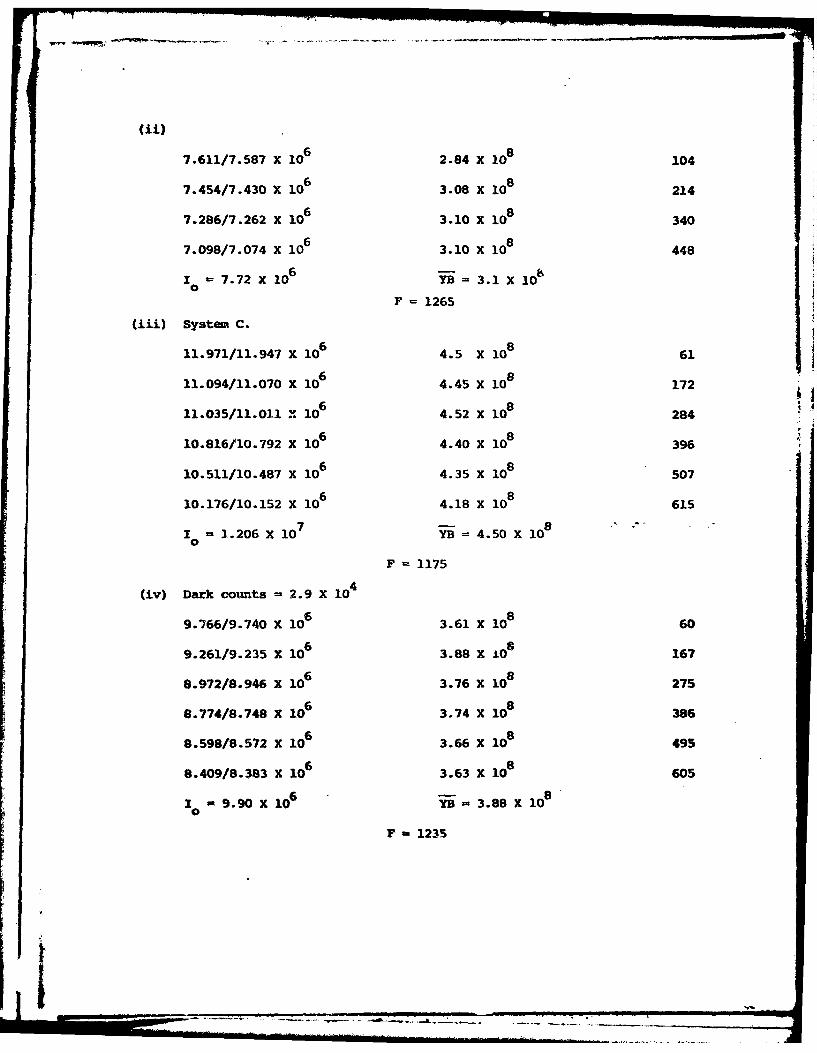
(ii)=
7.611/7.587 X 106 2.84 X 10⁸ 104
7.454/7.430 X 106 3.08 X 10⁸ 214
7.286/7.262 X 106 3.10 X 10⁸ 340
7.098/7.074 X 106 3.10 X 10⁸ 448
I ,=7.72 X 10 6 YB=3.1 X 100
F = 1265
(iii) System C.
11.971/11.947 X 106 4.5 X 10⁸ 61
11.094/11.070 X 106 4.45 X 10⁸ 172
11.035/11.011 X 106 4.52 X 10⁸ 284
10.816/10.792 X 106 4.40 X 10⁸ 396
10.511/10.487 X 106 4.35 X 10⁸ 507
10.176/10.152 X 106 4.18 X 10⁸ 615
I = 1.206x X 0 YB =4.50 X 10⁸
F 1175
(iv) Dark counts = 2.9 X 10 4
9.766/9.740 x 106 3.61 X 10⁸ 60
9.261/9.235 X 106 3.88 X .O8 167
8.972/8.9" x 106 3.76 X 10⁸ 275
8.774/8.748 X 106 3.74 X 10⁸ 386
8.598/8.572 X 106 3.66 X 10⁸ 495
8.409/8.383 X 106 3.63 X 10⁸ 605
S9.90 X106 YB= 3.88 X 10⁸
F, 1235
Ii I I III - -- .. . . .. . . - '

(v) Dark counts = 2.9 X 10⁴
8.088/8.059 X 106 3.25 X l08 61
8.039/8.009 X 106 3.32 X 10⁸ 173
7.879/7.849 X 106 3.33 X 10⁸ 285
7.709/7.680 X 10 6 3.31 X 10⁸ 395
7.515/7.486 X 106 3.26 X 10⁸ 503
7.292/7.263 X 106 3.27 X 10⁸ 612
10 = 8.27 X 10 6 B= 3.33 X 10⁶
F = 1268
Collecting F values
(i) 1453
(ii) 1265
F = (1236 ± 43) hv/count
(iii) 1175
(iv) 1235
(v) 1268, this is a std. deviation of 3.5%
The precision of calibration of the yellow box YB was 6%. Including this
the final results are:
Photometer A, F - 720 ± 50 hv/count
Photometer B, F - 1350 * 190 hv/count
Photometer C, F = 1240 ± 130 hy/count
These sensitivities are less than the F values derived from known solid angles
and the manufacturer's photocathode sensitivity curve. The low values may in
part be accounted for by the photomultiplier sensitivity being less than its
peak value over much of the range of bioluminescence emission, and variations
in overall photomultiplier sensitivity.
Preliminary Report on Calibration of the Luminol Standard
Chemiluminescence with a Standard Silicon-detector
Radiometer at the National Bureau of Standards.
Objectives:
This first visit in May was carried out with the purpose of determining
the opti-um experimental conditions; a later trip in July was used to perform
more definitive experiments. This work was carried out in collaboration with
Dr. Edward F. Zaleswski, N.B.S. who provided and operated the N.B.S. silicon
diode absolute radiometer.
Materials:
A solution of luminol (Aldrich), unpurified, in pH 11.6 NaHCO was prepared.3
It possessed an optical density of 36.6 at 347 nm, concentration by-weight 5.67 mM.
This was stored in a brown bottle and carried to the N.B.S. 1 ml sample of luminol
in a clear bottom fluorescence cuvette was used and the reaction initiated by
addition of 10 Vl of 30% H2 0 2 and 5 Ul of 10 mg/ml of horseradish peroxidase in
pH 11.6 buffer.
Apparatus:
The silicon detector radiometer was arranged so that it viewed upwards through
the bottom of a 1 cm fluorescence cuvette. The cuvette sat on top of a pair of
vertically stacked apertures of 0.5 sq. cm.
This geometry is analogous to the problem of heat flow between two apertures
and is capable of solution. Further details of the analysis of this problem will
be forthcoming from Dr. Zaleswski.
The cuvette and detector were surrounded by a cylinder constructed of black
cord with a loose fitting top. This did not prove to be light tight so that the

experiments had to be carried in total darkness. The output from the radiometer
was amplified using the N.B.S. supplied current amplifier and fed to the input of
a sensitive strip chart recorder. The strip chart output was integrated by means
of a planimeter.
Results:
Reaction of a neat 5.67 mM luminol solution showed that adequate light was
available in the initial stages of the reaction. (This reaction was not carried
to completion because of excessive time required). Accordingly the stock solution
was diluted I'100 X (The type of pipette used for the dilution was such that
approx. 0.12 rather than 0.10 ml was diluted to 10.0 ml; so that the concentration
was uncertain).
These yielded for the first 3 experiments an integrated current output of
1) 4.52 X 10²¹
212) 4.45 X 101
3) 4.02 X 10²¹ electrons/mole
A further experiment was carried out using a 1oX dilution of this solution (accurate
this time) and yielded
214) 3.94 X 10 electrons/mole for 5.67 pM luminol.
The diluted solutions were stored overnight in darkness in clear glass flasks.
On the second day it was determined that 1OX dilution yielded 1) 3.31 X 10
electrons/mole. This is considerably lower than the day before and may indicate*
luminol photo-decomposition.
A fresh solution Gwo of 5.67 VM stock to 23 ml was prepared, yielding
2) 2.65 X 10²¹ electrons/mole
213) 2.76 X 10 electrons/mole
214) 2.82 X 10 electrons/mole
5) 2.88 X 10 2 electrons/mole

It is notable that experiments 3) and 4) were carried out with a spacer
separating the two apertures so that in principle only about 1/2 as much is
transmitted from sample to detector. They yield results negligibly different
from the others suggesting that the assumptions regarding geometry are
reasonable.
Conversion of the electrons/mole to photons per mole requires knowledge
of the silicon detector quantum efficiency averaged over the luminol emission
band. Approximating this to be 0.53, the value of the quantur ,fficiency at the
420 nm maximum of luminol, this suggests for the final (250X a set) that the
luminol quantum efficiency of chemiluminscence in 0.9%.
This is more than close enough to the literature value c , at least
for a first attempt.
Conclusions:
1) The problem is feasible.
2) A convenient luminol concentration is 10-30 UM at which concentration
the reaction takes 45-60 minutes to completion.
3) The reaction may be readily initiated by adding p1 amounts of H 0 and2 2
catalyst to 1 ml of luminol solution, thus obviating corrections due to volume
changes.
4) The sensitivity of the detector is such that the base initiated luminol
reaction in DMSO may be studied.
Second Visit
Measurements of these reactions were repeated under more optimized conditions
and the results found to be in satisfactory agreement with the first set of experi-
ments. The data has not been analyzed in detail as of the time of writing this
Report.

. ..fn . _
Attention was also given to the optical geometry of the experiment.
Accurate determination of this is a major source of uncertainty (±30%) for
these photometric measurements. Preliminary results indicate that the
assumptions made in the calculation of this geometry are approximately valid.
This problem however still requires more attention before confidence can be
given to its solution.
Conclusion
The measurement of the chemiluminescence quantum yield of the luminol
reaction in aqueous solution using the NBS Standard Photometer, is in satis-
factory agreement with the previously published value, 1-2%, of Lee and Seliger.
A large uncertainty arises from the optical geometrical correction. Further
work will be able to solve this problem. The experiments will be able to be
done also for the DMSO reaction, and also using more purified luminol samples.
An integrating sphere geometry should also be tried since this should reduce
or eliminate the optical geometric uncertainty.
U. . . . . . . . .

IA


![L. - ERIC · Skill DevelopmentMaps and Globes Social Studies for the Elementary School. Proficiency Module #8. Georgia Univ., Athens. Dept. of Social Science Education. [72] 39p.](https://static.documents.pub/doc/80x56/6078dad974eb7c0fe31983c5/l-eric-skill-developmentmaps-and-globes-social-studies-for-the-elementary-school.jpg)


