Germ Cell Transplantation and Testis Tissue Xenografting in MiceLin Tang* , Jose Rafael Rodriguez-Sosa* , Ina DobrinskiDepartment of Comparative Biology and Experimental Medicine, University of Calgary*These authors contributed equally
Citation: Tang, L., Rodriguez-Sosa, J.R., Dobrinski, I. Germ Cell Transplantation and Testis Tissue Xenografting in Mice. J. Vis. Exp. (60), e354510.3791/3545, DOI : 10.3791/3545 (2012).
Abstract
Germ cell transplantation was developed by Dr. Ralph Brinster and colleagues at the University of Pennsylvania in 19941,2. These ground-breaking studies showed that microinjection of germ cells from fertile donor mice into the seminiferous tubules of infertile recipient mice resultsin donor-derived spermatogenesis and sperm production by the recipient animal2. The use of donor males carrying the bacterial β-galactosidasegene allowed identification of donor-derived spermatogenesis and transmission of the donor haplotype to the offspring by recipient animals1.Surprisingly, after transplantation into the lumen of the seminiferous tubules, transplanted germ cells were able to move from the luminalcompartment to the basement membrane where spermatogonia are located3. It is generally accepted that only SSCs are able to colonize theniche and re-establish spermatogenesis in the recipient testis. Therefore, germ cell transplantation provides a functional approach to study thestem cell niche in the testis and to characterize putative spermatogonial stem cells. To date, germ cell transplantation is used to elucidate basicstem cell biology, to produce transgenic animals through genetic manipulation of germ cells prior to transplantation4,5, to study Sertoli cell-germcell interaction6,7, SSC homing and colonization3,8, as well as SSC self-renewal and differentiation9,10.
Germ cell transplantation is also feasible in large species11. In these, the main applications are preservation of fertility, dissemination of elitegenetics in animal populations, and generation of transgenic animals as the study of spermatogenesis and SSC biology with this technique islogistically more difficult and expensive than in rodents. Transplantation of germ cells from large species into the seminiferous tubules of miceresults in colonization of donor cells and spermatogonial expansion, but not in their full differentiation presumably due to incompatibility of therecipient somatic cell compartment with the germ cells from phylogenetically distant species12. An alternative approach is transplantation of germcells from large species together with their surrounding somatic compartment. We first reported in 2002, that small fragments of testis tissue fromimmature males transplanted under the dorsal skin of immunodeficient mice are able to survive and undergo full development with the productionof fertilization competent sperm13. Since then testis tissue xenografting has been shown to be successful in many species and emerged as avaluable alternative to study testis development and spermatogenesis of large animals in mice14.
Video Link
The video component of this article can be found at http://www.jove.com/video/3545/
Protocol
PART A. Germ cell transplantation in mice
1. Preparation of recipient mice
1. Recipients should be immunologically tolerant (either genetically matched to donors or immune-deficient) to the donor testis cells.2. Recipients should be either naturally devoid of spermatogenesis (e.g. W/ Wv mice) or depleted of endogenous germ cells. Germ cell depletion
can be achieved by irradiation or chemotherapeutic drugs such as Busulfan. In this protocol, we describe the method of preparing recipientmice with Busulfan.
3. Treat recipients at 4-6 week of age through i.p injection of Busulfan. The optimal dose of Busulfan is strain dependent. In commonly usedrecipient strains, a dose of 40-50 mg/kg is sufficient to deplete endogenous germ cells (e.g. 44 mg/kg for nude mice, 50 mg/kg for B6/129 F1recipients).
4. Dissolve Busulfan powder in DMSO and then add an equal volume of sterile distilled water to make a final concentration of 4 mg/ml. Keepsolution warm at 35-40°C before use to avoid precipitation of Busulfan. Discard solution if precipitation is observed.
5. After Busulfan treatment, allow at least one month before using recipients. Recipients can be used between 1 month and 3 months post-Busulfan treatment.
2. Preparation of microinjection pipettes
1. Choose borosilicate glass pipettes (capillary tubes) with a 1.0 mm outer diameter, a 0.75 mm inner diameter, and a length of 3 inches.2. Siliconize glass pipettes with Sigmacote, rinse pipettes with methanol and blow dry.3. Pull the pipettes using a pipette puller. Different pipette puller machines have different settings, therefore, test a few settings. Generally, a
setting similar to that used for making enucleation pipettes may work.4. Break the pipette tips under dissecting microscope prior to use to achieve a diameter of approximately 50 μm at the tip.
3. Preparation of donor cells for transplantation
1. Choise of donor strain is dependent on the experimental question studied. If quantification of spermatogenesis is used as endpoint, use ofdonors with an easily identifiable genetic marker such as Lac-Z (e.g. B6.129S7-Gt(ROSA)26Sor/J from Jackson Laboratory) or GFP (e.gC57BL/6-Tg(CAG-EGFP)1Osb/J from Jackson Laboratory).
2. Prepare 7 ml of collagenase in DMEM at 1 mg/ml, 4 ml of trypsin-EDTA (0.25% trypsin plus 1mM EDTA), and 2 ml of DNase in DMEM at 7mg/ml. This amount is for digesting 2 donor mouse testes.
3. Collect testes aseptically and remove the tunica albuginea. Spread seminiferous tubules gently with fine forceps to facilitate enzymaticdigestion.
4. Transfer tubules into collagenase, incubate at 37 °C for 5-10 min, and agitate frequently.5. Wash tubules twice by spinning (200-300 g for 3 min) and re-suspending in PBS w/o Ca2++.6. Re-suspend tubules in trypsin-EDTA and shake until they become sticky and cloudy. Monitor the digestion of tubules at 37 °C as it should
occur within 1-2 min.7. Add DNase and shake well. Incubate for 1-2 min.8. Add 3 ml of FBS to stop the action of enzymes.9. Filter cell suspension using a nylon mesh with 40-70 μm pore size to remove cell/tissue chunks.10. Collect cells at 600 g for 5 min and re-suspend cells in a small amount of DMEM (<100 μl).11. Count cells and adjust the volume to a desired final concentration (usually 100 x 106). Keep cell suspension on ice prior to use.12. Each Busulfan-treated mouse testis will be filled with only 10-15 μl of cell suspension. Due to potential waste and leakage, aim to have 30-50
μl per testis.
4. Transplantation procedure (Figure 1)
1. Anaesthetize recipients following approved protocols.2. Place in dorsal recumbency and surgically prepare the abdominal area. Make a ~1cm midline abdominal incision to expose the abdominal
wall. Lift abdominal wall by using small forceps at the point of the white line to avoid accidentally injuring abdominal organs, and proceed tomake a ~0.5 cm incision at the midline of the abdominal wall to expose the peritoneal cavity.
3. Use one iris forceps to hold the abdominal wall, and use another pair of iris forceps to search for the fat pads attached to the epididymisand testis in the peritoneal cavity. Gently pull the fat pad out until the testis is exteriorized and the testicular artery and epididymis are clearlyvisible. Work on one testis at a time.
4. Place a thin sterile drape made from autoclaved index cards underneath the fat pad/testis for better visual identification (optional). The drapealso works to absorb fluid.
5. Add a drop of Trypan Blue dye into the cell suspension and carefully load the cell suspension into the polyethylene tubing connected to a 1 mlsyringe. Attach the pulled pipette into the tubing and gently force the cell suspension into the pipette by applying pressure to the syringe.
6. Identify the efferent ducts (that connect the testis to the epididymis) and gently remove fat tissue around the ducts. Work carefully as theducts and the membrane around them are translucent.
7. Carefully insert the pipette into a duct in the bundle of efferent ducts, gently thread a few mm toward the testis.8. While avoiding moving the injection pipette, reach for the syringe, gently depress the plunger of the syringe to ensure that the suspension
flows into the rete testis and seminiferous tubules begin to fill.9. The injection rate and flow of cell suspension is regulated by thumb pressure. Avoid sudden increase in pressure; monitor the movement of
suspension in tubules.10. Stop the injection when almost all surface tubules have been filled before the testis starts to become ischemic.11. Return the testis to the abdominal cavity. Repeat procedure on the contralateral testis. Suture the abdominal wall with 6-0 silk suture and
close the skin with metal wound clips. Monitor mice on a warming pad until full recovery.
5. Analysis of the recipient testes
1. Allow 2-4 months after transplantation before analysis.2. Euthanize the recipient mouse according to animal care and use guidelines and collect the testis and epididymis into PBS. When a Lac-Z
transgenic donor strain has been used, the epididymis can be used as a positive staining control as it has endogenous beta-galactosidaseactivity.
3. For detection of donor cells by Lac-Z staining, fix the testis for 1-2 hour at 4 °C in 4% paraformaldehyde (PFA) in PBS. Rinse 3 x 30 min atroom temperature in lacZ rinse buffer.
4. Incubate overnight at 37 °C in lacZ staining solution. Testis can be stained as a whole or after dispersing the tubules.5. Fix and store in 10% neutral buffered formalin. If sections are needed use neutral fast red as counter-stain.
(From Dobrinski and Rathi 200815, and Rodriguez-Sosa et al. 201116).
1. Collection of donor tissue
1. Obtain testis tissue by castration or biopsy from a donor male.2. Place testis in PBS or biopsies into culture medium, maintaining sterile conditions.3. Keep the collected tissue on ice and transport to the laboratory.
2. Preparation of donor tissue
1. Perform in the tissue culture hood to maintain sterility.2. Wash each testis in ice-cold PBS containing antibiotics two to three times before transferring into a culture-dish with PBS. In the case of
biopsies, wash testis fragments two to three times with ice-cold culture medium containing antibiotics by spinning at 150g for 2 min andresuspending in fresh ice-cold culture medium.
3. For intact testes, remove tunica vaginalis by making an incision along the surface and extrude the testis. Remove from testis all annexstructures (spermatic cord, epididymis, connective tissue). Wash testes once in cold PBS and transfer into a culture-dish with PBS.
4. Carefully remove the tunica albuginea of the testis by using a scalpel blade and a pair of scissors. If the testis is very small, the tunica can beremoved by squeezing the testicular tissue out of the tunica through a small incision made on one end while holding the tunica with a pair ofsmall forceps on the other end.
5. Depending on the size of the testis either the whole testis tissue can be cut into small pieces of around 1 - 2 mm3 in size using curved forcepsand a scalpel blade, or large pieces of testis tissue can first be removed from the testis and then cut into smaller pieces. All this should bedone in ice-cold culture medium and under sterile conditions in a small culture dish (60 x15 mm).
6. Transfer the prepared tissue fragments to ice-cold culture medium in small culture-dishes on ice until grafting.
3. Castration of recipient mouse
1. Anaesthetize mouse as above and prepare sterile surgical field by clipping the hair (not necessary in nude mice), wiping with 70% Ethanoland Betadine solution.
2. Make a 0.5 -1 cm ventral midline skin incision to expose the abdominal wall.3. Carefully expose the testis, the testicular artery and epididymis as described in PartA, 4.2-4.3.4. Detach the tail of the epidydimis from the gubernaculum by blunt dissection.5. Ligate the testicular artery, and the vas deferens together with the blood vessel with silk, and section the ligated structures by cutting between
the testis and the ligature.6. Repeat the procedure for the second testis.7. Suture the abdominal wall with one or two surgical stitches.8. Close the skin incision with one or two Michel clips.
4. Ectopic xenografting
1. Position the mouse in ventral recumbency and prepare a sterile surgical field on its back as above.2. Depending on how many grafts are to be inserted (generally 4-8/mouse), make ~0.5 cm long skin incisions on each side of the mid line of the
back of the mouse.3. Use forceps to hold a border of the skin incision, and make a subcutaneous cavity by teasing apart the connective tissue using scissors.4. Using an iris forceps place a piece of testis tissue deep into the subcutaneous cavity, holding the border of the skin incision with another iris
forceps.5. Close the skin incision with one Michel clip and keep mouse on heating pad until it starts to recover from anaesthesia.6. Transfer the mouse to a cage with additional insulation and cover and monitor until mice are fully recovered.
5. Collection of testis xenografts for analysis and sperm harvesting
1. Euthanize the host mouse according to animal care and use guidelines, and make a midline skin incision on the back skin running from thetail to the neck and open skin. This exposes the grafts which can be located either on the subcutaneous tissue or attached to the skin.
2. Carefully remove the grafts using a pair forceps and a pair of scissors.3. Record number of grafts recovered, size and weight of individual grafts.4. Retrieve the seminal vesicles from the abdomen of the mouse and record their weight as an indication of testosterone production by the
grafted tissue.5. For histological analysis
a. Suspend xenografts into sample vial containing Bouin's solution (or other fixative) in a volume ~10x that of the xenograft, and label vialappropriately.
b. Incubate overnight in the refrigerator followed by washing at least 3 times in 70% ethanol at intervals of 24 hours preferably.c. Proceed for processing and embedding in paraffin.
6. For sperm harvestinga. Wash xenografts by spinning them down at 300g for 1 min and resuspending them in culture medium containing antibiotics.
b. Cut grafts into small pieces and mince carefully with the forceps in a tissue culture dish containing 3 - 5 ml of culture medium.c. Filter minced tissue through the 40 μm cell strainer.
PART C. Representative Results
1. Germ cell transplantation
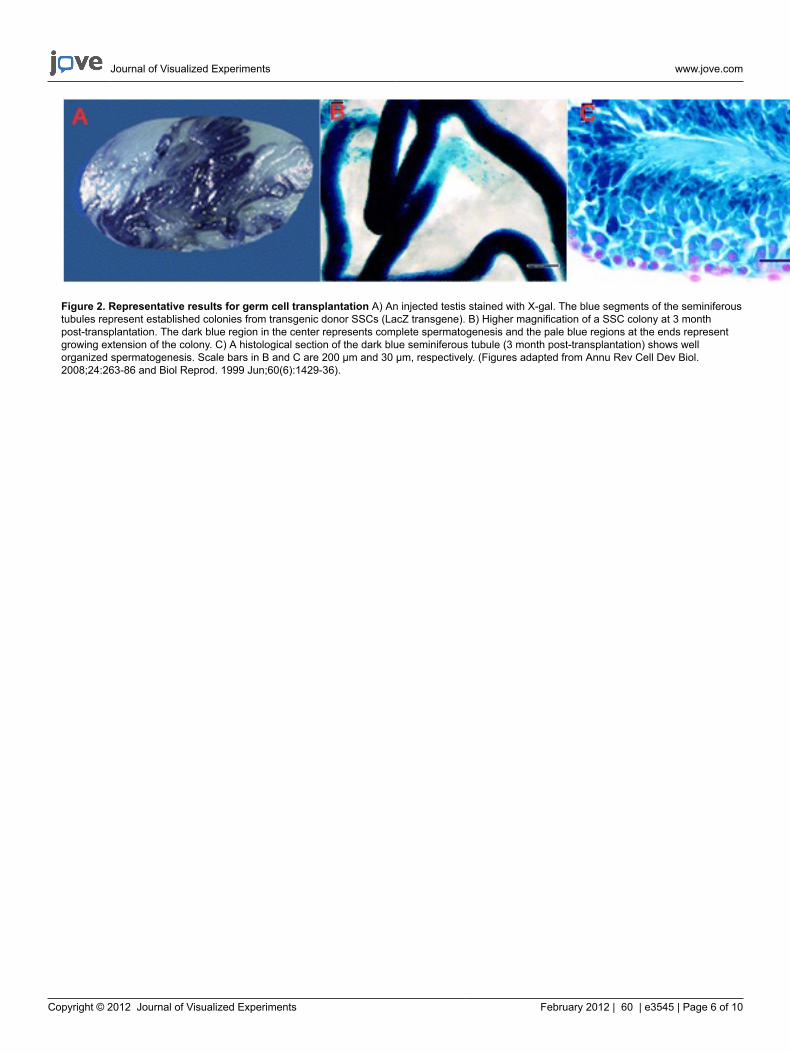
If the transplanted donor cell suspension contains spermatogonial stem cells carrying a genetic marker such as the LacZ transgene, colonizationof donor SSCs in the recipient testis can be visualized by X-gal staining as distinctive blue segments of the seminiferous tubules after 2-3 monthpost-transplantation3 (Figure 2A). A well- established colony should have a long dark blue stretch of completely filled segments with two or morelayers of blue cells closer to the center, and relatively weaker stained regions at both ends where a network of single, paired or small groups ofblue cells is apparent3 (Figure 2B). A cross section of the dark blue seminiferous tubule region should reveal well-established and well-organizedspermatogenesis with blue germ cells at various differentiation stages (Figure 2C).
2. Testis tissue xenografting
The viability of testis tissue after transplantation is inversely correlated with the developmental stage of the donor; the best outcome is obtainedwhen tissue from newborn males is used, while adult tissue shows a high tendency to degenerate and die17-21. Generally, xenograft successdecreases as donor tissue undergoes meiosis of the first wave of spermatogenesis. Time to full development of immature donor tissue andcomplete spermatogenesis is species specific, and often somewhat shorter in comparison with testes in situ. The number of seminiferous tubuleswith complete spermatogenesis is variable according to the species. While in sheep, goats and pigs that number is greater than 50%, in cattleand cats it is less than 15% 14(Figure 3).
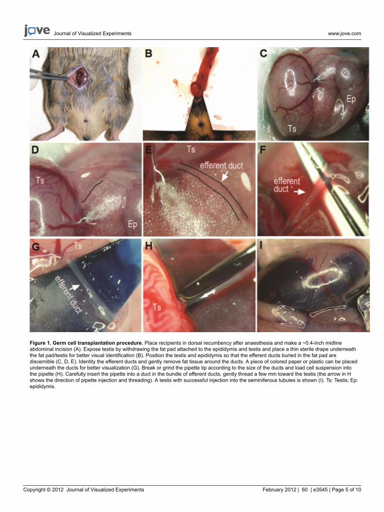
Figure 1. Germ cell transplantation procedure. Place recipients in dorsal recumbency after anaesthesia and make a ~0.4-inch midlineabdominal incision (A). Expose testis by withdrawing the fat pad attached to the epididymis and testis and place a thin sterile drape underneaththe fat pad/testis for better visual identification (B). Position the testis and epididymis so that the efferent ducts buried in the fat pad arediscernible (C, D, E). Identity the efferent ducts and gently remove fat tissue around the ducts. A piece of colored paper or plastic can be placedunderneath the ducts for better visualization (G). Break or grind the pipette tip according to the size of the ducts and load cell suspension intothe pipette (H). Carefully insert the pipette into a duct in the bundle of efferent ducts, gently thread a few mm toward the testis (the arrow in Hshows the direction of pipette injection and threading). A testis with successful injection into the seminiferous tubules is shown (I). Ts: Testis; Ep:epididymis.
Figure 2. Representative results for germ cell transplantation A) An injected testis stained with X-gal. The blue segments of the seminiferoustubules represent established colonies from transgenic donor SSCs (LacZ transgene). B) Higher magnification of a SSC colony at 3 monthpost-transplantation. The dark blue region in the center represents complete spermatogenesis and the pale blue regions at the ends representgrowing extension of the colony. C) A histological section of the dark blue seminiferous tubule (3 month post-transplantation) shows wellorganized spermatogenesis. Scale bars in B and C are 200 μm and 30 μm, respectively. (Figures adapted from Annu Rev Cell Dev Biol.2008;24:263-86 and Biol Reprod. 1999 Jun;60(6):1429-36).
Figure 3. Ectopic xenografting of immature testis tissue from large animals into immunodeficient mice. Fragments of immature donortestis (~1 mm3) transplanted under the dorsal skin of immunodeficient mice (A) are able to survive and respond to mouse gonadotropins. Asa result, testis tissue undergoes complete development, including formation of fertilization competent sperm (B). Once testis xenografts are
collected (C) they can be used for analysis or to obtain sperm (D) for ICSI (E) and embryo production (F). Bars equal 50 μm (B, E and F) or 10μm (D). (Modified from Rodriguez-Sosa and Dobrinksi 200914).
Discussion
1. Germ cell transplantation
Germ cell transplantation provides the only functional assay for unequivocal confirmation of the presence of spermatogonial stem cells (SSCs) ina cell population. Only SSCs can home to and colonize the SSC niche at the basement membrane and initiate donor-derived spermatogenesis.Germ cell transplantation made it possible to study and manipulate SSCs in an unprecedented manner. The technique has been used to producetransgenic animals through genetic manipulation of SSCs4; to elucidate the pattern, efficiency and kinetics of SSC colonization3,8; to study thesignalling pathways that regulate SSC self-renewal and differentiation9,10; to characterize surface markers on SSCs for their identification22,23; tostudy the niche environment for SSCs6,7,24. Furthermore, reciprocal transplantation has been employed to investigate whether a phenotype ofinfertility originated from a defect in Sertoli cells or in germ cells25,26.
For transplantation to work efficiently, choice and treatment of recipient animals is important. Recipients should be either genetically matchedto donors or immune-suppressed. Recipients should also lack endogenous spermatogenesis: either due to a mutation as in W/ Wv mice, orrendered infertile as a result of germ cell depletion by irradiation or chemotherapeutic drugs such as Busulfan. Moreover, a good preparation ofdonor cell suspensions and proficiency in transplantation procedure are important for the success of the technique as well.
Germ cell transplantation has its own limitations. There is no fast read-out for results. The analysis of recipient testes needs to wait at least twomonth as reestablishment of complete spermatogenesis in the otherwise infertile recipients happens two month after transplantation3. It is aqualitative or semi-quantitative assay due to large variations in cell number injected and degree of recipient germ cell suppression. Although theconcept of germ cell transplantation has been adapted to other animal species, the procedure itself is technically different and somewhat morechallenging in non-rodent species as a result of the anatomic differences across species11.
2. Testis tissue xenografting
Testis tissue xenografting works across many mammalian donor species and is a relatively simple technique. As in other types oftransplantations, the sooner after collection the tissue is transplanted the bigger chance of success. Therefore, preservation and handling ofthe tissue from collection to transplantation is important. However, in our experience testis tissue does not require special handling other thanbeing kept refrigerated. Testis tissue can be maintained at refrigerator temperature up to 24 - 36 hr, and then fragments can be prepared fortransplantation. Furthermore, fragments of fresh testis can be maintained in standard culture medium at 4 °C overnight prior transplantationwithout a noticeable effect on the grafting outcome27. Testis tissue can also be cryopreserved if long-term storage is desired. Studies performedin goat13, pig13,27, and monkey28 have shown that freezing and subsequent thawing of testis tissue does not affect significantly its capabilityto develop and produce sperm after ectopic xenografting in mice. Successful cryopreservation of testis tissue can be achieved by automated-freezing28 or conventional slow-freezing in an alcohol bath13,27, using DMSO as cryoprotectant in standard tissue culture medium containingFBS. For transplantation, cryopreserved testis tissue is then thawed by standard methods and subsequently washed in culture medium beforetransplantation27. Once the tissue has been transplanted the recipient mouse serves as an in vivo incubator and no major interventions arerequired. However, in some cases supplementation with exogenous gonadotropins may be required; testis tissue from 6-month-old rhesusmonkeys required injecting recipient mice subcutaneously with 10 IU of hCG twice a week to attain full spermatogenesis at 6-7 months29.
As mentioned above, the best outcome is obtained when tissue from newborn males is used. Tissue from males in which postmeiotic germ cellsare present shows a tendency to degenerate. However, with immature animals complete recapitulation of testis development is possible and hasnumerous clinical and research applications. In a clinical setting, testis tissue xenografting can be used for fertility preservation, particularly inimmature males in which sperm recovery is not an option. Small pieces in the form of biopsies can be collected and frozen for long-term storage.When desired, the fragments can be thawed and grafted into mice27,28. Another alternative is the cryopreservation of the sperm that is harvestedfrom xenografts. Microinjection with snap-frozen sperm from pig testis xenografts resulted in generation of morphologically normal embryos,although at a lower efficiency in comparison to testicular, epididymal, or ejaculated sperm27. After tissue has developed, it can be collected forharvesting sperm and sperm can be used to produce embryos in vitro13,16,30. A limitation of this, however, is the fact that resulting sperm do notundergo epididymal maturation and therefore require ICSI for fertilization. Therefore, use of xenograft-derived sperm for fertilization is limited tospecies where ICSI has been established.
In research, testis tissue xenografting is an attractive alternative to study testis development and spermatogenesis of large species in a rodentmodel. For example, a single donor testis can be transplanted to multiple mice. Recipient mice can then be exposed to different treatments,and/or sacrificed at different time points for xenograft collection. This not only eliminates donor effects, but also reduces the number of largemales required for a particular experiment or study. This is particularly important in large animals where studies involving numerous males arelogistically difficult and expensive, and may carry ethical limitations, particularly in primates. However, applications of testis tissue xenograftingare limited as manipulation of specific cell types before transplantation is not easily possible and efficiency of spermatogenesis is low in certaindonor species14.
Work from the authors laboratory has been supported by USDA/CSREES/NRICGP (2007-35203-18213); NIH/NCRR (2 R01 RR17359-06), NIH/NIEHS (1 R21 ES014856-01A2) and Alberta Innovates – Health Solutions.
References
1. Brinster, R.L. & Avarbock, M.R. Germline transmission of donor haplotype following spermatogonial transplantation. Proceedings of theNational Academy of Sciences of the United States of America. 91 (24), 11303 (1994).
2. Brinster, R.L. & Zimmermann, J.W. Spermatogenesis following male germ-cell transplantation. Proceedings of the National Academy ofSciences of the United States of America. 91 (24), 11298 (1994).
3. Nagano, M., Avarbock, M.R., & Brinster, R.L. Pattern and kinetics of mouse donor spermatogonial stem cell colonization in recipient testes.Biology of Reproduction. 60 (6), 1429 (1999).
4. Nagano, M., Brinster, C.J., & Orwig, K.E., et al. Transgenic mice produced by retroviral transduction of male germ-line stem cells.Proceedings of the National Academy of Sciences of the United States of America. 98 (23), 13090 (2001).
5. Ryu, B.Y., Orwig, K.E., & Oatley, J.M., et al. Efficient generation of transgenic rats through the male germline using lentiviral transduction andtransplantation of spermatogonial stem cells. Journal of Andrology. 28 (2), 353 (2007).
6. Hess, R.A., Cooke, P.S., & Hofmann, M.C., et al. Mechanistic insights into the regulation of the spermatogonial stem cell niche. Cell Cycle. 5(11), 1164 (2006).
7. Oatley, M.J., Racicot, K.E., & Oatley, J.M. Sertoli cells dictate spermatogonial stem cell niches in the mouse testis. Biology of Reproduction.84 (4), 639 (2011).
8. Nagano, M.C. Homing efficiency and proliferation kinetics of male germ line stem cells following transplantation in mice. Biology ofReproduction. 69 (2), 701 (2003).
9. Kubota, H., Avarbock, M.R., & Brinster, R.L. Growth factors essential for self-renewal and expansion of mouse spermatogonial stem cells.Proceedings of the National Academy of Sciences of the United States of America. 101 (47), 16489 (2004).
10. Oatley, J.M., Avarbock, M.R., & Telaranta, A.I., et al. Identifying genes important for spermatogonial stem cell self-renewal and survival.Proceedings of the National Academy of Sciences of the United States of America. 103 (25), 9524 (2006).
11. Dobrinski, I. Germ cell transplantation and testis tissue xenografting in domestic animals. Animal Reproduction Science. 89 (1-4), 137 (2005).12. Dobrinski, I., Avarbock, M.R., & Brinster, R.L. Transplantation of germ cells from rabbits and dogs into mouse testes. Biology of Reproduction.
61 (5), 1331 (1999).13. Honaramooz, A., Snedaker, A., & Boiani M., et al. Sperm from neonatal mammalian testes grafted in mice. Nature. 418 (6899), 778 (2002).14. Rodriguez-Sosa, J.R. & Dobrinski, I. Recent developments in testis tissue xenografting. Reproduction. 138 (2), 187 (2009).15. Dobrinski, I. & Rathi, R. Ectopic grafting of mammalian testis tissue into mouse hosts. Methods in Molecular Biology (Clifton, N.J). 139, 450
(2008).16. Rodriguez-Sosa, J.R., Schlatt, S., & Dobrinski, I. Testicular tissue transplantation for fertility preservation. In Fertility Preservation: Emerging
Technologies and Clinical Applications, Seli, E. & Agarwal, A., eds. Springer, New York, NY, 331 (2011).17. Rathi, R., Honaramooz, A., & Zeng, W., et al. Germ cell fate and seminiferous tubule development in bovine testis xenografts. Reproduction.
130 (6), 923 (2005).18. Rathi, R., Honaramooz, A., & Zeng, W., et al. Germ cell development in equine testis tissue xenografted into mice. Reproduction. 131 (6),
1091 (2006).19. Kim, Y., Selvaraj, V., & Pukazhenthi, B., et al. Effect of donor age on success of spermatogenesis in feline testis xenografts. Reproduction,
Fertility, and Development. 19 (7), 869 (2007).20. Arregui, L., Rathi, R., & Zeng, W., et al. Xenografting of adult mammalian testis tissue. Animal Reproduction Science. 106 (1-2), 65 (2008).21. Schlatt, S., Honaramooz, A., & Ehmcke, J., et al. Limited survival of adult human testicular tissue as ectopic xenograft. Human Reproduction.
21 (2) 384 (2006).22. Shinohara, T., Avarbock, M.R., & Brinster, R.L. beta1- and alpha6-integrin are surface markers on mouse spermatogonial stem cells.
Proceedings of the National Academy of Sciences of the United States of America. 96 (10), 5504 (1999).23. Kanatsu-Shinohara, M., Toyokuni, S., & Shinohara, T. CD9 is a surface marker on mouse and rat male germline stem cells. Biology of
Reproduction. 70 (1), 70 (2004).24. Ryu, B.Y., Orwig, K.E., & Oatley, J.M., et al. Effects of aging and niche microenvironment on spermatogonial stem cell self-renewal. Stem
Cells. 24 (6), 1505 (2006).25. Costoya, J.A., Hobbs, R.M., & Barna, M., et al. Essential role of Plzf in maintenance of spermatogonial stem cells. Nature Genetics. 36 (6),
653 (2004).26. Morrow, C.M., Hostetler, C.E., & Griswold, M.D., et al. ETV5 is required for continuous spermatogenesis in adult mice and may mediate blood
testes barrier function and testicular immune privilege. Annals of the New York Academy of Sciences. 1120, 144 (2007).27. Zeng, W., Snedaker, A.K., & Megee, S., et al. Preservation and transplantation of porcine testis tissue. Reproduction, Fertility and
Development. 21 (3) 489 (2009).28. Jahnukainen, K., Ehmcke, J., & Hergenrother, S.D., et al. Effect of cold storage and cryopreservation of immature non-human primate
testicular tissue on spermatogonial stem cell potential in xenografts. Human Reproduction. 22 (4) 1060 (2007).29. Rathi, R., Zeng, W., & Megee, S., et al. Maturation of testicular tissue from infant monkeys after xenografting into mice. Endocrinology. 149
30. Honaramooz, A., Li, M.W., & Penedo, M.C., et al. Accelerated maturation of primate testis by xenografting into mice. Biology of Reproduction.70 (5), 1500 (2004).