Germicidal Efficacy and Mammalian Skin Safety of 222-nm UV Light Manuela Buonanno a , Brian Ponnaiya a , David Welch a , Milda Stanislauskas b , Gerhard Randers-Pehrson a , Lubomir Smilenov a , Franklin D. Lowy c , David M. Owens b,d , and David J. Brenner a,1 a Center for Radiological Research, Columbia University Medical Center, New York, New York b Department of Dermatology, Columbia University Medical Center, New York, New York c Department of Medicine and Infectious Diseases, Columbia University Medical Center, New York, New York d Department of Pathology and Cell Biology, Columbia University Medical Center, New York, New York Abstract We have previously shown that 207-nm ultraviolet (UV) light has similar antimicrobial properties as typical germicidal UV light (254 nm), but without inducing mammalian skin damage. The biophysical rationale is based on the limited penetration distance of 207-nm light in biological samples (e.g. stratum corneum) compared with that of 254-nm light. Here we extended our previous studies to 222-nm light and tested the hypothesis that there exists a narrow wavelength window in the far-UVC region, from around 200–222 nm, which is significantly harmful to bacteria, but without damaging cells in tissues. We used a krypton-chlorine (Kr-Cl) excimer lamp that produces 222-nm UV light with a bandpass filter to remove the lower- and higher-wavelength components. Relative to respective controls, we measured: 1. in vitro killing of methicillin- resistant Staphylococcus aureus (MRSA) as a function of UV fluence; 2. yields of the main UV- associated premutagenic DNA lesions (cyclobutane pyrimidine dimers and 6-4 photoproducts) in a 3D human skin tissue model in vitro; 3. eight cellular and molecular skin damage endpoints in exposed hairless mice in vivo. Comparisons were made with results from a conventional 254-nm UV germicidal lamp used as positive control. We found that 222-nm light kills MRSA efficiently but, unlike conventional germicidal UV lamps (254 nm), it produces almost no premutagenic UV- associated DNA lesions in a 3D human skin model and it is not cytotoxic to exposed mammalian skin. As predicted by biophysical considerations and in agreement with our previous findings, far- UVC light in the range of 200–222 nm kills bacteria efficiently regardless of their drug-resistant proficiency, but without the skin damaging effects associated with conventional germicidal UV exposure. 1 Address for correspondence: Center for Radiological Research, Columbia University Medical Center, 630 West 168th St., New York, NY 10032; [email protected]. HHS Public Access Author manuscript Radiat Res. Author manuscript; available in PMC 2017 August 10. Published in final edited form as: Radiat Res. 2017 April ; 187(4): 483–491. doi:10.1667/RR0010CC.1. Author Manuscript Author Manuscript Author Manuscript Author Manuscript
Transcript
Germicidal Efficacy and Mammalian Skin Safety of 222-nm UV Light
Manuela Buonannoa, Brian Ponnaiyaa, David Welcha, Milda Stanislauskasb, Gerhard Randers-Pehrsona, Lubomir Smilenova, Franklin D. Lowyc, David M. Owensb,d, and David J. Brennera,1
aCenter for Radiological Research, Columbia University Medical Center, New York, New York
bDepartment of Dermatology, Columbia University Medical Center, New York, New York
cDepartment of Medicine and Infectious Diseases, Columbia University Medical Center, New York, New York
dDepartment of Pathology and Cell Biology, Columbia University Medical Center, New York, New York
Abstract
We have previously shown that 207-nm ultraviolet (UV) light has similar antimicrobial properties
as typical germicidal UV light (254 nm), but without inducing mammalian skin damage. The
biophysical rationale is based on the limited penetration distance of 207-nm light in biological
samples (e.g. stratum corneum) compared with that of 254-nm light. Here we extended our
previous studies to 222-nm light and tested the hypothesis that there exists a narrow wavelength
window in the far-UVC region, from around 200–222 nm, which is significantly harmful to
bacteria, but without damaging cells in tissues. We used a krypton-chlorine (Kr-Cl) excimer lamp
that produces 222-nm UV light with a bandpass filter to remove the lower- and higher-wavelength
components. Relative to respective controls, we measured: 1. in vitro killing of methicillin-
resistant Staphylococcus aureus (MRSA) as a function of UV fluence; 2. yields of the main UV-
associated premutagenic DNA lesions (cyclobutane pyrimidine dimers and 6-4 photoproducts) in a
3D human skin tissue model in vitro; 3. eight cellular and molecular skin damage endpoints in
exposed hairless mice in vivo. Comparisons were made with results from a conventional 254-nm
UV germicidal lamp used as positive control. We found that 222-nm light kills MRSA efficiently
but, unlike conventional germicidal UV lamps (254 nm), it produces almost no premutagenic UV-
associated DNA lesions in a 3D human skin model and it is not cytotoxic to exposed mammalian
skin. As predicted by biophysical considerations and in agreement with our previous findings, far-
UVC light in the range of 200–222 nm kills bacteria efficiently regardless of their drug-resistant
proficiency, but without the skin damaging effects associated with conventional germicidal UV
exposure.
1Address for correspondence: Center for Radiological Research, Columbia University Medical Center, 630 West 168th St., New York, NY 10032; [email protected].
HHS Public AccessAuthor manuscriptRadiat Res. Author manuscript; available in PMC 2017 August 10.
Published in final edited form as:Radiat Res. 2017 April ; 187(4): 483–491. doi:10.1667/RR0010CC.1.
Author M
anuscriptA
uthor Manuscript
Author M
anuscriptA
uthor Manuscript
INTRODUCTION
The use of ultraviolet (UV) light for inactivating bacteria and viruses is well established (1,
2). However, UV radiations emitted by typical germicidal lamps with a peak emission at 254
nm represent a human health hazard, causing skin cancer (3, 4) and cataracts (5, 6).
We have developed an approach to kill bacteria without harming human cells in skin tissue
models (7) and mouse skin in vivo (8) that employs single-wavelength UVC light generated
by inexpensive filtered excilamps (9). The approach is based on the limited penetration
distance of UVC light in the wavelength range of 200–222 nm in biological samples.
Specifically, while far-UVC light has enough range to traverse microbes that are much
smaller in size than human cells [less than 1 μm in diameter (10, 11), compared to the
diameter of typical human cells ranging from about 10–25 μm (10)], it is strongly absorbed
by the proteins in the cytoplasm of human cells (12, 13) and is drastically attenuated before
reaching the human cell nucleus. It follows that far-UVC light is not able to penetrate the
stratum corneum of skin and reach the underlying critical basal cells or melanocytes (4).
Another organ especially sensitive to UV damage is the lens; however, the lens is positioned
distal to the cornea that is sufficiently thick [~500 μm (14))]. Therefore, penetration of far-
UVC ~200-nm light through the cornea to the lens is predicted to be essentially zero (15).
The potential use of UVC light for microbe sterilization purposes in the presence of humans
paves the way to numerous clinical applications, including reduction of surgical site
infections (SSI) that are the second most common healthcare-associated infections resulting
in read-missions, prolonged hospital stays, increased morbidity and mortality, and an overall
higher medical cost (16, 17). A key factor contributing to the severity of SSI is the incidence
of drug-resistant bacteria such as methicillin-resistant Staphylococcus aureus (MRSA) (18,
19).
To address the issue of reducing SSI, we have developed an approach that involves the use of
inexpensive excimer lamps that, appropriately filtered, emit monoenergetic wavelengths in
the far-UVC range. A crucial property of UVC-mediated germicidal killing is that it is
essentially independent of acquired drug resistance (20, 21).
We have previously shown that 207-nm light emitted by a filtered krypton-bromine (Kr-Br)
excilamp has bactericidal efficacy while being minimally cytotoxic to human cells in a 3D
skin tissue model in vitro (7) and in a hairless mouse skin model in vivo (8). Thus,
continuous exposure of the wound to far-UVC light during surgery may inactivate the
microbes alighting directly onto the surgical wound from the air. If proven to be safe to eyes
as well as skin, continuous operation of far-UVC light would not require the use of
cumbersome protective clothing, hoods and eye shields for the surgical staff and the patient
(22, 23).
Here we extended those studies to a filtered krypton-chlorine (Kr-Cl) excimer lamp that
produces essentially monoenergetic UV light at 222 nm and established that a wavelength
window exist in the far-UVC region (from 200–222 nm) that inactivates bacteria efficiently
but is not cytotoxic or mutagenic to mammalian cells.
Buonanno et al. Page 2
Radiat Res. Author manuscript; available in PMC 2017 August 10.
Author M
anuscriptA
uthor Manuscript
Author M
anuscriptA
uthor Manuscript
We describe measurements of MRSA survival and of typical UV-induced premutagenic
DNA lesions in a 3D human skin model immediately after exposure to different fluences of
222-nm light. We compared the results to those measured in samples exposed to similar
fluences from a typical germicidal lamp emitting at 254 nm.
We further tested eight cellular and molecular endpoints relevant to skin damage in vivo in
dorsal skin of hairless mice 48 h after exposure to 222-nm or 254-nm light used as positive
control (8), at a fluence at which the 254-nm light is predicted to produce a significant
decrease in SSI rates (22).
In agreement with our previous results using 207-nm light (7, 8), here we showed that 222-
nm light has similar antimicrobial properties as a conventional germicidal lamp but without
causing mammalian skin damage.
The finding that a far-UVC wavelength window (200–225 nm) is differentially cytotoxic to
bacteria relative to mammalian cells is novel and can be used for various applications that
would require the presence of humans, including room sterilization and reduction of surgical
site infections, without the need of additional personal protective equipment.
MATERIALS AND METHODS
UV Lamps
We used an excimer lamp based on a krypton-chlorine (Kr-Cl) gas mixture that emits
principally at 222 nm. The lamp (High Current Electronics Institute, Tomsk, Russia) was air
cooled with a 6,000-mm2 exit window (24). A custom bandpass filter (Omega Optical,
Brattleboro, VT) was used to remove essentially all but the dominant 222-nm wavelength
emission.
A UV spectrometer (Photon Control, BC, Canada) sensitive in the wavelength range from
200–360 nm was used to characterize the wavelength spectra emitted by the excimer lamp,
and a deuterium lamp standard with a NIST-traceable spectral irradiance (Newport Corp,
Stratford, CT) was used to calibrate the UV spectrometer.
Studies were also carried out with a conventional mercury germicidal lamp (Hygeaire,
Atlantic Ultraviolet Corporation, Hauppauge, NY) with peak emission at 254 nm and used
as positive control. An ILT1400 sensor equipped with a SEL220 detector (International
Light Technologies, Peabody, MA) was used to measure the fluence rate from both lamps.
For acute exposures (MRSA and 3D tissues), the 222- and 254-nm lamps were positioned 9
and 99 cm, respectively, from the sample at which corresponded a power density of 0.036
mJ/cm2. For chronic exposures of mice, delivery of 157 mJ/cm2 in a 7 h period was obtained
by locating the 222- and the 254-nm lamps 41 and 205 cm from the samples, respectively;
the corresponding power densities were 0.0062 mJ/cm2 for the 222-nm lamp and 0.0008
mJ/cm2 in the case of the conventional germicidal lamp.
Figure 1 shows the measured spectra emitted from the Kr-Cl excimer lamp that, together
with the main excimer emission at 222 nm, also includes lower fluences of higher
wavelength light (i.e. ~237 and ~258 nm). To remove these more penetrating higher
Buonanno et al. Page 3
Radiat Res. Author manuscript; available in PMC 2017 August 10.
Author M
anuscriptA
uthor Manuscript
Author M
anuscriptA
uthor Manuscript
wavelengths and therefore potentially more harmful to human cells, we used a custom
bandpass filter (Omega Optical, Brattleboro, VT). As shown in the inset in Fig. 1, the
filtered lamp effectively emitted only the characteristic single wavelength of 222 nm.
IN VITRO STUDIES
MRSA Cell Survival
We used methicillin-resistant S. aureus (MRSA USA300, multilocus sequence type 8, clonal
complex 8, staphylococcal cassette chromosome mec type IV), which is a major cause of
both community and nosocomial infections (25–27). Survival of MRSA as a function of
UVC fluence was assessed immediately after exposure with the standard colony forming
unit (CFU) assay, as previously reported (7).
UV-Associated Premutagenic DNA Lesions in a 3D Human Skin Model
We used a 3D human skin model EpiDerm-FT (MatTek Corp., Ashland, MA) consisting of
stratum corneum and 8–12 human cell layers to reproduce human epidermis and dermis
(28). We measured induction of the two most abundant premutagenic DNA lesions in the
epidermis, cyclobutane pyrimidine dimers (CPD) and pyrimidine-pyrimidone 6-4
photoproducts (6-4PP) (4), as a function of UVC fluence in 3D human skin constructs
immediately after exposure using the immunohistochemical approach previously described
(7).
IN VIVO STUDIES
Mouse Irradiations
We used six- to eight-week-old male hairless mice (SKH1-Elite strain 477, Charles River
Laboratories, Stone Ridge, NY); the strain has UV action spectra for histological, physical
and visible changes similar to those of human skin (29, 30). Moreover, the typical thickness
of the stratum corneum of SKH1 mice (5–10 μm) (31, 32) is comparable to that of human
skin (5–20 μm) (33).
One group of three mice was exposed to fluence of 157 mJ/cm2 from 222 nm light delivered
in a 7 h period by a filtered Kr-Cl excimer lamp while another group of three mice was sham
irradiated to zero UV fluence. Mice exposed to 157 mJ/cm2 delivered in 7 h period from 254
nm light represented the positive controls (8).
All animal procedures were carried out in accordance with federal guidelines and protocols
approved by the Columbia University Medical Center IACUC.
Mouse Skin Safety-Specific Endpoints
Following the same protocols described in our previous mouse skin safety study (8), 48 h
after UV exposure we measured the following endpoints: 1. Epidermal thickness measured
in hematoxylin and eosin stained samples; 2. The percentage of proliferating keratinocytes
expressing the Ki-67 antigen (34, 35). 3. Yields of UV-induced cyclobutane pyrimidine
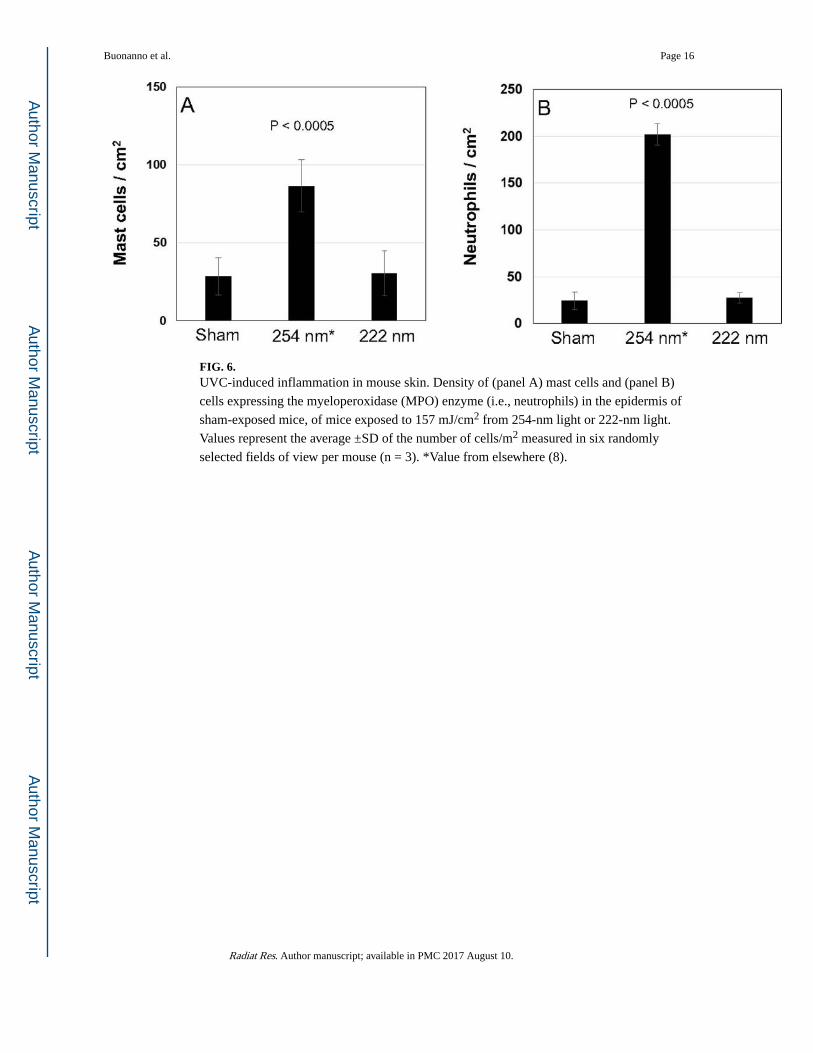
dimers (CPD) and 6-4 photoproducts (6-4PP); 4. The number of neutrophils and mast cells
Buonanno et al. Page 4
Radiat Res. Author manuscript; available in PMC 2017 August 10.
Author M
anuscriptA
uthor Manuscript
Author M
anuscriptA
uthor Manuscript
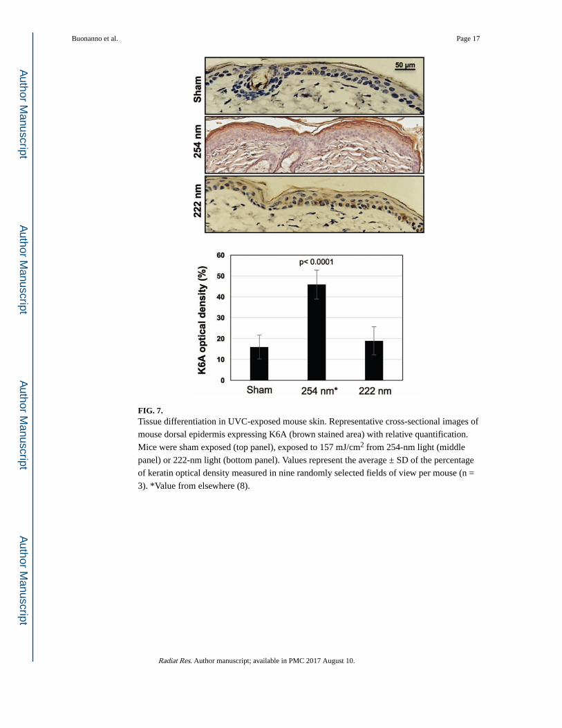
as markers of skin inflammation (36); 5. The expression of keratin (K) K6A as marker of
skin differentiation (37, 38).
Tissues were examined with an Olympus IX70 microscope equipped with a Photometrics®
PVCAM high-resolution, high-efficiency digital camera; Image-Pro Plus 6.0 software and
Fiji/Image J software were used to analyze the images. For each mouse, each endpoint was
measured in at least six randomly selected fields of view.
Statistical Analysis
Comparisons of mean values between treatment groups and controls were performed using
Student’s t test, and comparison of proportions were assessed with standard χ2 tests.
IN VITRO RESULTS
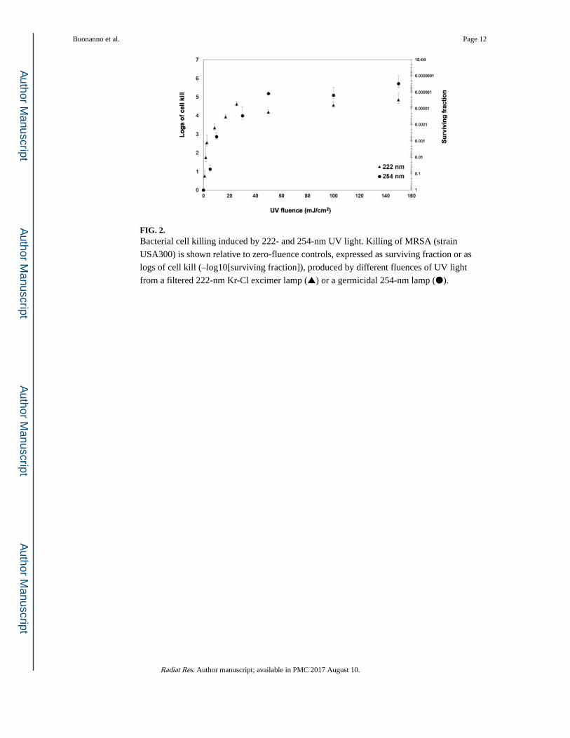
MRSA Survival
We measured MRSA survival immediately after exposure to different fluences of 222-nm
light generated by a filtered Kr-Cl excimer lamp (Fig. 2). We compared the results to
survival of MRSA exposed to similar fluences from a conventional germicidal UV lamp
(254 nm) (7). As illustrated in Fig. 2, at relatively high fluences at which the 254-nm light is
predicted to produce a significant decrease in SSI rates (1), 222-nm light kills MRSA almost
as efficiently as a conventional germicidal UV lamp. However, 254-nm light is almost
equally efficient at killing human cells (7).
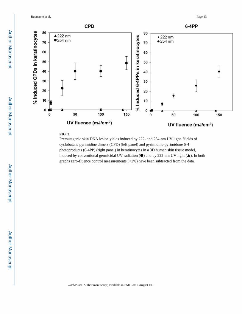
Induction of Premutagenic DNA Lesions in Human Skin
Figure 3 shows the comparison of the induced yields of CPD (left panel) and 6-4PP (right
panel) in 3D skin tissue models exposed to 222-nm or 254-nm light. Unlike conventional
germicidal light (254 nm), exposure to the 222-nm light at any of the studied fluences did
not induce yields of either lesions that was significantly higher than the sham-irradiated
samples.
IN VIVO RESULTS
Epidermal Thickness and Keratinocyte Proliferation
At 48 h after exposure, fixed dorsal skin sections were stained with hematoxylin and eosin
(H&E) for analysis of epidermal thickness (Fig. 4A). As shown by the typical H&E-stained
cross-sections of dorsal skin of sham-exposed mice (top panel), of mice exposed to 157
mJ/cm2 from 254-nm light (middle panel) or 222-nm light (bottom panel), we found that,
unlike the 254-nm light, the epidermal thickness of skin of mice exposed to the 222-nm
excimer lamp was not statistically different from skin of unexposed mice (P = 0.18) (Fig. 4B
germicidal lamps, has considerable promise to be a safe and inexpensive modality for SSI
reduction, while being cytotoxic to both drug-resistant and drug-sensitive microbes (22).
Acknowledgments
We are very grateful to Lynn Shostack for her support and advice. We thank Ms. Elizabeth Flores of the Department of Medicine and Infectious Disease at Columbia University for her expertise and for the use of the laboratory equipment. We thank Drs. Alan W. Bigelow and Yanping Xu for their insight in the UV dosimetry. GR-P receives royalty payments from Ushio Inc. (Tokyo, Japan) pursuant to exclusive license and research agreements with Columbia University.
Buonanno et al. Page 7
Radiat Res. Author manuscript; available in PMC 2017 August 10.
Author M
anuscriptA
uthor Manuscript
Author M
anuscriptA
uthor Manuscript
References
1. Yin R, Dai T, Avci P, Jorge AE, de Melo WC, Vecchio D, et al. Light based anti-infectives: ultraviolet C irradiation, photodynamic therapy, blue light, and beyond. Curr Opin Pharmacol. 2013; 13(5):731–62. [PubMed: 24060701]
2. McDevitt JJ, Rudnick SN, Radonovich LJ. Aerosol Susceptibility of Influenza Virus to UV-C Light. Appl Environ Microbiol. 2012; 78(6):1666–9. [PubMed: 22226954]
3. Mitchell DL, Nairn RS. The (6-4) photoproduct and human skin cancer. Photo-dermatol. 1988; 5(2):61–4.
4. Pfeifer GP, Besaratinia A. UV wavelength-dependent DNA damage and human non-melanoma and melanoma skin cancer. Photochem Photobiol Sci. 2012; 11(1):90–7. [PubMed: 21804977]
5. Jose JG, Pitts DG. Wavelength dependency of cataracts in albino mice following chronic exposure. Exp Eye Res. 1985; 41(4):545–63. [PubMed: 4085580]
6. Soderberg PG. Acute cataract in the rat after exposure to radiation in the 300 nm wavelength region. A study of the macro-, micro- and ultrastructure. Acta Ophthalmol. 1988; 66(2):141–52. [PubMed: 3389086]
7. Buonanno M, Randers-Pehrson G, Bigelow AW, Trivedi S, Lowy FD, Spotnitz HM, et al. 207-nm UV light - A promising tool for safe low-cost reduction of surgical site infections. I: In vitro studies. PLoS ONE. doi:101371/journalpone0076968. eCollection 2013.
8. Buonanno M, Stanislauskas M, Ponnaiya B, Bigelow AW, Randers-Pehrson G, Shuryak I, et al. 207-nm UV light - A promising tool for safe low-cost reduction of surgical site infections. II: In-vivo safety studies. PLoS One. 2016 Jun 8.11(6):e0138418.doi: 10.1371/journal.pone.0138418 [PubMed: 27275949]
9. Sosnin EA, Oppenlander T, Tarasenko VF. Applications of capacitive and barrier discharge excilamps in photoscience. J Photochem Photobiol C: Photochem Rev. 2006; 7:145–63.
10. Metzler, DE., Metzler, CM. Biochemistry: The Chemical Reactions of Living Cells. 2nd. San Diego: Academic Press; 2001.
11. Lorian V, Zak O, Suter J, Bruecher C. Staphylococci, in vitro and in vivo. Diagn Microbiol Infect Dis. 1985; 3(5):433–44. [PubMed: 4028668]
13. Setlow, J. The molecular basis of biological effects of ultraviolet radiation and photoreactivation. In: Ebert, M., Howard, A., editors. Current topics in radiation research II. Amsterdam: North Holland Publishing Company; 1966. p. 195-248.
14. Doughty MJ, Zaman ML. Human corneal thickness and its impact on intraocular pressure measures: a review and meta-analysis approach. Surv Ophthalmol. 2000; 44(5):367–408. [PubMed: 10734239]
15. Kolozsvari L, Nogradi A, Hopp B, Bor Z. UV absorbance of the human cornea in the 240- to 400-nm range. Invest Ophthalmol Vis Sci. 2002; 43(7):2165–8. [PubMed: 12091412]
16. Anderson DJ. Surgical site infections. Infect Dis Clin North Am. 2011; 25(1):135–53. [PubMed: 21315998]
17. Allegranzi B, Bischoff P, de Jonge S, Kubilay NZ, Zayed B, Gomes SM, et al. New WHO recommendations on preoperative measures for surgical site infection prevention: an evidence-based global perspective. Lancet Infect Dis. 2016
18. Fry DE, Barie PS. The changing face of Staphylococcus aureus: a continuing surgical challenge. Surg Infect. 2011; 12(3):191–203.
19. Anderson DJ, Kaye KS. Staphylococcal surgical site infections. Infect Dis Clin North Am. 2009; 23(1):53–72. [PubMed: 19135916]
20. Conner-Kerr TA, Sullivan PK, Gaillard J, Franklin ME, Jones RM. The effects of ultraviolet radiation on antibiotic-resistant bacteria in vitro. Ostomy Wound Manage. 1998; 44(10):50–6. [PubMed: 9866596]
Buonanno et al. Page 8
Radiat Res. Author manuscript; available in PMC 2017 August 10.
Author M
anuscriptA
uthor Manuscript
Author M
anuscriptA
uthor Manuscript
21. Rao BK, Kumar P, Rao S, Gurung B. Bactericidal effect of ultraviolet C (UVC), direct and filtered through transparent plastic, on gram-positive cocci: an in vitro study. Ostomy Wound Manage. 2011; 57(7):46–52. [PubMed: 21904015]
22. Ritter MA, Olberding EM, Malinzak RA. Ultraviolet lighting during orthopaedic surgery and the rate of infection. J Bone Joint Surg Am. 2007; 89(9):1935–40. [PubMed: 17768189]
23. Berg M, Bergman BR, Hoborn J. Ultraviolet radiation compared to an ultra-clean air enclosure. Comparison of air bacteria counts in operating rooms. J Bone Joint Surg Br. 1991; 73(5):811–5. [PubMed: 1894672]
25. Anderson DJ, Sexton DJ, Kanafani ZA, Auten G, Kaye KS. Severe surgical site infection in community hospitals: epidemiology, key procedures, and the changing prevalence of methicillin-resistant Staphylococcus aureus. Infect Control Hosp Epidemiol. 2007; 28(9):1047–53. [PubMed: 17932825]
26. Hidron AI, Edwards JR, Patel J, Horan TC, Sievert DM, Pollock DA, et al. NHSN annual update: antimicrobial-resistant pathogens associated with healthcare-associated infections: annual summary of data reported to the National Healthcare Safety Network at the Centers for Disease Control and Prevention, 2006-2007. Infect Control Hosp Epidemiol. 2008; 29(11):996–1011. [PubMed: 18947320]
27. Lessa FC, Mu Y, Ray SM, Dumyati G, Bulens S, Gorwitz RJ, et al. Impact of USA300 methicillin-resistant Staphylococcus aureus on clinical outcomes of patients with pneumonia or central line-associated bloodstream infections. Clin Infect Dis. 2012; 55(2):232–41. [PubMed: 22523264]
28. Kubilus J, Hayden PJ, Ayehunie S, Lamore SD, Servattalab C, Bellavance KL, et al. Full thickness epiderm: a dermal-epidermal skin model to study epithelial-mesenchymal interactions. Altern Lab Anim. 2004; 32(Suppl 1A):75–82. [PubMed: 23577437]
29. Bissett DL, Hannon DP, Orr TV. Wavelength dependence of histological, physical, and visible changes in chronically UV-irradiated hairless mouse skin. Photochem Photobiol. 1989; 50(6):763–9. [PubMed: 2626490]
30. Bissett DL, Hannon DP, Orr TV. An animal model of solar-aged skin: histological, physical, and visible changes in UV-irradiated hairless mouse skin. Photochem Photobiol. 1987; 46(3):367–78. [PubMed: 3671514]
31. Sato K, Sugibayashi K, Morimoto Y. Species differences in percutaneous absorption of nicorandil. J Pharm Sci. 1991; 80(2):104–7. [PubMed: 1828835]
32. Bronaugh RL, Stewart RF, Congdon ER. Methods for in vitro percutaneous absorption studies. II. Animal models for human skin. Toxicol Appl Pharmacol. 1982; 62(3):481–8. [PubMed: 7071863]
33. Russell LM, Wiedersberg S, Delgado-Charro MB. The determination of stratum corneum thickness: an alternative approach. Eur J Pharm Biopharm. 2008; 69(3):861–70. [PubMed: 18424094]
34. van Diest PJ, Brugal G, Baak JP. Proliferation markers in tumours: interpretation and clinical value. J Clin Pathol. 1998; 51(10):716–24. [PubMed: 10023332]
35. Scholzen T, Gerdes J. The Ki-67 protein: from the known and the unknown. J Cell Physiol. 2000; 182(3):311–22. [PubMed: 10653597]
37. Horio T, Miyauchi H, Sindhvananda J, Soh H, Kurokawa I, Asada Y. The effect of ultraviolet (UVB and PUVA) radiation on the expression of epidermal keratins. Br Dermatol. 1993; 128(1):10–5.
38. Sano T, Kume T, Fujimura T, Kawada H, Higuchi K, Iwamura M, et al. Long-term alteration in the expression of keratins 6 and 16 in the epidermis of mice after chronic UVB exposure. Arch Dermatol Res. 2009; 301(3):227–37. [PubMed: 18979106]
39. Green H, Boll J, Parrish JA, Kochevar IE, Oseroff AR. Cytotoxicity and mutagenicity of low intensity, 248 and 193 nm excimer laser radiation in mammalian cells. Cancer Res. 1987; 47(2):410–3. [PubMed: 3791231]
Buonanno et al. Page 9
Radiat Res. Author manuscript; available in PMC 2017 August 10.
Author M
anuscriptA
uthor Manuscript
Author M
anuscriptA
uthor Manuscript
40. Coohill TP. Virus-cell interactions as probes for vacuum-ultraviolet radiation damage and repair. Photochem Photobiol. 1986; 44(3):359–63. [PubMed: 3786457]
41. Crolla RM, van der Laan L, Veen EJ, Hendriks Y, van Schendel C, Kluytmans J. Reduction of surgical site infections after implementation of a bundle of care. PLoS One. 2012; 7(9):e44599. [PubMed: 22962619]
42. Lidwell OM, Lowbury EJ, Whyte W, Blowers R, Stanley SJ, Lowe D. Airborne contamination of wounds in joint replacement operations: the relationship to sepsis rates. J Hosp Infect. 1983; 4(2):111–31. [PubMed: 6195220]
43. Gosden PE, MacGowan AP, Bannister GC. Importance of air quality and related factors in the prevention of infection in orthopaedic implant surgery. J Hosp Infect. 1998; 39(3):173–80. [PubMed: 9699136]
44. Stocks GW, O’Connor DP, Self SD, Marcek GA, Thompson BL. Directed air flow to reduce airborne particulate and bacterial contamination in the surgical field during total hip arthroplasty. J Arthroplasty. 2011; 26(5):771–6. [PubMed: 20851565]
45. de Carvalho CC. Biofilms: recent developments on an old battle. Recent Pat Biotechnol. 2007; 1(1):49–57. [PubMed: 19075832]
46. Rahim K, Saleha S, Zhu X, Huo L, Basit A, Franco OL. Bacterial contribution in chronicity of wounds. Microb Ecol. 2016
47. Uplekar M, Weil D, Lonnroth K, Jaramillo E, Lienhardt C, Dias HM, et al. WHO’s new end TB strategy. Lancet. 2015; 385(9979):1799–801. [PubMed: 25814376]
48. Reed NG. The history of ultraviolet germicidal irradiation for air disinfection. Public Health Rep. 2010; 125(1):15–27.
49. Nardell EA, Bucher SJ, Brickner PW, Wang C, Vincent RL, Becan-McBride K, et al. Safety of upper-room ultraviolet germicidal air disinfection for room occupants: results from the Tuberculosis Ultraviolet Shelter Study. Public Health Rep. 2008; 123(1):52–60. [PubMed: 18348480]
50. Escombe AR, Moore DA, Gilman RH, Navincopa M, Ticona E, Mitchell B, et al. Upper-room ultraviolet light and negative air ionization to prevent tuberculosis transmission. PLoS Med. 2009; 6(3):e43. [PubMed: 19296717]
51. Linnes JC, Rudnick SN, Hunt GM, McDevitt JJ, Nardell EA. Eggcrate UV: a whole ceiling upper-room ultraviolet germicidal irradiation system for air disinfection in occupied rooms. Indoor Air. 2014; 24(2):116–24. [PubMed: 23889191]
52. Wengraitis S, Reed NG. Ultraviolet spectral reflectance of ceiling tiles, and implications for the safe use of upper-room ultraviolet germicidal irradiation. Photochem Photobiol. 2012; 88(6):1480–8. [PubMed: 22731691]
53. Sliney D. Balancing the risk of eye irritation from UV-C with infection from bioaerosols. Photochem Photobiol. 2013; 89(4):770–6. [PubMed: 23668297]
Buonanno et al. Page 10
Radiat Res. Author manuscript; available in PMC 2017 August 10.
Author M
anuscriptA
uthor Manuscript
Author M
anuscriptA
uthor Manuscript
FIG. 1. Nonfiltered and filtered measured emission spectra from a Kr-Cl excimer lamp (main peak
222 nm).
Buonanno et al. Page 11
Radiat Res. Author manuscript; available in PMC 2017 August 10.
Author M
anuscriptA
uthor Manuscript
Author M
anuscriptA
uthor Manuscript
FIG. 2. Bacterial cell killing induced by 222- and 254-nm UV light. Killing of MRSA (strain
USA300) is shown relative to zero-fluence controls, expressed as surviving fraction or as
logs of cell kill (–log10[surviving fraction]), produced by different fluences of UV light
from a filtered 222-nm Kr-Cl excimer lamp (▲) or a germicidal 254-nm lamp (●).
Buonanno et al. Page 12
Radiat Res. Author manuscript; available in PMC 2017 August 10.
Author M
anuscriptA
uthor Manuscript
Author M
anuscriptA
uthor Manuscript
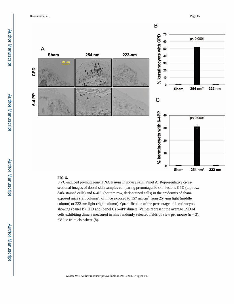
FIG. 3. Premutagenic skin DNA lesion yields induced by 222- and 254-nm UV light. Yields of
cyclobutane pyrimidine dimers (CPD) (left panel) and pyrimidine-pyrimidone 6-4
photoproducts (6-4PP) (right panel) in keratinocytes in a 3D human skin tissue model,
induced by conventional germicidal UV radiation (●) and by 222-nm UV light (▲). In both
graphs zero-fluence control measurements (<1%) have been subtracted from the data.
Buonanno et al. Page 13
Radiat Res. Author manuscript; available in PMC 2017 August 10.
Author M
anuscriptA
uthor Manuscript
Author M
anuscriptA
uthor Manuscript
FIG. 4. Epidermal thickness and keratinocyte proliferation in mouse skin exposed to UVC light.
Panel A: Representative cross-sectional images of H&E-stained mouse dorsal skin
comparing the epidermal thickness in sham-exposed mice (top panel), in mice exposed to
157 mJ/cm2 from 254-nm light (middle panel) or from 222-nm light (bottom panel). Panel
B: Quantification of epidermal thickness. Panel C: Ki-67-positive keratinocytes (dark
brown-stained cells) in typical cross-sections of skin of sham-exposed mice (top panel), of
mice exposed to 254-nm light (middle panel) or to 222-nm light (bottom panel). Panel D:
Quantification of the percentage of keratinocytes expressing Ki-67 antigen. Values represent
the average ± SD of epidermal thickness measured in nine randomly selected fields of view
per mouse (n = 3). *Values from elsewhere (8).
Buonanno et al. Page 14
Radiat Res. Author manuscript; available in PMC 2017 August 10.
Author M
anuscriptA
uthor Manuscript
Author M
anuscriptA
uthor Manuscript
FIG. 5. UVC-induced premutagenic DNA lesions in mouse skin. Panel A: Representative cross-


![US EPA, Pesticide Product Label, THYMOX DISINFECTANT … · Germicidal Spray Method] [and] [Virucidal*] [according to the ASTM Standard Test Method for Efficacy of Virucidal Agents][.]](https://static.documents.pub/doc/80x56/5c8aeadc09d3f22e408cb553/us-epa-pesticide-product-label-thymox-disinfectant-germicidal-spray-method.jpg)


