313 RESEARCH ARTICLE INTRODUCTION A subpopulation of astrocytes in the subventricular zone (SVZ) have been identified as the multipotent neural stem cells of the adult mammalian brain (Doetsch et al., 1999; Laywell et al., 2000; Quinones-Hinojosa et al., 2006; Sanai et al., 2004). These neural stem cells express glial fibrillary acidic protein (GFAP) and are located along the length of the lateral ventricles. In the human brain, intense expression of a splice variant of GFAP, termed GFAPd, was found at this specific location (Roelofs et al., 2005), which suggests that GFAPd-expressing cells might be the neural stem cells of the adult human brain. Recently, studies in rodents have shown that the adult neural stem cells in the SVZ are derived from radial glia (Bonfanti and Peretto, 2007; Merkle et al., 2004). Radial glia are bipolar cells with a soma in the ventricular zone (VZ), a short process making contact with the ventricular surface and a long radial fibre reaching the pial surface. In humans, however, there is, at present, no direct proof that adult SVZ astrocytes are derived from radial glia, although they have similar features. First, a number of SVZ neural progenitors maintain direct contact with the ventricles, like radial glia (Sanai et al., 2004; Tramontin et al., 2003). Second, neural progenitors from the SVZ migrate a long distance to the olfactory bulb in the adult rodent brain (Lois and Alvarez-Buylla, 1994); however, this is still under debate for the human brain (Curtis et al., 2007; Sanai et al., 2004; Sanai et al., 2007). This could be compared to the migration of neural progenitors from their site of birth in the VZ and SVZ to their final destination in the developing brain. Third, like radial glia, adult human SVZ neural stem cells express vimentin, nestin and GFAP (Sanai et al., 2004). The development of the layered structure of the cortex can be nicely monitored in sections of the developing cerebral cortex from human foetuses of various ages throughout gestation. In the course of early development, cortical neurons emerge from the radial glia in the VZ, which are direct descendants of the primitive neuroepithelium. The classical view is that radial glia are astrocyte precursor cells important for guiding neuronal migration (Bystron et al., 2008; Rakic, 1971). However, recent studies have shown that they can also act as neural stem cells, being capable of divisions that lead to the genesis of astrocytes, neurons (Götz et al., 2002; Hartfuss et al., 2001; Malatesta et al., 2000; Noctor et al., 2001; Noctor et al., 2002) and oligodendrocytes (Merkle et al., 2004). Radial glia express the intermediate filament (IF) proteins vimentin and nestin, and also, in primates, GFAP. As development proceeds, a second proliferative zone becomes discernable between the VZ and the cell- sparse intermediate zone, that is, the SVZ. The number of rapidly proliferating progenitor cells in the SVZ grows significantly during the last trimester, in parallel with a reduction in the number of cells in the VZ. Finally, the VZ will disappear before birth and the SVZ will persist into adulthood (Tramontin et al., 2003). A disturbance of the layered development of the human brain is observed in the developmental neuronal migration disorder type II lissencephaly, which is neuropathologically characterized by loss of sulci and broadening gyri, with often irregular appearance of the brain surface in affected regions (cobblestone appearance). This type of cortical dysplasia results from a failure of the arrest of radial neuronal migration due to defects in the integrity of the pial/glial Development 137, 313-321 (2010) doi:10.1242/dev.041632 1 Department of Astrocyte Biology & Neurodegeneration, Netherlands Institute for Neuroscience, An Institute of the Royal Netherlands Academy of Arts and Sciences, Amsterdam, The Netherlands. 2 Department of Neuropathology, Academic Medical Center, University of Amsterdam, Amsterdam, The Netherlands. 3 Department of Biotechnologies and Biosciences, University Bicocca of Milan, Milan, Italy. 4 Service Histologie-Embryologie-Cytogénétique, Groupe Hospitalier Necker Enfants-Malades, Université Paris-Descartes, Paris, France. 5 Department of Neuropsychiatric Disorders, Netherlands Institute for Neuroscience, Amsterdam, The Netherlands. 6 Stichting Epilepsie Instellingen Heemstede, Heemstede, The Netherlands. *Author for correspondence ([email protected]) Accepted 16 November 2009 SUMMARY A subpopulation of glial fibrillary acidic protein (GFAP)-expressing cells located along the length of the lateral ventricles in the subventricular zone (SVZ) have been identified as the multipotent neural stem cells of the adult mammalian brain. We have previously found that, in the adult human brain, a splice variant of GFAP, termed GFAPd, was expressed specifically in these cells. To investigate whether GFAPd is also present in the precursors of SVZ astrocytes during development and whether GFAPd could play a role in the developmental process, we analyzed GFAPd expression in the normal developing human cortex and in the cortex of foetuses with the migration disorder lissencephaly type II. We demonstrated for the first time that GFAPd is specifically expressed in radial glia and SVZ neural progenitors during human brain development. Expression of GFAPd in radial glia starts at around 13 weeks of pregnancy and disappears before birth. GFAPd is continuously expressed in the SVZ progenitors at later gestational ages and in the postnatal brain. Co-localization with Ki67 proved that these GFAPd-expressing cells are able to proliferate. Furthermore, we showed that the expression pattern of GFAPd was disturbed in lissencephaly type II. Overall, these results suggest that the adult SVZ is indeed a remnant of the foetal SVZ, which develops from radial glia. Furthermore, we provide evidence that GFAPd can distinguish resting astrocytes from proliferating SVZ progenitors. KEY WORDS: GFAP, Radial glia, Human brain development, Neural progenitors, Lissencephaly GFAPd in radial glia and subventricular zone progenitors in the developing human cortex Jinte Middeldorp 1 , Karin Boer 2 , Jacqueline A. Sluijs 1 , Lidia De Filippis 3 , Férechté Encha-Razavi 4 , Angelo L. Vescovi 3 , Dick F. Swaab 5 , Eleonora Aronica 2,6 and Elly M. Hol 1, * DEVELOPMENT DEVELOPMENT
Transcript
313RESEARCH ARTICLE
INTRODUCTIONA subpopulation of astrocytes in the subventricular zone (SVZ) havebeen identified as the multipotent neural stem cells of the adultmammalian brain (Doetsch et al., 1999; Laywell et al., 2000;Quinones-Hinojosa et al., 2006; Sanai et al., 2004). These neuralstem cells express glial fibrillary acidic protein (GFAP) and arelocated along the length of the lateral ventricles. In the human brain,intense expression of a splice variant of GFAP, termed GFAPd, wasfound at this specific location (Roelofs et al., 2005), which suggeststhat GFAPd-expressing cells might be the neural stem cells of theadult human brain.
Recently, studies in rodents have shown that the adult neural stemcells in the SVZ are derived from radial glia (Bonfanti and Peretto,2007; Merkle et al., 2004). Radial glia are bipolar cells with a somain the ventricular zone (VZ), a short process making contact with theventricular surface and a long radial fibre reaching the pial surface.In humans, however, there is, at present, no direct proof that adultSVZ astrocytes are derived from radial glia, although they havesimilar features. First, a number of SVZ neural progenitors maintaindirect contact with the ventricles, like radial glia (Sanai et al., 2004;Tramontin et al., 2003). Second, neural progenitors from the SVZmigrate a long distance to the olfactory bulb in the adult rodent brain
(Lois and Alvarez-Buylla, 1994); however, this is still under debatefor the human brain (Curtis et al., 2007; Sanai et al., 2004; Sanai etal., 2007). This could be compared to the migration of neuralprogenitors from their site of birth in the VZ and SVZ to their finaldestination in the developing brain. Third, like radial glia, adulthuman SVZ neural stem cells express vimentin, nestin and GFAP(Sanai et al., 2004).
The development of the layered structure of the cortex can benicely monitored in sections of the developing cerebral cortex fromhuman foetuses of various ages throughout gestation. In the courseof early development, cortical neurons emerge from the radial gliain the VZ, which are direct descendants of the primitiveneuroepithelium. The classical view is that radial glia are astrocyteprecursor cells important for guiding neuronal migration (Bystronet al., 2008; Rakic, 1971). However, recent studies have shown thatthey can also act as neural stem cells, being capable of divisions thatlead to the genesis of astrocytes, neurons (Götz et al., 2002; Hartfusset al., 2001; Malatesta et al., 2000; Noctor et al., 2001; Noctor et al.,2002) and oligodendrocytes (Merkle et al., 2004). Radial gliaexpress the intermediate filament (IF) proteins vimentin and nestin,and also, in primates, GFAP. As development proceeds, a secondproliferative zone becomes discernable between the VZ and the cell-sparse intermediate zone, that is, the SVZ. The number of rapidlyproliferating progenitor cells in the SVZ grows significantly duringthe last trimester, in parallel with a reduction in the number of cellsin the VZ. Finally, the VZ will disappear before birth and the SVZwill persist into adulthood (Tramontin et al., 2003).
A disturbance of the layered development of the human brain isobserved in the developmental neuronal migration disorder type IIlissencephaly, which is neuropathologically characterized by loss ofsulci and broadening gyri, with often irregular appearance of thebrain surface in affected regions (cobblestone appearance). This typeof cortical dysplasia results from a failure of the arrest of radialneuronal migration due to defects in the integrity of the pial/glial
Development 137, 313-321 (2010) doi:10.1242/dev.041632
1Department of Astrocyte Biology & Neurodegeneration, Netherlands Institute forNeuroscience, An Institute of the Royal Netherlands Academy of Arts and Sciences,Amsterdam, The Netherlands. 2Department of Neuropathology, Academic MedicalCenter, University of Amsterdam, Amsterdam, The Netherlands. 3Department ofBiotechnologies and Biosciences, University Bicocca of Milan, Milan, Italy. 4ServiceHistologie-Embryologie-Cytogénétique, Groupe Hospitalier Necker Enfants-Malades,Université Paris-Descartes, Paris, France. 5Department of Neuropsychiatric Disorders,Netherlands Institute for Neuroscience, Amsterdam, The Netherlands. 6StichtingEpilepsie Instellingen Heemstede, Heemstede, The Netherlands.
SUMMARYA subpopulation of glial fibrillary acidic protein (GFAP)-expressing cells located along the length of the lateral ventricles in thesubventricular zone (SVZ) have been identified as the multipotent neural stem cells of the adult mammalian brain. We havepreviously found that, in the adult human brain, a splice variant of GFAP, termed GFAPd, was expressed specifically in these cells. Toinvestigate whether GFAPd is also present in the precursors of SVZ astrocytes during development and whether GFAPd could play arole in the developmental process, we analyzed GFAPd expression in the normal developing human cortex and in the cortex offoetuses with the migration disorder lissencephaly type II. We demonstrated for the first time that GFAPd is specifically expressed inradial glia and SVZ neural progenitors during human brain development. Expression of GFAPd in radial glia starts at around 13weeks of pregnancy and disappears before birth. GFAPd is continuously expressed in the SVZ progenitors at later gestational agesand in the postnatal brain. Co-localization with Ki67 proved that these GFAPd-expressing cells are able to proliferate. Furthermore,we showed that the expression pattern of GFAPd was disturbed in lissencephaly type II. Overall, these results suggest that the adultSVZ is indeed a remnant of the foetal SVZ, which develops from radial glia. Furthermore, we provide evidence that GFAPd candistinguish resting astrocytes from proliferating SVZ progenitors.
GFAPd in radial glia and subventricular zone progenitors inthe developing human cortexJinte Middeldorp1, Karin Boer2, Jacqueline A. Sluijs1, Lidia De Filippis3, Férechté Encha-Razavi4,Angelo L. Vescovi3, Dick F. Swaab5, Eleonora Aronica2,6 and Elly M. Hol1,*
DEVELO
PMENT
DEVELO
PMENT
314
barrier. Consequential histopathological microscopic features arerepresented by a highly disorganized cerebral cortex structure, withfocal absence of a glia limitans, disturbance of radial fibres, andneuronal and glial heterotopias in the leptomeninges (Squier, 1993).
In the current study, we explored the developmental path of adultSVZ astrocytes in the normal developing human brain and in brainsof type II lissencephalic foetuses by analyzing GFAPd expression.We hypothesized that, if the presence of GFAPd is important for thedevelopment of SVZ neural stem cells, it would also be expressedin its precursors during development. We furthermore expect thatthe localization of these precursors is disturbed in lissencephalicbrains due to a disruption of radial glia and that this can be observedby studying the GFAPd-expressing cells. In addition, continuousexpression of GFAPd in this lineage would provide evidence that theadult human SVZ astrocytes originate from radial glia.
MATERIALS AND METHODSHuman brain materialThe subjects included in this study were obtained from the brain collectionsof the Department of Neuropathology of the Academic Medical Center,University of Amsterdam, The Netherlands and the Service Histologie-Embryologie-Cytogénétique Hôpital Necker-Enfants Malades, Paris,France. Informed consent was obtained for the use of brain tissue and foraccess to medical records for research purposes. The expression of GFAPdwas evaluated during brain development; the following ages were included:gestational week (gw) 9, 10, 13, 16, 17, 20, 22, 23, 25, 29, 31, 36 and 40,based on last menstrual period and ultrasound scanning. The tissue wasobtained from spontaneous or medically induced abortions, with appropriatematernal written consent for brain autopsy. We also obtained normal-appearing control cortex/white matter at autopsy from three young cases (3months, 7 months and 8.5 years), without a history of seizures or otherneurological diseases. In addition, we obtained tissue from three cases withtype II lissencephaly of 21, 22 and 23 gw. All autopsies were performedwithin 12 hours of death. More details of the human brain material used aresummarized in Table S1 in the supplementary material.
Tissue preparationTissue was fixed in 10% buffered formalin and embedded in paraffin.Paraffin-embedded tissue was sectioned at 6 m and mounted onorganosilane-coated slides (Sigma, St Louis, MO, USA), and used forimmunocytochemical staining as described below.
Immunocytochemical analysisFor single labelling, paraffin-embedded sections were deparaffinized,rehydrated and incubated for 20 min in 0.3% H2O2 diluted in methanol toquench the endogenous peroxidase activity. Antigen retrieval was performedby incubation for 10 min at 121°C in citrate buffer (0.01 M, pH 6.0) in apressure cooker. Sections were washed with phosphate-buffered saline(PBS: 137 mM NaCl, 2.7 mM KCl, 1.8 mM KH2PO4 and 4 mM Na2HPO4,pH 7.4) and incubated for 30 min in 10% normal goat serum (Harlan Sera-Lab, Loughborough, Leicestershire, UK). After incubation with polyclonalrabbit pan-GFAP, which recognizes all GFAP isoforms (DAKO, Glostrup,Denmark; 1:4000), and GFAPd [bleeding 100501; 1:500 (Roelofs et al.,2005)] primary antibodies in PBS overnight at 4°C, we washed the sectionsin PBS. We then used the ready-for-use Powervision peroxidase system(Immunologic, Duiven, The Netherlands) and 3,3�-diaminobenzidine(DAB; Sigma) as a chromogen to visualize the antibodies. Sections werecounterstained with haematoxylin, dehydrated and coverslipped.
To determine more specifically the cell type of GFAPd-expressing cellsin the developing brain, we performed several co-localization studies. Wecombined GFAPd with monoclonal mouse antibodies against vimentin(clone V9; DAKO; 1:1000), a known marker of radial glia in the VZ duringhuman brain development (Honig et al., 1996), nestin (MAB5326,Chemicon; 1:200), a marker of both a subpopulation of radial glia (Zecevic,2004) and SVZ neural progenitors (Lendahl et al., 1990), Ki67 (cloneMIB-1, DAKO; 1:200), a marker of proliferation (Scholzen and Gerdes,2000), and with polyclonal rabbit antibody against Sox2 (AB5603,
Chemicon; 1:200), a transcription factor important for the maintenance ofneural stem cells (Graham et al., 2003). For double labelling with polyclonalgoat antibody against the C terminus of GFAP, which could, in principle,also detect other low-expressed GFAP isoforms, for example, GFAP135(Hol et al., 2003) (GFAP C-term; Santa Cruz Biotechnology; 1:200), weblocked with normal swine serum instead of normal goat serum. Afterincubation overnight with the primary antibodies at 4°C, sections wereincubated for two hours at room temperature with Alexa Fluor 568-conjugated anti-rabbit IgG and Alexa Fluor® 488 anti-mouse or anti-goatIgG (1:100, Molecular Probes, The Netherlands). Sections were mountedwith Vectashield containing DAPI (which targets DNA in the cell nucleus;blue emission) and analyzed using a laser scanning confocal microscope(Leica TCS Sp2, Wetzlar, Germany). Sections incubated without the primaryantibody or with pre-immune serum (for GFAPd) were essentially blank,except for the outer layer of the VZ/ependymal layer, which, in some cases,appeared a bit darker when treated with the pre-immune serum.
GFAP plasmids and transfectionsTo obtain recombinant protein samples for western blotting, expressionplasmids pcDNA3-GFAP and pcDNA3-GFAPd (Roelofs et al., 2005) weretransfected separately into SH-SY5Y neuroblastoma cells, which do notexpress GFAP endogenously. These cells were cultured in high-glucoseDMEM, supplemented with 10% heat-inactivated foetal calf serum, 100U/ml penicillin and 100 g/ml streptomycin, at 37°C with 5% CO2. The cellculture medium was refreshed two hours before lipofectamine (Invitrogen)transfection with pcDNA3-GFAP or pcDNA3-GFAPd plasmids, accordingto the manufacturer’s protocol.
Western blotProtein was isolated from GFAP- and GFAPd-transfected SH-SY5Y cellsby homogenization with lysis buffer (0.1 M NaCl, 0.01 M Tris-HCl pH 7.6,1 mM EDTA pH 8.0) supplemented with a protease inhibitor cocktail(Roche Diagnostics, Mannheim, Germany). The samples were dissolved in2� loading buffer (2�: 100 mM Tris, 4% SDS, 20% glycerol, 200 mMDTT, 0.006% bromophenol blue) and boiled for five minutes. Subsequently,they were run on a 7.5% SDS-PAGE gel and blotted semi-dry onnitrocellulose. Blots were probed overnight with polyclonal rabbit pan-GFAP (DAKO; 1:20 000), GFAPd [bleeding 100501; 1:500 (Roelofs et al.,2005)] or polyclonal goat C-terminal GFAP (GFAP C-term; Santa CruzBiotechnology; 1:200) antibodies diluted in Supermix (0.05 M Tris, 0.9%NaCl, 0.25% gelatin and 0.5% Triton X-100, pH 7.4) antibodies. The nextday, the blots were washed with TBS-T (TBS; 100 mM Tris-HCl pH 7.4,150 mM NaCl, with 0.2% tween-20) and incubated with secondary antibodyanti-rabbit IRDye800 or anti-goat IRDye800 (1:5000; RocklandImmunochemicals, Gilbertsville, USA) in Supermix for one hour at roomtemperature. After three washes in TBS, bands were visualized with theOdyssey Infrared Imaging System (LI-COR Biosciences, Lincoln, NE,USA).
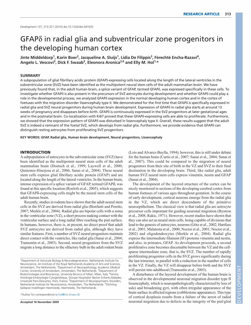
RESULTSDifferent GFAP antibodies recognize specific GFAPisoformsA western blot analysis of recombinant GFAP or GFAPd proteinwas performed to verify the specificity of the different GFAPantibodies used in this study. As indicated in a schematic picture(Fig. 1A), the epitope of the pan-GFAP antibody is not known. TheGFAP C-term and GFAPd antibodies are directed to a sequence inthe C terminus of, respectively, GFAP and GFAPd. Atapproximately 55 kDa, protein bands for both GFAP and GFAPdprotein were detected with the pan-GFAP antibody, whereas theGFAPd antibody specifically detected the GFAPd protein, as waspreviously shown (Roelofs et al., 2005). The antibody directedagainst the C terminus of GFAP only detected GFAPrecombinant protein, not GFAPd (Fig. 1B).
RESEARCH ARTICLE Development 137 (2)
DEVELO
PMENT
DEVELO
PMENT
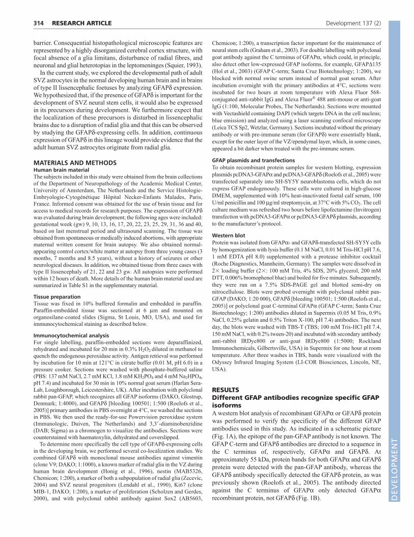
GFAPd expression during human braindevelopment visualizes the development of theneurogenic astrocytic ribbonGFAPd expression was not observed in brain sections at 10 gw andyounger (Fig. 2A). In addition, the pan-GFAP antibody failed to showany GFAP expression in adjacent sections (Fig. 2B). The first GFAPdexpression was detected at 13 gw in cells lining the VZ along thelateral ventricles (Fig. 2C). These cells had a bipolar morphology, withsomata in the VZ, short processes towards the ventricles and longerfibres extending into the brain parenchyma. These radial-glia-like cellswere also intensely stained by the pan-GFAP antibody, which moreclearly labelled the long fibres extending from the VZ into the brainparenchyma (Fig. 2D). Around 17 gw, the SVZ clearly expressedGFAPd and possibly other GFAP isoforms as well, because we alsoobserved pan-GFAP expression (Fig. 2E,F). The VZ cell fibres, whichwere also still visible at this stage in development, seemed to extendthrough the SVZ cell layer, which contained more round and unipolarcells. GFAPd expression at this stage was clearly less pronounced thanthat of pan-GFAP, pointing to stronger expression of other GFAPisoforms. This difference became even more evident at older ages,when additional transitional profiles of radial glia appeared and cellsbecame more differentiated (Fig. 2G-J). At 22 gw, the VZ cells stillexpressed GFAPd, as did the cells in the SVZ (Fig. 2G). However,with the pan-GFAP antibody, much more intense labelling wasdetected and also more cells outside the SVZ were positive for pan-GFAP (Fig. 2H), whereas very little GFAPd expression was found incells outside the VZ and SVZ. At the perinatal age of 36 gw, the VZcells were no longer present and GFAPd expression was limited to theSVZ and some processes in the ependymal layer (Fig. 2I), whereaspan-GFAP expression was also found throughout the rest of the brain(Fig. 2J). This GFAPd expression pattern is similar to that observedpostnatally (not shown) and in the adult brain (Roelofs et al., 2005).
GFAPd-positive cells are a subpopulation of allGFAP-expressing cellsAlthough several GFAP isoforms, including GFAPd, were expressedin the developing VZ and SVZ, superficial layers of the developingcortex lacked GFAPd expression, but did clearly express other
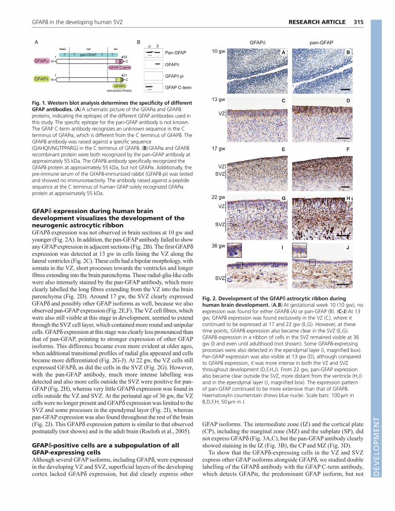
GFAP isoforms. The intermediate zone (IZ) and the cortical plate(CP), including the marginal zone (MZ) and the subplate (SP), didnot express GFAPd (Fig. 3A,C), but the pan-GFAP antibody clearlyshowed staining in the IZ (Fig. 3B), the CP and MZ (Fig. 3D).
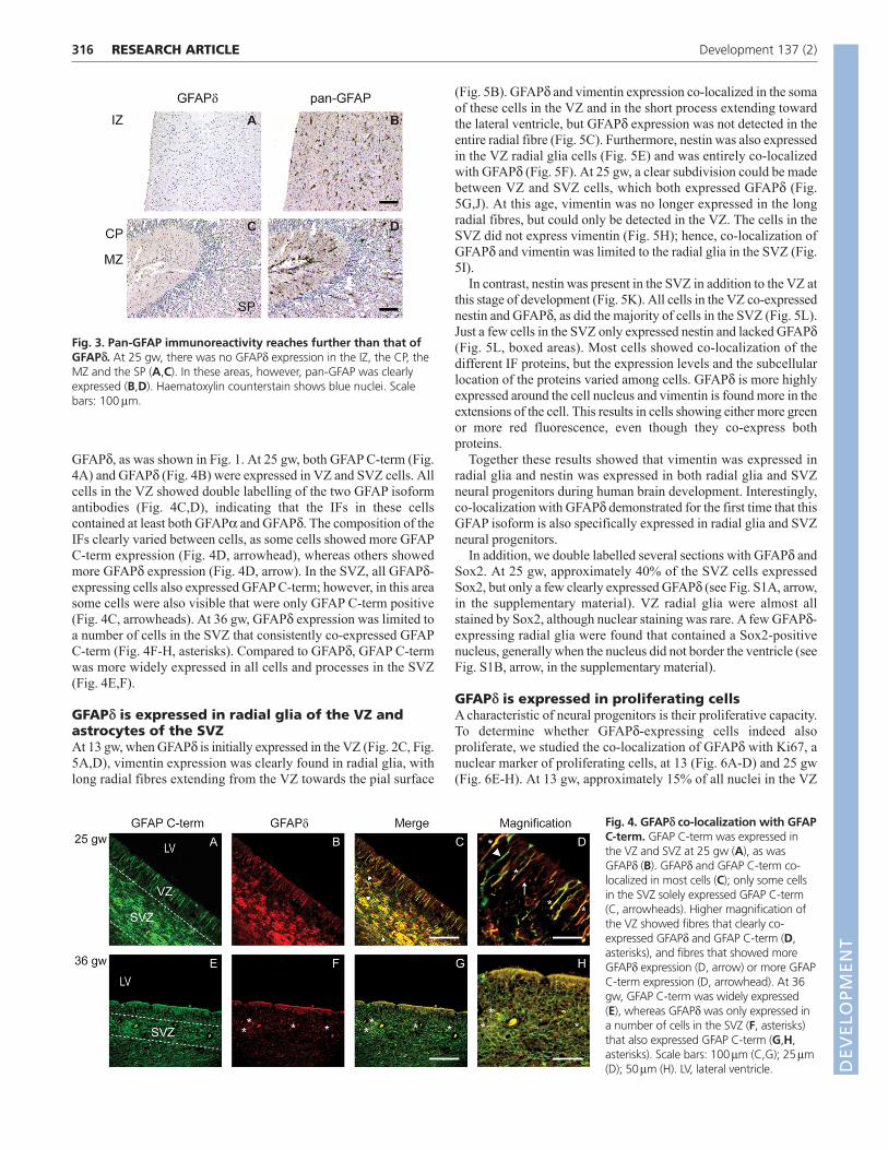
To show that the GFAPd-expressing cells in the VZ and SVZexpress other GFAP isoforms alongside GFAPd, we studied doublelabelling of the GFAPd antibody with the GFAP C-term antibody,which detects GFAP, the predominant GFAP isoform, but not
315RESEARCH ARTICLEGFAPd in the developing human SVZ
Fig. 1. Western blot analysis determines the specificity of differentGFAP antibodies. (A)A schematic picture of the GFAP and GFAPdproteins, indicating the epitopes of the different GFAP antibodies used inthis study. The specific epitope for the pan-GFAP antibody is not known.The GFAP C-term antibody recognizes an unknown sequence in the Cterminus of GFAP, which is different from the C terminus of GFAPd. TheGFAPd antibody was raised against a specific sequence(QAHQIVNGTPPARG) in the C terminus of GFAPd. (B)GFAP and GFAPdrecombinant protein were both recognized by the pan-GFAP antibody atapproximately 55 kDa. The GFAPd antibody specifically recognized theGFAPd protein at approximately 55 kDa, but not GFAP. Additionally, thepre-immune serum of the GFAPd-immunized rabbit (GFAPd pi) was testedand showed no immunoreactivity. The antibody raised against a peptidesequence at the C terminus of human GFAP solely recognized GFAPprotein at approximately 55 kDa.
Fig. 2. Development of the GFAPd astrocytic ribbon duringhuman brain development. (A,B)At gestational week 10 (10 gw), noexpression was found for either GFAPd (A) or pan-GFAP (B). (C-J)At 13gw, GFAPd expression was found exclusively in the VZ (C), where itcontinued to be expressed at 17 and 22 gw (E,G). However, at thesetime points, GFAPd expression also became clear in the SVZ (E,G).GFAPd expression in a ribbon of cells in the SVZ remained visible at 36gw (I) and even until adulthood (not shown). Some GFAPd-expressingprocesses were also detected in the ependymal layer (I, magnified box).Pan-GFAP expression was also visible at 13 gw (D), although comparedto GFAPd expression, it was more intense in both the VZ and SVZthroughout development (D,F,H,J). From 22 gw, pan-GFAP expressionalso became clear outside the SVZ, more distant from the ventricle (H,J)and in the ependymal layer (J, magnified box). The expression patternof pan-GFAP continued to be more extensive than that of GFAPd.Haematoxylin counterstain shows blue nuclei. Scale bars: 100m inB,D,F,H; 50m in J.
DEVELO
PMENT
DEVELO
PMENT
316
GFAPd, as was shown in Fig. 1. At 25 gw, both GFAP C-term (Fig.4A) and GFAPd (Fig. 4B) were expressed in VZ and SVZ cells. Allcells in the VZ showed double labelling of the two GFAP isoformantibodies (Fig. 4C,D), indicating that the IFs in these cellscontained at least both GFAP and GFAPd. The composition of theIFs clearly varied between cells, as some cells showed more GFAPC-term expression (Fig. 4D, arrowhead), whereas others showedmore GFAPd expression (Fig. 4D, arrow). In the SVZ, all GFAPd-expressing cells also expressed GFAP C-term; however, in this areasome cells were also visible that were only GFAP C-term positive(Fig. 4C, arrowheads). At 36 gw, GFAPd expression was limited toa number of cells in the SVZ that consistently co-expressed GFAPC-term (Fig. 4F-H, asterisks). Compared to GFAPd, GFAP C-termwas more widely expressed in all cells and processes in the SVZ(Fig. 4E,F).
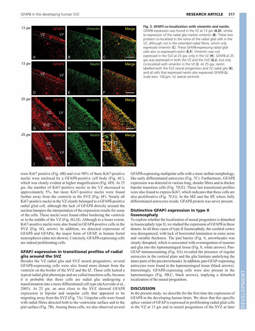
GFAPd is expressed in radial glia of the VZ andastrocytes of the SVZAt 13 gw, when GFAPd is initially expressed in the VZ (Fig. 2C, Fig.5A,D), vimentin expression was clearly found in radial glia, withlong radial fibres extending from the VZ towards the pial surface
(Fig. 5B). GFAPd and vimentin expression co-localized in the somaof these cells in the VZ and in the short process extending towardthe lateral ventricle, but GFAPd expression was not detected in theentire radial fibre (Fig. 5C). Furthermore, nestin was also expressedin the VZ radial glia cells (Fig. 5E) and was entirely co-localizedwith GFAPd (Fig. 5F). At 25 gw, a clear subdivision could be madebetween VZ and SVZ cells, which both expressed GFAPd (Fig.5G,J). At this age, vimentin was no longer expressed in the longradial fibres, but could only be detected in the VZ. The cells in theSVZ did not express vimentin (Fig. 5H); hence, co-localization ofGFAPd and vimentin was limited to the radial glia in the SVZ (Fig.5I).
In contrast, nestin was present in the SVZ in addition to the VZ atthis stage of development (Fig. 5K). All cells in the VZ co-expressednestin and GFAPd, as did the majority of cells in the SVZ (Fig. 5L).Just a few cells in the SVZ only expressed nestin and lacked GFAPd(Fig. 5L, boxed areas). Most cells showed co-localization of thedifferent IF proteins, but the expression levels and the subcellularlocation of the proteins varied among cells. GFAPd is more highlyexpressed around the cell nucleus and vimentin is found more in theextensions of the cell. This results in cells showing either more greenor more red fluorescence, even though they co-express bothproteins.
Together these results showed that vimentin was expressed inradial glia and nestin was expressed in both radial glia and SVZneural progenitors during human brain development. Interestingly,co-localization with GFAPd demonstrated for the first time that thisGFAP isoform is also specifically expressed in radial glia and SVZneural progenitors.
In addition, we double labelled several sections with GFAPd andSox2. At 25 gw, approximately 40% of the SVZ cells expressedSox2, but only a few clearly expressed GFAPd (see Fig. S1A, arrow,in the supplementary material). VZ radial glia were almost allstained by Sox2, although nuclear staining was rare. A few GFAPd-expressing radial glia were found that contained a Sox2-positivenucleus, generally when the nucleus did not border the ventricle (seeFig. S1B, arrow, in the supplementary material).
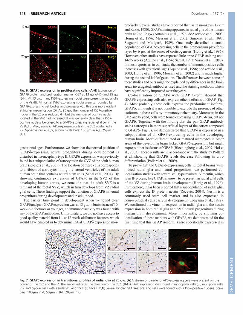
GFAPd is expressed in proliferating cellsA characteristic of neural progenitors is their proliferative capacity.To determine whether GFAPd-expressing cells indeed alsoproliferate, we studied the co-localization of GFAPd with Ki67, anuclear marker of proliferating cells, at 13 (Fig. 6A-D) and 25 gw(Fig. 6E-H). At 13 gw, approximately 15% of all nuclei in the VZ
RESEARCH ARTICLE Development 137 (2)
Fig. 3. Pan-GFAP immunoreactivity reaches further than that ofGFAPd. At 25 gw, there was no GFAPd expression in the IZ, the CP, theMZ and the SP (A,C). In these areas, however, pan-GFAP was clearlyexpressed (B,D). Haematoxylin counterstain shows blue nuclei. Scalebars: 100m.
Fig. 4. GFAPd co-localization with GFAPC-term. GFAP C-term was expressed inthe VZ and SVZ at 25 gw (A), as wasGFAPd (B). GFAPd and GFAP C-term co-localized in most cells (C); only some cellsin the SVZ solely expressed GFAP C-term(C, arrowheads). Higher magnification ofthe VZ showed fibres that clearly co-expressed GFAPd and GFAP C-term (D,asterisks), and fibres that showed moreGFAPd expression (D, arrow) or more GFAPC-term expression (D, arrowhead). At 36gw, GFAP C-term was widely expressed(E), whereas GFAPd was only expressed ina number of cells in the SVZ (F, asterisks)that also expressed GFAP C-term (G,H,asterisks). Scale bars: 100m (C,G); 25m(D); 50m (H). LV, lateral ventricle. D
EVELO
PMENT
DEVELO
PMENT
were Ki67 positive (Fig. 6B) and over 90% of these Ki67-positivenuclei were enclosed by a GFAPd-positive cell body (Fig. 6C),which was clearly evident at higher magnification (Fig. 6D). At 25gw, the number of Ki67-positive nuclei in the VZ decreased toapproximately 5%, but more Ki67-positive nuclei were foundfurther away from the ventricle in the SVZ (Fig. 6F). Nearly allKi67-positive nuclei in the VZ clearly belonged to a GFAPd-positiveradial glial cell, although the lack of GFAPd directly around thenucleus hampers the interpretation of the expression results for someof the cells. These nuclei were found either bordering the ventricleor in the middle of the VZ (Fig. 6G,H). Although to a lesser extent,Ki67-positive nuclei were also found in GFAPd-positive cells in theSVZ (Fig. 6G, arrow). In addition, we detected expression ofGFAPd and GFAP, the major form of GFAP, in human foetalneurospheres (data not shown). Concisely, GFAPd-expressing cellsare indeed proliferating cells.
GFAPd expression in transitional profiles of radialglia around the SVZBesides the VZ radial glia and SVZ neural progenitors, severalGFAPd-expressing cells were also found more distant from theventricle on the border of the SVZ and the IZ. These cells lacked atypical radial glial phenotype and are called transition cells, becauseit is probable that these cells are radial glia undergoing atransformation into a more differentiated cell type (deAzevedo et al.,2003). At 25 gw, an area close to the SVZ showed GFAPdexpression in bipolar and unipolar cells that appeared to bemigrating away from the SVZ (Fig. 7A). Unipolar cells were foundwith radial fibres directed both to the ventricular surface and to thepial surface (Fig. 7B). Among those cells, we also observed several
GFAPd-expressing multipolar cells with a more stellate morphology,like early differentiated astrocytes (Fig. 7C). Furthermore, GFAPdexpression was detected in various long, slender fibres and in thickerbipolar transition cells (Fig. 7D,E). These last transitional profileswere also found to express Ki67, which indicates that these cells arealso proliferative (Fig. 7F,G). In the MZ and the SP, where fullydifferentiated astrocytes reside, GFAPd protein was never present.
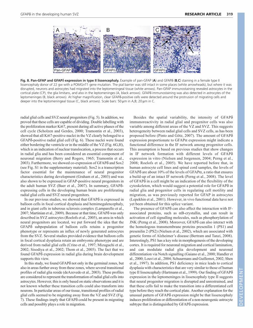
Distinctive GFAPd expression in type IIlissencephalyTo explore whether the localization of neural progenitors is disturbedin lissencephaly type II, we studied the expression of GFAPd in thesedonors. In all three cases of type II lissencephaly, the cerebral cortexwas disorganized, with lack of horizontal lamination in some areasand variable thickness. The pial barrier (Fig. 8, arrowheads) wasclearly disrupted, which is associated with overmigration of neuronsand glia into the leptomeningeal tissue (Fig. 8, white arrows). Pan-GFAP immunostaining (Fig. 8A) revealed the presence of regularastrocytes in the cortical plate and the glia limitans underlying theintact parts of the pia (arrowheads). In addition, pan-GFAP-expressingastrocytes were found in the leptomeningeal tissue (black arrows).Interestingly, GFAPd-expressing cells were also present in theleptomeninges (Fig. 8B,C, black arrows), implying a disturbedlocalization of the neural progenitors.
DISCUSSIONIn the present study, we describe for the first time the expression ofGFAPd in the developing human brain. We show that this specificsplice variant of GFAP is expressed in proliferating radial glial cellsin the VZ at 13 gw and in neural progenitors of the SVZ at later
317RESEARCH ARTICLEGFAPd in the developing human SVZ
Fig. 5. GFAPd co-localization with vimentin and nestin.GFAPd expression was found in the VZ at 13 gw (A,D), similarto expression of the radial glia marker vimentin (B). These twoproteins co-localized in the soma of the radial glial cells in theVZ, although not in the extended radial fibres, which onlyexpressed vimentin (C). These GFAPd-expressing radial glialcells also co-expressed nestin (E,F). Vimentin was notexpressed in the SVZ at 25 gw, only in the VZ (H). GFAPd at 25gw was expressed in both the VZ and the SVZ (G,J), but onlyco-localized with vimentin in the VZ (I). At 25 gw, nestinlabelled both the SVZ neural progenitors and VZ radial glia (K),and all cells that expressed nestin also expressed GFAPd (L).Scale bars: 100m. LV, lateral ventricle.
DEVELO
PMENT
DEVELO
PMENT
318
gestational ages. Furthermore, we show that the normal position ofGFAPd-expressing neural progenitors during development isdisturbed in lissencephaly type II. GFAPd expression was previouslyfound in a subpopulation of astrocytes in the SVZ of the adult humanbrain (Roelofs et al., 2005). The location of these cells correspondsto a ribbon of astrocytes lining the lateral ventricles of the adulthuman brain that contains neural stem cells (Sanai et al., 2004). Byshowing continuous expression of GFAPd in the SVZ of thedeveloping human cortex, we conclude that the adult SVZ is aremnant of the foetal SVZ, which in turn develops from VZ radialglial cells. These findings support the function of GFAPd in neuralprogenitors during development and in adulthood.
The earliest time point in development when we found clearGFAPd and pan-GFAP expression was at 13 gw. In brain tissue of 10-week-old foetuses or younger, no immunoreactivity was found withany of the GFAP antibodies. Unfortunately, we did not have access togood-quality material from 11- or 12-week-old human foetuses, whichwould have enabled us to determine initial GFAPd expression more
precisely. Several studies have reported that, as in monkeys (Levittand Rakic, 1980), GFAP staining appeared in radial glia of the humanbrain at 9 to 12 gw (Antanitus et al., 1976; deAzevedo et al., 2003;Honig et al., 1996; Messam et al., 2002; Simonati et al., 1997;Stagaard and Mollgard, 1989). One study described a smallpopulation of GFAP-expressing cells in the premordium plexiformlayer by 6 gw, at the onset of corticogenesis (Honig et al., 1996).However, other studies have reported little or no GFAP staining until14-25 weeks (Aquino et al., 1996; Sarnat, 1992; Sasaki et al., 1988).In most reports, as in our study, the number of immunopositive cellsincreases with gestational age (Aquino et al., 1996; deAzevedo et al.,2003; Honig et al., 1996; Messam et al., 2002) and is much higherduring the second half of gestation. The differences between some ofthese studies and ours might be explained by differences in the brainareas investigated, antibodies used and the staining methods, whichhave significantly improved over the years.
Co-localization of GFAPd with GFAP C-term showed thatGFAPd-expressing cells also express other isoforms of GFAP (Fig.4). Most probably, these cells express the predominant isoform,GFAP, although it is not possible to exclude the presence of otherisoforms in these cells by immunocytochemistry. Moreover, in theSVZ and beyond, cells were found expressing GFAP C-term, but notGFAPd. Together with the finding that the pan-GFAP antibodystains astrocytes in more superficial layers of the cortex comparedto GFAPd (Fig. 3), we demonstrated that GFAPd is expressed in asubpopulation of all GFAP-expressing cells in the developinghuman brain. More differentiated or matured astrocytes in otherareas of the developing brain lacked GFAPd expression, but mightexpress other isoforms of GFAP (Blechingberg et al., 2007; Hol etal., 2003). These results are in accordance with the study by Pollardet al. showing that GFAPd levels decrease following in vitrodifferentiation (Pollard et al., 2009).
To prove that the GFAPd-expressing cells in foetal brains wereindeed radial glia and neural progenitors, we performed co-localization studies with several cell type markers. Vimentin, whichis an IF protein, like GFAP, is known to be present in radial glial cellsof the VZ during human brain development (Honig et al., 1996).Furthermore, it has been reported that a subpopulation of radial glialcells express the IF protein nestin (Zecevic, 2004). Nestin is acommonly used stem cell marker and is also expressed inneuroepithelial cells early in development (Tohyama et al., 1992).We confirmed the vimentin expression in radial glia and the nestinexpression in both radial glia and SVZ neural progenitors duringhuman brain development. More importantly, by showing co-localization of these markers with GFAPd, we demonstrated for thefirst time that this GFAP isoform is also specifically expressed in
RESEARCH ARTICLE Development 137 (2)
Fig. 6. GFAPd expression in proliferating cells. (A-H)Expression ofGFAPd protein and proliferation marker Ki67 at 13 gw (A-D) and 25 gw(E-H). At 13 gw, many Ki67-expressing nuclei were present in radial gliaof the VZ (B). Almost all Ki67-expressing nuclei were surrounded byGFAPd-expressing cell bodies and processes (C); this was more evidentat higher magnification (D). At 25 gw, the number of Ki67-positivenuclei in the VZ was reduced (F), but the number of positive nucleilocated in the SVZ had increased. It was generally clear that a Ki67-positive nucleus belonged to a GFAPd-expressing radial glial cell in theVZ (G,H). Also, some GFAPd-expressing cells in the SVZ contained aKi67-positive nucleus (G, arrow). Scale bars: 100m in A,E; 25m inD,H.
Fig. 7. GFAPd expression in transitional profiles of radial glia at 25 gw. (A)A stream of parallel GFAPd-expressing cells were present on theborder of the SVZ and the IZ. The arrow indicates the direction of the SVZ. (B-E)GFAPd expression was found in monopolar cells (B), multipolar cells(C), and bipolar cells with slender (D) and thick (E) fibres. (F,G)Several bipolar GFAPd-expressing cells were found with a Ki67-positive nucleus. Scalebars: 100m in A; 50m in B-F; 20m in G. D
EVELO
PMENT
DEVELO
PMENT
radial glial cells and SVZ neural progenitors (Fig. 5). In addition, weproved that these cells are capable of dividing. Double labelling withthe proliferation marker Ki67, present during all active phases of thecell cycle (Scholzen and Gerdes, 2000; Tramontin et al., 2003),showed that all Ki67-positive nuclei in the VZ clearly belonged to aGFAPd-positive radial glial cell (Fig. 6). These nuclei were foundeither bordering the ventricle or in the middle of the VZ (Fig. 6G,H),which is an indication of nuclear translocation, a process that occursin radial glia and has been considered an essential component ofneuronal migration (Berry and Rogers, 1965; Tramontin et al.,2003). Furthermore, we showed co-expression of GFAPd and Sox2(see Fig. S1 in the supplementary material). Sox2 is a transcriptionfactor essential for the maintenance of neural progenitorcharacteristics during development (Graham et al., 2003) and wasalso shown to be expressed in GFAP-positive neural progenitors inthe adult human SVZ (Baer et al., 2007). In summary, GFAPd-expressing cells in the developing human brain are proliferatingradial glial cells and SVZ neural progenitors.
In our previous studies, we showed that GFAPd is expressed inballoon cells in focal cortical dysplasia and hemimegalencephaly,and in giant cells in tuberous sclerosis complex (Lamparello et al.,2007; Martinian et al., 2009). Because at that time, GFAPdwas onlydescribed in SVZ astrocytes (Roelofs et al., 2005), an area in whichneural progenitors are located, we put forward the idea that theGFAPd subpopulation of balloon cells retains a progenitorphenotype or represents an influx of newly generated astrocytesfrom the SVZ. Several studies provided evidence that balloon cellsin focal cortical dysplasia retain an embryonic phenotype and arederived from radial glial cells (Crino et al., 1997; Mizuguchi et al.,2002; Sisodiya et al., 2002; Thom et al., 2005). The fact that wefound GFAPd expression in radial glia during brain developmentsupports this view.
In this study, we found GFAPd not only in the germinal zones, butalso in areas further away from these zones, where several transitionalprofiles of radial glia reside (deAzevedo et al., 2003). These profilesare considered to represent the transformation of radial glial cells intoastrocytes. However, this is only based on static observations and it isnot known whether these transition cells could also transform intoneurons. In particular areas of our tissue, transitional profiles of radialglial cells seemed to be migrating away from the VZ and SVZ (Fig.7). These findings imply that GFAPd could be present in migratingcells and possibly plays a role in migration.
Besides the spatial variability, the intensity of GFAPdimmunoreactivity in radial glial and progenitor cells was alsovariable among different areas of the VZ and SVZ. This suggestsheterogeneity between radial glial cells and SVZ cells, as has beenproposed before (Pinto and Götz, 2007). The amount of GFAPdexpression proportionate to GFAP expression might indicate afunctional difference in the IF network among progenitor cells.This assumption is based on previous studies that show changesin IF network formation with different levels of GFAPdexpression in vitro (Nielsen and Jorgensen, 2004; Perng et al.,2008; Roelofs et al., 2005). We have reported before that, inhuman astrocyte cell lines and spinal cord samples, the levels ofGFAPd are about 10% of the levels of GFAP, a ratio that ensuresa build-up of an intact IF network (Perng et al., 2008). The levelof GFAPd in a cell might be an indication of the flexibility of thecytoskeleton, which would suggest a potential role for GFAPd inradial glia and progenitor cells in regulating cell motility andmigration, as was previously reported for GFAP in astrocytes(Lepekhin et al., 2001). However, in vivo functional data have notyet been obtained for this splice variant.
The presence of GFAPd can also affect the interaction with IF-associated proteins, such as B-crystallin, and can result inactivation of cell signalling molecules, such as phosphorylation ofJNK (Perng et al., 2008). Moreover, GFAPd can also interact withthe homologous transmembrane proteins presenilin 1 (PS1) andpresenilin 2 (PS2) (Nielsen et al., 2002), which are associated withgenetic forms of Alzheimer’s disease (Bertram and Tanzi, 2008).Interestingly, PS1 has a key role in morphogenesis of the developingcortex. It is required for neuronal migration and cortical lamination,and can modulate radial glia development and neuronaldifferentiation via Notch signalling (Gaiano et al., 2000; Handler etal., 2000; Louvi et al., 2004; Schuurmans and Guillemot, 2002; Shenet al., 1997). In addition, PS1 deficiency in mice leads to corticaldysplasia with characteristics that are very similar to those of humantype II lissencephaly (Hartmann et al., 1999). Our finding of GFAPdexpression in the leptomeninges in lissencephaly type II suggeststhat neural progenitor migration is disrupted and unrestrained, andthat these cells fail to make the transition into a differentiated celltype before they reach the cortical plate. Another explanation for theunusual pattern of GFAPd expression might be that lissencephalyinduces proliferation or differentiation of a non-neurogenic astrocytesubtype that is distinguished by GFAPd expression.
319RESEARCH ARTICLEGFAPd in the developing human SVZ
Fig. 8. Pan-GFAP and GFAPd expression in type II lissencephaly. Example of pan-GFAP (A) and GFAPd (B,C) staining in a female type IIlissencephaly donor of 22 gw with a POMGnT1 gene mutation. The pial barrier was still intact in some places (white arrowheads), but where it wasdisrupted, neurons and astrocytes had migrated into the leptomeningeal tissue (white arrows). Pan-GFAP immunostaining revealed astrocytes in thecortical plate (CP), the glia limitans, and also in the leptomeninges (A, black arrows). GFAPd immunostaining was also detected in astrocytes of theleptomeninges (B, black arrows). At higher magnification, clear GFAPd-positive cells were detected around the protrusion of migrating cells anddeeper into the leptomeningeal tissue (C, black arrows). Scale bars: 50m in A,B; 20m in C.
DEVELO
PMENT
DEVELO
PMENT
320
Whether the interaction of GFAPd with presenilin could beinvolved in this disturbed development requires furtherinvestigation. Regulation of neuronal migration by PS1 showedstriking similarities to regulation by cyclin-dependent kinase 5(Cdk5) (Ohshima et al., 2002). Cdk5 is associated with the IFprotein nestin (Sahlgren et al., 2003), which is co-expressed withGFAPd in the developing cortex (Fig. 5F,L).
In conclusion, we show that GFAPd is a protein that might haveimportant functions in radial glia and neural progenitors in thehuman developing cortex. The interaction of GFAPd with proteinsinvolved in development and cell signalling implies that this specificGFAP isoform has a function in human brain development. Furtherresearch is needed to find out the exact role of GFAPd in neural stemcells, their migration and the commitment of their progeny.
AcknowledgementsWe acknowledge the financial support of the Internationale StichtingAlzheimer Onderzoek (ISAO; 04511 to E.M.H.), NWO-ALW-Vici (865.09.003 toE.M.H.), Hersenstichting Nederland (HsN; 13F05.08 to E.M.H.), NationalEpilepsy Fund (NEF; 05-11 to K.B. and E.A. and 09-05 to E.A.) and EU FP7project NeuroGlia (grant agreement number 202167 to E.A.).
Competing interests statementThe authors declare no competing financial interests
Supplementary materialSupplementary material for this article is available athttp://dev.biologists.org/lookup/suppl/doi:10.1242/dev.041632/-/DC1
ReferencesAntanitus, D. S., Choi, B. H. and Lapham, L. W. (1976). The demonstration of
glial fibrillary acidic protein in the cerebrum of the human fetus by indirectimmunofluorescence. Brain Res. 103, 613-616.
Aquino, D. A., Padin, C., Perez, J. M., Peng, D., Lyman, W. D. and Chiu, F. C.(1996). Analysis of glial fibrillary acidic protein, neurofilament protein, actin andheat shock proteins in human fetal brain during the second trimester. Brain Res.Dev. Brain Res. 91, 1-10.
Baer, K., Eriksson, P. S., Faull, R. L., Rees, M. I. and Curtis, M. A. (2007). Sox-2is expressed by glial and progenitor cells and Pax-6 is expressed by neuroblasts inthe human subventricular zone. Exp. Neurol. 204, 828-831.
Berry, M. and Rogers, A. W. (1965). The migration of neuroblasts in thedeveloping cerebral cortex. J. Anat. 99, 691-709.
Bertram, L. and Tanzi, R. E. (2008). Thirty years of Alzheimer’s disease genetics:the implications of systematic meta-analyses. Nat. Rev. Neurosci. 9, 768-778.
Blechingberg, J., Holm, I. E., Nielsen, K. B., Jensen, T. H., Jorgensen, A. L.and Nielsen, A. L. (2007). Identification and characterization of GFAPkappa, anovel glial fibrillary acidic protein isoform. Glia 55, 497-507.
Bonfanti, L. and Peretto, P. (2007). Radial glial origin of the adult neural stemcells in the subventricular zone. Prog. Neurobiol. 83, 24-36.
Bystron, I., Blakemore, C. and Rakic, P. (2008). Development of the humancerebral cortex: Boulder Committee revisited. Nat. Rev. Neurosci. 9, 110-122.
Crino, P. B., Trojanowski, J. Q. and Eberwine, J. (1997). Internexin, MAP1B, andnestin in cortical dysplasia as markers of developmental maturity. ActaNeuropathol. 93, 619-627.
Curtis, M. A., Kam, M., Nannmark, U., Anderson, M. F., Axell, M. Z.,Wikkelso, C., Holtas, S., van Roon-Mom, W. M., Bjork-Eriksson, T.,Nordborg, C. et al. (2007). Human neuroblasts migrate to the olfactory bulbvia a lateral ventricular extension. Science 315, 1243-1249.
deAzevedo, L. C., Fallet, C., Moura-Neto, V., Daumas-Duport, C., Hedin-Pereira, C. and Lent, R. (2003). Cortical radial glial cells in human fetuses:depth-correlated transformation into astrocytes. J. Neurobiol. 55, 288-298.
Doetsch, F., Caille, I., Lim, D. A., Garcia-Verdugo, J. M. and Alvarez-Buylla, A.(1999). Subventricular zone astrocytes are neural stem cells in the adultmammalian brain. Cell 97, 703-716.
Gaiano, N., Nye, J. S. and Fishell, G. (2000). Radial glial identity is promoted byNotch1 signaling in the murine forebrain. Neuron 26, 395-404.
Götz, M., Hartfuss, E. and Malatesta, P. (2002). Radial glial cells as neuronalprecursors: a new perspective on the correlation of morphology and lineagerestriction in the developing cerebral cortex of mice. Brain Res. Bull. 57, 777-788.
Graham, V., Khudyakov, J., Ellis, P. and Pevny, L. (2003). SOX2 functions tomaintain neural progenitor identity. Neuron 39, 749-765.
Handler, M., Yang, X. and Shen, J. (2000). Presenilin-1 regulates neuronaldifferentiation during neurogenesis. Development 127, 2593-2606.
Hartfuss, E., Galli, R., Heins, N. and Götz, M. (2001). Characterization of CNSprecursor subtypes and radial glia. Dev. Biol. 229, 15-30.
Hartmann, D., De Strooper, B. and Saftig, P. (1999). Presenilin-1 deficiencyleads to loss of Cajal-Retzius neurons and cortical dysplasia similar to humantype 2 lissencephaly. Curr. Biol. 9, 719-727.
Hol, E. M., Roelofs, R. F., Moraal, E., Sonnemans, M. A., Sluijs, J. A., Proper,E. A., de Graan, P. N., Fischer, D. F. and Van Leeuwen, F. W. (2003).Neuronal expression of GFAP in patients with Alzheimer pathology andidentification of novel GFAP splice forms. Mol. Psychiatry 8, 786-796.
Honig, L. S., Herrmann, K. and Shatz, C. J. (1996). Developmental changesrevealed by immunohistochemical markers in human cerebral cortex. Cereb.Cortex 6, 794-806.
Lamparello, P., Baybis, M., Pollard, J., Hol, E. M., Eisenstat, D. D., Aronica, E.and Crino, P. B. (2007). Developmental lineage of cell types in cortical dysplasiawith balloon cells. Brain 130, 2267-2276.
Laywell, E. D., Rakic, P., Kukekov, V. G., Holland, E. C. and Steindler, D. A.(2000). Identification of a multipotent astrocytic stem cell in the immature andadult mouse brain. Proc. Natl. Acad. Sci. USA 97, 13883-13888.
Lendahl, U., Zimmerman, L. B. and McKay, R. D. (1990). CNS stem cells expressa new class of intermediate filament protein. Cell 60, 585-595.
Lepekhin, E. A., Eliasson, C., Berthold, C. H., Berezin, V., Bock, E. and Pekny,M. (2001). Intermediate filaments regulate astrocyte motility. J. Neurochem. 79,617-625.
Levitt, P. and Rakic, P. (1980). Immunoperoxidase localization of glial fibrillaryacidic protein in radial glial cells and astrocytes of the developing rhesus monkeybrain. J. Comp Neurol. 193, 815-840.
Lois, C. and Alvarez-Buylla, A. (1994). Long-distance neuronal migration in theadult mammalian brain. Science 264, 1145-1148.
Louvi, A., Sisodia, S. S. and Grove, E. A. (2004). Presenilin 1 in migration andmorphogenesis in the central nervous system. Development 131, 3093-3105.
Malatesta, P., Hartfuss, E. and Götz, M. (2000). Isolation of radial glial cells byfluorescent-activated cell sorting reveals a neuronal lineage. Development 127,5253-5263.
Martinian, L., Boer, K., Middeldorp, J., Hol, E. M., Sisodiya, S. M., Squier, W.,Aronica, E. and Thom, M. (2009). Expression patterns of GFAP delta inepilepsy-associated lesional pathologies. Neuropathol. Appl. Neurobiol. 35, 394-405.
Merkle, F. T., Tramontin, A. D., Garcia-Verdugo, J. M. and Alvarez-Buylla, A.(2004). Radial glia give rise to adult neural stem cells in the subventricular zone.Proc. Natl. Acad. Sci. USA 101, 17528-17532.
Messam, C. A., Hou, J., Berman, J. W. and Major, E. O. (2002). Analysis of thetemporal expression of nestin in human fetal brain derived neuronal and glialprogenitor cells. Brain Res. Dev. Brain Res. 134, 87-92.
Mizuguchi, M., Yamanouchi, H., Becker, L. E., Itoh, M. and Takashima, S.(2002). Doublecortin immunoreactivity in giant cells of tuberous sclerosis andfocal cortical dysplasia. Acta Neuropathol. 104, 418-424.
Nielsen, A. L. and Jorgensen, A. L. (2004). Self-assembly of the cytoskeletal glialfibrillary acidic protein is inhibited by an isoform-specific C terminus. J. Biol.Chem. 279, 41537-41545.
Nielsen, A. L., Holm, I. E., Johansen, M., Bonven, B., Jorgensen, P. andJorgensen, A. L. (2002). A new splice variant of glial fibrillary acidic protein,GFAP epsilon, interacts with the presenilin proteins. J. Biol. Chem. 277, 29983-29991.
Noctor, S. C., Flint, A. C., Weissman, T. A., Dammerman, R. S. and Kriegstein,A. R. (2001). Neurons derived from radial glial cells establish radial units inneocortex. Nature 409, 714-720.
Noctor, S. C., Flint, A. C., Weissman, T. A., Wong, W. S., Clinton, B. K. andKriegstein, A. R. (2002). Dividing precursor cells of the embryonic corticalventricular zone have morphological and molecular characteristics of radial glia.J. Neurosci. 22, 3161-3173.
Ohshima, T., Ogawa, M., Takeuchi, K., Takahashi, S., Kulkarni, A. B. andMikoshiba, K. (2002). Cyclin-dependent kinase 5/p35 contributes synergisticallywith Reelin/Dab1 to the positioning of facial branchiomotor and inferior oliveneurons in the developing mouse hindbrain. J. Neurosci. 22, 4036-4044.
Perng, M. D., Wen, S. F., Gibbon, T., Middeldorp, J., Sluijs, J., Hol, E. M. andQuinlan, R. A. (2008). Glial fibrillary acidic protein filaments can tolerate theincorporation of assembly-compromised GFAP-delta, but with consequences forfilament organization and alphaB-crystallin association. Mol. Biol. Cell 19, 4521-4533.
Pinto, L. and Götz, M. (2007). Radial glial cell heterogeneity-the source of diverseprogeny in the CNS. Prog. Neurobiol. 83, 2-23.
Pollard, S. M., Yoshikawa, K., Clarke, I. D., Danovi, D., Stricker, S., Russell,R., Bayani, J., Head, R., Lee, M., Bernstein, M. et al. (2009). Glioma stem celllines expanded in adherent culture have tumor-specific phenotypes and aresuitable for chemical and genetic screens. Cell Stem Cell 4, 568-580.
Quinones-Hinojosa, A., Sanai, N., Soriano-Navarro, M., Gonzalez-Perez, O.,Mirzadeh, Z., Gil-Perotin, S., Romero-Rodriguez, R., Berger, M. S., Garcia-Verdugo, J. M. and Alvarez-Buylla, A. (2006). Cellular composition andcytoarchitecture of the adult human subventricular zone: a niche of neural stemcells. J. Comp. Neurol. 494, 415-434.
RESEARCH ARTICLE Development 137 (2)
DEVELO
PMENT
DEVELO
PMENT
Rakic, P. (1971). Guidance of neurons migrating to the fetal monkey neocortex.Brain Res. 33, 471-476.
Roelofs, R. F., Fischer, D. F., Houtman, S. H., Sluijs, J. A., Van Haren, W., VanLeeuwen, F. W. and Hol, E. M. (2005). Adult human subventricular,subgranular, and subpial zones contain astrocytes with a specializedintermediate filament cytoskeleton. Glia 52, 289-300.
Sahlgren, C. M., Mikhailov, A., Vaittinen, S., Pallari, H. M., Kalimo, H., Pant,H. C. and Eriksson, J. E. (2003). Cdk5 regulates the organization of Nestin andits association with p35. Mol. Cell. Biol. 23, 5090-5106.
Sanai, N., Tramontin, A. D., Quinones-Hinojosa, A., Barbaro, N. M., Gupta,N., Kunwar, S., Lawton, M. T., McDermott, M. W., Parsa, A. T., Manuel-Garcia, V. J. et al. (2004). Unique astrocyte ribbon in adult human braincontains neural stem cells but lacks chain migration. Nature 427, 740-744.
Sanai, N., Berger, M. S., Garcia-Verdugo, J. M. and Alvarez-Buylla, A. (2007).Comment on ‘Human neuroblasts migrate to the olfactory bulb via a lateralventricular extension’. Science 318, 393.
Sarnat, H. B. (1992). Regional differentiation of the human fetal ependyma:immunocytochemical markers. J. Neuropathol. Exp. Neurol. 51, 58-75.
Sasaki, A., Hirato, J., Nakazato, Y. and Ishida, Y. (1988). Immunohistochemicalstudy of the early human fetal brain. Acta Neuropathol. 76, 128-134.
Scholzen, T. and Gerdes, J. (2000). The Ki-67 protein: from the known and theunknown. J. Cell. Physiol. 182, 311-322.
Schuurmans, C. and Guillemot, F. (2002). Molecular mechanisms underlying cellfate specification in the developing telencephalon. Curr. Opin. Neurobiol. 12, 26-34.
Shen, J., Bronson, R. T., Chen, D. F., Xia, W., Selkoe, D. J. and Tonegawa, S.(1997). Skeletal and CNS defects in presenilin-1-deficient mice. Cell 89, 629-639.
Simonati, A., Rosso, T. and Rizzuto, N. (1997). DNA fragmentation in normaldevelopment of the human central nervous system: a morphological studyduring corticogenesis. Neuropathol. Appl. Neurobiol. 23, 203-211.
Sisodiya, S. M., Thom, M., Lin, W. R., Bajaj, N. P., Cross, J. H. and Harding, B.N. (2002). Abnormal expression of cdk5 in focal cortical dysplasia in humans.Neurosci. Lett. 328, 217-220.
Squier, M. V. (1993). Development of the cortical dysplasia of type II lissencephaly.Neuropathol. Appl. Neurobiol. 19, 209-213.
Stagaard, M. and Mollgard, K. (1989). The developing neuroepithelium inhuman embryonic and fetal brain studied with vimentin-immunocytochemistry.Anat. Embryol. (Berl) 180, 17-28.
Thom, M., Martinian, L., Sisodiya, S. M., Cross, J. H., Williams, G., Stoeber,K., Harkness, W. and Harding, B. N. (2005). Mcm2 labelling of balloon cells infocal cortical dysplasia. Neuropathol. Appl. Neurobiol. 31, 580-588.
Tohyama, T., Lee, V. M., Rorke, L. B., Marvin, M., McKay, R. D. andTrojanowski, J. Q. (1992). Nestin expression in embryonic humanneuroepithelium and in human neuroepithelial tumor cells. Lab. Invest. 66, 303-313.
Tramontin, A. D., Garcia-Verdugo, J. M., Lim, D. A. and Alvarez-Buylla, A.(2003). Postnatal development of radial glia and the ventricular zone (VZ): acontinuum of the neural stem cell compartment. Cereb. Cortex 13, 580-587.
Zecevic, N. (2004). Specific characteristic of radial glia in the human fetaltelencephalon. Glia 48, 27-35.
321RESEARCH ARTICLEGFAPd in the developing human SVZ