Page 1
UPDATE IN GNATHIC PATHOLOGY. GUEST EDITORS: ANGELA CHI, DMD AND JOHN WRIGHT, DDS
Giant Cell Lesions of the Craniofacial Bones
Adrienne M. Flanagan • Paul M. Speight
Received: 10 October 2014 / Accepted: 5 November 2014 / Published online: 20 November 2014
� The Author(s) 2014. This article is published with open access at Springerlink.com
Introduction
Multinucleate giant cells of one type or another are com-
monly encountered in oral and maxillofacial lesions. These
include the common occurrence of foreign-body-type giant
cells in some reactive lesions, and giant cells associated
with granulomatous inflammation as a consequence of
infection. However in these cases the giant cells do not
represent the primary pathology. In this brief review, we
will focus on lesions that may arise in the jaws and in
which osteoclasts-like giant cells are a characteristic or
defining feature. The classification of this group of lesions
remains problematic, because for some lesions, for exam-
ple, central giant cell granulomas of the jaw, the true nature
or cause of the lesions has not been established. In other
cases, molecular pathology is now beginning to unravel the
pathogenesis of these lesions, and also their relationships to
each other. We will not include a discussion of the
pathology of hyperparathyroidism except to emphasize that
when an osteoclast-rich tumor is encountered within the
jaw bones, consideration should be given to the exclusion
of hyperparathyroidism. This is usually straightforward
based on radiology and appropriate serology.
Aneurysmal Bone Cyst
General Features
Aneurysmal bone cyst (ABC) is an osteolytic tumor arising
in the intramedullary cavity [1]. There are two variants,
primary ABC which is characterized by a USP6 gene
rearrangement [2, 3], and secondary ABC which may arise
as a reactive process in association with almost any other
benign or, less commonly, malignant bone tumor [4]. ABC
present as radiolucent lesions with a characteristic bal-
looning of the cortex, and are most commonly encountered
in individuals\30 years of age, although the diagnosis has
been confirmed by fluorescent in situ hybridization (FISH)
using a break-apart probe for USP6, presenting in a 57 year
old [2, 5].
Although lesions may recur, the treatment of choice is a
conservative procedure, most commonly curettage.
Histopathology
ABC is characterized microscopically by a spectrum of
features which are present to varying degrees [1, 6]: the
tumor may be dominated by cystic spaces which are often
blood-filled surrounded by thin septa, in which there may
be osteoid deposition, lined by spindle-shaped cells which
do not express endothelial cell markers (CD31, CD34 and
ERG negative by immunohistochemistry). Solid areas may
dominate in some tumors. Osteoclasts are often ‘lined up’
within the septa and can protrude into the cystic spaces.
The osteoid may have a blue hue (blue bone), which is
characteristic of this tumor. ABC are also composed to a
lesser or greater extent by solid areas of monotonous
spindle cells, which can be mitotically active, although the
figures are normal in configuration. The tumor cells do not
A. M. Flanagan (&)
UCL Cancer Institute, 72 Huntley Street, London, UK
e-mail: [email protected]
P. M. Speight
School of Clinical Dentistry, University of Sheffield, Sheffield,
UK
e-mail: [email protected]
123
Head and Neck Pathol (2014) 8:445–453
DOI 10.1007/s12105-014-0589-6
Page 2
show cytological atypia and necrosis is not generally a
feature. The amount of osteoid deposition is highly variable
but can be quite extensive. Distinguishing primary and
secondary ABC can be impossible purely on histological
grounds in the absence of sampling of the primary tumor,
such as fibrous dysplasia, osteoblastoma, chondromyxoid
fibroma, giant cell tumor of bone, and conventional carti-
laginous tumors, amongst others (Fig. 1; Table 1).
Molecular Pathology
Approximately 75 % of primary ABC harbor a balanced
chromosomal translocation involving USP6 on 17p13 [4,
5]. A variety of fusion partners including CDH11, ZNF9,
COL1A1, TRAP150, and OMD have been reported [3, 4].
The spindle cells in ABC harbor the genetic alteration and
not the osteoclasts or their precursors [4], and experimental
evidence suggests that the oncogenic impact of the USP6
rearrangement results in alteration of cell migration and
cytokinesis [7]. Although rare in the craniofacial bones, the
characteristic fusion gene involving (6:17)(p21;p13) has
been detected by cytogenetics in an intranasal tumor in a
6 year old [8]. Secondary ABC does not harbor a USP6
alteration, although the detection of a genetic aberration
characteristic of the primary tumor, such as GNAS R201
alterations involving R201H (*57 %), R201C (*38 %),
Fig. 1 Light photomicrographs
and X-ray of fibrous dysplasia
harboring a GNAS
mutation (R201C) with
secondary ABC change. a A
low power magnification of a
fibro-osseous lesion merging
with a cystic lesion. b Bony
trabeculae, not lined by
osteoblasts, embedded in the
bland spindle cells. c Cystic
spaces, the wall of which are
composed of spindle cell in
which numerous osteoclasts are
present. d X-ray of skull
showing osteolytic lesion with
cortical break-though
Table 1 Genetic alterations in osteoclast-rich tumors
Diagnosis Genetic
alteration—type
Specific alteration
Sporadic
Aneurysmal bone
cyst (primary)
Rearrangement/
fusion gene
t(16;17)(q22;p13)
[3, 4]
Giant cell granuloma
(peripheral and
central)
No known
Chondroblastoma Substitution H3F3B and H3F3A
p.Lys36Met
(p.K36M) [11]
Giant cell tumour of
bone
Substitution H3F3A p.Gly34Trp
(p.G34W) [11]
Fibrous dysplasia Substitution GNAS1 [9]
Germline
Cherubism Substitution,
occasional
deletion reported
SH3BP2 [40]
Noonan syndrome Substitution PTPN11, SOS1, RAF1
[29]
Leopard syndrome Substitution PTPN11 [34, 36]
Craniofacial
cutaneous
syndrome
Substitution BRAF, MAP2K1 [37]
Neurofibromatosis
type 1
Substitution, indels NF1 [25, 38]
446 Head and Neck Pathol (2014) 8:445–453
123
Page 3
and Q227L (*5 %) in fibrous dysplasia [9] (Fig. 1), a
GRM1 alteration in chondromyxoid fibroma [10], and H3.3
alterations in giant cell tumor of bone and chondroblastoma
[11], can help in reaching a diagnosis.
It is noteworthy that USP6 rearrangements have also
been detected in close to 90 % of nodular fasciitis [5, 12], a
soft tissue tumor, often a reaction to trauma, that resolves
spontaneously. The USP6 alteration has also been detected
in some cases diagnosed as myositis ossificans, and it has
been suggested that these would be better classified as soft
tissue ABCs [12]. It is noteworthy that whereas MYH9, on
chromosome 22q12.3, is the common fusion partner (65 %
of cases) with USP6 in nodular fasciitis, it has not been
reported in ABC [5, 12].
Chondroblastoma
General Features
Chondroblastoma is classified as a benign intramedullary
cartilaginous tumor, accounting for approximately 1 % of
all primary bone tumors. The tumors occur at the ends of
the long bone, and at the apophysis, and can present in the
immature skeleton [1, 6].
There are only a small number of reports of chondro-
blastoma occurring in the bones of the craniofacial region
with the temporal bone being most commonly affected.
The largest series reported included 30 cases collected
from multiple institutions by Bertoni et al. [13], and there
are also other case reports and small series [14]. Whereas
the majority of chondroblastoma of the long bones present
in the second and early third decade, those reported in the
craniofacial region present more commonly later—in the
third and fifth decade. The treatment of choice is curettage.
Histopathology
Chondroblastomas in the bones of the craniofacial region
have the same histological features as those at other sites.
The tumor has a biphasic appearance comprising chondroid-
rich, and osteoclast-rich components. The islands or sheets
of cartilage are generally sharply demarcated from the
osteoclast-rich areas, and the proportion of the two compo-
nents varies considerable from tumor to tumor (Fig. 2). This
can result in difficulties in reaching a diagnosis, particularly
on a needle core biopsy, with the differential diagnosis being
determined by the component that is sampled. The chondroid
area of chondroblastoma shares similarities with other pri-
mary cartilaginous tumors such as chondromyxoid fibroma,
conventional and mesenchymal chondrosarcoma and
chordoma, whereas diagnoses including GCG and ABC
would be considered if the osteoclast-rich area were sampled
(vide infra). However, pericellular calcification is a well-
recognised feature of chondroblastoma, and can be a helpful
in arriving at a diagnosis, as this is not characteristic of other
cartilaginous tumors, although in chondroblastoma, this
calcification process can be focal.
The multinucleate ‘giant cells’ in chondroblastoma are
considered to be osteoclasts: in this tumor, just as in giant
cell tumor of bone, the osteoclasts are abnormally large and
contain as many as 50 or more nuclei. They can be so
numerous that the intervening tumor mononuclear cells
may be overlooked. The mononuclear cell population has a
characteristic grooved nucleus; the nuclear chromatin is
smooth, and mitotic figures, although present in small
numbers, are normal. Host bone entrapment is not seen,
although there can be extensive endosteal erosion at the
tumor-host bone interface [1, 6].
Tumor cells in the chondroid component of chondro-
blastoma express S100, and cytokeratin expression is also a
feature of this tumor type [15], but these findings are of
little value in distinguishing chondroblastoma from other
lesions with histological similarities, because of the lack of
sensitivity and specificity of these markers. Brachyury
expression is valuable in distinguishing cartilaginous
tumors from chordoma [16].
Molecular Pathology
Through whole genome sequencing, using massively par-
allel sequencing technology of 6 chondroblastomas, we
Fig. 2 Haematoxylin and eosin-stained sections of a chondroblas-
toma. a A low-power view showing sheets of a ‘monotonous’ cell
population in which scattered larger cells (osteoclasts—arrows) are
noted. Islands of cartilaginous matrix asterisk are present. b The
cartilaginous component sharply demarcated from spindle and round
cells without atypia in which there is conspicuous eosinophilic
cytoplasm (arrow) and osteoclasts (arrowhead). Courtesy of Lester
D. R. Thompson
Head and Neck Pathol (2014) 8:445–453 447
123
Page 4
identified the presence of recurrent H3.3 alterations. An
extension study revealed that 95 % (73/77) of tumors with
typical features of chondroblastoma harbored a p.Lys36
Met (p.K36 M) substitution in either the replication-inde-
pendent histone variants, H3.3, which encode H3F3A or
the H3F3B genes [11]. These are present on chromosome
1 and 17 respectively and the 2 genes share identical
protein sequences but have different exonic and intronic
DNA sequences. The H3F3B gene was more commonly
affected than the H3F3A gene in chondroblastoma but
other than these alterations, the genomes revealed no
other recurrent aberrations and there was a relatively low
number of somatic changes. Copy number and rear-
rangement analysis showed that the tumors overall were
diploid and had low numbers of structural changes [11].
To date, there have been no reports of H3.3 alterations in
either chondroblastomas of gnathic bones, or bones of the
skull.
Cystic change (secondary ABC) is well recognized as
occurring in association with many primary bone tumors,
including chondroblastoma, and can represent a major
element of the tumor. We have found that the p.K36M is
detected in a number of cases where this represents a major
element of the tumor and has aided in reaching the correct
diagnosis (unpublished).
The p.K36M mutations are mutually exclusive with the
H3.3 mutations reported in giant cell tumor of bone at
extra-gnathic sites (vide infra) [11]. Furthermore, the
alterations are mutually exclusive with USP6 rearrange-
ments, which are detected in *75 % of primary ABC
(vide supra), and were not detected in chondromyxoid
fibromas, which are characterized by a complex alteration
in chromosome 6 involving the glutamate receptor gene
GRM1 in 80 % of cases. This results in over-expression
of GRM1 through the recombining of several partner
genes, through promoter swapping and gene fusion events
[10]. H3.3 alterations are also reported in only 1/75
conventional or dedifferentiated cartilaginous tumors and
are mutually exclusive of isocitrate dehydrogenase (IDH)
type 1 or IDH2 substitutions which occur in 60 % of
conventional or dedifferentiated cartilaginous tumors [11,
17]). Therefore H3.3 K36M alterations represent a valu-
able adjunct in reaching a diagnosis of chondroblastoma,
and allow chondroblastoma to be distinguished from other
cartilaginous and osteoclast-rich lesions (Table 1). How-
ever, it is not possible to rely entirely on the genetic
alteration when making a diagnosis as no single bio-
marker is 100 % specific or sensitive and H3.3 alterations
are also present rarely in osteosarcoma. Specifically, one
osteosarcoma with a H3.3 p.G34W, and 2 osteosarcomas
with a H3.3 p.G34R (one in H3F3A and one in H3F3B)
have been reported among 110 osteosarcomas [11, 18].
Giant Cell Granuloma
Central Giant Cell Granuloma
General Features
Central giant cell granuloma (GCG) is an intramedullary
bone lesion involving the mandible and maxilla, the former
being affected more frequently. Although they present over
a wide age range, they occur more frequently under the age
of 20. They are classified clinically as non-aggressive and
aggressive lesions, the former being slow-growing, gener-
ally painless lesions without evidence of tooth resorption
and cortical perforation. In contrast, the aggressive lesion is
painful with or without paresthesia and is associated with
tooth resorption and cortical perforation: it also has a
higher risk of local recurrence following curettage which is
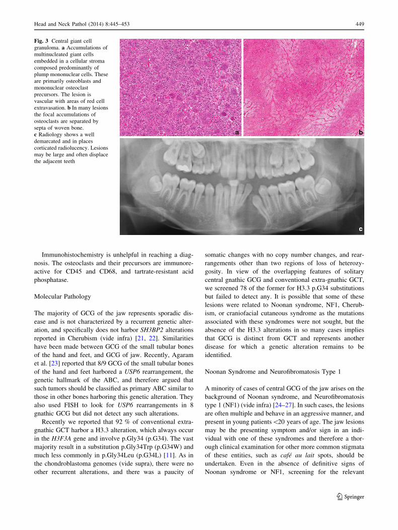
the treatment of choice (Fig. 3) [6].
Giant cell granuloma are sited most commonly in the
anterior jaw. They can be solitary or involve the jaw more
extensively. Although gnathic GCG shares similarities with
conventional giant cell tumor of extra-gnathic sites, they
are considered to be separate entities on the basis that there
are sufficient differences in the histology and behavior
(vide infra) to warrant this distinction. The recent finding
of H3.3 alteration in 92 % of giant cell tumors of bone and
not in GCG supports this (vide infra) [11].
Histopathology Giant cell granuloma is composed of a
monotonous mononuclear cell population in which low
numbers of mitotic figures are noted. The characteristic
feature is a variable numbers of giant cells, which have been
shown to be osteoclasts [19]. These may contain up to 20
nuclei but are generally not as large as those seen in con-
ventional extra-gnathic giant cell tumors of bone. The
osteoclasts are commonly grouped in clusters associated
with thin-walled vascular channels, with evidence of hem-
orrhage and haemosiderin deposition (Fig. 3). The stromal
cells comprise a mixture of spindled fibroblasts and tartrate-
resistant acid phosphatase-positive polygonal cells which
represent osteoclast precursors [19], and variable numbers of
osteoblasts [20]. In GCG the osteoclasts are usually more
widely distributed, and do not occur as dense collections with
almost no detectable intervening stroma as seen in conven-
tional extra-gnathic GCG. The tumors are contained by a thin
rim of peripheral bone, and a limited amount of osteoid and
woven bone can be seen within the main tumor mass. The
histological features of solitary gnathic GCG are similar to
those seen in osteoclast-rich lesions of the jaw, which occur
on the genetic background of Noonan syndrome, Neurofi-
bromatosis type 1 (NF1), Cherubism and craniofacial cuta-
neous syndrome (vide infra) [6].
448 Head and Neck Pathol (2014) 8:445–453
123
Page 5
Immunohistochemistry is unhelpful in reaching a diag-
nosis. The osteoclasts and their precursors are immunore-
active for CD45 and CD68, and tartrate-resistant acid
phosphatase.
Molecular Pathology
The majority of GCG of the jaw represents sporadic dis-
ease and is not characterized by a recurrent genetic alter-
ation, and specifically does not harbor SH3BP2 alterations
reported in Cherubism (vide infra) [21, 22]. Similarities
have been made between GCG of the small tubular bones
of the hand and feet, and GCG of jaw. Recently, Agaram
et al. [23] reported that 8/9 GCG of the small tubular bones
of the hand and feet harbored a USP6 rearrangement, the
genetic hallmark of the ABC, and therefore argued that
such tumors should be classified as primary ABC similar to
those in other bones harboring this genetic alteration. They
also used FISH to look for USP6 rearrangements in 8
gnathic GCG but did not detect any such alterations.
Recently we reported that 92 % of conventional extra-
gnathic GCT harbor a H3.3 alteration, which always occur
in the H3F3A gene and involve p.Gly34 (p.G34). The vast
majority result in a substitution p.Gly34Trp (p.G34W) and
much less commonly in p.Gly34Leu (p.G34L) [11]. As in
the chondroblastoma genomes (vide supra), there were no
other recurrent alterations, and there was a paucity of
somatic changes with no copy number changes, and rear-
rangements other than two regions of loss of heterozy-
gosity. In view of the overlapping features of solitary
central gnathic GCG and conventional extra-gnathic GCT,
we screened 78 of the former for H3.3 p.G34 substitutions
but failed to detect any. It is possible that some of these
lesions were related to Noonan syndrome, NF1, Cherub-
ism, or craniofacial cutaneous syndrome as the mutations
associated with these syndromes were not sought, but the
absence of the H3.3 alterations in so many cases implies
that GCG is distinct from GCT and represents another
disease for which a genetic alteration remains to be
identified.
Noonan Syndrome and Neurofibromatosis Type 1
A minority of cases of central GCG of the jaw arises on the
background of Noonan syndrome, and Neurofibromatosis
type 1 (NF1) (vide infra) [24–27]. In such cases, the lesions
are often multiple and behave in an aggressive manner, and
present in young patients\20 years of age. The jaw lesions
may be the presenting symptom and/or sign in an indi-
vidual with one of these syndromes and therefore a thor-
ough clinical examination for other more common stigmata
of these entities, such as cafe au lait spots, should be
undertaken. Even in the absence of definitive signs of
Noonan syndrome or NF1, screening for the relevant
Fig. 3 Central giant cell
granuloma. a Accumulations of
multinucleated giant cells
embedded in a cellular stroma
composed predominantly of
plump mononuclear cells. These
are primarily osteoblasts and
mononuclear osteoclast
precursors. The lesion is
vascular with areas of red cell
extravasation. b In many lesions
the focal accumulations of
osteoclasts are separated by
septa of woven bone.
c Radiology shows a well
demarcated and in places
corticated radiolucency. Lesions
may be large and often displace
the adjacent teeth
Head and Neck Pathol (2014) 8:445–453 449
123
Page 6
germline alterations should be considered, particularly if
there is more than one jaw lesion, and if the lesion is
extensive. This is because phenotypes of these syndromes
may be very mild [28]. However, clotting disorders and
cardiac defects in Noonan syndrome are important to detect
as they can be managed pro-actively. The jaw lesions in
Noonan syndrome and NF1 can be so extensive that they
can mimic Cherubism, and therefore this diagnosis should
be borne in mind and excluded (vide infra) [25, 29, 30].
Noonan syndrome and NF1 are among the most com-
monly encountered germline alterations, with an incidence
of 1 in 2,500—1 in 3,000 of the population. Noonan syn-
drome, a complex clinical genetic disorder, is inherited as
an autosomal dominant trait caused by alterations in
PTPN11, SOS1, RAF1, KRAS, NRAS, and BRAF genes and
characterized by short stature, craniofacial dysmorphism,
short neck with webbing, deformity of the sternum, cardiac
and clotting anomalies, and cryptorchidism [31]. The most
common germline alterations involve PTPN11 (*50 %),
SOS1 (10–15 %), and RAF1 (5–10 %); with KRAS muta-
tions only occurring in 2 % of those affected. The severity
and the spectrum of the phenotypic changes are significant,
with some individuals having almost no clinical stigmata of
the disease. A small proportion of individuals with Noonan
syndrome also exhibit multiple gnathic GCG previously
reported as Noonan-like/multiple GCG, but this phenotype
is now recognized to be allelic with Noonan syndrome
[32], as mutations in PTPN11, SOS1, and RAF1 have been
reported in this syndrome [33, 34]. LEOPARD syndrome is
also allelic with Noonan syndrome and is associated with
two recurrent PTPN11 mutations in exons 7 (Tyr279Cys)
and 12 (Thr468Met), although other less common altera-
tions are also seen. We have reported on an individual with
LEOPARD syndrome and multiple GCG of the jaw caused
by alterations in PTPN11 [35, 36]. Very occasional patients
with craniofacial cutaneous syndrome, classified as a
RASopathy (vide infra), and multiple giant cell tumors of
the jaw syndrome have also been reported with BRAF or
MAP2K1 mutations [37].
Neurofibromatosis type 1, Noonan syndrome, LEOP-
ARD syndrome and craniofacial cutaneous syndrome are
considered RASopathies as a consequence of germline
mutations in genes encoding specific proteins of the RAS/
mitogen-activated protein kinase (MAPK) pathway [27, 30,
35]. The activation of this pathway during development
results in patients with these four disorders exhibiting
overlapping phenotypes: Noonan syndrome and NF1 are
both associated with freckling/cafe au lait spots, and
occasionally multiple GCG of the jaw, in addition to dys-
morphic craniofacial features, congenital cardiac defects,
skin abnormalities, varying degrees of intellectual disabil-
ity, and increased risk of malignancies (acute leukemia). It
has on some occasions been difficult to distinguish these
two syndromes and some individuals were therefore clas-
sified as having neurofibromatosis-Noonan syndrome.
However, there is now evidence that this syndrome is
allelic to NF1 in most patients [25, 38].
Cherubism
Cherubism is a rare benign disease characterized by sym-
metric enlargement of the jaw and is limited to the man-
dible and maxillary bones [1]. It is inherited as an
autosomal dominant trait, and caused by mutations in the
SH3-domain binding protein 2 (SH3BP2), sited in 4p16.3,
resulting in variable penetrance and expressivity. Approx-
imately 50 % appear to arise as de novo mutations. Males
and females are affected equally, and there is no ethnic
predilection. The lesion presents in children up to the age
of 6 [36, 39].
The disease obtained its name from the apparent upward
gaze of the eyes, as seen in Baroque paintings of Cherubs
by Ruben, resulting from displacement of the orbits and
retraction of the eyelids as a consequence of lesional tissue
involving the floor of the orbit. Involvement of the optic
nerve and proptosis may also occur, and lymphadenopathy
can also be seen in children. The disease stabilizes at
puberty and if the phenotype is not severe may largely
resolve but the dysmorphism may persist in those severely
affected.
The radiological appearance of the established disease is
characteristic with massive expansion of the jaws associ-
ated with multilocular radiolucencies (Fig. 4), although in
the early stages changes may be seen only at the mandib-
ular angles. The histology reveals an osteoclast and spindle
cell lesion, with overlapping features with those of GCG of
the jaw (Fig. 4).
In contrast to Noonan syndrome and NF1, Cherubism is
a non-complex genetic disease, which only affects the jaw
and is not associated with other stigmata such as freckling
or cafe au lait spots. A major distinguishing factor between
Cherubism and these other syndromes is that the jaw lesion
is always symmetric and always presents early (\6 years
old) [36]. In the absence of SH3BP2 mutation in an indi-
vidual considered to have Cherubism, genetic screening for
mutations in genes implicated in Noonan syndrome
(PTPN11, SOS1, RAF1, KRAS, NRAS, and BRAF), NF1,
and craniofacial cutaneous syndrome (BRAF, MAP2K1)
should be undertaken [31]. As with all osteoclast-rich
lesions, hyperparathyroidism should be excluded in the first
instance.
SH3BP2 is an adaptor protein encoded by 13 exons, and
is involved in signal transduction by forming complexes
with other proteins. The majority of the mutations occur in
exon 9 within a 6 amino acid sequence (RSPPDG), which
is a proline-rich domain proximal to the SH2 domain of
450 Head and Neck Pathol (2014) 8:445–453
123
Page 7
SH3BP2, although alterations have also been reported in
exon 3 and 4, and mutations in exon 3 appear to be asso-
ciated with a severe phenotype [40]. Other more rare
mutations have been more reported more recently.
A mouse model has been developed to study Cherub-
ism and has revealed that the Cherubism mutation
(Pro416Arg) results in bone resorption, and increased
levels of TNF-a, a cytokine known to increase osteoclast
recruitment [41]. The mutations result in activation of
certain signaling pathways as a result of SH3BP2 stabil-
ization through inhibition of tankyrase-mediated destruc-
tion of the protein. However, this did not explain why in
humans the disease is restricted to the jaw [42]. Recently,
Yoshitaka et al. [43] reported that macrophages harboring
the SH2BP3 alterations are hyper-responsive to pathogen-
associated and damage-associated molecular patterns, both
of which activate Toll-like receptors, and that the disease
in mice is rescued by depletion of toll-like receptors. They
speculate that the presence of a large amount of toll-like
ligands, such as oral bacteria, present during development
of the jaw bones cause the anatomical-specific develop-
ment of human Cherubism lesions. A recent report shows
that an anti-TNF-a antagonist (Etanercept) can prevent or
ameliorate the disease progression in Cherubism mice
[43].
The treatment of Cherubism is determined by the
severity of the disease and surgery may be considered.
Radiotherapy is not generally advised.
Open Access This article is distributed under the terms of the
Creative Commons Attribution License which permits any use, dis-
tribution, and reproduction in any medium, provided the original
author(s) and the source are credited.
Fig. 4 Cherubism, radiology and histology. a Radiology showing an
extreme case of cherubism with extensive bilateral replacement of the
jaws with radiolucent fibrous tissue. The lesions are typically
multilocular giving a characteristic ‘‘soap bubble’’ appearance, and
‘free floating teeth’. b The giant cells and areas of haemorrhage are
similar to those seen in giant cell granuloma, although in cherubism
the stroma tends to be composed of fibrous connective tissue with
fibroblasts arranged in sheets with a slight storiform or fascicular
pattern. c In some areas osteoclasts are not conspicuous and blood
vessels are prominent, and in some cases may be surrounded by a cuff
of hyalinized collagen (Courtesy of John Wright)
Head and Neck Pathol (2014) 8:445–453 451
123
Page 8
References
1. Fletcher CDM, World Health Organization, International Agency
for Research on Cancer. WHO classification of tumours of soft
tissue and bone. 4th ed. Lyon: IARC Press; 2013. p. 468.
2. Oliveira AM, Hsi BL, Weremowicz S, Rosenberg AE, Dal Cin P,
Joseph N, et al. USP6 (Tre2) fusion oncogenes in aneurysmal
bone cyst. Cancer Res. 2004;64:1920–3.
3. Oliveira AM, Perez-Atayde AR, Dal Cin P, Gebhardt MC, Chen CJ,
Neff JR, et al. Aneurysmal bone cyst variant translocations upregulate
USP6 transcription by promoter swapping with the ZNF9, COL1A1,
TRAP150, and OMD genes. Oncogene. 2005;24:3419–26.
4. Oliveira AM, Perez-Atayde AR, Inwards CY, Medeiros F, Derr
V, Hsi BL, et al. USP6 and CDH11 oncogenes identify the
neoplastic cell in primary aneurysmal bone cysts and are absent
in so-called secondary aneurysmal bone cysts. Am J Pathol.
2004;165:1773–80.
5. Amary MF, Ye H, Berisha F, Tirabosco R, Presneau N, Flanagan
AM. Detection of USP6 gene rearrangement in nodular fasciitis:
an important diagnostic tool. Virchows Arch. 2013;463:97–8.
6. Barnes L, International Academy of Pathology, World Health
Organization, International Agency for Research on Cancer.
Pathology and genetics of head and neck tumours. Lyon: IARC
Press; 2005. p. 430.
7. Rueckert C, Haucke V. The oncogenic TBC domain protein
USP6/TRE17 regulates cell migration and cytokinesis. Biol Cell.
2012;104:22–33.
8. Winnepenninckx V, Debiec-Rychter M, Jorissen M, Bogaerts S,
Sciot R. Aneurysmal bone cyst of the nose with 17p13 involve-
ment. Virchows Arch. 2001;439:636–9.
9. Idowu BD, Al-Adnani M, O’Donnell P, Yu L, Odell E, Diss T,
et al. A sensitive mutation-specific screening technique for
GNAS1 mutations in cases of fibrous dysplasia: the first report of
a codon 227 mutation in bone. Histopathology. 2007;50:691–704.
10. Nord KH, Lilljebjorn H, Vezzi F, Nilsson J, Magnusson L,
Tayebwa J, et al. GRM1 is upregulated through gene fusion and
promoter swapping in chondromyxoid fibroma. Nat Genet.
2014;46:474–7.
11. Behjati S, Tarpey PS, Presneau N, Scheipl S, Pillay N, Van Loo
P, et al. Distinct H3F3A and H3F3B driver mutations define
chondroblastoma and giant cell tumor of bone. Nat Genet.
2013;45:1479–82.
12. Erickson-Johnson MR, Chou MM, Evers BR, Roth CW, Seys
AR, Jin L, et al. Nodular fasciitis: a novel model of transient
neoplasia induced by MYH9-USP6 gene fusion. Lab Invest.
2011;91:1427–33.
13. Bertoni F, Unni KK, Beabout JW, Harner SG, Dahlin DC.
Chondroblastoma of the skull and facial bones. Am J Clin Pathol.
1987;88:1–9.
14. Hong SM, Park YK, Ro JY. Chondroblastoma of the temporal
bone: a clinicopathologic study of five cases. J Korean Med Sci.
1999;14:559–64.
15. Bousdras K, O’Donnell P, Vujovic S, Henderson S, Boshoff C,
Flanagan AM. Chondroblastomas but not chondromyxoid fibro-
mas express cytokeratins: an unusual presentation of a chondro-
blastoma in the metaphyseal cortex of the tibia. Histopathology.
2007;51:414–6.
16. Vujovic S, Henderson S, Presneau N, Odell E, Jacques TS, Ti-
rabosco R, et al. Brachyury, a crucial regulator of notochordal
development, is a novel biomarker for chordomas. J Pathol.
2006;209:157–65.
17. Amary MF, Bacsi K, Maggiani F, Damato S, Halai D, Berisha F,
et al. IDH1 and IDH2 mutations are frequent events in central
chondrosarcoma and central and periosteal chondromas but not in
other mesenchymal tumours. J Pathol. 2011;224:334–43.
18. Joseph CG, Hwang H, Jiao Y, Wood LD, Kinde I, Wu J, et al.
Exomic analysis of myxoid liposarcomas, synovial sarcomas, and
osteosarcomas. Genes Chromosom Cancer. 2014;53:15–24.
19. Flanagan AM, Nui B, Tinkler MA, Williams DM, Chambers TJ.
The multinucleate cells in giant cell granulomas of the jaw are
osteoclasts. Cancer. 1988;62:1139–45.
20. Liu B, Yu SF, Li TJ. Multinucleated giant cells in various forms
of giant cell containing lesions of the jaws express features of
osteoclasts. J Oral Pathol Med. 2003;32:367–75.
21. Idowu BD, Thomas G, Frow R, Diss TC, Flanagan AM. Muta-
tions in SH3BP2, the cherubism gene, were not detected in
central or peripheral giant cell tumours of the jaw. Br J Oral
Maxillofac Surg. 2008;46:229–30.
22. Teixeira RC, Horz HP, Damante JH, Garlet GP, Santos CF,
Nogueira RL, et al. SH3BP2-encoding exons involved in cher-
ubism are not associated with central giant cell granuloma. Int J
Oral Maxillofac Surg. 2011;40:851–5.
23. Agaram NP, LeLoarer FV, Zhang L, Hwang S, Athanasian EA,
Hameed M, et al. USP6 gene rearrangements occur preferentially
in giant cell reparative granulomas of the hands and feet but not
in gnathic location. Hum Pathol. 2014;45:1147–52.
24. Dunlap C, Neville B, Vickers RA, O’Neil D, Barker B. The
Noonan syndrome/cherubism association. Oral Surg Oral Med
Oral Pathol. 1989;67:698–705.
25. van Capelle CI, Hogeman PH, van der Sijs-Bos CJ, Heggelman
BG, Idowu B, Slootweg PJ, et al. Neurofibromatosis presenting
with a cherubism phenotype. Eur J Pediatr. 2007;166:905–9.
26. Tartaglia M, Gelb BD, Zenker M. Noonan syndrome and clini-
cally related disorders. Best Pract Res Clin Endocrinol Metab.
2011;25:161–79.
27. Reig I, Boixeda P, Fleta B, Morenoc C, Gamez L, Truchuelo M.
Neurofibromatosis-Noonan syndrome: case report and clinico-
pathogenic review of the Neurofibromatosis–Noonan syndrome
and RAS–MAPK pathway. Dermatol Online J. 2011;17:4.
28. Chen PC, Yin J, Yu HW, Yuan T, Fernandez M, Yung CK, et al.
Next-generation sequencing identifies rare variants associated
with Noonan syndrome. Proc Natl Acad Sci USA. 2014;111:
11473–8.
29. Jafarov T, Ferimazova N, Reichenberger E. Noonan-like syn-
drome mutations in PTPN11 in patients diagnosed with cherub-
ism. Clin Genet. 2005;68:190–1.
30. Martinez-Quintana E, Rodriguez-Gonzalez F. RASopathies: from
Noonan to LEOPARD syndrome. Rev Esp Cardiol (Engl Ed).
2013;66:756–7.
31. Tartaglia M, Zampino G, Gelb BD. Noonan syndrome: clinical
aspects and molecular pathogenesis. Mol Syndromol. 2010;1:2–26.
32. Lee JS, Tartaglia M, Gelb BD, Fridrich K, Sachs S, Stratakis CA,
et al. Phenotypic and genotypic characterisation of Noonan-like/
multiple giant cell lesion syndrome. J Med Genet. 2005;42:e11.
33. Karbach J, Coerdt W, Wagner W, Bartsch O. Case report: Noo-
nan syndrome with multiple giant cell lesions and review of the
literature. Am J Med Genet A. 2012;158A:2283–9.
34. Sarkozy A, Carta C, Moretti S, Zampino G, Digilio MC, Pan-
taleoni F, et al. Germline BRAF mutations in Noonan, LEOP-
ARD, and cardiofaciocutaneous syndromes: molecular diversity
and associated phenotypic spectrum. Hum Mutat. 2009;30:
695–702.
35. Bezniakow N, Gos M, Obersztyn E. The RASopathies as an
example of RAS/MAPK pathway disturbances: clinical presen-
tation and molecular pathogenesis of selected syndromes. Dev
Period Med. 2014;18:285–96.
36. Flanagan AM, Delaney D, O’Donnell P. Benefits of molecular
pathology in the diagnosis of musculoskeletal disease : part II of a
two-part review: bone tumors and metabolic disorders. Skelet
Radiol. 2010;39:213–24.
452 Head and Neck Pathol (2014) 8:445–453
123
Page 9
37. Neumann TE, Allanson J, Kavamura I, Kerr B, Neri G, Noonan J,
et al. Multiple giant cell lesions in patients with Noonan syn-
drome and cardio-facio-cutaneous syndrome. Eur J Hum Genet.
2009;17:420–5.
38. Yazdizadeh M, Tapia JL, Baharvand M, Radfar L. A case of
neurofibromatosis–Noonan syndrome with a central giant cell
granuloma. Oral Surg Oral Med Oral Pathol Oral Radiol Endod.
2004;98:316–20.
39. Reichenberger EJ, Levine MA, Olsen BR, Papadaki ME, Lietman
SA. The role of SH3BP2 in the pathophysiology of cherubism.
Orphanet J Rare Dis. 2012;7(Suppl 1):S5.
40. Ueki Y, Tiziani V, Santanna C, Fukai N, Maulik C, Garfinkle J,
et al. Mutations in the gene encoding c-Abl-binding protein
SH3BP2 cause cherubism. Nat Genet. 2001;28:125–6.
41. Coste E, Greig IR, Mollat P, Rose L, Gray M, Ralston SH, et al.
Identification of small molecule inhibitors of RANKL and TNF
signalling as anti-inflammatory and antiresorptive agents in mice.
Ann Rheum Dis. 2013. doi:10.1136/annrheumdis-2013-203700.
42. Levaot N, Voytyuk O, Dimitriou I, Sircoulomb F, Chandrakumar
A, Deckert M, et al. Loss of Tankyrase-mediated destruction of
3BP2 is the underlying pathogenic mechanism of cherubism.
Cell. 2011;147:1324–39.
43. Yoshitaka T, Ishida S, Mukai T, Kittaka M, Reichenberger EJ,
Ueki Y. Etanercept administration to neonatal SH3BP2 knock-in
cherubism mice prevents TNF-alpha-induced inflammation and
bone loss. J Bone Miner Res. 2014;29:1170–82.
Head and Neck Pathol (2014) 8:445–453 453
123