+ All Categories
Home > Documents > Git Prelab Tnx Full

Git Prelab Tnx Full

Date post: 06-Apr-2018
Category:
Upload: crisanto-macaraeg
View: 245 times
Download: 0 times
Share this document with a friend
8
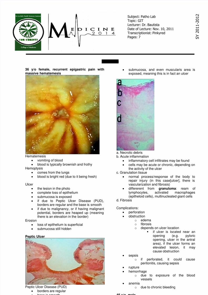
GIT Prelab 36 y/o female, recurrent epigastric pain with massive hematemesis Hematemesis vomiting of blood blood is typically brownish and frothy Hemoptysis comes from the lungs blood is bright red (due to it being fresh) Ulcer the lesion in the photo complete loss of epithelium submucosa is exposed if due to Peptic Ulcer Disease (PUD), borders are regular and the base is smooth if due to malignancy, or if having malignant potential, borders are heaped up (meaning there is an elevation in the border) Erosion loss of epithelium is superficial submucosa still hidden Peptic Ulcer Peptic Ulcer Disease (PUD) borders are regular  base is smooth submucosa, and even muscularis area is exposed, meaning this is in fact an ulcer  a. Necrotic debris b. Acute inflammation inflammatory cell infiltrates may be found cells may be acute or chronic, depending on the activity of the ulcer c. Granulation tissue normal process/response of the body to repair injury (in this case[ulcer], there is vascularization and fibrosis) differerent from granuloma: ream of lymphocytes, activated macrophages (epitheloid cells), multinucleated giant cells d. Fibrosis Complications: perforation  obstruction o edema o fibrosis o depends on ulcer location  if ulcer is located near an opening (e.g. pyloric opening, ulcer in the antral area), if the ulcer forms an elevated lesion, it may cause obstruction  sepsis o if perforated, it could cause peritonitis, causing sepsis   rupture  hemorrhage o due to exposure of the blood vessels  anemia o due to chronic bleeding 45 y/o, male Subject: Patho Lab Topic: GIT Lecturer: Dr. Bautista Date of Lecture: Nov. 10, 2011 Transcriptionist: Pinkyred Pages: 7    S    Y    2    0    1    1      2    0    1    2
Transcript

8/3/2019 Git Prelab Tnx Full

http://slidepdf.com/reader/full/git-prelab-tnx-full 1/7


GIT Prelab

36 y/o female, recurrent epigastric pain withmassive hematemesis

Hematemesis vomiting of blood

blood is typically brownish and frothyHemoptysis

comes from the lungs

blood is bright red (due to it being fresh)

Ulcer

the lesion in the photo

complete loss of epithelium

submucosa is exposed

if due to Peptic Ulcer Disease (PUD),borders are regular and the base is smooth

if due to malignancy, or if having malignantpotential, borders are heaped up (meaningthere is an elevation in the border)

Erosion

loss of epithelium is superficial

submucosa still hidden

Peptic Ulcer

Peptic Ulcer Disease (PUD)

borders are regular 

base is smooth 

submucosa, and even muscularis area isexposed, meaning this is in fact an ulcer 

a. Necrotic debris

b. Acute inflammation inflammatory cell infiltrates may be found

cells may be acute or chronic, depending onthe activity of the ulcer

c. Granulation tissue

normal process/response of the body torepair injury (in this case[ulcer], there isvascularization and fibrosis)

differerent from granuloma: ream oflymphocytes, activated macrophages(epitheloid cells), multinucleated giant cells

d. Fibrosis

Complications: perforation 

obstruction o edema o fibrosis o depends on ulcer location 

if ulcer is located near anopening (e.g. pyloricopening, ulcer in the antralarea), if the ulcer forms anelevated lesion, it maycause obstruction 

sepsis o if perforated, it could cause

peritonitis, causing sepsis 

rupture 

hemorrhage o due to exposure of the blood

vessels 

anemia o due to chronic bleeding 

45 y/o, male

Subject: Patho LabTopic: GITLecturer: Dr. Bautista

Date of Lecture: Nov. 10, 2011Transcriptionist: PinkyredPages: 7    S

   Y

   2   0   1   1  -   2   0   1   2

8/3/2019 Git Prelab Tnx Full

http://slidepdf.com/reader/full/git-prelab-tnx-full 2/7


Benign lesion

well demarcated borders 

no area of necrosis  punctate areas of hemorrhages are possible

even in benign lesions 

Gastrointestinal Stromal Tumor (GIST)

mucosa may be normal or ulcerated (thereis a visible bulge [elevated area], but themucosa is still normal)

neoplasm in the stroma pushes the mucosaupward or outward, but mucosal lining isnormal

histologically

uniform population of spindle cells

very few pleomorphisms

cells look alike

cytoplasms may be delineated

75 y/o male, epigastric fullness, weight loss,black tarry stool

Melena

black tarry stoolBased on the history, lesion is in the pyloric area ofthe stomach

lesions in the pyloric area are likely to causeobstructions because the opening is small,and there is a muscular sphincter

Malignant lesion

necrosis

bleeding

irregular border

Adenocarcinoma, pyloric area

Adenocarcinoma

most common GIT tumor

more common in the colon

Well-differentiated carcinoma

glands and granular formation are seen

glands are compact, forming cribriformpatterns, sieve-like (there are “holes” allover)

8/3/2019 Git Prelab Tnx Full

http://slidepdf.com/reader/full/git-prelab-tnx-full 3/7


22 y/o male, severe abdominal pain

Meckel’s diverticulum

shown in the photo

congenital

pathogenesis: failure of vitelline duct toinvolute

a true diverticulum

involves all layers

lined by gastric mucosa, sometimes withnon-malignant pancreatic tissue

usually located at the antimesenteric side(opposite side of where the mesentery is)

Colonic diverticulum

pathogenesis: invagination of the wall,usually in the colon (due to it having 2layers: inner circular, outer longitudinal)

not a true diverticulum

does not involve all layers

68 y/o male, severe abdominal pain, vomitingand diarrhea

gangrenous

could be an infarction caused byo hypoxic injury

mechanical obstruction intussusception volvulus adhesion hernia

would not causeinfarctionimmediately

would incarceratefirst, thenstrangulate

o reperfusion injury

histologicallyinfarction/gangrene

mucosa is already necrotic anddeluded(being detached from the lining)

there are thrombosed veins in submucosa

inflammation extends to serosa(inflammatory cells: acute or chronicdepends on the onset of injury)

congestion/hemorrhages

mucosa is normal affects only the submucosa

8/3/2019 Git Prelab Tnx Full

http://slidepdf.com/reader/full/git-prelab-tnx-full 4/7


33 y/o male, abdominal enlargement

patient also manifested with chronic cough (>2weeks) and hemoptysis

diagnosis: ileocecal tuberculosis

On closer view, granuloma and giant cells may beseen

chronic granulomatous formation

64 y/o, abdominal enlargement, small scantyhard stool

Multiple cervical lymphadenopathy- NonHodgkin’s lymphoma 

@ scanning view- take note of diffuse proliferation ofmalignant cell, no demarcation@ hpo- uniform population of malignant cell,presence of prominent nucleoli, pleomorphism

*NHL

Mixed hemorrhoids-possible liver failure -portal hypertension, backflow of blood at tributaries,@ egd-possible esophageal varices

Infectious- causative agent: EntamoebahistolyticaAmebic colitis- flask shape ulcer, @ base- cyst andtrophozoite

Juvenile Polyp

@ microscope-cystic dilation of glands

No malignant potential

8/3/2019 Git Prelab Tnx Full

http://slidepdf.com/reader/full/git-prelab-tnx-full 5/7


  Villus- fingerlike projection 

  @ submucosa invading-high malignantpotential

Right sided malignancy- Carcinoma ( epithelialorigin) @

Ileocecal junctionMUCINOUS ADENOCARCINOMA

*L sided vs R sided malignancy- napkin ring  Right sided- fungating, capacious at cecum, withspace, with enlargement but no obstruction, itoutgrows the blood supply causing necrotic sitearea, which is site for bleedingManifestation- anemia due to bleedingLeft sided- napkin ring lesion, narrowing of lumen,presentation-obstruction, goat stool like stool

Pinkish/purplish-mucin

Floating cells- malignant cells floating inlakes /pools of mucin

Invades the submucosa, full thickness ofcolonic wall or sometimes extending tocolonic fat

Pigmented: melanoma- can be at back,perineal area, scalp

Clusters of malignant cells with pigment

  Malignant version of gist

Gist- leiomyoma+ spindle cells

Grossly with hemorrhages

8/3/2019 Git Prelab Tnx Full

http://slidepdf.com/reader/full/git-prelab-tnx-full 6/7


Diagnosis: Appendicitis 

RUQ pain : differentialsFemale: ectopic pregnancy, salphingitisMale: meckel’s diverticulum ( 2ft from ileocecalarea)- may rupture and inflamed and may present asRUQ pain

Section taken from the appendix

  Malignant-glandular

  Adenocarcinoma of the Appendix

More common tumor/ malignancy of theappendix: carcinoid, usually found at thetip of the appendix

 ___________END OF TRANX____________ 

8/3/2019 Git Prelab Tnx Full

http://slidepdf.com/reader/full/git-prelab-tnx-full 7/7


Recommended