Global Transcriptional Analysis of clpP Mutations of Type 2Streptococcus pneumoniae and Their Effects on Physiology
and VirulenceGregory T. Robertson, Wai-Leung Ng, Joseph Foley, Raymond Gilmour, and Malcolm E. Winkler*
Infectious Diseases Research, Lilly Research Laboratories, Indianapolis, Indiana 46285
Received 1 February 2002/Accepted 5 April 2002
Streptococcus pneumoniae is an important human pathogen that contains single copies of genes encoding theClpP and FtsH ATP-dependent proteases but lacks the Lon and HslV proteases. We constructed and charac-terized the phenotypes of clpP, clpC, and clpX deletion replacement mutants, which lack the ClpP proteasesubunit or the putative ClpC or ClpX ATPase specificity factor. A �clpP mutant, but not a �clpC or �clpXmutant, of the virulent D39 type 2 strain of S. pneumoniae grew poorly at 30°C and failed to grow at 40°C.Despite this temperature sensitivity, transcription of the heat shock regulon determined by microarray analysiswas induced in a �clpP mutant, which was also more sensitive to oxidative stress by H2O2 and to puromycinthan its clpP� parent strain. A �clpP mutant, but not a �clpC mutant, was strongly attenuated for virulencein the murine lung and sepsis infection models. All of these phenotypes were complemented in a �clpP/clpP�
merodiploid strain. Consistent with these complementation patterns, clpP was found to be in a monocistronicoperon, whose transcription was induced about fivefold by heat shock in S. pneumoniae as determined byNorthern and real-time reverse transcription-PCR analyses. Besides clpP, transcription of clpC, clpE, and clpL,but not clpX or ftsH, was induced by heat shock or entry into late exponential growth phase. Microarray analysisof �clpP mutants showed a limited change in transcription pattern (�80 genes) consistent with these pheno-types, including repression of genes involved in oxidative stress, metal ion transport, and virulence. Inaddition, transcription of the early and late competence regulon was induced in the �clpP mutant, andcompetence gene expression and DNA uptake seemed to be constitutively induced throughout growth. To-gether, these results indicate that ClpP-mediated proteolysis plays a complex and central role in numerouspneumococcal stress responses, development of competence, and virulence.
Four distinct classes of ATP-dependent proteases have beenfound in bacteria, including the multimeric Clp (ClpAP,ClpXP, or ClpCP) and HslUV (ClpYQ) proteases and thesingle-chain AAA (FtsH) and Lon family proteases (Lon) (19).The Clp proteases (caseinolytic proteases) were first identifiedin Escherichia coli and consist of an ATPase specificity factor(ClpA or ClpX in E. coli and ClpX or ClpC in Bacillus subtilis)and a proteolytic domain (ClpP) that contains a consensusserine protease active site (17, 19, 36). This multimeric enzymecomplex assembles into a structure that is remarkably similarto that of the eukaryotic proteosome (7). In E. coli, ClpP-mediated proteolysis, which is regulated by heat shock, re-moves abnormal proteins that accumulate during stress condi-tions, recycles amino acids from nonessential proteins duringstarvation, and contributes to the clearance of truncated pep-tides produced from stalled ribosomes by the SsrA system (20,47). In addition, ClpP-mediated proteolysis in E. coli controlsthe half-life of regulatory proteins, including the stationaryphase sigma factor �s (59), the heterodimeric UmuD SOSprotein (11), and several phage proteins (reviewed in reference17). Despite the significant roles played by the ClpP system instress responses in E. coli, the phenotype of E. coli clpP mu-tants is essentially identical to that of wild-type cells (5), espe-
cially in the degradation of abnormal proteins (18). Presum-ably, this results from the presence of other redundant ATP-dependent proteases that likely play a more critical role in thisprocess (i.e., Lon and HslUV).
ClpP-mediated proteolysis appears to be more central forstress tolerance and global regulation in bacteria other than E.coli. For example, clpP mutants of B. subtilis and Lactococcuslactis are impaired for survival during heat shock and otherstress conditions and exhibit clear defects in the degradation ofabnormal proteins (12, 14, 39). Microbial developmental pro-cesses, such as natural genetic competence and sporulation inB. subtilis (39), biofilm development in Pseudomonas fluore-scens (41), and viability and cell cycle progression in Cau-lobacter crescentus (25), are dependent on ClpP-mediated pro-teolysis. Moreover, a growing number of studies demonstrate arequirement for ClpP-mediated proteolysis for virulence anddisease progression of important bacterial pathogens. For ex-ample, ClpP function is required for virulence factor expres-sion, intracellular survival, and pathogenesis of Listeria mono-cytogenes in mice (13). ClpP function has also been linked tothe acid tolerance response and virulence of the intracellularpathogen Salmonella enterica serovar Typhimurium (21, 56)and to virulence factor expression in Yersinia enterocolitica(42). In some of these cases, the association between ClpPfunction and virulence may reflect the general role played byClpP-mediated proteolysis in stress survival.
The S. pneumoniae genome has recently been shown tocontain only single copies of clpP and ftsH genes and to lack lon
* Corresponding author. Mailing address: Lilly Research Laborato-ries, Drop Code 1543, Lilly Corporate Center, Indianapolis, IN 46285.Phone: (317) 433-0095. Fax: (317) 276-9159. E-mail: [email protected].
and hslUV genes (23). In this regard, S. pneumoniae is uniqueamong eubacteria, which contain both clpP and lon or multiplecopies of clpP when lon is absent (7). S. pneumoniae containsputative orthologs of the ClpC, ClpE, ClpL, and ClpX ATPasespecificity factors that presumably associate with the ClpP pro-tease subunit (reviewed in reference 48). S. pneumoniae is aserious human pathogen that is a causal agent of bacterialpneumonia, bronchitis, meningitis, and otitis media and is par-ticularly devastating to the elderly and the very young (55).Antibiotic-resistant clinical isolates of S. pneumoniae have re-cently become common, and multiple-antibiotic-resistantstrains are now found worldwide (27, 52). Normal diseaseprogression for S. pneumoniae begins with entry and coloniza-tion of the nasopharnyx followed by progression to other sites,such as the lung (55). The infection process exposes S. pneu-moniae to numerous stress conditions, including temperatureshift between the upper respiratory tract (30°C) and deepertissues (37°C), pH changes, exposure to reactive oxygen speciesgenerated by host phagocytes, and nutritional deprivation (60).
The role of ClpP- or FtsH-mediated ATP-dependent prote-olysis in the stress responses and virulence of S. pneumoniae islargely unknown. Recently, signature-tagged mutagenesis wasused to identify ClpC as a putative virulence factor of S. pneu-moniae in mice (45); however, ClpP did not turn up in thisstudy. Pneumococcal ClpC was reported to regulate growthinhibition at high temperature, autolysis in the presence ofantibiotics, transformation, and adherence (3); however, an-other report did not confirm these phenotypes (4). Chastanetand coworkers recently reported that ClpP and ClpE play arole in the thermotolerance of S. pneumoniae (4). Their bio-chemical and genetic evidence in a heterologous B. subtilissystem suggest that CtsR regulates the heat-shock response ofclpC, clpL, clpE, and clpP and unexpectedly groESL in S. pneu-moniae (4). They further demonstrated that the expression oftwo competence genes is induced in a clpP mutant, suggestinga negative regulatory role for ClpP in competence develop-ment under inappropriate conditions (4). Here, we report fur-ther characterization of the phenotypes of S. pneumoniae clpP,clpC, and clpX mutants, including global microarray analysis oftranscription patterns of clpP mutants and the heat shock regu-lon of S. pneumoniae. Our results show that ClpP-mediatedproteolysis plays complex roles in the response of S. pneu-moniae to thermal and oxidation stress, the regulation of thenatural competence pathway, and virulence.
MATERIALS AND METHODS
Bacterial strains and growth conditions. S. pneumoniae strains were cultivatedstatically in brain heart infusion (BHI) broth or on trypticase soy agar II bloodagar plates (TSA-BA; Becton Dickson BBL) at 37°C in an atmosphere of 5%CO2 unless otherwise stated. Chemically defined medium (CDM) for the growthof S. pneumoniae cultures (50) lacking methionine and glucose was formulated byJRH Biosciences (Denver, Pa.). CDM was supplemented with 1.0% (wt/vol)glucose, 0.1% (wt/vol) choline chloride, 0.075% (wt/vol) L-cysteine HCl, and0.25% (wt/vol) NaHCO3. For antibiotic selection, media was supplemented with0.3 �g of erythromycin per ml, 100 �g of spectinomycin per ml, 2.5 �g ofchloramphenicol per ml, or 2.5 �g of novobiocin per ml as appropriate.
The virulent type 2 encapsulated S. pneumoniae strain D39 and its unencap-sulated derivative R6 (49) used in these studies were assigned unique straindesignations to track isogenic derivatives (Table 1). Strains CPM7 [CP1250ssbB::(pEVP3)::ssbB] and CP1500 were used as the ssbB::lacZ reporter strainand the source of genomic donor DNA in transformation experiments, respec-tively (Table 1) (34). S. pneumoniae mutants were constructed by transformation
of linear deletion replacement amplicons following treatment with competencestimulatory peptide (CSP) (34). Briefly, 1.0- to 1.5-kb DNA fragments corre-sponding to the 5� and 3� regions of target genes to be deleted (�60 to 80% ofinternal coding sequences) and either the 1.0-kb ermAM (erythromycin resis-tance [Err]) (35) or aad9 (spectinomycin resistance [Spr]) (33) cassettes wereamplified by PCR using oligonucleotide primers containing short stretches ofDNA sequences that were complementary to a DNA fragment designed to beadjoining (Table 2). The resulting three DNA fragments were mixed, allowed toanneal at the overlapping complementary sequences, and extended and ampli-fied using a second round of PCR (22). Recombinant rTth polymerase and XLreaction buffer (Applied Biosystems) were used in these amplifications. Result-ant linear gene amplicons containing deletion replacements were transformedinto early exponential phase cultures of S. pneumoniae (OD620 of �0.05 to 0.1)diluted 1 to 10 in BHI medium containing 10% heat-inactivated horse serum(Sigma), 10 mM glucose, and 100 ng of synthetic CSP-1 per ml and incubated for15 min at 30 or 37°C to induce competence. Following transformation, bacteriawere allowed to recover for 2 h to allow phenotypic expression of the antibioticresistance marker, and recombinant clones were recovered on TSA-BA in thepresence of selective antibiotics in nutrient broth soft-agar overlay (0.8% [wt/vol]Bacto Nutrient Broth, 0.4% [wt/vol] Bacto Agar) at 30 or 37°C.
Phenotypic characterization of S. pneumoniae clpP mutants. The ability of S.pneumoniae to grow at reduced and elevated temperatures was determined intriplicate on TSA-BA plates incubated at 30, 37, or 40°C for 24 h. The sensitivityof S. pneumoniae EL161 (D39 parent) and its derivatives to puromycin, H2O2, orHCl was evaluated using a disk diffusion assay, as EL59 (R6 parent) was inca-pable of forming an adequate lawn on TSA plates without blood supplementa-tion. Bacteria were harvested from TSA-BA plates following overnight incuba-tion at 37°C in an atmosphere of 5% CO2 and suspended in sterile 0.9% (wt/vol)NaCl to yield an optical density at 620 nm (OD620) of �0.13 (�1 � 107 CFU perml). One milliliter of suspension was mixed with 3 ml of nutrient broth soft agarand poured onto the surfaces of BHI agar plates. Sterile 8-mm-diameter diskswere placed in the center of the lawns of test organisms and saturated with 10 �lof 4.8 mg of puromycin (Sigma) per ml, 4.9 M H2O2 (Mallinckrodt), or 1 N HCl(Red Bird Inc.). Average zones of inhibition were determined after 24 h ofgrowth at 37°C under 5% CO2, and statistical comparisons were made usingone-way analysis of variance (ANOVA), where P values of �0.05 were consid-ered significant. The effects of clpP, clpC, and clpX mutations on autolysis ofpneumococcus was determined by measuring the loss of OD of 6-ml BHI cul-tures at mid-exponential phase (OD660 of �0.13) over time following addition ofa final concentration of 0.01% (wt/vol) Triton X-100.
Infection of ICR outbred mice. In conducting experiments with animals, theinvestigators adhered to the Guide for the Care and Use of Laboratory Animals,prepared by the Committee on Care and Use of Laboratory Animals of theInstitute of Laboratory Animal Resources, National Research Council (40a).
Bacteria grown overnight on TSA-BA plates were harvested into 0.9% (wt/vol)NaCl and adjusted to a MacFarland standard of 0.5 (�1.0 � 108 CFU per ml)which was further diluted fivefold in 0.9% NaCl. After brief isofluorane anes-thetization, male ICR outbred mice (Harlan Sprague Dawley) were infected byintratracheal inoculation with 50 �l of bacterial suspension. At 0, 8, 24, and 48 hfollowing inoculation, four mice per group were sacrificed by CO2 asphyxiationand the lungs aseptically harvested and homogenized in 0.9% NaCl. Total num-bers of viable bacteria per lung were determined by serial dilution and spreadingonto TSA-BA plates. Statistical comparisons were made using ANOVA where Pvalues of �0.05 were considered significant. For 50% lethal dose (LD50) studies,bacteria were grown as described above with the exception that 0.5 MacFarlandstandard suspensions were serially diluted 10-fold in BHI medium to yield in-fection doses ranging from �102 to 106 CFU per ml. Male ICR outbred micewere injected in the peritoneal cavity with 50 �l of bacterial suspensions andobserved daily for clinical signs of disease. Moribund animals were sacrificed byCO2 asphyxiation, and the LD50 was calculated based on the mean infecting doserequired to promote evidence of morbidity in 50% of test subjects (10 mice perdose per strain) in a given period of time.
Extraction of total RNA from S. pneumoniae. Total RNA was extracted fromS. pneumoniae by using a variation of the hot-phenol method described by Tsuiet al. (53). Briefly, 10 ml of S. pneumoniae culture was mixed directly with 5 mlof 100°C lysis buffer containing 2% (wt/vol) sodium dodecyl sulfate (SDS) and 16mM EDTA (pH 8.0) and held for 2 min at 100°C to induce cell lysis. The lysedculture was extracted with 10 ml of acidified phenol (Gibco BRL) at 65°C for 5min with vigorous agitation. The aqueous phase was further extracted once withphenol-chloroform-isoamyl alcohol (25:24:1; Gibco BRL) and once with chlo-form-isoamyl alcohol (Gibco BRL). Total RNA was concentrated from theaqueous phase by using Centriplus YM-30 cartridges (Millipore) and purified
VOL. 184, 2002 ClpP PROTEOLYSIS AND ADAPTIVE RESPONSES IN S. PNEUMONIAE 3509
using an RNeasy (Qiagen) column with on-column DNase I treatment per-formed according to the manufacturer’s instructions.
Heat shock of S. pneumoniae. S. pneumoniae cells were cultivated staticallyovernight in CDM at 37°C in an atmosphere of 5% CO2. Cultures were dilutedto an OD620 of �0.03 in 40 ml of prewarmed CDM and allowed to incubatewithout shaking at 37°C until the culture reached early to middle exponentialgrowth phase (OD620 of �0.3). Cultures were divided into three 10-ml portionswhich were heated at 37°C or for 5 and 15 min at 45°C, after which RNA wasextracted as described above.
Northern analysis and quantitative real time reverse transcription (RT)-PCR.Aliquots of 5 �g of total S. pneumoniae RNA, purified as described above, wereseparated by formaldehyde agarose gel electrophoresis and transferred using aNorthern Max kit (Ambion, Inc.) according to the manufacturer’s instructions. A0.5-kb internal fragment of clpP generated by PCR with the primer set clpP-5�
and clpP-3� (Table 2) was labeled with psoralen-biotin by using a Brightstarpsoralen-biotin nonisotopic labeling kit (Ambion) and used as a probe for North-ern analysis according to the conditions given for the Northern Max kit. Bandson the exposed audioradiographic film were quantified using Sigma Scan version2.0 (Jandel). Relative increases (n-fold) in clpP band intensities of RNA isolatedfrom cultures following a shift from 37 to 45°C for 5 and 15 min were comparedto that of the 37°C control.
S. pneumoniae clpP mRNA was quantified by a real-time, 5� exonuclease PCR(TaqMan) assay (31) using a primer-probe combination that recognized a por-tion of the S. pneumoniae clpP gene. For comparison purposes, primer-probe setsfor fabK and 16S rRNA were also employed. The primers and probes are listedin Table 2. Primer-probe sets were selected using Primer Express software (PEBiosystems, Foster City, Calif.) and were obtained from PE Biosystems. Theprimers were used in concentrations of 300 nM and the probes were used in
TABLE 1. Bacterial strains and DNA amplicons used in strain construction
Strain Genotype and/or phenotypea Description Source or reference
EL59 R6, avirulent unencapsulated parent Derived from D39 isolate 49EL161 D39, virulent encapsulated type 2
parentSubclone of original clinical isolate 49
CP1500 hex nov-r1 bry-r str-1 ery-r1 ery-r2 Nvr Donor of novobiocin resistance point marker 34CPM7 CP1250 ssbB�::(pEVP3)::ssbB�
ssbB::lacZ Cmr SsbB�Rx derivative with ssbB::lacZ reporter construct 34
EL539b EL59 clpP::aad9 Spr EL59 transformed with linear clpP::aad9 amplicon This studyEL555 EL59 bgaA::ermAM Err EL59 bearing a bgaA::ermAM mutation D. LeBlanc strain
collectionEL556 EL59 lytA::aad9 Spr EL59 transformed with linear lytA::aad9 amplicon This studyEL677 EL161 clpP::aad9 Spr EL161 transformed with linear clpP::aad9 amplicon This studyEL824 CPM7 clpP::aad9 Spr CPM7 transformed with linear clpP::aad9 amplicon This studyEL854 EL59 clpC::ermAM Err EL59 transformed with linear clpC::ermAM amplicon This studyEL856 EL161 clpC::ermAM Err EL161 transformed with linear clpC::ermAM amplicon This studyEL873 EL59 clpX::ermAM Err EL59 transformed with linear clpX::ermAM amplicon This studyEL923 EL677 bgaA::ermAM::T1T2::clpP�
Spr Err ClpP�EL677 transformed with linear bgaA::ermAM::T1T2::clpP� This study
EL925 EL539 bgaA::ermAM::T1T2::clpP�
Spr Err ClpP�EL539 transformed with linear bgaA::ermAM::T1T2::clpP� This study
EL961 EL161 bgaA::ermAM Err EL161 transformed with linear bgaA::ermAM from EL555 This study
a Nvr, resistant to novobiocin; Cmr, resistant to chloramphenicol; Spr, resistant to spectinomycin; Err, resistant to erythromycin.b Strain construction was carried out by transformation of indicated recipient with linear double stranded synthetic PCR amplicon.c Template DNA for amplicon generation.d Primer pair set used to generate a given amplicon (see Table 2).
concentrations of 200 nM in 25-�l reaction mixtures. TaqMan reactions were setup using an EZ RT-PCR kit (PE Biosystems). Each reaction contained twofoldserial dilutions of either 200 ng or 20 ng of total RNA, depending on theprimer-probe set used. The quantitative RT-PCRs were performed on a ABIPRISM 7700 (PE Biosystems) and included the following steps: 2 min treatmentwith uracil N-glycosylase at 50°C, 30 min of reverse transcription at 60°C, 5 minat 95°C, and then 40 cycles of amplification using 95°C for 15 seconds followedby 60°C for 60 s. Three replicates were performed for each sample. The resultswere calculated using the comparative critical threshold (CT) method (userbulletin no. 2 [http://docs.appliedbiosystems.com/pebiodocs/04303859.pdf]; Ap-plied Biosystems), in which the amount of target is normalized to a reference(non-heat shock control) relative to an internal calibrator target RNA (16SrRNA). Transcript amounts from a control target gene (fabK) were analyzedusing the same parameters. Data are presented as the relative changes in exper-
imental target amounts compared to those of the 37°C control following 5- and15-min shifts from 37 to 45°C or in the clpP mutant (EL539).
Microarray analysis. Fragmentation and labeling of RNA was performed asinstructed by Affymetrix (Santa Clara, Calif.). Briefly, 60 �g of total RNA wasfragmented in 1.1� T4 polynucleotide kinase buffer (New England Biolabs) for10 min at 95°C. The fragmented RNA was then end-labeled with 0.1 mM[-S]ATP by using T4 polynucleotide kinase for 1 h at 37°C. Labeled RNA wasprecipitated and allowed to react with 2 mM PEO-iodoacetyl-biotin (Pierce) in30 mM MOPS (morpholinepropanesulfonic acid) (pH 7.5) for 1 h at 37°C. Thebiotinylated RNA was purified using a DNA/RNA midi kit (Qiagen) accordingto the manufacturer’s instructions. RNA was concentrated by precipitation withisopropanol. An aliquot of 10 �g of fractionated biotin-labeled RNA was usedfor hybridization with each S. pneumoniae R6 microarray, which were customdesigned and manufactured by Affymetrix. The custom arrays represent �95%
TABLE 2. Synthetic oligonucleotides used in these studies
Name of primer Sequence (5� 3 3�)a
Primer set for clpP probe generation for Northern blottingclpP-5� ............................................................................................GCCGTGGAGAACGTTCTTAclpP-3� ............................................................................................GGGCGCTCATCCAGTTA
a Abbreviations: 6FAM, 6-carboxyfluorescein; TAMRA, N, N, N�, N�-tetramethyl-6-carboxyrhodamine. The underlined portion of each oligonucleotide representsshort heterologous extensions that are complementary to the fragments of DNA to be linked by overlap-extension PCR.
VOL. 184, 2002 ClpP PROTEOLYSIS AND ADAPTIVE RESPONSES IN S. PNEUMONIAE 3511
of S. pneumoniae R6 predicted open reading frames (ORFs) (23) tiled onto twolow-density 50-�m feature gene arrays. The genes that were not represented onthe chip included one copy of those from identical or redundant ORFs (i.e.,comX1 and comX2), ORFs that were �200 bp in length, and ORFs encodingtRNAs. Each gene on the microarray was represented by a probe set whichconsisted of 20 pairs of perfect-match and mismatch oligonucleotides. Hybrid-ization, staining with R-phycoerythrin (Molecular Probes), and washing condi-tions were performed as suggested by Affymetrix. Stained microarrays werescanned in a Hewlett-Packard GeneArray Scanner at 570 nm. Data were ana-lyzed using Affymetrix Microarray Suite 4.0. The absence, presence, and relativeexpression level of a given gene is subject to multiple standardized algorithms,which can be viewed in detail at the Affymetrix website. The hybridizationintensity of each probe pair was calculated based on the difference in signalbetween the perfect-match and the mismatch oligonucleotides. The relativechange (n-fold) of the normalized hybridization intensity of the experimental(e.g., R6 clpP mutant) probe set is relative to that of the control (e.g., R6parent) probe set. Only probe pairs having hybridization intensities that werewithin the standard deviation of the mean of the hybridization intensity of theentire probe set were included in the relative change (n-fold) calculation. Formicroarray analysis of heat shock gene induction, the results represent a singlemicroarray analysis and only changes in predicted heat-inducible genes arereported.
Competence and �-galactosidase assays. For an assay of natural competencedevelopment, bacterial cultures of similar ODs after overnight incubation in BHIbroth at 37°C were diluted 1 to 10 in 8 ml of BHI medium or CDM to yield anOD620 of �0.01. Cultures were incubated at 37°C in an atmosphere of 5% CO2,and growth was monitored by direct measurement of changes at OD620. At30-min intervals, 100 �l of cell suspension was removed and mixed with 0.2 �g ofCP1500 genomic DNA carrying a marker for novobiocin resistance (Table 1).Cultures were incubated for 15 min at 37°C and then treated with 20 U of DNaseI (Sample Buffer II; Genomic Solutions) to remove remaining extracellularDNA. Cultures were incubated at 37°C for an additional 45 min to allow phe-notypic expression of the antibiotic marker. Nvr clones were recovered fromdilutions of the cell suspensions in novobiocin-containing nutrient broth soft-agar overlays poured onto TSA-BA plates. The number of Nvr colonies permilliliter of culture was recorded following overnight incubation at 37°C in anatmosphere of 5% CO2.
For ssbB::lacZ expression studies and experimental induction of competencewith CSP-1, bacteria were grown overnight in BHI broth at 37°C and diluted into3 ml of fresh BHI medium to an OD620 of �0.002. Cultures were incubated untilthe OD620 reached 0.07, at which point genomic DNA from donor strain CP1500(Table 1) was added to 0.2 �g per ml with and without 50 ng of synthetic CSP-1per ml. Cultures were incubated for 20 min at 37°C and then treated with 20 Uof DNase I to remove remaining extracellular DNA. Thirty minutes after addi-tion of donor DNA, 1 ml of cell suspension was removed, lysed with 0.1%(vol/vol) Triton X-100 at 37°C for 2 min, and assayed for �-galactosidase activityby using ONPG (o-nitrophenyl-�-D-galactopyranoside) as the substrate. Resultsof �-galactosidase assays are expressed as Miller units normalized to the OD620
of the culture when the assay was initiated. The remaining culture was allowedto incubate in BHI medium at 37°C for an additional 90 min to allow phenotypicexpression of the antibiotic resistance marker, and Nvr clones were recovered asdescribed above.
Database searches for HrcA and CtsR DNA binding motifs. The 5� leaderregions of genes whose transcriptions were induced in the absence of clpP weresearched using Vector NTI version 5.0 to find the consensus nucleotide bindingsequences for CtsR (RGTCAAWnAnRGTCARn) (9) and HrcA (TTAGCACTC-N9-GAGTGCTAA) (40), where R equals G or A, W equals A or T, and nequals any base. Additional searches were performed on the genome sequence ofS. pneumoniae R6 by using the nucleic acid pattern searching program fuzznuc,developed and distributed through the European Molecular Biology Open Soft-ware Suite (EMBOSS [http://www.uk.embnet.org/Software/EMBOSS/index.html]).
RESULTS
Temperature requirements for construction of S. pneu-moniae clp mutants. The lack of multiple classes of ATP-dependent proteases in S. pneumoniae R6 led to investigationof the contribution of ClpP to stress response and pathogenesisof this bacterium. Genes predicted to encode homologs ofClpP, ClpX, and ClpC were readily identifiable in the S. pneu-
moniae R6 genome. The predicted S. pneumoniae clpP andclpX gene products share �76% amino acid similarity withtheir counterparts in B. subtilis and E. coli (14, 18, 37). Thegene encoding the ClpC protein was previously cloned andcharacterized in S. pneumoniae (3) and shares 63% amino acidsimilarity with its ortholog from B. subtilis (28). Individualdeletion replacement mutations of clpP, clpX, and clpC wereconstructed by transforming S. pneumoniae with linear dele-tion cassettes for each gene bearing a selective antibiotic mark-er; thus, mutations were confined to the limited regions in theamplicons used for transformation. clpC mutants were ob-tained using standard transformation conditions at 37°C forboth the R6 (EL59) and D39 (EL161) parents. In contrast,isolation of clpP strains was significantly enhanced followingtransformation and recovery at 30°C in the R6 background andwas absolutely essential for isolation of these mutants in theD39 background (data not shown). clpX mutants of R6 wereobtained at a lower-than-expected frequency at 37°C, and theisolation of a clpX mutation in D39 was achieved only byhigh-efficiency transformation using genomic DNA isolatedfrom the R6 clpX strain (EL539). At present, the basis for theapparent decrease in the recovery of clpX and clpP trans-formants in the D39 background is not known. All mutantswere confirmed by diagnostic PCR analysis of genomic DNAfrom isolated clones (data not shown).
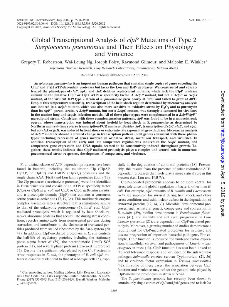
ClpP, but not ClpX or ClpC, is required for growth atreduced and elevated temperatures. The requirement for areduced temperature for the efficient recovery of clpP mutantsof S. pneumoniae suggested that ClpP proteolysis may beneeded for growth under certain stress conditions. To deter-mine whether ClpP-mediated proteolysis was required forgrowth under heat stress conditions, pneumococcal clp mu-tants or parental strains were tested for their ability to grow onsolid media at 30, 37, and 40°C. Unlike the R6 (EL59) or D39(EL161) parent strains (Fig. 1) (data not shown), the clpPmutants did not grow at 40°C. In contrast, clpC and clpX singlemutants were not restricted for growth at 40°C. These findingsare consistent with those reported recently by Chastanet andcoworkers for S. pneumoniae clpP mutants in liquid media (4).
We observed a slight reduction in colony size of clpPstrains on TSA-BA plates and a somewhat slower doublingtime (78 min versus 63 min) in CDM for the R6 clpP (EL539)mutant compared to its parent (EL59) at 37°C. Surprisingly,the D39 clpP::aad9 mutant (EL677) showed a marked reduc-tion in colony size at 30°C on TSA-BA plates, despite the factthat 30°C incubation was required for efficient initial recoveryof the D39 clpP mutant (see above). Taken together, thesefindings suggest that loss of the pneumococcal clpP gene prod-uct effects growth at temperature extremes and reducinggrowth rate may enhance the initial recovery of these mutants.Last, complementation of clpP mutant phenotypes had notbeen demonstrated previously (4). Therefore, we introducedan ectopic copy of the clpP� gene and its promoter into thedispensable (58) bgaA locus of clpP mutants (EL925 andEL923; Table 1). Growth of the clpP/clpP� merodiploids wasindistinguishable from that of the R6 and D39 parent strainson different media at 30, 37, and 40°C (Fig. 1; data not shown).Thus, loss of clpP function alone is responsible for all of theobserved growth defects at reduced and elevated tempera-tures.
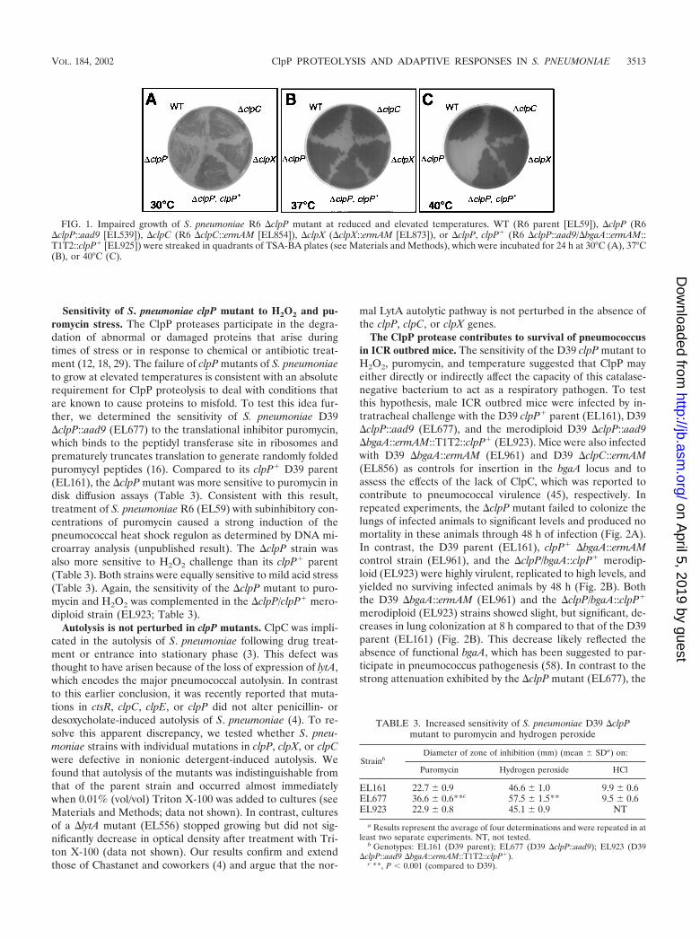
Sensitivity of S. pneumoniae clpP mutant to H2O2 and pu-romycin stress. The ClpP proteases participate in the degra-dation of abnormal or damaged proteins that arise duringtimes of stress or in response to chemical or antibiotic treat-ment (12, 18, 29). The failure of clpP mutants of S. pneumoniaeto grow at elevated temperatures is consistent with an absoluterequirement for ClpP proteolysis to deal with conditions thatare known to cause proteins to misfold. To test this idea fur-ther, we determined the sensitivity of S. pneumoniae D39clpP::aad9 (EL677) to the translational inhibitor puromycin,which binds to the peptidyl transferase site in ribosomes andprematurely truncates translation to generate randomly foldedpuromycyl peptides (16). Compared to its clpP� D39 parent(EL161), the clpP mutant was more sensitive to puromycin indisk diffusion assays (Table 3). Consistent with this result,treatment of S. pneumoniae R6 (EL59) with subinhibitory con-centrations of puromycin caused a strong induction of thepneumococcal heat shock regulon as determined by DNA mi-croarray analysis (unpublished result). The clpP strain wasalso more sensitive to H2O2 challenge than its clpP� parent(Table 3). Both strains were equally sensitive to mild acid stress(Table 3). Again, the sensitivity of the clpP mutant to puro-mycin and H2O2 was complemented in the clpP/clpP� mero-diploid strain (EL923; Table 3).
Autolysis is not perturbed in clpP mutants. ClpC was impli-cated in the autolysis of S. pneumoniae following drug treat-ment or entrance into stationary phase (3). This defect wasthought to have arisen because of the loss of expression of lytA,which encodes the major pneumococcal autolysin. In contrastto this earlier conclusion, it was recently reported that muta-tions in ctsR, clpC, clpE, or clpP did not alter penicillin- ordesoxycholate-induced autolysis of S. pneumoniae (4). To re-solve this apparent discrepancy, we tested whether S. pneu-moniae strains with individual mutations in clpP, clpX, or clpCwere defective in nonionic detergent-induced autolysis. Wefound that autolysis of the mutants was indistinguishable fromthat of the parent strain and occurred almost immediatelywhen 0.01% (vol/vol) Triton X-100 was added to cultures (seeMaterials and Methods; data not shown). In contrast, culturesof a lytA mutant (EL556) stopped growing but did not sig-nificantly decrease in optical density after treatment with Tri-ton X-100 (data not shown). Our results confirm and extendthose of Chastanet and coworkers (4) and argue that the nor-
mal LytA autolytic pathway is not perturbed in the absence ofthe clpP, clpC, or clpX genes.
The ClpP protease contributes to survival of pneumococcusin ICR outbred mice. The sensitivity of the D39 clpP mutant toH2O2, puromycin, and temperature suggested that ClpP mayeither directly or indirectly affect the capacity of this catalase-negative bacterium to act as a respiratory pathogen. To testthis hypothesis, male ICR outbred mice were infected by in-tratracheal challenge with the D39 clpP� parent (EL161), D39clpP::aad9 (EL677), and the merodiploid D39 clpP::aad9bgaA::ermAM::T1T2::clpP� (EL923). Mice were also infectedwith D39 bgaA::ermAM (EL961) and D39 clpC::ermAM(EL856) as controls for insertion in the bgaA locus and toassess the effects of the lack of ClpC, which was reported tocontribute to pneumococcal virulence (45), respectively. Inrepeated experiments, the clpP mutant failed to colonize thelungs of infected animals to significant levels and produced nomortality in these animals through 48 h of infection (Fig. 2A).In contrast, the D39 parent (EL161), clpP� bgaA::ermAMcontrol strain (EL961), and the clpP/bgaA::clpP� merodip-loid (EL923) were highly virulent, replicated to high levels, andyielded no surviving infected animals by 48 h (Fig. 2B). Boththe D39 bgaA::ermAM (EL961) and the clpP/bgaA::clpP�
merodiploid (EL923) strains showed slight, but significant, de-creases in lung colonization at 8 h compared to that of the D39parent (EL161) (Fig. 2B). This decrease likely reflected theabsence of functional bgaA, which has been suggested to par-ticipate in pneumococcus pathogenesis (58). In contrast to thestrong attenuation exhibited by the clpP mutant (EL677), the
FIG. 1. Impaired growth of S. pneumoniae R6 clpP mutant at reduced and elevated temperatures. WT (R6 parent [EL59]), clpP (R6clpP::aad9 [EL539]), clpC (R6 clpC::ermAM [EL854]), clpX (clpX::ermAM [EL873]), or clpP, clpP� (R6 clpP::aad9/bgaA::ermAM::T1T2::clpP� [EL925]) were streaked in quadrants of TSA-BA plates (see Materials and Methods), which were incubated for 24 h at 30°C (A), 37°C(B), or 40°C (C).
TABLE 3. Increased sensitivity of S. pneumoniae D39 clpPmutant to puromycin and hydrogen peroxide
StrainbDiameter of zone of inhibition (mm) (mean SDa) on:
clpC::ermAM mutant (EL856) was only marginally attenu-ated, replicated to high levels, and produced significant mor-bidity in infected ICR mice (Fig. 2A). The attenuation of theclpP mutant (EL677) was further evaluated using a murinemodel of septicemia. In these studies, we determined the av-erage infecting dose of the D39 parent (EL161) and the clpPmutant (EL677) required to produce signs of morbidity in 50%of the test subjects (LD50) within 24 h following direct intra-peritoneal injection. The LD50s of the D39 parent (EL161) andthe clpP mutant were 10 to 100 and �106, respectively (datanot shown), again indicating the strong requirement for clpPfunction in the pathogenicity of type 2 encapsulated pneumo-coccus in a murine host.
clpP transcript amounts increased in S. pneumoniae in re-sponse to heat shock. To date, all reported clpP gene productsfrom nonphotosynthetic bacteria are regulated by heat shock(46). Chastanet et al. (4) located a consensus binding site forCtsR (class three stress gene repressor) (9) 67 base pairs up-stream of the pneumococcal clpP ATG start codon (Fig. 3A;
Table 5), and they showed that purified CtsR binds to thisregion (4). They also reported a 30-fold induction of a S.pneumoniae clpP-bgaB fusion in a heterologous B. subtilis sys-tem in response to heat shock at 48°C (4). The magnitude ofthis heat shock response was considerably greater than thatreported for other stress-response proteases (15, 18). There-fore, we determined the extent of clpP heat shock induction byNorthern analysis and real-time quantitative RT-PCR (Taq-Man) in the R6 parent strain (EL59) after a shift to 45°C for 5and 15 min (Fig. 3B and 3C). Transcription of pneumococcalclpP increased in response to heat shock by 2.2- to 7.1-fold byreal-time RT-PCR (Fig. 3C) and 3.0- to 4.7-fold by Northernanalysis (Fig. 3B). In addition, the Northern analysis indicatedthat clpP is a monocistronic operon in S. pneumoniae. Thetranscript length of 0.7 kb corresponds to that expected for the5�-leader and clpP reading frame alone and not a bicistronictranscript of �0.9 kb containing both the clpP and orf-0657reading frames (Fig. 3A).
FIG. 2. Impaired virulence of S. pneumoniae D39 clpP mutant inlung model of infection. Male ICR outbred mice were infected byintratrachael inoculation with the indicated strains (see Materials andMethods). At each time point, four mice per group were sacrificed, andthe CFU per lung were determined by serial dilution and plating onTSA-BA (see Materials and Methods). Symbols: ■, D39 parent(EL161); �, D39 clpP::aad9 (EL677); F, D39 clpC::ermAM(EL856); E, D39 clpP::aad9/bgaA::ermAM::T1T2::clpP� merodip-loid (EL923); �, D39 bgaA::ermAM (EL961); vertical bars, standarddeviations; †, number of moribund animals at each time point; ‡,number of animals that became moribund between the 24- and 48-htime points.
FIG. 3. Transcription of clpP in S. pneumoniae R6 at 37°C and afterheat shock at 45°C. (A) Structure of the clpP locus of S. pneumoniaeR6 (drawn to scale). Arrows indicate predicted directions of transcrip-tion. The predicted �35 and CtsR-binding sites (underlined) are in-dicated. See text for details. (B) Northern blot of total RNA isolatedfrom S. pneumoniae R6 (EL59) probed for clpP (see Materials andMethods). Relative expression levels are indicated below the autora-diograph (see Materials and Methods). Lanes: 1, 37°C; 2, 45°C for 5min; 3, 45°C for 15 min. (C) Quantitative real-time RT-PCR of clpPand fabK transcription following heat shock (HS) at 45°C. Data rep-resent the relative change (n-fold) in mRNA amounts compared tothat of the 37°C control, calibrated to 16S rRNA amounts (see Mate-rials and Methods). fabK transcript amounts were included as a controlof a gene not known to be regulated by heat shock. No clpP transcriptwas detected from the clpP mutant (R6 clpP::aad9) (EL539).
Global transcriptional profile of �clpP mutant of strain R6.The phenotypes of clpP mutants suggest that the ClpP proteaseplays fundamental roles in the normal physiology, stress sur-vival, and pathogenesis of S. pneumoniae. To begin to addressthe genetic basis for these phenotypes, we performed an anal-ysis of global transcription of the R6 clpP mutant comparedto the R6 parent during exponential growth in CDM by usinga custom Affymetrix oligonucleotide microarray (Materialsand Methods). As an internal control of this method, clpPtranscripts were not detected (called absent, with a decrease ofat least 22-fold) from the R6 clpP mutant (EL539) comparedto the R6 parent strain (EL59) (Table 4). Furthermore, thelevel of transcription from the upp and orf-0657 genes, whichflank clpP (Fig. 3A), was the same in the R6 clpP and itsclpP� parent, indicating a lack of polarity on the surroundinggenes.
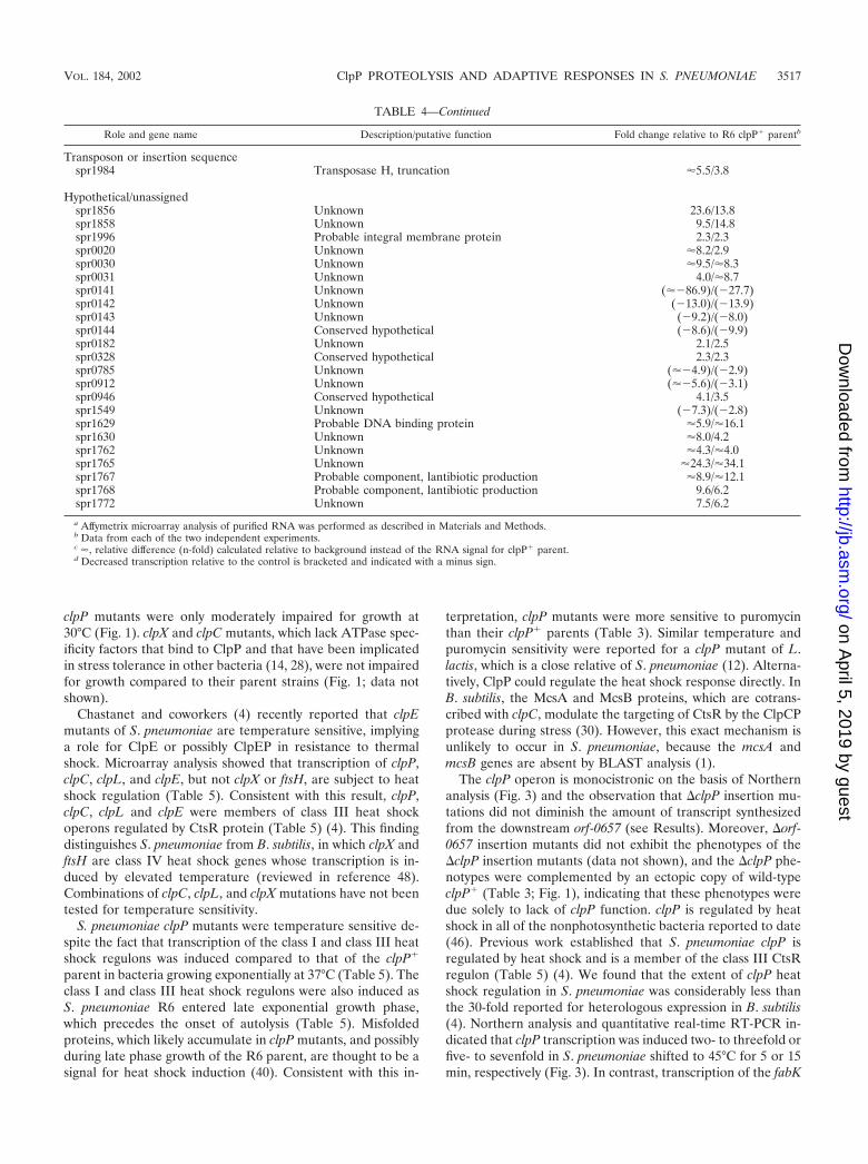
Changes in relative transcript amounts of �2.0-fold wereobserved for 150 and 200 genes, respectively, in two separateand independent experiments. Of these transcriptionalchanges, 79 were common to both experiments and thus wereconsidered reproducible (see Materials and Methods). Someof the identified genes were linked in the chromosome andshowed similar changes in expression; however, additional ex-periments would be necessary to demonstrate polycistronicoperons for these genes. The majority of altered genes couldbe clustered into several major classes (Table 4), including themajority of the known competence and heat shock regulons(all induced), genes required for virulence and oxidative stressresistance (all repressed), a large group of hypothetical genesof unknown functions, and several genes associated with me-tabolism and cell surface components. Repressed genes thatmediate resistance to oxidative stress included tpx (thiore-doxin-linked thiol peroxidase) and sodA (manganese-depen-dent superoxide dismutase). Repressed genes that influencepneumococcal virulence included sodA and psaABC (manga-nese transport) (2, 57). It is noteworthy that the transcriptionof several cell surface proteins that may mediate pathogenesiswere induced or repressed in the R6 clpP mutant, includinglytA (major autolysin), dlytC (D-alanyl carrier protein), andcbpD (choline-binding protein D) (Table 4).
Altered competence gene expression in clpP mutants. Chas-tanet and coworkers recently reported that expression of fu-sion constructs of the early comDCE and late ssbB competencegenes was increased in a clpP insertion mutant (4). This ob-servation led to the conclusion that ClpP may act as a negativeregulator that prevents competence gene expression under in-appropriate conditions. Our global transcription analysisshowed that transcription of the majority of genes in the S.pneumoniae competence regulon is highly induced in a clpPmutant during mid-exponential growth phase (Table 4). Totest whether this induction was transient, we assayed the as-similation of donor genomic DNA in a clpP mutant derivedfrom the competence indicator strain CPM7 (Table 1) (34). Inagreement with the microarray studies, the clpP mutant(EL824) became competent for transformation in CDM at amuch lower cell density than did its parental control (CPM7)and remained competent throughout logarithmic growth (Fig.4A). In contrast, competence in the parental CPM7 strain wastransient, resulting in typical pneumococcal transformation ki-netics (44) (Fig. 4A). We also found that natural genetic com-
petence did not develop for CPM7 grown in BHI but that thisblock in competence development could eventually be over-come in a clpP strain (EL824) (Fig. 4B). Independent exper-iments with BHI medium revealed strong ssbB::lacZ expres-sion (fivefold increase above the parental control) in the clpPmutant or after addition of synthetic CSP-1 to the clpP� strain,and this was accompanied by a comparable increase in thetransformation frequency (data not shown). Maximum expres-sion of ssbB::lacZ (ninefold increase compared to the un-treated parental control) and transformation frequency oc-curred in the clpP mutant treated with synthetic CSP-1 (datanot shown). Thus, our microarray and direct determinations ofcompetence development support and extend the conclusionof Chastanet et al. (4) that ClpP proteolysis plays a negativeregulatory role in natural competence development of S. pneu-moniae. Our results further suggest that ClpP proteolysis playsa role in the termination of competence as well.
Transcriptional induction of major heat shock genes in S.pneumoniae R6. Microarray analysis of the S. pneumoniae clpPstrain during normal exponential growth revealed a stronginduction of genes that classically comprise the bacterial heatshock regulon (Table 4). Computer searches had previouslydemonstrated the presence of consensus HrcA binding sites(putative class I heat shock genes) (40) immediately upstreamof the putative groES-groEL and hrcA-grpE-dnaK-dnaJ operonsand CtsR consensus binding sites (putative class III heat shockgenes) (9) preceding the clpP, ctsR-clpC, clpE, clpL, and groES-groEL loci (Table 5) (4, 26). Putative HcrA and CtsR bindingsites were not identified upstream of other pneumococcalgenes in the R6 genome (data not shown). Microarray analysisrevealed significant transcriptional induction of these pneumo-coccal classes I and III heat shock genes in bacteria shifted to45°C (Table 5). In contrast, transcription of clpX (putativespecificity factor for ClpP) and ftsH (ATP-dependent protease)were not induced by thermal shock (Table 5), despite the factthat they are class IV heat shock genes in B. subtilis (48). Last,de Saizieu and coworkers (8) found that expression of the clpLand groEL heat shock genes was induced as pneumococcusentered the late exponential phase of growth. We found thattranscription of the remaining class I and class III heat shockgenes were induced in late exponential compared to earlyexponential cultures of S. pneumoniae R6 grown in CDM (Ta-ble 5). Unlike the heat shock genes, expression of clpX and ftsHwas not affected by growth phase (Table 5).
DISCUSSION
Unlike most other bacterial species, S. pneumoniae containsonly two ATP-dependent proteases, ClpP and FtsH, which areencoded by single-copy genes (23). We report here that clpPplays complex, pleiotropic roles in pneumococcal stress resis-tance, gene expression, regulation of genetic competence, andvirulence in a murine host. clpP mutants of virulent S. pneu-moniae strain D39 exhibited a very limited range of growthcentered around 37°C. As noted before (4), clpP mutants of S.pneumoniae do not grow at 40°C, and we show further that thistemperature sensitivity is complemented by a wild-type copy ofclpP� (Fig. 1). We found that D39 clpP mutants were coldsensitive and grew poorly at 30°C (see Results), whereas R6
VOL. 184, 2002 ClpP PROTEOLYSIS AND ADAPTIVE RESPONSES IN S. PNEUMONIAE 3515
Competence and DNA metabolismcomD Competence factor dependent histidine kinase �17.0/�15.1comE Competence factor dependent response regulator 24.7/31.6comX1 Competence-specific sigma factor 3.5/3.4spr1859 ComYA-like protein 20.5/12.1cglD ComG-like protein 6.8/6.8cglC Pilin-like wall structure 1.6/5.3cglB Prepilin transport ATPase 52.8/28.9cglA Prepilin transport pore 34.1/�31.8comF Competence protein 2.4/�3.0celA DNA binding �5.7/4.9celB DNA uptake pore 2.4/2.5ssbB, cilA ssDNA binding 10.6/10.7dinF Efflux pump 4.8/3.1recA DNA recombination 5.4/2.4cinA Unknown 4.7/4.1comA Transport, ATP binding protein 4.1/1.5comB Transport �23.3/11.9
Peptide/amino acid transportaliA ABC transporter, oligopeptide transport (�2.2)/(�2.7)pncP ABC transporter, unknown substrate �11.4/�3.3spr0100 ABC transporter, amino acid transport 2.8/�2.9spr1773 ABC transporter, ATP-binding protein �8.9/�12.6
Sugar transport and metabolismmalR Transcriptional repressor, maltose operon �3.8/2.1manN PTS-EIIAB, mannose specific 2.1/2.2bglA 6-Phospho-�-glucosidase (�12.8)/(�2.9)spr0277 Conserved hypothetical (�7.6)/(�3.2)spr0278 PTS-EIIB, sugar specific (�7.6)/(�2.4)bglG Transcriptional antiterminator, BglG family (�6.0)/(�2.3)spr0280 PTS-EIIA, sugar specific (�6.7)/(�2.6)spr0281 Hypothetical (�7.9)/(�3.1)spr0282 PTS-EIIC, sugar specific (�7.4)/(�2.9)
Cell surfacelytA Major autolysin 2.7/1.7dlytC D-alanyl carrier protein �8.3/3.0cbpD Choline-binding protein D �9.4/4.0htrA Putative cell surface serine protease (�2.0)/(�4.5)
Metal transport/metabolismcopY Transcriptional regulator, copper transport �11.1/3.7ctpA P-type ATPase, copper transport �13.4/2.7psaB ATP-binding protein, Mn2� transport (�2.4)/(�2.0)psaC Membrane spanning permease, Mn2� transport (��2.5)/(�2.4)
clpP mutants were only moderately impaired for growth at30°C (Fig. 1). clpX and clpC mutants, which lack ATPase spec-ificity factors that bind to ClpP and that have been implicatedin stress tolerance in other bacteria (14, 28), were not impairedfor growth compared to their parent strains (Fig. 1; data notshown).
Chastanet and coworkers (4) recently reported that clpEmutants of S. pneumoniae are temperature sensitive, implyinga role for ClpE or possibly ClpEP in resistance to thermalshock. Microarray analysis showed that transcription of clpP,clpC, clpL, and clpE, but not clpX or ftsH, are subject to heatshock regulation (Table 5). Consistent with this result, clpP,clpC, clpL and clpE were members of class III heat shockoperons regulated by CtsR protein (Table 5) (4). This findingdistinguishes S. pneumoniae from B. subtilis, in which clpX andftsH are class IV heat shock genes whose transcription is in-duced by elevated temperature (reviewed in reference 48).Combinations of clpC, clpL, and clpX mutations have not beentested for temperature sensitivity.
S. pneumoniae clpP mutants were temperature sensitive de-spite the fact that transcription of the class I and class III heatshock regulons was induced compared to that of the clpP�
parent in bacteria growing exponentially at 37°C (Table 5). Theclass I and class III heat shock regulons were also induced asS. pneumoniae R6 entered late exponential growth phase,which precedes the onset of autolysis (Table 5). Misfoldedproteins, which likely accumulate in clpP mutants, and possiblyduring late phase growth of the R6 parent, are thought to be asignal for heat shock induction (40). Consistent with this in-
terpretation, clpP mutants were more sensitive to puromycinthan their clpP� parents (Table 3). Similar temperature andpuromycin sensitivity were reported for a clpP mutant of L.lactis, which is a close relative of S. pneumoniae (12). Alterna-tively, ClpP could regulate the heat shock response directly. InB. subtilis, the McsA and McsB proteins, which are cotrans-cribed with clpC, modulate the targeting of CtsR by the ClpCPprotease during stress (30). However, this exact mechanism isunlikely to occur in S. pneumoniae, because the mcsA andmcsB genes are absent by BLAST analysis (1).
The clpP operon is monocistronic on the basis of Northernanalysis (Fig. 3) and the observation that clpP insertion mu-tations did not diminish the amount of transcript synthesizedfrom the downstream orf-0657 (see Results). Moreover, orf-0657 insertion mutants did not exhibit the phenotypes of theclpP insertion mutants (data not shown), and the clpP phe-notypes were complemented by an ectopic copy of wild-typeclpP� (Table 3; Fig. 1), indicating that these phenotypes weredue solely to lack of clpP function. clpP is regulated by heatshock in all of the nonphotosynthetic bacteria reported to date(46). Previous work established that S. pneumoniae clpP isregulated by heat shock and is a member of the class III CtsRregulon (Table 5) (4). We found that the extent of clpP heatshock regulation in S. pneumoniae was considerably less thanthe 30-fold reported for heterologous expression in B. subtilis(4). Northern analysis and quantitative real-time RT-PCR in-dicated that clpP transcription was induced two- to threefold orfive- to sevenfold in S. pneumoniae shifted to 45°C for 5 or 15min, respectively (Fig. 3). In contrast, transcription of the fabK
TABLE 4—Continued
Role and gene name Description/putative function Fold change relative to R6 clpP� parentb
Transposon or insertion sequencespr1984 Transposase H, truncation �5.5/3.8
a Affymetrix microarray analysis of purified RNA was performed as described in Materials and Methods.b Data from each of the two independent experiments.c �, relative difference (n-fold) calculated relative to background instead of the RNA signal for clpP� parent.d Decreased transcription relative to the control is bracketed and indicated with a minus sign.
VOL. 184, 2002 ClpP PROTEOLYSIS AND ADAPTIVE RESPONSES IN S. PNEUMONIAE 3517
gene, which is involved in fatty acid biosynthesis (23) and is notpart of a heat shock regulon (Table 5), was not induced by thistemperature shift (Fig. 3).
ClpP proteases do not generally seem to mediate the deg-radation of oxidatively damaged proteins (6). Therefore, it wassomewhat unexpected to find that a S. pneumoniae clpP mu-tant is more sensitive to H2O2 compared to its clpP� parent(Table 3). Global transcriptional analysis of this catalase-lack-ing bacterium showed that expression of the tpx (thioredoxin-linked thiol peroxidase) and sodA (manganese-dependent su-peroxide dismutase) genes was decreased in the clpP mutantcompared to its clpP� parent (Table 4). This repression couldpossibly account for the H2O2 sensitivity of the clpP mutant.Further experiments are required to understand the link be-tween ClpP function and sensitivity to H2O2.
The microarray and direct determination of competencedevelopment reported here support and extend the generalconclusion of Chastanet and coworkers that ClpP acts as anegative regulator of competence expression (4). Moreover,our results are consistent with a role of ClpP in the terminationof competence, which is normally transient in S. pneumoniae(44). We found that transcription of the majority of the known
competence regulon is strongly induced in the clpP mutant ata time during exponential growth when the pathway is nor-mally repressed (Table 4). Expression of an ssbB-lacZ reportergene (data not shown) and DNA uptake (Fig. 4) were dere-pressed without the addition of CSP-1, and addition of syn-thetic CSP-1 further increased derepression and DNA uptake(data not shown). These results could be explained by in-creased expression of the ComDE two-component system,which senses CSP-1 levels and indirectly activates transcriptionof late genes in the competence pathway, or increased expres-sion of the CSP-1 pheromone, which is cotranscribed in thecomCDE operon (43). A direct role for ClpP-mediated prote-olysis in regulating the cellular amounts of ComDE is a test-able hypothesis, although the effect of ClpP could also beindirect. There is a precedent for ClpP-mediated proteolysis ofa competence transcription factor in B. subtilis in which acomplex forms containing the MecA adaptor protein, theComK competence transcription factor, and ClpCP (54). S.pneumoniae encodes a MecA homolog (73% amino acid sim-ilarity to B. subtilis), but lacks other factors, such as ComK andComS, involved in the B. subtilis MecA switch. Furthermore,our data combined with that of Chastanet and coworkers (4)suggest that ClpC and ClpE do not singly mediate the effect ofClpP on competence induction. The potential role of MecAand ClpX or ClpL in the regulation of S. pneumoniae compe-tence by ClpP will be tested in future experiments.
Finally, we found that a D39 clpP mutant was stronglyattenuated for virulence in the mouse lung (Fig. 2) and sepsismodels (�104 to 105 increase in LD50; see Results). This at-tenuation phenotype was complemented by an ecotopic copyof the clpP� gene (Fig. 2). In contrast, attenuation by a clpCmutation was mild compared to that of the clpP mutation(Fig. 2). It is unlikely that this attenuation is due to the tem-perature sensitivity of the D39 clpP mutant, because the bodytemperature of mice ranges between 37.1 to 37.4°C (24), whichsupports the growth of the clpP mutants in vitro (Fig. 1).Visual inspection of colonies of the D39 clpP mutant onplates did not suggest any diminution of capsule production,which is strongly linked to S. pneumoniae virulence (38).Global transcription analysis showed that the transcription lev-els of several likely pathogenicity factors was reduced in theclpP mutant compared to that of the clpP� strain, includingsodA (manganese-dependent superoxide dismutase), tpx (thi-oredoxin-linked thiol peroxidase), and psaABC (manganesetransporter) (Table 4). sodA and psaABC have previously beenlinked with the virulence of S. pneumoniae (2, 57), and anti-oxidizing enzymes, such as Tpx and Sod, have been linked tophagocyte resistance and virulence in several pathogens (10,32). In addition, the transcription of several genes encodingcell surface proteins, which have been suggested to play pos-sible roles in virulence (38), and proteins of unknown functions(23) are altered in clpP mutants. Knockout mutants of thehk06/rr06 two-component system, whose transcription is in-duced in the clpP mutant (Table 4), are attenuated for viru-lence in our lung and sepsis animal models (data not shown)and those reported elsewhere (51). Together, these data implythat the large attenuation in virulence of clpP mutants is acomplex phenotype with many contributing factors. It remainsto be determined how the loss of this major ATP-dependent
FIG. 4. ClpP negatively regulates competence development in S.pneumoniae. Cells grown in CDM (A) or BHI medium (B) weremonitored for changes in OD620 and for the capacity to assimilategenomic donor DNA encoding resistance to novobiocin (see Materialsand Methods). ●, OD620 of the ssbB::lacZ parent strain (CPM7); �,OD620 of the clpP::aad9 ssbB::lacZ strain (EL824); �, Nvr transfor-mants recovered from the ssbB::lacZ parent strain (CPM7); �, Nvr
transformants recovered from the clpP::aad9 ssbB::lacZ strain(EL824). The data are representative of two independent experiments.
protease affects gene expression in S. pneumoniae at the levelof protein stability and amount.
ACKNOWLEDGMENTS
We thank Alexander Tomasz, Janet Yother, Donald LeBlanc, andDonald Morrison for bacterial strains, Donald Morrison (University ofIllinois at Chicago) for helpful discussions on competence, Yong Yangfor bioinformatic assistance, John Glass and Jennifer Glass for assis-tance with quantitative real-time RT-PCR analyses, Genshi Zhao andDan Mytelka for reviewing the manuscript, and John Richardson fortechnical assistance.
This work was supported by resources provided by Lilly ResearchLaboratories, and Gregory T. Robertson was supported by a LillyPostdoctoral Fellowship.
REFERENCES
1. Altschul, S. F., W. Gish, W. Miller, E. W. Myers, and D. J. Lipman. 1990.Basic local alignment search tool. J. Mol. Biol. 215:403–410.
2. Berry, A. M., and J. C. Paton. 1996. Sequence heterogeneity of PsaA, a37-kilodalton putative adhesin essential for virulence of Streptococcus pneu-moniae. Infect. Immun. 64:5255–5262.
3. Charpentier, E., R. Novak, and E. Tuomanen. 2000. Regulation of growthinhibition at high temperature, autolysis, transformation and adherence inStreptococcus pneumoniae by clpC. Mol. Microbiol. 37:717–726.
4. Chastanet, A., M. Prudhomme, J. P. Claverys, and T. Msadek. 2001. Reg-ulation of Streptococcus pneumoniae clp genes and their role in competencedevelopment and stress survival. J. Bacteriol. 183:7295–7307.
5. Damerau, K., and A. C. St. John. 1993. Role of Clp protease subunits indegradation of carbon starvation proteins in Escherichia coli. J. Bacteriol.175:53–63.
6. Davies, K. J., and S. W. Lin. 1988. Oxidatively denatured proteins aredegraded by an ATP-independent proteolytic pathway in Escherichia coli.Free Radic. Biol. Med. 5:225–236.
7. De Mot, R., I. Nagy, J. Walz, and W. Baumeister. 1999. Proteasomes andother self-compartmentalizing proteases in prokaryotes. Trends Microbiol.7:88–92.
8. de Saizieu, A., U. Certa, J. Warrington, C. Gray, W. Keck, and J. Mous. 1998.
Bacterial transcript imaging by hybridization of total RNA to oligonucleotidearrays. Nat. Biotechnol. 16:45–48.
9. Derré, I., G. Rapoport, and T. Msadek. 1999. CtsR, a novel regulator ofstress and heat shock response, controls clp and molecular chaperone geneexpression in gram-positive bacteria. Mol. Microbiol. 31:117–131.
10. Fang, F. C., M. A. DeGroote, J. W. Foster, A. J. Baumler, U. Ochsner, T.Testerman, S. Bearson, J. C. Giard, Y. Xu, G. Campbell, and T. Laessig.1999. Virulent Salmonella typhimurium has two periplasmic Cu, Zn-super-oxide dismutases. Proc. Natl. Acad. Sci. USA 96:7502–7507.
11. Frank, E. G., D. G. Ennis, M. Gonzalez, A. S. Levine, and R. Woodgate. 1996.Regulation of SOS mutagenesis by proteolysis. Proc. Natl. Acad. Sci. USA93:10291–10296.
12. Frees, D., and H. Ingmer. 1999. ClpP participates in the degradation ofmisfolded protein in Lactococcus lactis. Mol. Microbiol. 31:79–87.
13. Gaillot, O., E. Pellegrini, S. Bregenholt, S. Nair, and P. Berche. 2000. TheClpP serine protease is essential for the intracellular parasitism and viru-lence of Listeria monocytogenes. Mol. Microbiol. 35:1286–1294.
14. Gerth, U., E. Kruger, I. Derré, T. Msadek, and M. Hecker. 1998. Stressinduction of the Bacillus subtilis clpP gene encoding a homologue of theproteolytic component of the Clp protease and the involvement of ClpP andClpX in stress tolerance. Mol. Microbiol. 28:787–802.
15. Goff, S. A., and A. L. Goldberg. 1985. Production of abnormal proteins in E.coli stimulates transcription of lon and other heat shock genes. Cell 41:587–595.
16. Goldberg, A. L. 1972. Correlation between rates of degradation of bacterialproteins in vivo and their sensitivity to proteases. Proc. Natl. Acad. Sci. USA69:2640–2644.
17. Gottesman, S. 1996. Proteases and their targets in Escherichia coli. Annu.Rev. Genet. 30:465–506.
18. Gottesman, S., W. P. Clark, V. de Crecy-Lagard, and M. R. Maurizi. 1993.ClpX, an alternative subunit for the ATP-dependent Clp protease of Esch-erichia coli. Sequence and in vivo activities. J. Biol. Chem. 268:22618–22626.
19. Gottesman, S., and M. R. Maurizi. 1992. Regulation by proteolysis: energy-dependent proteases and their targets. Microbiol. Rev. 56:592–621.
20. Gottesman, S., E. Roche, Y. Zhou, and R. T. Sauer. 1998. The ClpXP andClpAP proteases degrade proteins with carboxy-terminal peptide tails addedby the SsrA-tagging system. Genes Dev. 12:1338–1347.
21. Hensel, M., J. E. Shea, C. Gleeson, M. D. Jones, E. Dalton, and D. W.Holden. 1995. Simultaneous identification of bacterial virulence genes bynegative selection. Science 269:400–403.
22. Horton, R. M., Z. L. Cai, S. N. Ho, and L. R. Pease. 1990. Gene splicing by
TABLE 5. Organization and transcriptional induction of R6 heat shock genes by temperature shift to 45°C, deletionof clpP, and entry of cultures into late exponential phasea
a Growth of cultures, RNA extraction, and Affymetrix microarray analysis were performed as described in Materials and Methods.b Relative induction (n-fold) was calculated for the first gene of each operon. �, relative change (n-fold) calculated relative to that of the background instead of 37°C
control.c Data are from each of two independent experiments.d Number of nucleotides (nt) between the 3� end of the putative regulatory element and the start codon for the first gene of each operon is given. NA, neither
regulatory element was detected for clpX and ftsH.e The consensus sequence was previously determined (9). Capitalized bases are strictly conserved. Lower case bases are not conserved.f (rev), listed sequence is located on the noncoding strand.g The consensus sequence was previously determined (39). (R), conservation of either A or G.
VOL. 184, 2002 ClpP PROTEOLYSIS AND ADAPTIVE RESPONSES IN S. PNEUMONIAE 3519
overlap extension: tailor-made genes using the polymerase chain reaction.BioTechniques 8:528–535.
23. Hoskins, J., W. E. Alborn, Jr., J. Arnold, L. C. Blaszczak, S. Burgett, B. S.DeHoff, S. T. Estrem, L. Fritz, D. J. Fu, W. Fuller, C. Geringer, R. Gilmour,J. S. Glass, H. Khoja, A. R. Kraft, R. E. Lagace, D. J. LeBlanc, L. N. Lee,E. J. Lefkowitz, J. Lu, P. Matsushima, S. M. McAhren, M. McHenney, K.McLeaster, C. W. Mundy, T. I. Nicas, F. H. Norris, M. O’Gara, R. B. Peery,G. T. Robertson, P. Rockey, P. M. Sun, M. E. Winkler, Y. Yang, M. Young-Bellido, G. Zhao, C. A. Zook, R. H. Baltz, S. R. Jaskunas, P. R. Rosteck, Jr.,P. L. Skatrud, and J. I. Glass. 2001. Genome of the bacterium Streptococcuspneumoniae strain R6. J. Bacteriol. 183:5709–5717.
24. Jacoby, R. O., and J. G. Fox. 1984. Biology and diseases of mice, p. 31–89. InJ. G. Fox, B. J. Cohen, and F. M. Loew (ed.), Laboratory animal medicine.Academic Press, Orlando, Fla.
25. Jenal, U., and T. Fuchs. 1998. An essential protease involved in bacterialcell-cycle control. EMBO J. 17:5658–5669.
26. Kim, S. N., S. W. Kim, S. N. Pyo, and D. K. Rhee. 2001. Molecular cloningand characterization of groESL operon in Streptococcus pneumoniae. Mol.Cell 11:360–368.
27. Klugman, K. P., and C. Feldman. 2001. Streptococcus pneumoniae respira-tory tract infections. Curr. Opin. Infect. Dis. 14:173–179.
28. Kruger, E., U. Volker, and M. Hecker. 1994. Stress induction of clpC inBacillus subtilis and its involvement in stress tolerance. J. Bacteriol. 176:3360–3367.
29. Kruger, E., E. Witt, S. Ohlmeier, R. Hanschke, and M. Hecker. 2000. The clpproteases of Bacillus subtilis are directly involved in degradation of misfoldedproteins. J. Bacteriol. 182:3259–3265.
30. Kruger, E., D. Zuhlke, E. Witt, H. Ludwig, and M. Hecker. 2001. Clp-mediated proteolysis in Gram-positive bacteria is autoregulated by the sta-bility of a repressor. EMBO J. 20:852–863.
31. Kruse, N., M. Pette, K. Toyka, and P. Rieckmann. 1997. Quantification ofcytokine mRNA expression by RT PCR in samples of previously frozenblood. J. Immunol. Methods 210:195–203.
32. Kwatia, M. A., D. J. Botkin, and D. L. Williams. 2000. Molecular andenzymatic characterization of Schistosoma mansoni thioredoxin peroxidase.J. Parasitol. 86:908–915.
33. LeBlanc, D. J., L. N. Lee, and A. Abu-Al-Jaibat. 1992. Molecular, genetic,and functional analysis of the basic replicon of pVA380–1, a plasmid of oralstreptococcal origin. Plasmid 28:130–145.
34. Lee, M. S., and D. A. Morrison. 1999. Identification of a new regulator inStreptococcus pneumoniae linking quorum sensing to competence for genetictransformation. J. Bacteriol. 181:5004–5016.
35. Macrina, F. L., J. A. Tobian, K. R. Jones, R. P. Evans, and D. B. Clewell.1982. A cloning vector able to replicate in Escherichia coli and Streptococcussanguis. Gene 19:345–353.
36. Maurizi, M. R. 1992. Proteases and protein degradation in Escherichia coli.Experientia 48:178–201.
37. Maurizi, M. R., W. P. Clark, Y. Katayama, S. Rudikoff, J. Pumphrey, B.Bowers, and S. Gottesman. 1990. Sequence and structure of ClpP, the pro-teolytic component of the ATP-dependent Clp protease of Escherichia coli.J. Biol. Chem. 265:12536–12545.
38. Mitchell, T. J. 2000. Virulence factors and the pathogenesis of diseasecaused by Streptococcus pneumoniae. Res. Microbiol. 151:413–419.
39. Msadek, T., V. Dartois, F. Kunst, M. L. Herbaud, F. Denizot, and G. Rap-oport. 1998. ClpP of Bacillus subtilis is required for competence develop-ment, motility, degradative enzyme synthesis, growth at high temperatureand sporulation. Mol. Microbiol. 27:899–914.
40. Narberhaus, F. 1999. Negative regulation of bacterial heat shock genes. Mol.Microbiol. 31:1–8.
40a.National Research Council. 1985. Guide for the care and use of laboratoryanimals. NIH publication no. 86–23. National Institutes of Health, Bethesda,Md.
41. O’Toole, G. A., and R. Kolter. 1998. Initiation of biofilm formation inPseudomonas fluorescens WCS365 proceeds via multiple, convergent signal-ling pathways: a genetic analysis. Mol. Microbiol. 28:449–461.
42. Pederson, K. J., S. Carlson, and D. E. Pierson. 1997. The ClpP protein, asubunit of the Clp protease, modulates ail gene expression in Yersinia en-terocolitica. Mol. Microbiol. 26:99–107.
43. Pestova, E. V., L. S. Havarstein, and D. A. Morrison. 1996. Regulation ofcompetence for genetic transformation in Streptococcus pneumoniae by anauto-induced peptide pheromone and a two-component regulatory system.Mol. Microbiol. 21:853–862.
44. Peterson, S., R. T. Cline, H. Tettelin, V. Sharov, and D. A. Morrison. 2000.Gene expression analysis of the Streptococcus pneumoniae competence regu-lons by use of DNA microarrays. J. Bacteriol. 182:6192–6202.
45. Polissi, A., A. Pontiggia, G. Feger, M. Altieri, H. Mottl, L. Ferrari, and D.Simon. 1998. Large-scale identification of virulence genes from Streptococcuspneumoniae. Infect. Immun. 66:5620–5629.
46. Porankiewicz, J., J. Wang, and A. K. Clarke. 1999. New insights into theATP-dependent Clp protease: Escherichia coli and beyond. Mol. Microbiol.32:449–458.
47. Reeve, C. A., A. T. Bockman, and A. Matin. 1984. Role of protein degrada-tion in the survival of carbon-starved Escherichia coli and Salmonella typhi-murium. J. Bacteriol. 157:758–763.
48. Schumann, W., Hecker, M., and T. Msadek. 2002. Regulation and functionof heat inducible genes in Bacillus subtilis, p. 359–368. In A. L. Sonenshein,J. A. Hoch, and R. Losick (ed.), Bacillus subtilis and its closest relatives: fromgenes to cells. ASM Press, Washington, D.C.
49. Smith, M. D., and W. R. Guild. 1979. A plasmid in Streptococcus pneu-moniae. J. Bacteriol. 137:735–739.
50. Talkington, D. F., D. C. Voellinger, L. S. McDaniel, and D. E. Briles. 1992.Analysis of pneumococcal PspA microheterogeneity in SDS polyacrylamidegels and the association of PspA with the cell membrane. Microb. Pathog.13:343–355.
51. Throup, J. P., K. K. Koretke, A. P. Bryant, K. A. Ingraham, A. F. Chalker,Y. Ge, A. Marra, N. G. Wallis, J. R. Brown, D. J. Holmes, M. Rosenberg, andM. K. Burnham. 2000. A genomic analysis of two-component signal trans-duction in Streptococcus pneumoniae. Mol. Microbiol. 35:566–576.
52. Tomasz, A. 1999. New faces of an old pathogen: emergence and spread ofmultidrug-resistant Streptococcus pneumoniae. Am. J. Med. 107:55S-62S.
53. Tsui, H.-C. T., A. J. Pease, T. M. Koehler, and M. E. Winkler. 1994. Detec-tion and quantitation of RNA transcribed from bacterial chromosomes.Methods Mol. Genet. 3:179–204.
54. Turgay, K., J. Hahn, J. Burghoorn, and D. Dubnau. 1998. Competence inBacillus subtilis is controlled by regulated proteolysis of a transcription fac-tor. EMBO J. 17:6730–6738.
55. Watson, D. A., D. M. Musher, and J. Verhoef. 1995. Pneumococcal virulencefactors and host immune responses to them. Eur. J. Clin. Microbiol. Infect.Dis. 14:479–490.
56. Webb, C., M. Moreno, M. Wilmes-Riesenberg, R. Curtiss III, and J. W.Foster. 1999. Effects of DksA and ClpP protease on sigma S production andvirulence in Salmonella typhimurium. Mol. Microbiol. 34:112–123.
57. Yesilkaya, H., A. Kadioglu, N. Gingles, J. E. Alexander, T. J. Mitchell, andP. W. Andrew. 2000. Role of manganese-containing superoxide dismutase inoxidative stress and virulence of Streptococcus pneumoniae. Infect. Immun.68:2819–2826.
58. Zahner, D., and R. Hakenbeck. 2000. The Streptococcus pneumoniae beta-galactosidase is a surface protein. J. Bacteriol. 182:5919–5921.
59. Zhou, Y., and S. Gottesman. 1998. Regulation of proteolysis of the station-ary-phase sigma factor RpoS. J. Bacteriol. 180:1154–1158.
60. Zugel, U., and S. H. Kaufmann. 1999. Role of heat shock proteins in pro-tection from and pathogenesis of infectious diseases. Clin. Microbiol. Rev.12:19–39.