Human-Biology Introduction 1 Glossary Human Biology - Introduction Anatomy: The science of biological structures. Physiology: The study of the functioning body organs. Human Body: The entire physical structure of a human being. B. Cavity: A space within the body that contains various internal organs. Homeostasis: The condition in which the body's internal environment remains relatively constant, within limits. Systems of the Body: A group of interacting elements in the body functioning as a complex whole. Cardiovascular S.: The constituting elements of the body (blood, heart, and blood vessels) which nourish all body tissues at (cellular level), removes metabolic waste products (in cooperation with excretory and respiratory systems) and distributes thermal energy throughout the body. Digestive S.: The organs involved in the mechanical and chemical breakdown of food into small molecules for absorption and use. Endocrine S.: The collection of endocrine glands of the body; in humans the Excretory S.: The components of the body involved in the elimination of metabolic waste products from the body; in humans the liver and the kidneys. Immune or Lymphatic S.: The network if cells, tissues, and organs that defends the body against microbial invaders. Made up by the lymph, lymph vessels and structures and organs containing lymphatic tissue (large numbers of white blood cells called lymphocytes). Nervous S.: The network of nerves that integrate and coordinate the activities of all the bodily systems. • Parasympathetic NS: The autonomic nervous system, having cell bodies of preganglionic neurons in nuclei in the brain stem and in the lateral gray matter of the sacral portion of the spinal cord; primarily concerned with activities that restore and conserve body energy (cranosacral division). • Sympathetic NS: The autonomic nervous system, having cell bodies of preganglionic neurons in the lateral gray columns of the thoracic segment and first two or three lumbar segments of the spinal cord; primarily concerned with processes involving the expenditure of energy (thoracolumbar division). Reproductive S.: The tissues and organs (gonads, testis and ovum) involved in the production and maturation of gametes (sperm and egg) and the supportive structures required to maintain the developing embryonic and fetal stages. Respiratory S.: The sections of the body involved in the overall exchange of gases between the atmosphere, blood, and body cells; involves pulmonary respiration, external respiration, and internal respiration mediated by the trachea, lungs (alveolar tissue, surfactant), and muscles of the rib cage. Skeletal S.: The passive and rigid body support to which muscles attach and apply force. Cartilage and osseous tissue comprise the skeletal system.
Transcript
Human-Biology Introduction1
Glossary Human Biology - IntroductionAnatomy: The science of biological structures.Physiology: The study of the functioning body organs.Human Body: The entire physical structure of a human being.
B. Cavity: A space within the body that contains various internal organs.Homeostasis: The condition in which the body's internal environment remains relatively constant, within limits.Systems of the Body: A group of interacting elements in the body functioning as a complex whole.
Cardiovascular S.: The constituting elements of the body (blood, heart, and blood vessels) which nourish allbody tissues at (cellular level), removes metabolic waste products (in cooperation with excretory andrespiratory systems) and distributes thermal energy throughout the body.Digestive S.: The organs involved in the mechanical and chemical breakdown of food into small molecules forabsorption and use.Endocrine S.: The collection of endocrine glands of the body; in humans theExcretory S.: The components of the body involved in the elimination of metabolic waste products from thebody; in humans the liver and the kidneys.Immune or Lymphatic S.: The network if cells, tissues, and organs that defends the body against microbialinvaders. Made up by the lymph, lymph vessels and structures and organs containing lymphatic tissue (largenumbers of white blood cells called lymphocytes).Nervous S.: The network of nerves that integrate and coordinate the activities of all the bodily systems.• Parasympathetic NS: The autonomic nervous system, having cell bodies of preganglionic neurons in
nuclei in the brain stem and in the lateral gray matter of the sacral portion of the spinal cord; primarilyconcerned with activities that restore and conserve body energy (cranosacral division).
• Sympathetic NS: The autonomic nervous system, having cell bodies of preganglionic neurons in thelateral gray columns of the thoracic segment and first two or three lumbar segments of the spinal cord;primarily concerned with processes involving the expenditure of energy (thoracolumbar division).
Reproductive S.: The tissues and organs (gonads, testis and ovum) involved in the production and maturationof gametes (sperm and egg) and the supportive structures required to maintain the developing embryonic andfetal stages.Respiratory S.: The sections of the body involved in the overall exchange of gases between the atmosphere,blood, and body cells; involves pulmonary respiration, external respiration, and internal respiration mediatedby the trachea, lungs (alveolar tissue, surfactant), and muscles of the rib cage.Skeletal S.: The passive and rigid body support to which muscles attach and apply force. Cartilage andosseous tissue comprise the skeletal system.
Human-Biology Introduction2
Tissue: A group of cells of the same type performing the same function within the body. The four types are:Bony T.: ????????????????????????????Connective T.: Connects and surrounds other tissues and whose cells are embedded in collagen matrix (largeamount of intercellular space filled with viscous solutions):• Types of CT: 1) Loose CT (fills space between muscles, and delicate membrane layer in underlying
organs, connecting their epithelial tissues). 2) Cartilage (fibers in a gel-like matrix, provide the stiffframework of nose, ear-rims, etc). 3) Adipose CT, simply fat (stores fat droplets, acts as a mechanicalbuffer around kidneys, koints, etc.). 4) Fibrous CT (collagen- and elastic fibers, accounting for tendons,ligaments etc.).
Epithelial T.: (Gk. epi, on; thele, nipple) Covers the body surface and lines the body cavities, ducts, vessels,and forming glands (see glands for exo- / endocrine; see epithel for functions).Can be squamous (flat), cuboidal (cube-shaped), columnar (column-like), or stratified (in layers).• Functions: 1) Reception of environmental signals i.e. cochlea of inner ear, alfactory epithel of the nose,
retina of the eye, etc. 2) Body protection, e.g. ciliated epithel in lungs and intestines; 3) Secretions ofsweat, milk, wax, etc. 4) Excretion of waste, absorption of nutrients in the gut. 5) Absorption of nutrients,drugs, etc. in the gut.
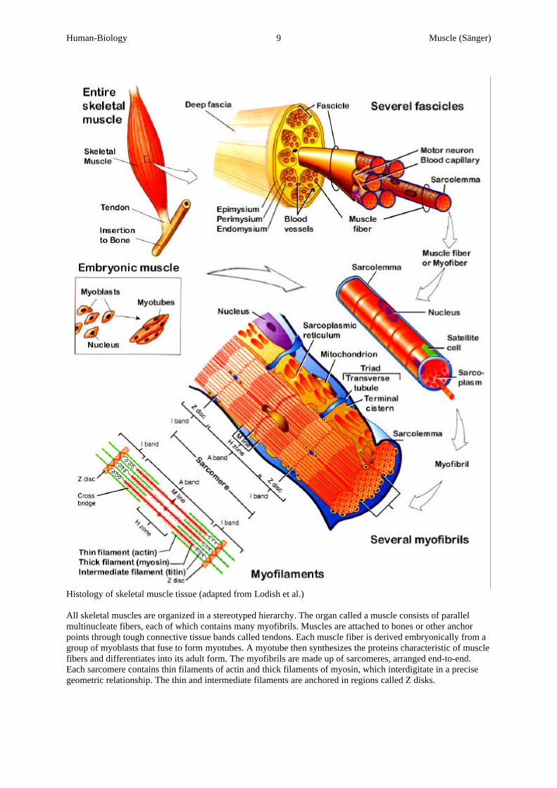
• Epithelial tissue accounts for two major glands: Endo- and exocrine glands - see glands.• Coelomic E.: Inner lining of the coelom.Muscle T.: Enables animals to move by contraction (myosin- and actin filaments slide past each other);• Giant muscle cell with many nuclei and more myofibrils; contracts when stimulated, consuming ATP;
locomotion due to shortening of actin-myosin filaments; types of MT:Cardiac M.: Specialized muscle tissue of the heart.Smooth M.: Type of muscle tissue in which the actin-myosin protein fibrils are not aligned; made ofspindle-shaped uninucleated cells and not striated; operating in glands, blood vessels, and internal organssuch as the intestine.Striated M.: Type of muscle tissue in which the repeating actin-myosin protein fibrils are aligned to givesthe appearance of cross striations, composed of long multinucleated cells; used for movement of skeletalapparatus.
Nervous T.: Contains neurons, cells which transmit electrochemical impulses to command, skeletal muscles orsecretory glands, sense environmental changes, and process information.
Human Biology III 1 Hearing (Pohlhammer)
Summary Human Biology - Somatic Senses, Taste and Smell
Components of Sensation: Typical event s for visual, auditory, gustatory, olfactory and somatic sensations are:Stimulation: A change in the environment that can activate certain sensory neurons.Transduction: A sensory receptor cell or organ that responds to the stimulus and transduces (converts) it to agenerator potential.Impulse Generation and Conduction: Upon arriving at the axon terminals, they stimulus triggers exocytosisof synaptic vesicles of neurotransmitter molecules. Once this chemical potential reaches threshold, thesucceeding dendrite elicits one or more nerve impulses and propagates them along its axons.Integration: A region of the CNS that receives and integrates the sensory nerve impulses into a sensation.
Cortex: External or surface layer of an organ; in this particular sense the outer areas of the brain associated tosensory capacities - the brain sees the picture, hears the music, feels the pain, not the receptors.
Pain: A protective mechanism for the body; it occurs whenever any tissues are being damaged, and it causes theindividual to react to remove the pain stimulus. The pain receptors in the skin and other tissues are all free-nerve endings. Pain stimuli release chemicals (prostaglandins and kinins) that stimulate these free nerveendings. Pain has been classified into two major types:• Fast P.: Fast pain is felt within about 0.1sec after a pain stimulus is applied; also described as sharp,
pricking, acute, and electric pain.• Slow P.: Slow pain begins after 1sec or more and then increases slowly over many seconds and sometimes
even minutes; also described as slow burning, aching, throbbing, nauseous, and chronic pain. P. Suppression: The brains capability to suppress pain stimuli by activating a special pain control system• Analgesia System: Enkephalin-secreting neurons suppress the incoming pain signals at the vertebral cord
level using ekephalins and serotinin.• Opiate System: Morphine-like agents (a dozen opiate molecules) attach to specialized receptors of a
neuron halting the ongoing firing activity of a pain receptor.Types of Pain:Phantom P.: The pain often experienced by patients who have had a limb amputated; they still experiencesensations such as itching, pressure, tingling, or pain in the extremity as of the limb were still there.Referred P.: A pain felt by a person in a part of his / her body that is considerably remote from the tissuecausing the pain.Visceral P.: Pain from the different viscera of the abdomen and chest; can be caused by Ischemia (formationof acidic metabolic end products or tissue-degradation), Gastritis (leakage of acidic gastric juices), Spasm ofthe Hollow Viscus (spasm of the gut, gallbladder, bile duct, ureter, or any other hollow viscus), oroverdistention of the Hollow Viscus (extreme overfilling of the viscus).
Receptor Cell: A neuronal cell that is specialized to respond to some particular sensory stimulation generally withlogarithmic characteristics - see also cell, Weber-Fechner law and range fractionation. Tasks of RCs:• Selective Recognition of stimuli: A low threshold-response to physical impact from the environment.• Transduction: General term for the modulation of one kind of energy into another; sense organs transduce
sensory stimuli (e.g. mechanical-, photonic-, chemical energy) into nerve impulses (AP).• Transformation: Conversion of the transductive AP into a digital signal (frequency encoded).
RC.-Response: Extero-RC response caused by a stimuli arriving from the external environment: Range Fractionation: The pattern in which receptors within one sensory modality are tuned to receiveinformation within relatively narrow, but not identical, intensity ranges, so the entire dynamic range of themodality is divided among different classes of receptors; i.e. certain receptors emit signals al relatively lowstimuli while others start firing only at strong mechanical stimuli. Spontaneously Active: In the absence of any stimulus, the RC or 2nd sensory fiber fires spontaneously andcovers the steep part of the curve relating the stimulus intensity to the frequency of APs, so even a very smallstimulus will increase or decrease the rate of firing; consequently doesn’t have a threshold e.g.: hair cells. Phasic R.: A quickly adapting RC, releasing many firing impulses but fading out as stimulus persist; commonin pressure, touch, and smell perception. Phaso-Tonic R.: A compound R with both phasic- and tonic characteristics. Tonic R.: Fires steadily during a maintained stimulus, although the firing frequency is highest at the beginningof the stimulation; common in the perception of pain, body position, and chemicals in the blood.
Human Biology III 2 Hearing (Pohlhammer)
RC.-Types: Extero-RC: Somatic sensory organs that provide information about the external environment; they detectstimuli arriving at the surface of the body from a distance:• Chemo-RC: A sensory receptor specifically sensitive to certain molecules (e.g. smell, taste, acidity, etc.).• Electromagnetic-RC: A sensory cell that is tuned to receive light energy (e.g. eye).• Mechano-RC: A sensory receptor tuned to respond to mechanical deformation, distortion or pressure (e.g.
tactile senses, ear, stretching, etc.) - see tactile senses.• Nociceptor RC: The type of receptors responsible for the sensation of pain (e.g. free nerve endings).• Thermo-RC: A free-nerve ending sensory cell, responsive to temperature changes (e.g. in fingertips, etc.) -
see thermal sensations.Interoceptive-RC: Internal receptors provide information about the internal environment; they responding tochanges w/n the body; connected to the vegetative NS.Proprioceptor C.: Internal receptors located in muscles, tendons, joints, and internal ear; they provideinformation about body position (see summary hearing and equilibrium), muscle tension, and the position andactivity of our joints.
Sensilla: see hair cell.S. Transduction: Elongation of a stereocilium that ctivates mechano-receptive ionic K+-channels, causingdepolarization, forcing Ca+ -channels to open triggering an AP.
Sensor: A mechanical, electrical, or biological device (receptor) that detects changes in its immediate environment.S. Adaptation: Property of sensory systems to become less sensitive during prolonged or repeatedstimulation.
Smell: The nose as a typical special sense chemical receptor housing the olfactory epithelium. Olfactory Epithelium: Button sized patches in the nasal passages capable of detecting a vast amount ofdifferent smells and odors. Olfactoric Transduction: Principle of signal amplification with a cascade receptor (D-R-G-AC-cAMP-INa);followed by an olfactoric projection in the brain; human = microsomat (10000 different odors).
Somatic Senses: The senses that includes the mechanoreceptive somatic senses (tactile and position sensations), thethermoreceptive senses (heat and cold detection), and the pain sensation.
Somato-Visceral Sensitivity: ?Tactile-Sense: These include touch, pressure, vibration and tickle senses:
• Itch and tickle: Stimulation of free nerve endings by certain chemicals.• Pressure: Results from the deformation of deeper tissues, (Pacinian C.).• Touch: Generally results from stimulation of tactile receptors in the skin of in tissues immediately beneath
the skin (hair end organs, Meissner C., Merkel D., Ruffini's end organs).• Vibration: Rapidly repetitive sensory signals, (hair end organs, Meissner C.).
Classes of Receptors: At least six different types of tactile receptors are known:Pressure:• Free nerve Endings (FnE): Sensors found everywhere in the skin and in many other tissues; e.g. the only
pressure sensitive receptor of the eye. According to their adaptation velocity, there are Myelinated FeE(slow adaptation, as in the case of cold temperature sensors) and Unmyelinated FeE (fast adaptation,typically the sensors responsible for tickling and itch).
• Pacinian Corpuscle: Quick pressure receptors found in the skin, muscle, joints, and connective tissue(adapt in 1/100 of a second); they consist of a nerve ending surrounded by a laminated capsule ofconnective tissue.
Touch (fast):• Hair-end-Organ: A nerve sensor in which the dendrites are wrapped around a hair follicle and sensitive to
any motion of these hairs (sensors of velocity which detect the change of ds/dt); e.g. wind, touch, etc.• Meissner Corpuscle: An egg-shaped and encapsulated nerve ending that excites a mass of dendrites
located in the dermal papillae of the skin. It has many internal branching terminal nerve filaments whichare present in the non-hairy part of the skin (glabrous skin), fingertips, lips, palms, soles, eyelids, tip oftongue, nipples, clitoris, and tip of penis. MC adapt in a fraction of a second after stimulation; therefore,particularly sensitive to movement of very light objects.
Human Biology III 3 Hearing (Pohlhammer)
Touch (slow):• Merkel's Disc (MD): A battery of Meissner Corpuscles innerveted by a single large myelinated fiber. MD
yields a steady state signal receptor that allow determination of continuos touch first by transmitting aninitially strong but partially adapting signal that decreases in intensity with time. Typical sensor of thefingertips (discriminative touch).
• Ruffini's end Organ: Multibranched, encapsulated nerve sensors that adapt very slowly, thus signalingcontinuos states of deformation of the skin and deeper tissues (heavy or continuos touch), as well as insignaling joint rotation.
Taste: A special gustatory sense; that enables humans to differ between, sour, salty, bitter, and sweet. Papilla: Small conical pumps, taste buds capable of receiving flavor molecules like sweet, salty bitter and sour.
Thermal Sensation: Free nerve ending-receptors located immediately under the skin at discrete but separated points.Different graduations of cold and heat can be perceived, progressing from freezing cold to cold to cool toindifferent to warm to hot to burning hot; these graduations are brought about by different temperaturesensors.• Cold Receptor: Operate within a temperature range of 10° to 40°C with a maximal firing frequency of 6
impulses/sec at 15°C. Cold receptors outnumber the warm receptors by a factor of 3 to 10 according to thelocation throughout the body.
• Pain Receptor: Both cold-pain fiber and heat-pain fiber start firing at <15°C or 45°C respectively with anincreasing firing rate when these temperatures are decreased / increased.
• Warm Receptor: Operate within a temperature range of 30° to 50°C with a maximal firing frequency of 10impulses/sec at 42°C.
Human Biology III 1 Hearing (Pohlhammer)
Summary Human Biology - Special Senses: Hearing and Equilibrium
Abnormalities of the Ear:Deafness: Significant or total loss of hearing caused by impairment of the cochlea, chochlear branch ofthe vestibulo-chochlear nerve (VIII), or by calcification of the tympanum ossicular system.Hyperacusia: Abnormally sensitive hearing due to paralysis of the stapedius muscle in the middle ear.Motion Sickness: Nausea and vomiting brought on by repetitive angular, linear, or vertical motion as aresult of excessive stimulation of the vestibular apparatus.Perforated Eardrum: A hole in the tympanic membrane, characterized initially by acute pain, ringing orroaring in the affected ear, hearing impairment, and sometimes dizziness. Can be caused by shockwavesof compressed air (explosions), scuba diving, trauma (ears swabs or skull fracture), or acute middle earinfections.Tinitus: A ringing, roaring, or clicking sound in the ears.
Auditory Centers of the brain: Several sites of sound processing are known so far:Contralateral Pathway: Signals from both ears are transmitted from the organ of Corti via the cochlearnerve through the superior olivary nucleus where nervous crossovers take place to join the contralateralside (trapezoid body, commissure of Probst, and the commissure connecting the two inferior collicoli).Reticular Activating System: It projects diffusely upward in the brainstem, downward into the spinalcord and the cerebellum to activate the entire nervous system in response to loud noise.Cochlear Nuclei: Certain fibers originating from the cochlea reach all the way to the brain of the auditorycortex and the inferior colliculi. Lesions in the posterior portion of the superior temporal gyrus (area ofWernicke, part of the auditory associative cortex) often make it impossible to interpret the meanings ofwords.
Ear: Frequency analyzing mechano-receptor, converting acoustical stimuli via a mechanical amplifier intoelectrical stimuli. This is done by the vibratory movement of the basilar membrane with respect to thetectorial membrane which produces shear on the stereocilia of the cochlea hair cells.Bony Labyrinth: A series of perilymph filled cavities within the petrous portion of the temporal bone,forming the vestibule, cochlea, and semicircular canals of the inner ear.Inner E.: Frequency analyzer; and transduction of vibratory liquid caused by a migrating sound wave;• Cochlea: A tapered tube wound into a spiral like the shell of a snail, containing hair cell receptors for
detecting sound; high pitch near the oval window; low pitch versus helicotrema. Elicitation ismediated via the vestibulo-chochlear nerve to the brain.Endocochlear Potential: Endolymph with exactly opposite ion concentration of the perilymph (<Na+,>K+) are exposed to an electrical potential of +80mV, with the positivity inside the scala media andnegativity outside. It is continuously generated by the transport of K+ into the scala media. Hair cellswith their negative intracellular potential of -70mV generate a total of ∆150mV at the tips of thestereocilia. This voltage further lowers the minimum threshold level for sound detection.Scala media: The cochlear duct (Ductus choclearis), a membrane labyrinth containing the organ ofCorti and the tectorial membrane; it is filled with endolymph, an extracellular fluid having a relativelyhigh concentration of K+ and low concentration of Na+.Scala tympani: The lower cochlear chamber connected with the scala vestibuli through thehelicotrema and deliminated by the round window; filled with perilymph, an extracellular fluid ofhigh Na+ (140mM) and low K+ concentration (7mM).Scala vestibule: The upper cochlear chamber connected with the scala tympani through thehelicotrema and deliminated by the oval window; filled with perilymph, an extracellular fluid of highNa+ (140mM) and low K+ concentration (7mM).
• Helicotrema: The apical end of the cochlea that connects the upper, perilymph filled chochlearchamber (scala tympani) with the lower one (scala vestibuli); it is the area of low frequency detection.
• Organ of Corti (spiral organ): The tissue within the cochlea housing the following structures:Basilar membrane: The delicate ribbon of tissue bearing the auditory hair cells in the cochlea. Thesetraverse ribbons, which increase in length from the proximal to the apical end. This causes theamplitude of a travelling wave to change along the length of the membrane (mechanical resonanceeffect of the travelling wave passing a particular frequency-location). Maximal basilar displacement isabout 1µm - anything in excess sheds off the stereocilia of the hair cells, causing loss of hearing.
Human Biology III 2 Hearing (Pohlhammer)
Haircell or Sensilla (HC): A spontaneously firing, mechano-sensory epithelial cell bearing stereocilia(nonmotile filament-filled projections in various lengths, that lack the internal structure of motile"9+2" cilia) and in some cases one long kinocilium (a true "9+2" or "9+0" cilium). Hair cells encodeboth frequency (i.e. pitch) and sound intensity. Neighboring stereocilia are attached via a thinspringlike link which modulates an ion-channel, allowing the free flow of ions; i.e. site of transductionof mechanical stimuli into electrochemical signals. Bending of the hair cells in one direction causesdepolarization, and bending them in the opposite direction results in hyperpolarization.The transduced signals travel via the cochlear branch of the vestibulo-cochlear nerve to the brain.Each cell has a mechanical resonance frequency that is determined by the length of the stereocilia inthe hair bundle (long cilia correspond to low frequency sound whereas short cilia to high frequencies)and an electrical resonance frequency which is determined by the balance of currents through voltagegated Ca2+ channels and through Ca2+-sensitive K+ channels in the basal membrane.Inner HC: 3 to 4 rows of external hair cells that accomplish the actual sound converting cells.Outer HC: A single row of hair cells that contribute to the tuning effect of the inner hair cells bygenerating acoustic emissions (self-induced vibrations to amplify responsiveness of the inner haircells).HC Transduction: Elongation of a stereocilium that activates mechano-receptive ionic K+-channels,causing depolarization, forcing Ca+ -channels to open triggering an AP.Nervus acusticus (Vestibulo-chochlear VIII nerve): Under neutral conditions, the nerve fiber leadingfrom the hair cells transmit continous impulses of 100Hz. Bent cilia modulate the frequency traffic(bending towards the kinocilium increases traffic to several hundred Hz, and vice versa). The cochlearbranch of this nerve arises in the spiral organ (of Corti), pass through internal auditory meatus, thenuclei in the medulla, and ends in the thalamus. Fibers synapse with neurons that relay impulses toauditory areas in the temporal lobe of the cerebral cortex (95% of nerve fibers innervate outer HC, 5%innervate inner HC).Afferent fibers conduct sensory signals from the transducing receptor to the processing centers of thebrain, whereas the efferent fibers carry signals from the brain to certain receptors to induce signalamplification as required to tune outer hair cells; e.g. efferent control of sound-sensitivity in a loudenvironment, which enables selective filtering of the someone’s voice.Tectorial membrane: A fine gelatinous sheet laying on the organ of Corti in contact with the cilia ofcochlear hair cells. The cilia are bent by shearing forces (i.e. a force perpendicular to the axis of thecilia) that arise when the hairs move through the gelatinous mucus that coats the tectorial membrane.Displacement of the tectorial membrane and balilar membrane occur simultaneously.
Middle E. or Tympanum: Impedance matching by the ossicular system; pressure conversion by 22:1.The ossicular system does not increase the movement distance of the stapes, it actually increases the forceof movement by about 1.3 times; the surface area of the tympanic membrane is about 55mm2, that of thestapes 3.2mm2. This 17 fold areal difference times the 1.3 fold ration causes 22 times as much pressure tobe exerted on the fluid of the cochlea.• Auditory Ossicle: The bones of the middle ear (malleus, incus, and stape) encapsulated in the
Tympanic antum, connecting the tympanic membrane and the oval window. These bones are requiredto avoid acoustical impedance mismatch which would otherwise occur when airborne sound (gaseousphase) should penetrate into the inner ear (liquid phase).Incus: The intermediate bone which articulates with the head of the stapes.Maleus: The handle, which is attached to the internal surface of the eardrum. Its head articulates withthe body of the incus. The tensor tympani muscle attached to the shaft of the maleus, limits movementand increases tension of the eardrum to prevent damage to the inner ear from loud noise.Stape: The final mechano-converting bone; its footplate fits into a membrane-covered opening (ovalwindow) in the thin bony partition between the middle and inner ear. The stapedius muscle dampenslarge vibrations resulting from loud noise; abnormally sensitive hearing results from paralysis of thismuscle.
• Eustachian tube (auditory tube): The bony tube (covered with hyaline cartilage) that connects themiddle ear with the nose and nasopharynx region of the throat; normally closed at its medial end,opens during swallowing and yawning.
• Oval Window: The connection between the inner ear and the cochlea; it is covered by the base of thestapes; approximately 0.1 x 0.05mm = 55mm2 (see inner ear).
• Round Window: A membrane-covered, separating the middle ear and the cochlea, through whichpressure waves leave after travelling through the cochlea; approx. 0.5mm in diameter.
Human Biology III 3 Hearing (Pohlhammer)
Outer E.: The external structure of the sound capturing device; average amplification x4 (frequencies in-between the 1k to 6kHz range, up to a 100 fold); it concentrates the oscillating air pressure onto aspecialized surface - the eardrum.• Auricle (Pinna): The outer structure of the human ear, which can be more or less leaborate and which
captures and funnels sound into the ear. The rim of the pinna is the helix, the inferior part is termedthe lobule.
• Ceruminous gland: A modified sudoriferous (sweat) gland in the external auditory meatus thatsecretes cerumen (ear wax).
• Meatus: The external 2.5cm long curved, audiotory tube, that lies in the temporal bone and leads tothe eardrum.
• Tragus: The tab that extends from the ventral (anterior) edge of the outer ear and partially covers theopening of the ear.
• Tympanic Membrane: The eardrum; a thin, semitransparent partition separating the externalauditory system from the middle ear.
Equilibrium: Positioning in space is achieved by a static detector (utricle and saccule) and a dynamic detector(semicircular canals with their ampullae).Bony Labyrinth: A series of perilymph filled cavities within the petrous portion of the temporal bone,forming the cochlea, semicircular ducts, and vestibule of the inner ear.Semicircular duct: The membranous semicircular canals filled with endolymph and floating in theperilymph of the bony semicircular canals. They contain cristae that are concerned with dynamicequilibrium (maintenance of head position in response to sudden movements such as rotation,acceleration, and deceleration).• Ampulla: A saclike dilution of one the semicircular canal housing cristae (the hair cells with its apical
tuft and cupula). The flow of endolymph through the appropriate duct of the ampulla excites thesensory cells.
• Semicircular canals: Three bony channels(anterior, lateral, and posterior), filled with perilymph, inwhich lie the membranous semicircular canals filled with endolymph. They contain receptors fordynamic equilibrium.
• Statoconia (Otolith): A particle of calcium carbonate (CaCO3) embedded in the otolithic membranethat functions in maintaining static equilibrium.
• Statoconic (Otolithic) Membrane: A thick, gelatinous, glycoprotein layer located directly over thehair cells of the macula (thickened region on the wall of the utricle and saccula); the hair cellprotruding into the membrane layer are deflected according to gravitational pull by the weight of thestatoconia, causing electrochemical stimuli - similar as in the hair cells of the cochlea.
• Vestibular Apparatus: Collective term for the organs of equilibrium, which includes the saccule,utricle, semicircular ducts, and the vestibular branch of the Nervous acousticus (see cochlea).The vestibule is a small space or cavity at the beginning of the inner ear canal, containing the saccule,utricle and the interface to the middle ear (oval window); both saccule and utricle contain the otolithicmembrane.Maculae (Gk. spot): The static sensory organ of the utricle and the saccule (containing hair cells,gelatinous layer, and statoconia) for detecting orientation of the head with respect to gravity; each ofthe two macculae is oriented in different directions so that at least some of the hair cells are stimulatedwhen the head bends forward, on the side, backwards, etc.Oval Window: A small, membrane-covered opening between the middle ear and inner ear into whichthe footplate of the stapes fit;Saccule: The inferior and smaller of the two chambers in the membranous labyrinth inside thevestibule of the inner war containing the receptor organ for static equilibrium (maintenance of theposition of the head).Utricle: The larger of the two divisions of the membranous labyrinth located inside the vestibule ofthe inner ear, containing a receptor organ for static equilibrium.
• Vestibular Nerve: The vestibular branch arises in the semicircular canals, saccule, and utricle andforms vestibular ganglion that join the cochlear branch to form the vestibular-cochlear (II) nerve;fibers end in pons and cerebellum.
Physical Background of Sound: Sound is an adiabatic pressure wave; the pressure differences betweencompression and rarefaction of a sound of a wave (constituting the wavelength) can not equalize eachother.Diffraction: The deviation of sound from rectilinear propagation. The bending of sound around anobstacle or through a narrow slit occurs in such a way that low frequencies experience a larger degree ofdiffraction than higher frequencies; important for frequency discrimination in the cochlea.Fourier Analysis: A mathematical method that will resolve any periodic wave form into a series ofsimple sine waves; i.e. superposition of fundamentals and their multiple harmonics.
Human Biology III 4 Hearing (Pohlhammer)
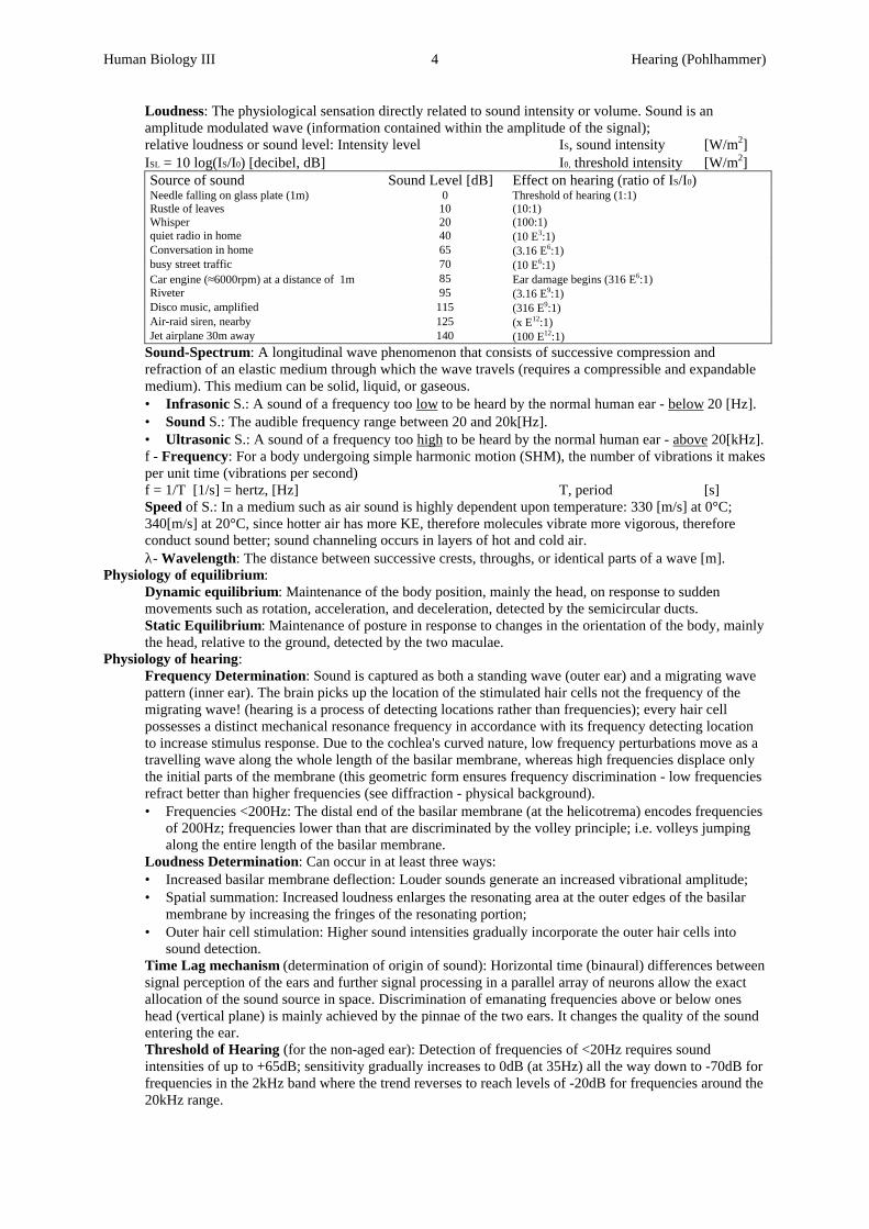
Loudness: The physiological sensation directly related to sound intensity or volume. Sound is anamplitude modulated wave (information contained within the amplitude of the signal);relative loudness or sound level: Intensity level IS, sound intensity [W/m2]ISL = 10⋅log(IS/I0) [decibel, dB] I0, threshold intensity [W/m2]Source of sound Sound Level [dB] Effect on hearing (ratio of IS/I0)Needle falling on glass plate (1m) 0 Threshold of hearing (1:1)Rustle of leaves 10 (10:1)Whisper 20 (100:1)quiet radio in home 40 (10⋅E3:1)Conversation in home 65 (3.16⋅E6:1)busy street traffic 70 (10⋅E6:1)Car engine (≈6000rpm) at a distance of 1m 85 Ear damage begins (316⋅E6:1)Riveter 95 (3.16⋅E9:1)Disco music, amplified 115 (316⋅E9:1)Air-raid siren, nearby 125 (x⋅E12:1)Jet airplane 30m away 140 (100⋅E12:1)
Sound-Spectrum: A longitudinal wave phenomenon that consists of successive compression andrefraction of an elastic medium through which the wave travels (requires a compressible and expandablemedium). This medium can be solid, liquid, or gaseous.• Infrasonic S.: A sound of a frequency too low to be heard by the normal human ear - below 20 [Hz].• Sound S.: The audible frequency range between 20 and 20k[Hz].• Ultrasonic S.: A sound of a frequency too high to be heard by the normal human ear - above 20[kHz].f - Frequency: For a body undergoing simple harmonic motion (SHM), the number of vibrations it makesper unit time (vibrations per second)f = 1/T [1/s] = hertz, [Hz] T, period [s]Speed of S.: In a medium such as air sound is highly dependent upon temperature: 330 [m/s] at 0°C;340[m/s] at 20°C, since hotter air has more KE, therefore molecules vibrate more vigorous, thereforeconduct sound better; sound channeling occurs in layers of hot and cold air.λ- Wavelength: The distance between successive crests, throughs, or identical parts of a wave [m].
Physiology of equilibrium:Dynamic equilibrium: Maintenance of the body position, mainly the head, on response to suddenmovements such as rotation, acceleration, and deceleration, detected by the semicircular ducts.Static Equilibrium: Maintenance of posture in response to changes in the orientation of the body, mainlythe head, relative to the ground, detected by the two maculae.
Physiology of hearing:Frequency Determination: Sound is captured as both a standing wave (outer ear) and a migrating wavepattern (inner ear). The brain picks up the location of the stimulated hair cells not the frequency of themigrating wave! (hearing is a process of detecting locations rather than frequencies); every hair cellpossesses a distinct mechanical resonance frequency in accordance with its frequency detecting locationto increase stimulus response. Due to the cochlea's curved nature, low frequency perturbations move as atravelling wave along the whole length of the basilar membrane, whereas high frequencies displace onlythe initial parts of the membrane (this geometric form ensures frequency discrimination - low frequenciesrefract better than higher frequencies (see diffraction - physical background).• Frequencies <200Hz: The distal end of the basilar membrane (at the helicotrema) encodes frequencies
of 200Hz; frequencies lower than that are discriminated by the volley principle; i.e. volleys jumpingalong the entire length of the basilar membrane.
Loudness Determination: Can occur in at least three ways:• Increased basilar membrane deflection: Louder sounds generate an increased vibrational amplitude;• Spatial summation: Increased loudness enlarges the resonating area at the outer edges of the basilar
membrane by increasing the fringes of the resonating portion;• Outer hair cell stimulation: Higher sound intensities gradually incorporate the outer hair cells into
sound detection.Time Lag mechanism (determination of origin of sound): Horizontal time (binaural) differences betweensignal perception of the ears and further signal processing in a parallel array of neurons allow the exactallocation of the sound source in space. Discrimination of emanating frequencies above or below oneshead (vertical plane) is mainly achieved by the pinnae of the two ears. It changes the quality of the soundentering the ear.Threshold of Hearing (for the non-aged ear): Detection of frequencies of <20Hz requires soundintensities of up to +65dB; sensitivity gradually increases to 0dB (at 35Hz) all the way down to -70dB forfrequencies in the 2kHz band where the trend reverses to reach levels of -20dB for frequencies around the20kHz range.
Human Biology III 5 Hearing (Pohlhammer)
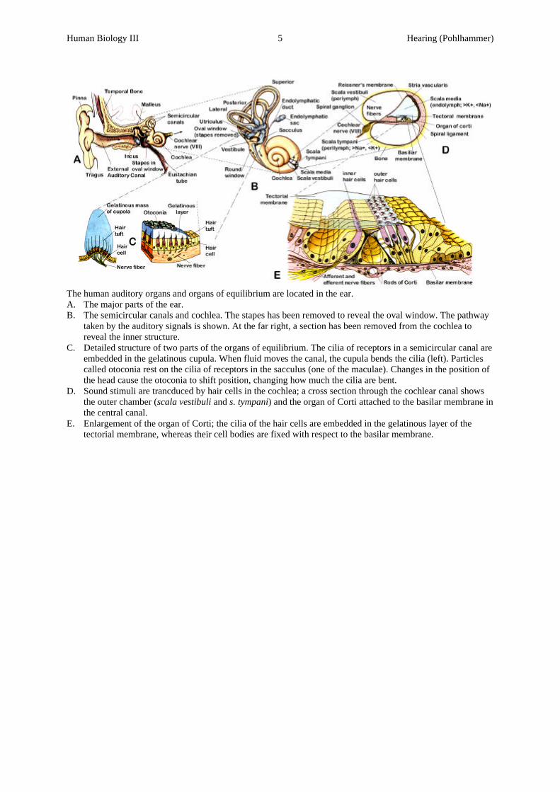
The human auditory organs and organs of equilibrium are located in the ear.A. The major parts of the ear.B. The semicircular canals and cochlea. The stapes has been removed to reveal the oval window. The pathway
taken by the auditory signals is shown. At the far right, a section has been removed from the cochlea toreveal the inner structure.
C. Detailed structure of two parts of the organs of equilibrium. The cilia of receptors in a semicircular canal areembedded in the gelatinous cupula. When fluid moves the canal, the cupula bends the cilia (left). Particlescalled otoconia rest on the cilia of receptors in the sacculus (one of the maculae). Changes in the position ofthe head cause the otoconia to shift position, changing how much the cilia are bent.
D. Sound stimuli are trancduced by hair cells in the cochlea; a cross section through the cochlear canal showsthe outer chamber (scala vestibuli and s. tympani) and the organ of Corti attached to the basilar membrane inthe central canal.
E. Enlargement of the organ of Corti; the cilia of the hair cells are embedded in the gelatinous layer of thetectorial membrane, whereas their cell bodies are fixed with respect to the basilar membrane.
Human Biology III 1 Vision (Pohlhammer)
Summary Human Biology - Special Senses: Vision Abnormalities of the Eye:
Achromatopsy: ???????????????? mismatched interpretation of red and green due to a defective expressionof the genome (X-chromosomes encode red and green; Y chromosome encodes blue) Astigmatism: An irregularity of the curvature of the lens or cornea of the eye causing the image to be partlyout of focus and producing faulty vision. Cataract: Loss of transparency of the lens of the eye or its capsule or both. Color Blindness: Absence of a single group of color-receptive cones from the retina leads to color blindnessof that particular color and the wavelengths in-between involving that particular hue.• Deuteranopy: Colorblindness of green; although green is missing the visual spectrum is not shortened.• Protanopy: Colorblindness of red with a shorthened visual spectrum at the long wavelength end; effects
more males (8%) than females (1.4%).• Tritanopy: Complete colorblindness for any of the three ground-colors (RGB); very rare. Conjunctivitis: Inflammation of the conjunctiva, the delicate membrane covering the eyeball and lining theeyelids; usually of microbial origin. Glaucoma: Eye disorder in which there is increases pressure due to an excess of fluid within the eye; i.e.excess liquids from both humors can not flow off via the trabeculae further through the canal of Schlemm intothe extracellular veins. As the pressure rises, the axons of the optic nerve (at the site of the optic disc, where itleaves the eye) are compressed, distorting or even blocking the flow of nutrients of the axons, whicheventually causes death of the involved neurons of the retina. Hyper(metr)opia (farsightedness): A condition in which visual images are focused behind the retina withresulting defective vision of near objects; can occur if the eyeball is too short or occasionally, to a lens systemthat is too weak. Myopia (near-sightedness): An eyeball that is too long or the refractive power of the lens too strong, causingthe focal point to center in front of the retina; defect in vision so that objects can be seen distinctly only whenvery close to the eyes. Night Blindness: A nutritional deficiency of vitamin A1 decreases the amount of available photopsin (cones)or rhodopsin (rods). The result is reduced photosensitivity of the eyes. Since bright light requires less (alltrans-retinol) vitamin A, it is stored in the cytoplasm of the rods and cones; to increase the photosensitivity atlow light, the stored vitamin A is reconverted to photopsin / rhodopsin; night blindness results out of aninsufficient supply of vitamin A from the cytoplasm to generate the extra photopsin / rhodopsin molecules. Presbynopia: The tendency for a human eye to become less able to focus to close objects with age due todenaturation of lens proteins; occurs as the lens becomes less compliant. Strabismus (squint, cross-eyedness): The lack of fusion of the eyes in one or more of the coordinates(horizontal, vertical, torsional strabismus) due to either malfunctioning extraocular muscles or impropermuscular control by the oculomotoric centers of the brain.
Eye: Organ of visual (photo-) reception that includes optical processing of light; Anatomical Structures of the E.:
Blind Spot: see optic disc. Cavity: The large fluid filled interior cavity of the eyeball divided into two smaller ones by the lens. The fluid, originating from the choroid plexus of the posterior chamber, passes forward between the iris andthe lens, through the pupil, into the anterior chamber.• Anterior C.: The section anterior (in front) of the lens filled with the aqueous humor; further divided into:
Anterior Chamber: The chamber behind the cornea and in front of the iris.Posterior Chamber: The chamber behind the iris and in front of the suspensory ligaments and lens.
• Posterior C.: The larger cavity behind the lens, filled with the vitreous humor. Fovea (area centralis): The area with the highest visual resolution due to small divergence and convergence inthe pathway linking photoreceptors to ganglia cells; area centralis is dominated by cones; with an averagediameter of 0.3mm it covers an area of approx. 1mm2. Horizontal Cell: A nerve cell whose fibers extends horizontally in the outer plexiform layer of the vertebrateretina; interconnecting adjacent photoreceptors, lowering resolution.
Human Biology III 2 Vision (Pohlhammer)
Humor: The intracellular fluid system which maintains sufficient pressure to keep the eye distended. Thisintraocular pressure, is produced mainly by the aqueous and to a lesser extent by the vitreous humor.• Aqueous H.: The watery fluid that fills the anterior cavity between the cornea and the lens of the eye. It is
formed by the ciliary processes of the ciliary body at a rate of 2-3µL/minute. This fluid is mainlycomposed of Na+, Cl-, CO3
2-, water and several nutrients such as amino acids, ascorbic acid and glucose.Intraocular Pressure (IOP): The pressure that maintains the shape of the eyeball and keeps the retinasmoothly applied to the choroid so the retina will form clear pictures. It averages 15mmHg (2kPa) and iscounterbalanced by the resistance of the outflow of aqueous humor through the anterior iridocorneal anglevia a meshwork of trabeculae and the canal of Schlemm and its production by the ciliary processes.The trabecular meshwork houses phagocyting cells which clean the fluid to prevent infection and blockageof the outflowing canals.
• Vitreous H. (vitreous body): A soft, jelly-like substance that fills the posterior cavity of the eyeball, lyingbetween the lens and the retina. It is composed primarily of greatly elongated proteoglycan molecules.
Lens: Transparent organ lying posterior to the pupil and iris of the eyeball and anterior to the vitreous humor.• Suspensory Ligament: Densely arranged connective tissue that attaches the lens to the ciliary body. Pupil: Opening of center of iris of eyeball for light transmission. Optic Disc (blind spot): A small area of the retina with no light receptor cells; it represents the openingsthrough which the fibers of the ganglion neurons emerge as the optic nerve. Optic Nerve (II): Nerve fibers and their associated connective tissue coursing together outside the centralnervous system, connecting the retina with the visual centers of the brain - see also visual pathway. Ora Serrata: The irregular margin of the retina lying internal and slightly posterior to the junction of thechoroid and ciliary body; i.e. the fringing edges of the retina. Scleral Venous Sinus (Canal of Schlemm): A circular venous sinus located at the junction of the sclera andthe cornea through which aqueous humor drains from the anterior chamber of the eyeball into the veins. Tunic: The tree anatomical divisions of the eyeball;• Fibrous Tunic: The outer coat of the eyeball, made up of the posterior sclera and the anterior cornea.
Cornea: The clear surface of the eye through which light passes as it enters the eye and is equipped withthe corneal lens, which focuses light entering the ommatidium (the functional unit of the compound eye,consisting of the lens, a focusing cone, and photoreceptor cells).Sclera: The white coat of fibrous tissue that forms the outer protective covering of the eyeball except inthe area of the anterior cornea.
• Nervous Tunic: The innermost coat; i.e. the retina, which lies in the posterior portion of the eye. Retina: The photosensitive inner surface of the eye. The entire structure is supplied with blood by retinalarteries and veins. Layers of the Retina (in order of incident light, according to the everse structure of the human eye): Innerlimiting membrane, layer of optic nerve fibers, ganglionic layer (ganglion cells), inner plexiform layer(amacrine cells), outer plexiform layer (fiber of Müller), outer nuclear layer cell body of rods and cones),outer limiting membrane, photosensitive layer (rods, and cones), pigment layer (black melanin layer toprevent light reflection -absent in albinos) - see visual pathway and also scan at the end.Plexiform Layer: Layer of nerve cells that mediate lateral interactions with the retina (preprocessing ofsignals originating from the retinal receptors).• Horizontal Cells: A nerve cell whose fibers extend horizontally in the outer plexiform layer of the
human retina and interconnects adjacent photoreceptors; these cells accomplish the task of lateralinhibition.
• Bipolar Cells: A neuron with two axons emerging from opposite sides of the soma; they transmitsignals from the photoreceptor cells to the retinal ganglion cells.
• Signal Convergence: A pattern in which inputs from many different neurons impinge upon a singleneuron. The retinal periphery groups 15-45 rods to 1 bipolar cell (increased sensitivity), whereas thefovea groups 1-20 cones to 1 bipolar cell (high resolution).
• Amacrine Cells: Neurons without axons, found in the inner plexiform layer and interconnect adjacentbipolar cells, and mediate stimuli down to the ganglion cells by a slope triggered firing pattern; i.e. fireonly at changes of signal states when objects move cross the retina, change of illumination, etc.
• Ganglion Cells: The afferent neurons of the optic nerve, that carry visual information from the innerplexiform layer to the higher centers of the brain. About every cone in the fovea is connected to aganglion cell, whereas several rods are routed down to one ganglion cell in the peripheral area - thisaccounts for the greater sensitivity of the peripheral retina to weak light and moving objects.
Human Biology III 3 Vision (Pohlhammer)
Cone: The bright-light visual receptor cell that has a tapered outer segment in which the lamellarphotosynthetic membranes (of free floating disks) remain continuos with the surface membrane; conesresponds to one out of three particular colors (red sensitive pigments = 445nm, green sensitive pigments =535nm, and blue sensitive pigments = 570nm); hue is calculated by differences of the RGB-values (short440nm-blue; medium 540nm-green; long 567nm-red) overall max. sensitivity in the yellowish-greenishspectrum, corresponds to approx. 555nm.Rod: The dim-light visual receptor cells many times more sensitive to light than cones (membranelamellae in the form of pigmented, free floating disks held in place by an outer segment - 4 times thelength of cones). Based on cellular physiology and on high degree of convergence onto second order cells;not sensitive to a particular frequency, rather to the full visible spectrum (illuminance detector - max.sensitivity at 505nm, which corresponds to the bluish-greenish spectrum).
• Vascular Tunic: The middle layer of the eyeball, composed of three portions:Choroid: The distal coat of the tunic, to which the outermost pigmented layer of the retina is attached. Itis a highly vascular tissue, which provides nutrients to the cones and rods (via diffusion).Ciliary Body: The lateral portions of the vascular tunic that includes the ciliary muscle and the ciliaryprocesses; it is also the production site of the aqueous humor.Iris: The pigmented circular diaphragm located behind the cornea of the vertebrate eye.
External Accessory Structures of the E.:Commissure: The angular junction of the eyelids at either corner of the eye.• Lateral C.: Further from the midline of the body, in this case outer junction of the eyelid.• Medial C.: Nearer to the midline of the body, in this case inner junction of the eyelid.Conjunctiva: The delicate membrane covering the eyeball and lining the eyes.Eyebrow: The hairy ridge above the eye, keeping sweat from dripping into the eye.Eyelash: Hairy fence-like structure at outer rim of the palpebra; keeps dust particles away.Lacrimal Canal: A duct, one on each eyelid, commencing at the punctum at the medial margin of an eyelidand conveying the tears medially into the nasolacrimal sac.Lacrimal Caruncle: Fleshy, yellowish projection of medial commissure containing modified sweat andsebaceous glands.Lacrimal Gland: Secretory cells located at the superior lateral portion of each orbit that secrete tears into theexcretory lacrimal ducts that open onto the surface of the conjunctiva.Lacrimal Sac: The superior expanded portion of the nasolacrimal duct that receives tears fr/ a lacrimal canal.Muscles of the eyes: Six extrinsic muscles enable eye movements.• Superior Rectus: Superior and central part of eyeball; rolls eyeball upward.• Inferior Rectus: Inferior and central part of eyeball; rolls eyeball downward.• Lateral Rectus: Lateral side of eyeball; rolls it laterally.• Medial Rectus: Medial side of eyeball; rolls it medially.• Superior Oblique: Insertion between superior and lateral recti of eyeball; rotates it on its axis, directing
cornea downward and laterally. Muscle deviated by trochlea (fibro-cartilaginous pulley).• Inferior Oblique: Insertion b/w inferior and lateral recti of eyeball; rotates on its axis; directs cornea
upward and downward.Palpebra (eyelid): Folds of skin and muscle lined by the conjunctiva. Aids in lubrication of cornea.
Physical Properties affecting vision: Brightness: Emission or reflection of light; synonymous for intensity of light.Depth of focus: The distance through which objects are in focus when a lens is in one fixed shape; itincreases when light is prevented from passing through the perimeter of the lens (site of increased opticalaberrations). Best possible depth of focus is obtained with extremely small pupils, i.e. at bright light.Diopters (power of a lens): The focal length (f) in meters of a convex lens given as 1/f [D], the shorter thefocal length the greater the power. A healthy human lens can cover a range of approx. +14 D. The refractivepower of the entire visual apparatus is about +59D. Concave lenses which have diverging properties but havethe same focal length as convex lenses, are assigned as "-"D. Hue: The property of color that is perceived and measured (wavelength in [nm]) on a scale ranging from redthrough yellow, green and blue to violet; and in particular a graduation of color, tint, shade.
Human Biology III 4 Vision (Pohlhammer)
Lens Equation: The lens of the eye is an optical instrument which focus or disperse incoming light wavesand has converging properties; i.e. a convex lens, which is thicker in the middle than at the edges, causingparallel rays passing through it to converge to the focal point: d, distance of object [m]L. Equation: 1/d +1/d’ = 1/f [m] d’, distance of image [m]L. Magnification: ML = -d’/d [m] f, distance of focus [m]L. Rays: Three principle rays characterize a lens’ behavior:
• The 1st incoming ray parallel to the lens’ axis will be deflected to pass the focal point past the lens.• The 2nd, center-seeking ray will straight pass through the center without a deflection.• The 3rd incoming ray striking the focal point will be deflected to a parallel beam past the lens.
L. Distortions:• Astigmatism: A defect caused when the radius of curvature is not uniformly the same throughout the
lens; i.e.: the inability to focus simultaneously light-trays arriving in different planes.• Chromatic Aberration: Chromatic distortion of an image produces by a lens or lens-system (red
refracts more than blue light).• Spherical Aberration: Parallel incoming rays at the edge of a lens do not meet at the focal point as do
rays which are closer to the axis of lens. Parallax: An apparent change in the direction of an object, caused by a change in the viewer's position.Quantum: Radiation of light is emitted in discrete bundles of energy; just as matter is quantified as a wholenumber of atoms, or electric charge is a whole number multiple of a single charge. Cones are able to detect asingle photon of light; the energy contained in a quantum of radiation is equal to Planck's constant divided bythe wavelength; since there are only few cones in the fovea, but many in the outer areas of the retina,peripheral minimal perception threshold exceeds that of the fovea, h, planks c. = 6.6⋅E-34 [J⋅s]With a simultaneous decrease in pint resolution (fewer cells/mm2): c, speed of light = 3⋅E8 [m]E = h⋅c / λ λ, wavelength [m]Refraction: The bending of an oblique ray of light when it passes from one transparent medium of onedensity to another with a different density, caused by a difference in the speed of light in those media. At theair-water interface, light entering the eye bends towards the perpendicular air-water line and vice versa.R. Index: The refractive power of a medium compared with that of air,designated as ndiamond = 2.4; nwater = 1.3: n, index of refraction [-]n = cvacuum/vof light in medium c, v, speed of light [m/s]n1⋅sinθ1 = n2⋅sinθ2 θx, angle (⊥ to surface) [degree]Resolution: Decides whether two remote sources can be clearly λ, wavelength [m]distinguished by the eye (also known as Rayleigh's criterion): 2⋅r, diameter of object [m]360° = 2⋅π [rad] 1’ = 2.909⋅10-4 [rad] 1“ = 4.848⋅10-6 [rad] θ, angle of resolution [rad]• Point R.: θR = 1.22⋅λ/2⋅r [rad] λ, wavelength [m]• Spatial R.: Integration by a post-synaptic neuron of simultaneous synaptic currents that arise from the
terminals of different pre-synaptic neurons;Saturation: Gradual blending of base colors; i.e. RGB (pink = reddish white ; brown = grayish yellow); withthis graduation, the eye can differentiate between 100E3 to 1E6 different colors.
Physiology of the eye: Adaptation (sensory): Decrease in sensitivity during sustained presentation of stimuli.• Dark A.: At dark conditions, large amount of rhodopsin are required to produce a photochemical response
in the form of a membrane potential. The retinal and opsin molecules in rods and cones are converted tolight sensitive pigments. Vitamin A is reconverted to retinal to provide extra light sensitive pigments.Dark adaptation after bright light exposure can take up to 45mins, in which the cones are activated in thefirst 10mins, followed by the activation of rods; in total, dark adaptation boosts sensitivity by a 25E3 fold.
• Light A.: At bright light, only very little photopsin / rhodopsin is required to trigger photochemicalresponse. Large portions of photochemicals in both rods and cones are reduced to retinal and subsequentlyto vitamin A which is stored in the cytoplasm of the cones and rods.
• Neuronal A.: The visual centers of the brain further modify vision - see visual pathway.• Pupilary Reflex: A neuronal reflex, that originates in the retina and controls the aperture of the iris. When
circular smooth muscle fibers of the iris contract, they decrease the proportion of incident light that isallowed to enter the eye; contraction of the radially oriented muscle fibers reverse this process.
Human Biology III 5 Vision (Pohlhammer)
Accommodation: Increase in curvature of the lens in order to bend the light-rays toward the central fovea(adjustment of focal length). The fibers of the zonula (ciliary processes) exert outwardly directed tensionaround the perimeter of the lens; radially arranged ciliary muscle (suspensory ligaments) adjust the amount oftension exerted on the lens. When the ciliary muscles relax, the lens flattens by elastic tension exerted by themuscle of the ciliary processes, which pull the perimeter of the lens outward - objects far from the eye appearsharp. Objects close to the eye are brought into focus when the ciliary muscles contract. Accommodation isdirectly controlled by the parasympathetic nerves.Binocular Convergence: A neuronal mechanism which positions the eyes so that the images formed fall onanalogous portions of the 2 retinas, avoiding double vision. When an object is close, each of the 2 eyes mustrotate toward the middle of the nose; when on object is far away, the 2 eyes rotate outward from the midline. Color Vision: Spectral sensitivities based on the on the degree of stimulation of each class of RGB cones;equal stimulation of all the red, green, and blue cones gives on the sensation of seeing white. Color visionpredominantly takes place in the fovea. Determination of Distance: Depth reception can occur in 3 different ways:• Moving Parallax: Moving objects close to the eye pass rapidly across the retina while the images of
distant objects remain almost completely stationary.• Retinal Size: An object of known size, according to its distance, projects a proportionally small image
onto the retina.• Stereopsis: Objects focused at close range result in a less parallel arranged optical axis than objects
viewed at infinity and produce images that are projected on different sites of the retina. Eye Movements: Movements of the eyes is controlled by a cerebral system which includes:• Muscular Control: Three pairs of muscle (controlled by nerve III, IV, and VI of the medial longitudinal
fasciculus) allow horizontal and vertical orientation of the eyeball.• Neuronal Pathways: Both voluntary and involuntary fixation areas in the brain control the oculomotoric
centers of the brain stem. Flicker Fusion-Frequency: The frequency at which images are projected onto the retina to observe harmonicmotion of single images; around 16-18 frames per second.Lateral Inhibition: Excitation and inhibition of a retinal area is brought about by the horizontal cells toincrease the contrasting capabilities of visual processing - see visual pathway.Light Intensity: Discrimination of light intensity requires a proportional electrical signal output from conesand rods. This electrotonic conduction (rather than an "all-or-non" response as in the case with actionpotentials) is essential for the interpretation of light intensities by the visual centers of the brain.Photoreception: Electromagnetic receptors that detects light on the retina of the eye (in order of signalprocessing); cones function best in bright light and provide high resolution (color receptors dominate the Areacentralis = fovea), whereas rods function best in dim light.• Photopsin: A colored (red, green or blue), light-sensitive photopigment molecule in cones - see rhodopsin
for signal transduction and resynthesization of the bleached molecule.• Rhodopsin: A purplish red, light-sensitive photopigment molecule of rods; a chromoprotein (combination
of scotopsin and retinal proteins) with 11-cis retinal as its prostethic group; found in the rods and cones ofthe retina. The cis-form is the activated light sensitive photopigment.Dark Current: A steady sodium current that leaks into the upper segment of the visual receptor cell (inboth rods and cones), while a sodium pumps at the base of each receptor cell complete this circle byactively exporting these ions. The dark current is reduced by photo-excitation which hyperpolarizes themembrane potential from -30mV to -55mV.Retinal: The carotenoid pigment portion of the photopigment rhodopsin. In the dark, the bonds of C-11are arranged in the cis configuration.Rhodopsin Isomerization: Rhodopsin changes its steric conformation into the straight, all-transconfiguration when it absorbs a photon - with still the same chemical but different physical structure. Thetrans form decomposes quickly (bleaching happens in msecs) to batho-rhodopsin, then to lumino-rhodopsin, and finally to meta-rhodopsin I+II. The later, via an enzymatic amplificating cascade, changesthe electrical resistance of the membrane, causing hyperpolarization.Rhodopsin Regeneration: Rhodopsin is reconstituted out of retinal and opsin via an isomerase-activityout of retinal and scotopsin, by returning the retinal to the 11-cis configuration; this can take severalminutes and is one reason for prolonged visionary images.
Human Biology III 6 Vision (Pohlhammer)
• Enzymatic Cascade: When light hits the photopigment, the enzymatic cascade is triggered and amplifiesthe signal by a 10E3 fold. The resulting hyperpolarized membrane potential is caused by increasednegativity due to decreased membrane conductance of Na+-ions (see dark current). This electricaldepolarization (electrotonic conduction - see light intensity) is proportional to the logarithm of the lightintensity. It is then pre-processed in horizontal and amacrine cells before it is conveyed down the opticnerve to the visual centers of the brain.Transduction of light: A photon hitting a rod excites rhodopsin; the so activated retinal increases theactivity of a G-protein on the discs - signal amplifying cascade - by a 250 fold, which then activates manyPDE- (phosphodiesterase) molecules reducing the intracellular concentration of cCMP (amplification of afurther 400 fold). A low concentration of cGMP causes the Na+ channels to close (dark current); themembrane-resting potential (MRP) becomes hyperpolarized (from -30mV to -55mV) which triggers anaction potential, that promotes the release of glutamate as the main neurotransmitter of cones and rods.
Range Fractionation: The pattern in which receptors within one sensory modality are tuned to receiveinformation within relatively narrow, but not identical, intensity ranges, so the entire dynamic range of themodality is divided among different classes of receptors. For example, the rods respond to dim light but aresaturated in bright light; cones are less sensitive to dim light but remain responsive in bright light. Receptive Field: Tat area of the retina, that when stimulated influences the activity of a given neuron is thereceptive field of that neuron; the area of the retina by which stimulation by light causes a ganglia cell toactivate or block; concentric on-off centers; fovea: 2.5µm; peripheral retina: 2mm.
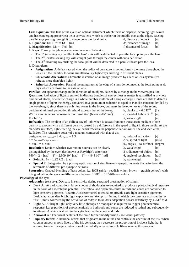
Visual Pathway and Information Processing: The nerve impulses leaving the retina from the nasal halves via theoptic nerves, cross to the opposite side where they join the fibers from the opposite temporal retinas to formthe optic tracts (see scan below). The fibers of each optic tract synapse in the dorsal lateral geniculate. Fromthere, the fibers pass by way of the optic radiation to the primary visual cortex of the occipital lobe.
Structures in order of signal processing:Hemiretina: The retinal halves of the eye that are superimposed in the visual processing centers of the brain.• Nasal H.: The field of vision of the left hand side connected to the right brain hemisphere.• Temporal H.: The field of vision of the right hand side connected to the left brain hemisphere. Optic Nerve (II): Chordlike bundle of nerve fibers and their associated connective tissue coursing togetheroutside the central nervous system, connecting the retina with the visual centers of the brain.Optic Chiasma: A swelling under the hypothalamus of the human brain where the two optic nerves meet;some axons cross the midline here and project to the contra-lateral side of the brain.Optic Tract: A bundle of axons that transmits nerve impulses from the retina of the eye between the opticchiasm and the thalamus.Lateral Geniculate: A region of the brain (thalamus) that processes visual information:• it relays visual information from the optical tract to the visual cortex by way of the optic radiation;
crossover in the optic chiasm allow the respective hemiretinal areas of the two eyes to connect withneurons that are approximately superimposed over one another;
• it gates the transmission of signals to the visual cortex; i.e. it is assumed that both gating circuits help tocontrol the visual information that is allowed to pass.
Optic Radiation: Axons of neuronal fibers synapting with the lateral geniculate, and project into the primaryvisual areas in the occipital lobes of the cerebral cortex.Visual Cortex (VC): The cerebral cortex in the occipital region of the cerebrum; devoted to processing visualinfo.• Primary VC: The terminus of direct visual signals from the fovea positioned at the outermost occipital
pole of the medial aspect of each occipetal cortex. Based on the retinal area, the fovea has several hundredtimes as much representation in the primary VC as do the peripheral portions of the retina.
• Secondary VC: These are the centers of the visual association areas and surround the primary VC.Secondary signals are transmitted to these areas for analysis of visual meanings; i.e. color interpretation,motion, position in space, 3-D rendering, which stay in close connection with both the somatic andmotoric cortex of the brain. Signal Summation: Spatial and temporal summation of incoming retinal signals in the visual centers ofthe brain to give a visual estimate of distance and velocity.
Human Biology III 7 Vision (Pohlhammer)
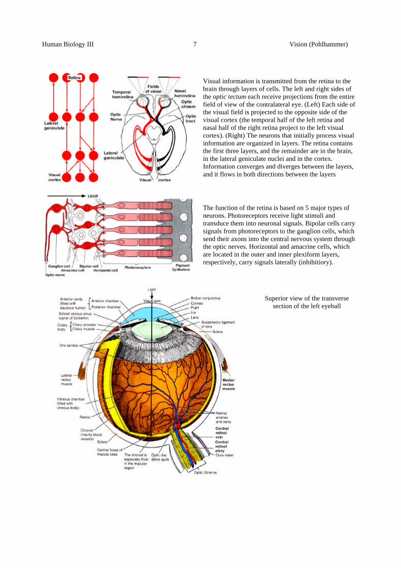
Visual information is transmitted from the retina to thebrain through layers of cells. The left and right sides ofthe optic tectum each receive projections from the entirefield of view of the contralateral eye. (Left) Each side ofthe visual field is projected to the opposite side of thevisual cortex (the temporal half of the left retina andnasal half of the right retina project to the left visualcortex). (Right) The neurons that initially process visualinformation are organized in layers. The retina containsthe first three layers, and the remainder are in the brain,in the lateral geniculate nuclei and in the cortex.Information converges and diverges between the layers,and it flows in both directions between the layers
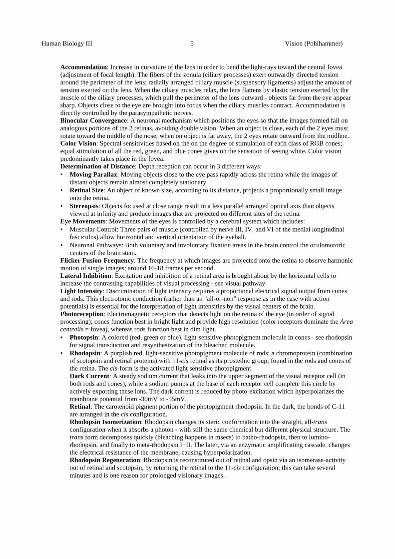
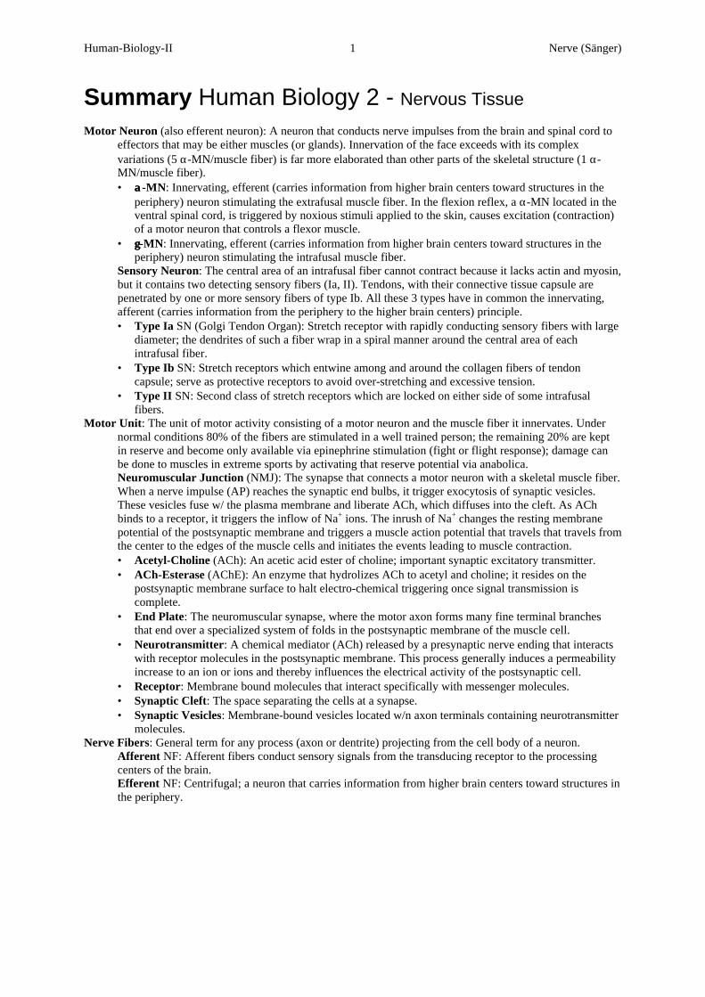
The function of the retina is based on 5 major types ofneurons. Photoreceptors receive light stimuli andtransduce them into neuronal signals. Bipolar cells carrysignals from photoreceptors to the ganglion cells, whichsend their axons into the central nervous system throughthe optic nerves. Horizontal and amacrine cells, whichare located in the outer and inner plexiform layers,respectively, carry signals laterally (inhibitiory).
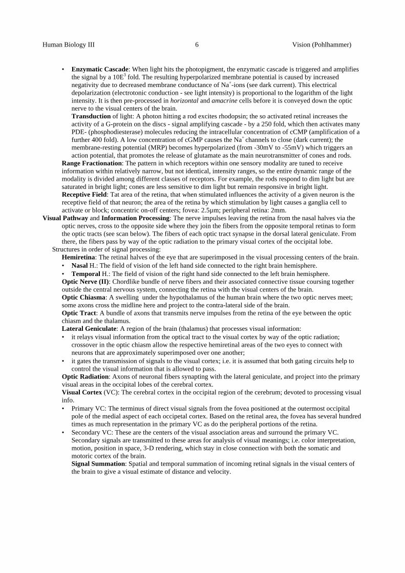
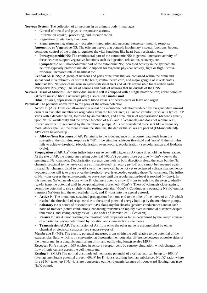
Superior view of the transversesection of the left eyeball
Human-Biology-II 1 Nerve (Sänger)
Summary Human Biology 2 - Nervous Tissue
Motor Neuron (also efferent neuron): A neuron that conducts nerve impulses from the brain and spinal cord toeffectors that may be either muscles (or glands). Innervation of the face exceeds with its complexvariations (5 α-MN/muscle fiber) is far more elaborated than other parts of the skeletal structure (1 α-MN/muscle fiber).• αα-MN: Innervating, efferent (carries information from higher brain centers toward structures in the
periphery) neuron stimulating the extrafusal muscle fiber. In the flexion reflex, a α-MN located in theventral spinal cord, is triggered by noxious stimuli applied to the skin, causes excitation (contraction)of a motor neuron that controls a flexor muscle.
• γγ-MN: Innervating, efferent (carries information from higher brain centers toward structures in theperiphery) neuron stimulating the intrafusal muscle fiber.
Sensory Neuron: The central area of an intrafusal fiber cannot contract because it lacks actin and myosin,but it contains two detecting sensory fibers (Ia, II). Tendons, with their connective tissue capsule arepenetrated by one or more sensory fibers of type Ib. All these 3 types have in common the innervating,afferent (carries information from the periphery to the higher brain centers) principle.• Type Ia SN (Golgi Tendon Organ): Stretch receptor with rapidly conducting sensory fibers with large
diameter; the dendrites of such a fiber wrap in a spiral manner around the central area of eachintrafusal fiber.
• Type Ib SN: Stretch receptors which entwine among and around the collagen fibers of tendoncapsule; serve as protective receptors to avoid over-stretching and excessive tension.
• Type II SN: Second class of stretch receptors which are locked on either side of some intrafusalfibers.
Motor Unit: The unit of motor activity consisting of a motor neuron and the muscle fiber it innervates. Undernormal conditions 80% of the fibers are stimulated in a well trained person; the remaining 20% are keptin reserve and become only available via epinephrine stimulation (fight or flight response); damage canbe done to muscles in extreme sports by activating that reserve potential via anabolica.Neuromuscular Junction (NMJ): The synapse that connects a motor neuron with a skeletal muscle fiber.When a nerve impulse (AP) reaches the synaptic end bulbs, it trigger exocytosis of synaptic vesicles.These vesicles fuse w/ the plasma membrane and liberate ACh, which diffuses into the cleft. As AChbinds to a receptor, it triggers the inflow of Na+ ions. The inrush of Na+ changes the resting membranepotential of the postsynaptic membrane and triggers a muscle action potential that travels that travels fromthe center to the edges of the muscle cells and initiates the events leading to muscle contraction.• Acetyl-Choline (ACh): An acetic acid ester of choline; important synaptic excitatory transmitter.• ACh-Esterase (AChE): An enzyme that hydrolizes ACh to acetyl and choline; it resides on the
postsynaptic membrane surface to halt electro-chemical triggering once signal transmission iscomplete.
• End Plate: The neuromuscular synapse, where the motor axon forms many fine terminal branchesthat end over a specialized system of folds in the postsynaptic membrane of the muscle cell.
• Neurotransmitter: A chemical mediator (ACh) released by a presynaptic nerve ending that interactswith receptor molecules in the postsynaptic membrane. This process generally induces a permeabilityincrease to an ion or ions and thereby influences the electrical activity of the postsynaptic cell.
• Receptor: Membrane bound molecules that interact specifically with messenger molecules.• Synaptic Cleft: The space separating the cells at a synapse.• Synaptic Vesicles: Membrane-bound vesicles located w/n axon terminals containing neurotransmitter
molecules.Nerve Fibers: General term for any process (axon or dentrite) projecting from the cell body of a neuron.
Afferent NF: Afferent fibers conduct sensory signals from the transducing receptor to the processingcenters of the brain.Efferent NF: Centrifugal; a neuron that carries information from higher brain centers toward structures inthe periphery.
Human-Biology-II 2 Nerve (Sänger)
Nervous System: The collection of all neurons in an animals body. It manages:• Control of mental and physical response reactions.• Information uptake, -processing, and -memorization.• Regulation of vital body functions.• Signal processing: stimulus - reception - integration and neuronal response - motoric response Autonomic or Vegetative NS: The efferent nerves that controls involuntary visceral functions; beyondconscious control of the brain; it regulates the vital functions like heart beat, respiration etc.:• Parasympatethic NS: The craniosacral part of the autonomic NS; in general, increased activity of
these neurons support vegetative functions such as digestion, relaxation, recovery, etc.• Sympatethic NS: Thorocolumnar part of the autonomic NS; increased activity in the sympathetic
neurons typically provides metabolic support for vigorous physical activity, fight or flight, stress-response, increased rate of heartbeat etc.
Central NS (CNS): A group of neurons and parts of neurons that are contained within the brain andspinal cord in vertebrates; or within the brain, ventral nerve cord, and major ganglia of invertebrates. Intrinsic NS: Network of neurons in gastro-intestinal tract and -ducts responsible for digestive tasks. Peripheral NS (PNS): The set of neurons and parts of neurons that lie outside of the CNS.
Nervous Tissue of Muscles: Each individual muscle cell is equipped with a single motor neuron; entire complex(skeletal muscle fiber + neuronal plate) also called a motor unit.Hilus: An area, depression, or pit where blood vessels of nerves enter or leave and organ.
Potential: The potential above zero to the peak of the action potential. Action P. (AP): Transient all-or-none reversal of a membrane potential produced by a regenerative inwardcurrent in excitable membranes originating from the hillock area; i.e.: nerve impulse, or spike; a typical APstarts with a depolarization, followed by an overshoot, and a final phase of repolarisation (depends greatlyupon Na+/K+ availability and the proper function of Na+- and K+-channels) and does not require ATP,instead used the PE generated by the membrane pumps. AP’s are considered to be sent like a frequencymodulated-signal i.e.: the more intense the stimulus, the denser the spikes are packed (FM-modulated)-AP’s can’t be added up.• All-Or-None Response of AP: Pertaining to the independence of response magnitude from the
strength of the stimulus; response is "all" if the stimulus achieves threshold and "none" if the stimulusfails to achieve threshold; (depolarization, overshooting, repolarization - see polarization and Hodgkincycle).
Propagation of AP: Ca2+-ions influx into a nerve cell will trigger an AP once threshold has been reached.At the site of AP, the membrane resting potential (-60mV) becomes more positive (+40mV) due to theopening of Na+-channels. Depolarization spreads passively in both directions along the axon but the Na+
channels proximal to the nerve cell are still inactivated (refractory period) and cannot be reopened again.Instead Na+ channels distal to the AP site of the nerve cell have not yet experienced voltage change, hencedepolarization will take place once the threshold-level is exceeded opening those Na+-channels. The influxof Na +-ions causes the axon-potential to overshoot until the repolarisation-level is reached (+40mv). Inthis moment Na+-channels close while K+-channels open to allow K+-ions to rush into the axon graduallyrepolarizing the potential until hyper-polarization is reached (-70mV). There K+-channels close again topermit the potential to rise slightly to the resting potential (-60mV). Continuously operating Na+/K+-pumpstransport Na+-ions into the extracellular fluid, and K+-ions into the axonal cytosol.• Active P.: The membrane sustained propagation from one end to the other of the nerve of an AP which
reached the threshold of response due to the stored potential energy built up by the membrane pumps.• Saltatory P.: A series of discontinued AP's along myelin sheaths (passive conductance) and at each
node of Ranvier (active conduction), enhancing transmission rapidly over internodial distances despitethin axons, and saving energy as well (see nodes of Ranvier, cell - Schwann).
• Passive P.: An AP not reaching the threshold will propagate as far as determined by the length constantof a particular nerve (determined by isolation and cross-section - see there).
• Transmission of AP: Transmission of AP from on to the other nerve is accomplished by eitherchemical or electrical synapses (see synapse-types of).
Membrane P. (MP): The electric potential measured from within the cell relative to the potential of theextracellular fluid, which is by convention at 0 potential i.e.: potential difference between opposite sides ofthe membrane; its a dynamic equilibrium of in- and outflowing ions;(see also MRP). Receptor P.: A change in MP elicited in sensory receptor cells by sensory simulation, which changes theflow of ionic current across the cell membrane. Resting P.: (MRP) The normal unstimulated membrane potential of a cell at rest; can be up to -100mV(average membrane potential at rest: -60mV for K+-ions) resulting from an unbalanced Na+/K+ ratio, wherefore e2 K+- taken up 3 Na+-ions are transported out i.e.: dynamic balance of in/out-ward flowing ions (seeNa/K pump).
Human-Biology-II 3 Nerve (Sänger)
Reversal P.: The MP at which no current flows through the membrane ion channels, even though thechannels are open; it is equal to the EP for ions that are conducted through open channels - compare EPSPand IPSP.
Reflex: An action that is generated without the participation of the highest neuronal centers and is thus nonvoluntary; an involuntary motor response mediated by a neuronal arc in response to sensory input. Itpermits the body to make exceedingly rapid adjustments to homeostatic imbalances. Reflexes occur in thegray matter; these are fast, predictable, automatic responses to changes in the environment (faster thanprocessing information via the cranial pathways of the brain); there are somatic reflexes (contraction ofskeletal muscles) and autonomic reflexes (unconsciouls perceived reflexes of the viscera).Relfex Arc (RA): A neuronal pathway that connects sensory input and motor output; consists of afferentinput to an integrated nerve center (CNS) that produces activity in efferent nerves to an effector organ:• Receptor: The distal end of a sensory neuron (dendrite); it responds to a specific stimulus by
producing a graded generator potential that leads to a nerve impulse once threshold level is reached.• Sensory neuron: The nerve impulse propagates into the sensory neuron in the gray matter of the
spinal cord (afferent pathway).• Integrating center: The region of the CNS within the gray matter that consists of one or more
associated neurons, which may rely the impulse to other association neurons.Monosynaptic RA: A reflex pathway in which the sensory neuron and the motor neuron pass via asingle synapse.Polysynaptic RA: A reflex pathway in which the sensory neuron synapses its information to otherassociated neurons as well as to the motor neuron; e.g. neurons that mediate info to the centers of thebrain.
• Motor neuron: Impulses triggered by the integrating center propagate out of the CNS along a motorneuron to the part of the body that will respond (efferent pathway).
• Effector: The responding part of the body such as a muscle or gland; its action is a reflex (somatic ifthe effector is a smooth-, cardiac muscle, or gland; or autonomic if it is a visceral reflex).
Relfex Types:• Crossed Extensor R.: A reflex in which extension of the joints in one limb occurs in conjunction with
contraction of the flexor muscles of the opposite limb.• Flexor R. (withdrawal R.): A polysynaptic, protective reflex in which the flexor muscles are
stimulated while extensor muscles are inhibited.• Stretch R. (tendon jerk or myotatic R.): A monosynaptic reflex triggered by sudden stretching of
muscle spindle within a muscle that elicits contraction of the same muscle.• Tendon R.: A polysynaptic, ipsilateral reflex that is designed to protect tendons and their associated
muscles from damage that might be brought about by excessive tension. The receptors involved arecalled tendon organs (Golgi tendon organs).
Human-Biology 1 Muscle (Sänger)
Summary Human Biology 2 - Muscle Tissue
Abnormalities:Gangrene: Death of a soft tissue, such as muscle, that results from interrupted blood supply.Muscular Dystrophy: An inherited muscle destroying disease; it affects only the skeletal (voluntarycontrolled) muscles and is caused by the absence of a sarcolemal protein, named dyshopin.Muscle Fatigue: Overstimulated groups of skeletal muscle, that have become progressively weaker whilethey no longer respond; occurs when muscles can't produce enough ATP to meet their needs, due toinsufficient oxygen supply, depletion of glycogen, buildup of lactic acid, failure of action potentials in themotor neuron to release ACh, and unexpected fatigue mechanisms in the central nervous system.Myoma: A tumor consisting of muscle tissue.Myostenia gravis: ????????????Necrosis: A pathological type of cell death that results from disease, injury, or lack of blood supply inwhich many adjacent cells swell, burst, and spill their contents into the interstitial fluid, triggering andinflammatory response.Paralysis: Loss or impairment of motor (muscular) function resulting from a lesion of nervous ormuscular origin.Tenosynovitis: Inflammation of the tendons, sheaths, and synovial membranes surrounding certain joints;affected sheaths often become swolen. Condition frequently follows trauma, strain, or excessive exercise.
Developmental Stages and phenotypical Expression of Muscles:Myogenesis: Birth of muscle fibers; 6 weeks after conception, the formation of myoblasts is followed bythe development of myotubes (fusion of myoblasts to a chainlike aggregation) and finalized in the 9th
week by myo-satellic cells. Further volumetric increase of muscle tissue (hypotrophy in the embryonicstage) induced by growth hormones and achieved by cell division.• Myoblast: Embryonic precursor for skeletal muscle fibers.• Myogenic Regulatory Function (MRF):• Pregnancy: After the 7th week in pregnancy, muscle fibers actively adapt to changed physiological
requirements by actively communicating between each other via neuronal stimuli and gap junctionsbetween muscle cells. This enables growth (triggering RNA-cascade to induce protein synthesis ofactin and myosin fibers) and degeneration during and after pregnancy.
• Regeneration process of an injured muscle fiber: An injury triggers a cascade of reactions; cellularinfiltration and inflammation of the injured fiber (by inflammatory cells and myonuclei) inducedigestion of damaged components (degenerative phase); this is followed by the proliferation andmigration of satellite cells (in skeletal muscle fiber) or pericytes (in smooth muscle fiber) which fuseinto a chainlike formation giving rise to myotubes (proliferation and fusionizing phase); finally, thesemyotubes form new myofibrils and mediated by myofibrillar proteins synthesize to new fiber fillingcompletely the injured site (myogenetic phase). Cardiac muscle tissue is not able to undergoregenerative processes and forms scars instead.
Synaptogenesis: The birth of the neuromuscular junction; prior to innervation ACh receptors aredistributed across the muscle fiber surface. As the nerve contacts the muscle fiber and electric activityensues, receptors aggregate in the region of the neuromuscular junction. As maturation progresses, extra-junctional receptors decrease significantly while junctional receptors increase in number and density.Termination: The finishing touches of synapse formation; approx. 50% of all synapses formed duringsynaptogenesis are faulty, hence are eliminated by a feedback loop via endogenic lysosomes or exogenicprotease activity (sort of macrophages):Stable ACh-receptors experience a positive feedback which promotes finalization whereas an unstableACh-receptor experiences a negative feedback by an excess of Ca2+ ions (toxic) which leads to theirelimination. Regeneration is incomplete if satellite cells are eliminated by a widespread destruction of thebasal lamina and the sarcolemma of the muscle fiber → transplantation.
Human-Biology 2 Muscle (Sänger)
Motion: Skeletal muscles produce movements by exerting force on tendons, which, in turn, pull on bones orother structures such as skin. Bones serve as levers, and joints as fulcrum of the levers;Lever System: A rigid rod that moves about on some fixed hinge point (F, fulcrum), an effector muscle(E), and a resisting moment (R):• 1st class Lever: Fulcrum is centered b/w an effort (effector muscle) and resistance (gravitanional pull
as the angular moment); e.g. neck muscle (E), cervical curve (F), and weight of head (R).• 2nd class Lever: Fulcrum at one end, the effort at the opposite end, and the resistance in-between
them; e.g. Gastrocnemius muscle (E), toe tips (F), tarsales (R).• 3rd class Lever: Fulcrum at one end, the effort at the opposite end, and the resistance in-between; e.g.
Biceps brachii (E), ellbow (F), gravitational pull of hands (R).Group Action: Movements that require several muscles acting together:• Agonist (Gk. agogos, leader) The prime mover, as in the case of the arm, the Biceps brachii.• Antagonist (Gk. antiagonistes, opposite) As the mover contracts, the antagonist relaxes; in the case of
the arm, the Triceps brachii muscle.• Synergist (Gk. syn, together; ergon, work): Steadying or stabilizing movement, to avoid unwanted
movements, and to increase the agonists efficiency.Muscle: A "tasty" organ composed of one of the three types of muscle tissue (approx. 700 skeletal, 1 cardiac,
and several visceral), specialized for active contraction to produce voluntary or involuntary movement ofparts of the body. In combination with the other three main body tissues, provide a useful tool forlocomotion and other vital body functions. Its modular structure, low energetic requirements at stand-by,and the availability of oxygen as fuel resulted in its global distribution.Chemical Constituents of muscles:• Inorganic C.: Up to 57% of a muscle cell consists of water; the remaining 25% are made of the
following minerals K+, Na+, Cl-, Mg2+, Ca2+, phosphate• Organic C.: Energy providing constituents are lipids, glucose, kreatin-phosphate, ATP, arginine-
phosphate, enzymes and proteins as the responsible contractile unit.Muscles participate in:• Body Stabilization: Muscle contraction maintain the body in stable positions, such as standing or
sitting. Postural muscles display sustained contraction when a person is awake (e.g. neck muscles tohold the head upright); sustained contraction of the sphincter (smooth muscles) may prevent outflowof the contents of hollow organs.
• Motion: Walking, running, localized motion such a s grasping rely on the integrated functioning ofbones, joints, and skeletal muscle.
• Movement w/n the body: Cardiac muscle contraction pump blood to all body tissues and help toregulate blood pressure. Peristalsis of smooth muscle contraction aid food and chyme locomotion,sperm ejaculation, urine excretion into the bladder and drainage of lymph and venous blood back tothe heart.
• Thermogenesis: A contracting muscle generates metabolic heat as a by-product. Much of the heat isused to maintain normal body temperature (85% of all body heat). Involuntary muscle contraction(shivering) can increase thermogenesis by several hundred percent.
Muscle Anatomy: Structural organization of muscle from a macroscopic to microscopic level (see scan below):Macroscopic elements of a Muscle: Connective tissue surrounds and protects underlying muscle tissue.• Fascia: A fibrous membrane of connective tissue that covers, supports, and separates muscles; a fascia
is held together by the epimysium, and itself encloses several fascicles.Deep F.: The inner sheath wrapped around a muscle (around the epi-, peri-, endomysium) to keep it inplace. Each of the mysial layers houses a dense network of blood capillaries for the transport of O2,CO2, lipids and nutrients as well as capillaries of the lymphatic system to allow drainage of excess(interstitial) liquid; all 3 may extend beyond the muscle fibers as a tendon:Endomysium: Invagination of the perimysium separating each individual muscle fiber (cell); it housesthe basic muscle fibers (cells) with its electro-chemical devices.Epimysium: A fibrous connective tissue around muscles, below the deep fascia enclosing perimysialfibers (enclosing a bundle of muscle fibers).Perimysium: Invagination of the epimysium that divides muscles into bundles (fascicle); each bundleencloses some muscle cells, which themselves are separated by the endomysium.Superficial F.: The subcutaneous, continuos outer sheath between the dermis of the skin and the deepfascia of the muscle. It represents the framework for nerves and blood vessels; composed of areolarconnective and adipose tissue, and stores water, fat, insulates, and cushions (mechanical protection);
Human-Biology 3 Muscle (Sänger)
• Fascicle: A small bundle or cluster bound by perimysium, enclosing several muscle fibers.Muscle Fiber (or myofibers, MF): A giant skeletal muscle cell, with many nuclei. It consists ofmyofibrils, sarcomeres, and their myofilaments (see microscopic elements - myofibril).Extrafusal MF: Contractile muscle fiber that make up the bulk of skeletal muscle.Intrafusal MF: The muscle fibers within a muscle spindle organ.
• Tendon: A band of tough fibrous connective tissue (continuation of endo-, epi-, and peri-mysium),that anchors skeletal muscle to bones.Insertion: Attachment of the other tendon form the muscle to the moveable bone.Origin: Attachment of a muscle tendon to the stationary bone.
Microscopic elements of a Muscle fiber: Several myofibril are bound together by following structures:• Myofibril: The contractile element of a skeletal muscle; a longitudinal unit of muscle fiber made up
of sarcomeres and surrounded by the sarcoplasmic reticulum, containing actin- and myosin-filaments.It encapsulates the sarcoplasm and the myofibril with its sarcomeres.Elastic Filament (Titin): The 3rd most plentiful protein in skeletal muscle (after actin and myosin); itanchors myosin to the Z-discs and thereby helps to stabilize the position of the thick filaments.Intermediate Filament (in smooth muscles only): An irregular network of protein filaments, thatprovide structural reinforcement, hold organelles in place, and give shape to the cell.Thin Filament (Actin): (Gk. actos, a ray) A ubiquitous protein, connected to the. G-actin is theglobular monomer that polymerizes to form F-actin, the backbone of the thin filaments; other actinproteins of the thin filament are tropomyosin and troponin complex.Thick Filament (Myosin): The 200 proteins that cross bridges in muscle fibers; it is also found inmany other cell types and is associated with cellular motility. Each myosin molecule consists of aglobular double head attached to a α-helical neck (heavy meromysin; 57nm long) and a long thin tail(light meromyosin; 93nm long).
• Sarcolemma (Gk. sarcos, flesh): The surface (plasma) membrane of a muscle fiber; it also serves asthe electro-chemical interface of the moto-neural stimulation.Basement membrane (Basal lamina): Thin, extracellular fibrous membrane surrounding thesarcolemma of a myofibril. It houses satellite cells that are essential in the repair of injured muscles.Mitochondrion: A microbody that provides cells with energy in form of ATP-molecules byaerobically (using O2) breaking down glucose molecules into H2O and CO2.
• Sarcomere: (Gk. meros, part of) The contractile unit of myofibrils bounded by transverse tubules; itextends form one Z-disc to the next:A-Band: The dark area of actin with the overlapping myosin segment.I Band: The less dense area with the remaining myosin segments that do not overlap with actin.H zone: The central non-overlapping myosin segment.M Line: The dividing protein line that halves the H zone.Z Disc: Narrow plate-shaped regions of dense material, that separate sarcomeres from each other.Dense Body (in smooth muscles only): The site of attachment of intermediate filaments (similar to Zdiscs) that are irregularly distributed throughout the cell.
• Triad: A complex of 3 units in a muscle fiber composed of the following structures:Sarcoplasmatic Reticulum (SR): A dense smooth membrane-limited network surrounding eachmyofibril (a fluid filled system of cisterns, equivalent of endoplasmatic reticulum in cells). Functionsto reabsorb calcium ions during relaxation and to release them to cause contraction.Terminal Cistern: The closed spaces that make up part of the sarcoplasmic reticulum on both sides ofthe Z line, making close contact with transverse tubules.Transverse Tubules: Small, cylindrical invaginations of the sarcolemma of striated muscle fibers(cells) that conduct muscle action potentials toward the center of the myofibrils.
Muscle Characteristics: Five principal characteristics enable a muscle to carry out its functions and thuscontribute to homeostasis:Conductivity: The ability to conduct action potentials along the plasma membrane.Contractility: The ability of muscle tissue to shorten (thicken), thus generating force to do work.Elasticity: Muscle tissue tends to return to its original shape after contraction or extension.Excitability: The ability to respond to certain stimuli by producing electrical action potentials triggeredby neurotransmitters (ACh), or hormones.Extensibility: Muscles can be extended (stretched) without damage to the tissue.
Muscle Contraction and Relaxation: A muscle fiber develops its greatest tension (maximum efficiency) whenthere is optimal overlap of the thick and thin filaments; efficiency decreases with decreasing or increasingwidth past the mid-average of the H-zone (see scan below).
Human-Biology 4 Muscle (Sänger)
Contraction: Upon stimulation of the neuro-muscular junction, free Ca2+ rushes out into the myoplasm(via appropriate release channels) and initiates the sliding filament mechanism.• Actomyosin Complex: The bound complex of cross-bridged actin and myosin filaments.• Adenosine-TriPhosphate (ATP): ATP is required to split the actomyosin complex into its
constituting partners; binding of ATP to the myosin head causes the myosin heads to detach from theactin filament, while ATP cleaves itself into ADP+P.
• Ca-binding proteins: Ca-ions mediating muscle contraction can react with the following proteins:Caldesmon: A Ca-binding regulatory protein in smooth muscle, which plays a role in the latchmechanism of some smooth muscles.Calmodulin: A troponin-like calcium-binding regulatory protein found in essentially all tissues.Calsequestrin: A Ca-binding protein that contributes to the regulation in muscle relaxation.Parvalbumin: Calcium-binding protein found in vertebrate muscle; it binds Ca2+ in the cytoplasm,thus accelerating muscle relaxation.Troponin: A complex of globular calcium-binding proteins associated with actin and tropomyosin inthe thin filaments of skeletal muscle. When troponin binds Ca2+, it undergoes a conformationalchange, allowing tropomyosin to reveal myosin binding sites on the actin filaments.
• Sliding Filament Mechanism: Decrease of sarcomere length by which actin and myosin slide pasteach other. Free Ca2+ entering the myoplasm binds to special mediator proteins that allow myosincross-bridges to bind to actin filaments. This results in a twisting of the myosin head which causes thesarcomere to shorten (the power stroke is characterized by the swiveling of the myosin heads towardsthe center of the sarcomere). Further excitation causes the myosin head to detach (the recovery strokeis initiated by the splitting ATP into ADP+P); the released energy is used to reshape the myosin headthus allowing reaction with the next actin, itself conformationally changed by other Ca-ion;Skeletal MC: The sliding filament mechanism of skeletal muscles fibers is mediated via external orautonomous signals. An ACh receptor initiates the process by releasing Ca2+ via the transverse tubulesinto the myoplasm (= sarcoplasm); the ions attach to troponin (part of the actin protein) causing aconformational change of the tropomyosin molecule, allowing myosin cross-bridges to bind to actinfilaments and initiate contraction; filaments pass each like an oar of a boat. Contraction continues ifATP is available and Ca2+ level in the sarcoplasm remains high.Smooth MC: Since these fibers lack a troponin; Ca2+ enters the myoplasm to bind with calmodulinforming a Ca2+/calmodulin complex that traps caldesmon. Caldesmon fixed in this reaction does notinterfere with the actin-myosin reaction, thus muscle contraction can occur. In yet another contractionmechanism, the Ca2+/calmodulim complex activates the myosin-light-chain kinase that uses ATP tophosphorylate myosin heads, enabling the formation of the actomyosin complex.Smooth muscles fibers lack transverse tubules (Ca2+ takes more time to diffuse to and from the tissue)thus increasing both contraction and relaxation latent periods. Hormones (e.g. epinephrine which havea relaxing effect), temperature, CO2, pH-changes, and ion concentrations also affect contraction.
Relaxation: Turning off the sliding filament mechanism is brought about by reabsorbtion of Ca-ions fromthe myoplasm back (Ca-pumps are powered by ATP) and storage in the sarcoplasmic reticulum(Calsequestrin traps Ca2+ in the SR). It is initiated once AChE breaks down ACh in the synaptic cleft,causing closure of the Ca2+ channels while Ca-pumps actively withdraw the ions into the SR; attachmentof ATP to myosin triggers detachment from actin filaments and reorientates the myosin heads; themissing Ca-ions cause a steric hindrance of the tropomyosin-troponin complex (in skeletal muscle) andcaldesmon binding to the actin-myosin complex, restricting muscle contraction (in smooth muscle). Sincemyosin heads can't dock with actin, the thin filaments slip back to their relaxed state.
Muscle Contraction and Tension: To enable gradual contraction patterns rather than the an all-or-nonereaction (typical contraction of a single muscle fiber). Sarcomeres are aligned in registers; the moremotor-neurons stimulate the muscle, the stronger is the contraction; several patterns are distinguished:Frequency Stimulation: Increase in strength of contraction by a second stimulus applied after therefracting period and before relaxation of a muscle.• Staircase Effect (Treppe): The gradual increase in the force of contraction of a muscle caused by
repetitive stimuli of the same strength.• Tetanus: An uninterrupted muscular contraction caused by high frequency motor impulses.Twitch MC: A brief contraction of all the muscle fibers in response to a single action potential in itsmotor neuron; a myogram (record of muscle contraction) distinguishes 3 distinct states of a twitch MC:• Contraction Period: Upward tracing of contracting force brought about by shortening of the
sarcomere; typically 10 to 100ms.• Latent Period: The brief period between application of the stimulus and the beginning of contraction,
in which Ca2+ is being released from the sarcoplasmic reticulum; typically 2ms.Relaxation Period: Downward tracing of contracting force due to the active withdrawal of Ca2+ backinto the sarcoplasmic reticulum, resulting in relaxation; typically 10-100ms long.
Human-Biology 5 Muscle (Sänger)
Muscle Metabolism: Muscle contraction requires energy in the form of ATP; relaxed muscle tissue requiresabout 20%, whereas contracting tissue can require as much as 200% more energy.Primary Energy Compound: ATP as the immediate energy resource accounts only for a few seconds ofmuscle activity; anything in excess requires an other recruitment mechanisms.• Phosphagen System: Phospho-creatine and phospho-arginine is 3 to 5 times more plentiful than ATP
and represents a high energy phosphate group that can be convert to ATP by giving of creatine orarginine respectively; it provides extra energy for further 15secs.
Secondary Energy Compounds: Stored proteins, carbohydrates, and fats are converted during the activestate to provide the tissue with pyruvate, glucose, amino acids, or fatty acids; a series of reactionsgenerates acetyl-coenzyme (an energy-rich molecules) that fuels the Krebs cycle in the assemblage ATPfrom ADP (see HB- or Biochem.-Metabolism).• Aerobic System: Release of energy by cellular respiration in mitochondria (O2-mediated breakdown
of fuel molecules) reduces pyruvic acid to water and CO2, with a total yield of 36 ATP from eachcleaved glucose molecule. Since this is a time-consuming process, activities lasting more than 30secsswap to the anaerobic system.Myoglobin: Once oxygen supply via the blood stream is insufficient, extra amounts of free oxygencan be recruited from the myoglobin storage sites from within the muscle fibers of type I and IIa.
• Anaerobic System (Glycogen-Lactic Acid): Conditions where oxygen levels are low or absent.Glucose is split into 2 molecules of pyruvic acid which yields 1 ATP each with the concomitantproduction of L-lactate (lactic acid). Given off to the bloodstream, the kidneys, heart, and liver canmetabolize lactic acid to generate ATP (liver cells are capable of converting this acid back to glucose).This system provides an extra 30 to 40secs of muscular activity.
Muscle Nomenclature: Arrangement of muscular fascicles (a small bundle or cluster of muscle fiber / cells) inskeletal muscle tissue is correlated with the power of a muscle and its range of motion;Naming skeletal muscles ExampleDirection: relative to the midline:• Rectus: Fiber runs parallel to the midline of the body;• Transverse: Fiber runs perpendicular to midline;• Oblique: Fiber runs diagonally to the midline;
Number of origins: Number of tendons of origin:• Biceps: means two origins• Triceps: means three origins;• Quadriceps: means four origins;
Biceps brachii;Triceps brachii;Quadriceps formis;
Shape: Relative shape of the muscle:• Deltoid: having a triangular shape;• Trapezius: ;having a trapezoid shape;• Serratus: ;having a saw-toothed shape;• Rhomboideus: ;having a rhomboid (diamond) shape;
Action: Principal action of the muscle:• Abductor: moves a bone away from the midline;• Adductor: ;moves a bone closer to the midline;• Depressor: ;produces a downward movement;• Extensor: increases the angle at a joint;• Flexor: decreases the angle at a joint;• Levator: ; produces an upward movement;• Pronator: turns the palm downward or posteriorly;• Rotator: moves a bone around its longitudinal axis;• Sphincter: decreases the size of an opening;• Supinator: turns the palm upward or anteriorly;• Tensor: makes a body part more rigid;
Fascicle: A small bundle or cluster of muscle fibers (cell); the length of muscles fibers can varyconsiderably and according to their task can extend even beyond three joints (multi-articular muscles i.e.Extensor digitorum longus) or septed (Rectus adbominalis).Arrangement and description of fascicles ExampleCircular: Fascicles are arranged in a concentric circular pattern toform sphincter muscles that enclose an orifice (opening)
Orbicularis oculi
Fusiform: Fascicles are nearly parallel with longitudinal axis ofmuscle and terminate at either end in flat tendons, but muscle taperstoward tendons where the diameter is less than that of the belly
Digastric muscle
Parallel: Fascicles are parallel with longitudinal axis of muscle andterminate at either end in flat tendons
Stylohhoid muscle
Pennate: Fascicle are short in relation to muscle length and thetendons extend nearly the entire length of the muscle• Unipennate: Fascicles are arranged on only one side of the tendon• Bipennate: Fascicles are arranged on both sides of a centrally
positioned tendon; Biceps means two origins;• Multipennate: Fascicles attach obliquely from many directions to
several tendons; Triceps means 3 origins, and Quadriceps 4 origins;
Muscle Spindle: An encapsulated proprioceptor in a skeletal muscle, consisting of specialized intrafusal musclefibers and nerves endings; stimulated by changes in length or tension of muscle fibers. The ends of thespindles are anchored to endomysium and perimysium.Extrafusal MF: Regular muscle fibers that surround the muscle spindle; are innervated by large α-motorneurons.Intrafusal MF: 3 to 10 specialized muscle fibers (cells), partially enclosed in a spindle-shaped connectivetissue capsule; these fibers make up a muscle spindle. Contract when stimulated by the γ-motor neurons.
Muscle Tonus: Small contractions that give firmness to a relaxed skeletal muscle.Concentric Contraction: Contraction that shortens the muscle.Eccentric C: Gradual extension of a muscle while working against a force.Isometric C: Contraction during which a muscle does not shorten significantly.Isotonic C: Contraction in which the force generated remains constant while the muscle shortens.Tonic C: A steady, slow contraction achieved w/o an action potential; e.g. muscles of the eyeball.
Muscle Types:Apart from a cardiac muscle cell, all other types can be repaired if damaged.
Characteristic Skeletal M. Cardiac M. Smooth M.Cell appearance,diameter andlength
Long cylindrical fiber w/many peripherally locatednuclei; striated;unbranched; 10-100µm;0.1-300mm long.
Branched cylinder usuallyw/ one centrally locatednucleus; striated; intercala-ted discs join neighboringfibers; 14µm; 50-100µm lg
Spindle-shaped fiber w/one, centrally positionednucleus; no striations;3-8µm in diameter;30-200µm long
Location Attached to bones Heart Walls of hollow viscera; airways,blood vessels, iris and ciliary body ofeye, urethra, viscera, arrector of hairfollicle.
Connect. tissuecomponents
Epi-, peri-, andendomysium
Endomysium Endomysium
Transversetubules
Aligned w/ each A-I bandjunction
Aligned w/ each Z-disc -
Gap junctions - Yes, via intercalated discs Yes, in visceralCa2+ source Sarcoplasmic reticulum Sarcoplasmic reticulum
and extracellular fluidSarcoplasmic reticulumand extracellular fluid
Reg.-Proteins forcontraction
Troponin and tropomyosin Troponin and tropomyosin Calmodulin and myosinlight chain kinase
Nervous control Voluntary (somatic NS) Involuntary (autonom.NS) Involuntary (autinom. NS)Regeneration Limited, via satellite cells
that induce hypertrophyNone - scar formation Considerable by pericytes
inducing hyperplasia
Human-Biology 7 Muscle (Sänger)
Cardiac M.: A self-contracting (involuntary - not under conscious control) striated muscle where theintrinsic contracting waves (self-generated action potentials) propagate along the entire tissue; startingfrom the sinoatrial node = pacemaker region, via the bundle of HIS and Purkinjie fibers, down to theapex. CM-cells are typically Y-shaped, allowing interconnections among each other; intercellularpropagation is mediated by gap junctions of intercalated discs; the CNS implies a modulating task only(propagation of approx. 0.8m/s);• Sympaticus stimulation: Managed by the sympatethic NS, resulting in an increased heartbeat.• Vagus stimulation: Managed by the parasympatethic NS, resulting in a lowered heartbeat.Being the only blood propelling organ, CM-cells are rich in mitochondria; therefore, require hugeamounts of glycogen, gluconitrates, lipids, and O2 (aerobic production of ATP). Healing of damaged CMcells (heart attack) by scar formation only; i.e. hypertrophy (an excessive enlargement or overgrowth oftissue w/o cell division).• Autorhytmic cells: Specialized intrinsic cells, capable of stimulating CM fibers; these cells can also
be moduoated by neuronal and hormonal influences, causing an in/decrease of the heart beat.• Intercalated discs: Ends of nerve fibers interconnect neighboring cells by irregular transverse thick
endings, which contain desmosomes that hold the fibers together. Gap junctions embedded in thesediscs allow muscle action potentials to spread from one muscle fiber to the next and thus allowcontraction of entire bundles as a functional unit.
Smooth M. (SM): A muscle of mesenchymatic origin without sarcomeres (contraction is slower, but lastslonger) and hence without striations. These non-striated tissue of involuntary control is subject tohormonal and neuronal influence - in some even autorhythmic. Each fiber has a ratio of 10-15 : 1 of thickto thin filaments. Shortening of SM produces a bubblelike expansion of the sarcolemma, in which fiberstwist like a helix. Myofilaments are non-uniformly distributed within small, mononucleated, spindle-shaped cells. Damaged SM are replaced by enlarging pericytes of adjacent muscle cells or via "satellite"cells (dormant stem cells); e.g. postnatal period of a woman.• Multi-unit SM: Individual fibers, each dotted with its own neuron terminal and gap junctions to
neighboring cells allow single muscle cell stimulation; multi-unit SM are found in arteries, airways,arrector pilli muscles that attach to hair follicles, radiar and circular muscle of the iris and cilliarybody that adjust the refractive power of the lens.
• Single-unit SM (visceral SM): The type of muscle found in the intestinal tract, because of theirextended rate of response. SM-cells communicate electrically through gap junctions, desmosomes andtend to contract in sequential waves. Besides the intestine, they are also found in the other hollowviscera (stomach, uterus, and urinary bladder) and wrapped around arterioles, and veins.
Striated M. (skeletal): Muscle fibers with a high myoglobin an mitochondria content appear red (redmuscle fiber) whereas those with a lower content appear white (white muscle fiber); a fibers ability toslow-twitch or fast-twitch depends how rapidly it splits ATP. Based on these structural and functionalcharacteristics, skeletal muscle fibers are classified into three types (type I, IIa, IIb):Structural Feature Slow oxidative (Type I) Fast oxidative (Type IIa) Fast glycolytic (Type IIb)Diameter of fiber Smallest Intermediate LargestMyoglobin cont. Large Large SmallMitochondria Many Many FewCapillaries Many Many FewColor Red Red to pink White (pale)Functional FeaturesATP production Aerobic process Aerobic process Glycolytic processATP hydrolysis Slow Fast FastVelocity of contract. Slow Fast FastFatigue resistance High Intermediate LowGlycogen storage Low Intermediate HighOrder of recruitment First Third SecondActivities Maintaining posture, endurance
activities; fatigue resistant; thestronger the stimulus, the stronger
muscle contraction
Walking, running, sprinting;Fatigue resistant; can arise from
endurance type exercise out oftype IIb;
Rapid, intense movements of shortduration; fatigue easily; react in"all or nothing" pattern; rise inwith weight lifting activities;
Endurance-type exercises, such as running or swimming, cause a gradual transformation of some fastglycolytic (type IIb) fibers into fast oxidative (type IIa) fibers.
Human-Biology 8 Muscle (Sänger)
Narcosis: Muscle relaxation by inhibition of electromotoric transmission; i.e. blockage of ACh receptor sites.Oxygen Demand of Muscle Tissue: Oxygen is required in the final metabolic steps in the conversion of fuel
packages fed into the Krebs cycle and the successively attached electron transport chain. Any depletion ofoxygen deviates the energy rich harvest via the aerobic in favor of the anaerobic pathway, with theconcomitant production of lactate. To avoid excess production of lactate, oxygen is stored in thefollowing tissues: myoglobin of muscles, hemoglobin, increased influx of air into the lungs, dissolved invarious body tissues.
Regeneration of Muscle Tissue: Skeletal muscle fibers have little potential to divide; growth of skeletal muscleis due to enlargement of existing cells (hypertrophy), rather than an increase in the number of fibers(hyperplasia); regeneration of damaged tissue is mediated by:Satellite Cells: Dormant stem cells that fuse with each other to form new skeletal muscle fibers; theyundergo fibrosis in that a scar forms (fibrous connective tissue); this especially applies for damagedcardiac muscle tissue.Pericytes: Stem cells w/n the endothelium of blood capillaries and veins, which accounts for the highpower of regeneration in smooth muscle fibers; e.g. hyperplasmia in the uterus lining.
Rigor Mortis: Rigidity that develops in a dying muscle as Ca2+ leaks out of the sarcoplasmic reticulum andbinds with troponin; quickly, ATP becomes depleted and cross bridges remain attached (myosin headsdetach only once ATP binds to myosin); the cell dies in absence of O2 and ATP. It disappears again oncetissue decomposition initiates.
Tendon: see Muscle fiber - connective tissue.Thermogenesis: Muscular contraction of skeletal muscle tissue to generate heat; partly used to maintain body
temperature. Shivering induces involuntary thermogenesis.Tonus: Sustained resting contraction of muscle, produced by basal neuromotor activity; see muscle tonus.
Human-Biology 9 Muscle (Sänger)
Histology of skeletal muscle tissue (adapted from Lodish et al.)
All skeletal muscles are organized in a stereotyped hierarchy. The organ called a muscle consists of parallelmultinucleate fibers, each of which contains many myofibrils. Muscles are attached to bones or other anchorpoints through tough connective tissue bands called tendons. Each muscle fiber is derived embryonically from agroup of myoblasts that fuse to form myotubes. A myotube then synthesizes the proteins characteristic of musclefibers and differentiates into its adult form. The myofibrils are made up of sarcomeres, arranged end-to-end.Each sarcomere contains thin filaments of actin and thick filaments of myosin, which interdigitate in a precisegeometric relationship. The thin and intermediate filaments are anchored in regions called Z disks.
Human-Biology 2 1 Skeleton (Sänger)
Summary Human Biology - Bones and SkeletonAbnormalities of the Bones and Skeleton:
Abnormal Curvature of the Vertebral column: Various conditions may exaggerate the normal curvature:• Kyphtosis (hunchback): An exaggerated thoracic curve, due to degeneration of the invertebral discs.• Lordosis (swayback): Is an exaggeration of the lumbar curve, due to increased weight of the abdomen
resulting out of pregnancy, extreme obesity, etc.).• Scolosis: Is a lateral bending of the vertebral column in the thoracic region due to a poor posture or one
leg being shorter than the other.Atrophic Bone: Decrease in size due to certain failures, abnormality of nutrition, or lack of use.Arthritis: A form of rheumatism in which the joints have become inflamed. Osteoarthritis: It apparentlyresults from a combination of aging, irritation of the joints, and wear and abrasion; a degenerative disease farmore common than rheumatoid arthritis and usually less damaging.Bursitis: Inflammation of the bursa.Fracture: A break, rupture, or crack in bones when exceeding the maximal permissible load.Herniated (slipped) disc: A rupture of an intervertebral disc; the nucleus polposus protrudes posteriorly intothe vertebral cavity toward the spinal cord and nerves causing considerable pain.Osteoporosis: Age-related disorder characterized by decreased bone mass and increased susceptibility tofractures; can also be induced via lactose intolerance, pregnancy, diabetes, or inheritance; effects backbone(humpback), femur, hip-bones; most common in Caucasians.Spina Bifida: A congenital defect of the vertebral column, in which the lamina fail to unite at the midline;can cause paralysis of certain inner organs.Paget's Disease: A disorder characterized by greatly accelerated remodeling process in which osteoclasticresorption is massive and new bone formation by osteoblasts id extensive. As a result, there is an irregularthickening and softening of the bones.Rheumatism: A painful state of the supporting body structures; bones, ligaments, joints, tendons, or muscles.Rickets: Characterized by an inability of the body to transport Ca2+ and P from the digestive tract into theblood for utilization by bones. Epiphyseal cartilage becomes wider than normal, and bones stay soft. Usuallycaused by a deficiency of vitamin D, Ca2+ , and P.
Articulation: see joint.Bone (Osteon): The main supporting tissue of humans, composed if a matrix of collagen hardened by calcium-
phosphate, and consists of approx. 25% water, 25% proteins, and 50% mineral salts.Bone Anatomy: Structural parts of the skeletal systems are bone tissues, cartilage, red and yellow bonemarrow, and periosteum. An adult bone (e.g. tibia) which shape is genetically determined, is made up of thefollowing anatomical structures:• Articular Cartilage: A thin layer of hyaline cartilage covering the epiphysis where the bone forms a joint
with another bone. It reduces friction and absorbs shock waves.• Diaphysis (Gk. dia, through; physis, growth): The shaft or long, main portion of the bone.• Endosteum (Gk. endo, within): A layer of osteoblasts that lines the medullary cavity and contains
scattered osteoclasts (cells that may assume a role in the removal of the bone - see bone formation).• Epiphysis (Gk. epi, above): The extremities or ends of the bone.• Methaphysis (Gk. meta, after): The region in a mature bone where diaphysis joins the epiphysis.• Marrow Cavity (Medullary, Gk. central part): The space within the diaphysis that contains the fatty
yellow marrow in adults. Yellow marrow consists primarily of fat cells and a few scattered blood cells (seebone tissue - spongy bone).
• Periosteum: (Gk. peri, around; osteo, bone) A dense, white fibrous membrane around the surface of thedense bone. It consists of two layers:Fibrous Layer: Outer layer composed of connective tissue containing blood vessels, lymphatic vessels,and nerves that pass into the bone.Osteogenic Layer: The inner layer that contains elastic fibers, blood vessels, and osteoprogenitor cellscapable of developing into osteoblasts - cells responsible for forming new bone during growth and repair(see bone formation).
Human-Biology 2 2 Skeleton (Sänger)
Bone Formation and Growth: Process by which bone forms in the body, called ossification or osteogenesis. Manyother tissues in the body need Ca2+ in order to perform their functions (e.g. nerve cells, muscle cells, bloodcells); bones are therefore used as a reservoir for metabolic requirements. A delicate homeostasis in removingand depositing Ca is maintained. Hormones (e.g. growth hormone, estrogen), vitamin (e.g. D), mineralimbalance (e.g. Ca, P), and mechanical (e.g. sports) factors induce mineralization resulting in a net increase ofthe substantia compacta (dense or compact bone).• Osteoclast: (Gk. clast, to break) A large, multinuclear cell that destroys or resorbs bone tissue; these cells
are important in the development, growth, maintenance, and repair of bony tissue.• Osteoprogenitor (Gk. pro, precursor; gen, to produce): An unspecialized stem cell of mesenchymal
origin, that can undergo mitosis and further differentiate into an osteoblast; they are found in the inner partof the periosteum, endosteum, and in the canals of bones.Osteoblast (Gk. osteo, bone; blast, bud): A bone-forming, collagen secreting cell, w/ no mitotic potentials.Osteoblasts are usually found on the surface of bones; as they become trapped by the surrounding bonymatrix, they become trapped in their secretions and mutate to osteocytes.Osteocyte (Gk. cyte, cell): A mature bone cell derived from osteoblast that has lost its ability to producenew bone tissue, but maintain their daily cellular activity (exchange of nutrients and metabolic wasteprosucts via the blood).
Calcification (mineralization, ossification): Deposition of mineral salts in the framework formed by collagenfibers of the matrix induces crystallization and subsequent hardening of the tissue. Without collagen fibersthat serve as reinforcement rods, bones would be very brittle. Two alternative ways to form dense bony tissueare known and involve the replacement of preexisting connective tissue:• Endochondral Ossification (Gk. endo, within; chondro, cartilage): Also called intra-cartilaginous
ossification, in which hyaline cartilage is replaced by bone from within. Once blood vessels penetrate theperichondrium, they stimulate the cells of the internal layer to enlarge and become osteoblasts; these cellsbegin to form a collar of compact bone (stage of periosteum). Further activity of these cells elevate the pH(alkaline) causing precipitation of Ca2+ (calcification) in the intercellular substance. Consequently,cartilaginous tissue dies due to hindered diffusion of nutrients, giving rise to dense bone tissue. Mostbones of the body, including the skull, are formed in this way.
• Intramembranous Ossification (L. intra, within; membrana, membrane): Formation of bone directly onor within the loose fibrous membrane without first going through a cartilage stage. Osteoblasts frommesenchymal origin secrete intercellular material, generating collagenous fibers that forms the trabecularmatrix. This spongy bones filled with vascularized connective tissue which differentiates into red bonemarrow, mesenchyme tissue at the outside condenses, and incorporates calcium salts to form dense bonetissue. Almost all flat bones of the skull (incl. clavicle) are formed this way.
Bone Function: The skeletal system performs several basic functions:Blood Cell Production: Red marrow in certain bones is capable of producing blood cells, a process calledhematopoiesis or hemopoiesis. Red marrow consists of blood cells in immature stages, fat cells, andmacrophages. Red marrow produces red blood cells, some white blood cells, and platelets.Mineral Homeostasis: Bones store several minerals that can be distributed to other parts of the body upondemand (bones release Ca2+ for muscle contraction and nerve activity). The principal stored minerals arecalcium carbonate (CaCO3), hydroxy apatate (Ca3(PO4)2⋅(OH2), and small amounts of MgOH, F, SO4
2-; seealso calcium homeostasis.Movement: Bones serve as levers to which muscles are attached. When the muscles contract, the bonesacting as levers produce movement.Protection: Many internal organs are protected from injury by the skeleton, e.g. the brain is protected by thecranial bones, the spinal cord by the vertebrate, the heart and lungs by the rib cage, and internal reproductiveorgans by the pelvic bones.Storage of Energy: Lipids stored in cells of the yellow bone marrow (adipose cells) are an importantchemical energy reserve.Support: The skeleton provides a framework for the body an, as such, it supports soft tissues and provides apoint of attachment for many muscles.
Human-Biology 2 3 Skeleton (Sänger)
Bone Tissue: Depending on the size and distribution of the spaces, the refions of a bone may be categorized as:Compact B. (Dense B. or Substantia compacta): The dense bony tissue with no apparent spaces in which thelayers of lamellae are fitted tightly together. Compact bones are found immediately deep the periosteum andexternal to spongy bone; histological structures of compact bones are:• Interstitial Lamella: The areas in-between osteons consisting of osteocytes and caniculi; they are thought
to be fragments of older osteons that have been partially destroyed by osteoclasts during rebuilding orgrowth.
• Osteon (Harvesian System): Any cylindrical unit within the compact bones consisting of the central(Harvesian) canal and its concentrically arranged structures (caniculi, lacunae, lamella, and osteocytes -see bone formation).Canaliculus: A microscopic radiating channel that connects neighboring lacunae; it ensures sufficientnutrient supply to adjacent osteocytes and is filled with extracellular fluid.Concentric Lamella: Concentric rings of dense bone that houses osteocytes and surround the centralcanal.Lacuna: A small, hollow space in which osteocytes are embedded.Haversian (central) Canal: A circular channel running longitudinally in the center of an osteon(Haversian system) of mature compact bone, containing blood and lymph vessels and nerves.Volkmann’s (perforating) Canal: A minute passageway, perpendicularly orientated, by means of whichblood vessels and nerves from the periosteum penetrate into compact bone.
Spongy B. (Cancellous B. or Substantia spnogiosa): An irregular latticework of thin plates of bone calledtrabeculae. Fibrous cord of connective tissue serving as supporting fiber by forming a septum extending intoan organ from its wall capsule. It does not contain true osteons.• Marrow: A soft, spongelike material in the cavities of bone.
Red marrow produces blood cells;Yellow marrow consisting mainly of fatty tissue, and has no blood-producing function; upon metabolicrequirements, metamorphosis into red marrow is possible.
• Trabeculae (Gk. little beam): The irregular latticework of thin plates of bone forming spaces that enclosered bone marrow; the site of hemopoiesis (red blood cell production).
Bone Types: Almost all the bones of the body may be classified into four principal types:Flat B.: Generally thin bones composed of two nearly flat plates of compact bone tissue; e.g. cranial bones,scapulae, sternum, and ribs.Irregular B.: Complex shaped bones showing a wide range of spongy and compact bone tissue; e.g. certaincranial bones and the vertebrae.• Sesamoid B.: Small pressure resistant bones of the tendons; e.g. kneecap, thumb, and toes.• Sutural B.: Bones located in the joints of certain cranial plates; they vary in number from person to
person.Long B.: Have greater length than width and are slightly curved for strength; they consist mostly of compactbone tissue; e.g. femur, fibula, humerus, phalanges, radius, tibia, and ulna.Short B.: Somewhat cube shaped, spongy bones as those of the carpals and tarsals.
Calcium Homeostasis: Bone is the major reservoir of Ca2+ in the body. Hormones like PTH or calcitonin mediatebetween the blood Ca-level (buffering system) and the bones; bony Ca2+ is released by osteoclasts when bloodCa-levels are low (risk of respiratory arrest) and reabsorbed by osteoblasts when Ca-levels are too high 8riskof cardiac arrest).Calcitonin (CT): A hormone excreted from the thyroid gland that inhibits osteoclast activity and speeds upCa2+ uptake by the bones from the bloodstream, thus lowering blood Ca-levels.Para-Thyroid Hormone (PTH): Besides promoting the recovery of Ca2+ from primary urine, it significantlyincreases the number and activity of osteoblasts, thus elevating blood Ca-levels.
Human-Biology 2 4 Skeleton (Sänger)
Cartilage: A type of connective tissue consisting of chondrocytes in lacunae embedded in a dense network ofcollagen and elastic fibers, firmly embedded in chondriotin sulfate (jelly-like substance). The elasticcartilaginous tissue protects the underlying bony tissue from abrasive mechanical influences.Types of C.:• Elastic C.: Chondrocytes are located in a threadlike network of elastic fibers. It provides strength and
maintains the shape of organs, i.e. epiglottis of the larynx, the external part of the ear (pinna), and theauditory (eustachian) tube.
• Fibro-C.: Rigid cartilage that provides strength; chondrocytes are scattered through many bundles ofvisible collagenous fibers; found at the symphisis pubis (point where the hip bones fuse anteriorly at themidline), in the intervertrebal discs between vertebrae and the menisci of the knee.
• Hyaline C.: Also called gristle, appears as a bluish-white, glossy, homogenous mass that providesflexibility and support; the most abundant kind of cartilage in the body; it is found at joints over the endsof the long bones (known as articular cartilage) and forms the costal cartilages at the ventral ends of theribs. It also forms the nose, larynx, trachea, bronchi, and bronchial tubes. Most of the embryonic skeletonis made up of this type of cartilage.
Cartilage Formation and Growth: Process by which cartilage forms in the body. Unlike other connective tissues,cartilage has no blood vessels or nerves (avascular, nutrition of chondrocytes occurs via diffusion only);hence, cartilage is a slow growing, metabolically inactive tissue in which quick regeneration of damagedcartilage is possible only via injective means.Chondroblast: (Gk. chondro, cartilage; blast, cell) A cartilage-forming cell, that matures into a chondrocyte.• Chondrocyte: Cell of mature cartilage.Chondroclast: (Gk. clast, to break) A cell that destroys or resorbs cartilaginous tissue.
Joint or articulation: A point of connection or articulation between more or less movable bodily parts.Functional Classification of J.: Takes into account the degree of movement these joints permit.• Amphiatrosis Gk. amphi, on both sides; athros, joint): Slightly moveable joints.• Diarthrosis (Gk. diathros, movable): Articulating, freely moving, opposing bones; see synovial joint.• Synarthrosis (Gk. syn, together): An immovable joint ; e.g. gomphosis, suture, and synchondrosis.Structural Classification of J.: Based on the presence or absence of a joint cavity (space b/w articulating
bones) and the kind of connective tissue that binds the bones together:• Cartilaginous J.: A joint without a joint cavity where the articulating bones are held tightly together by
hyaline cartilage, allowing little or no movement.Synchondrosis (Gk. syn, together; chondros, cartilage): A cartilaginous, hyaline joint found in theepiphyseal plates of the diaphysis; e.g. passage between dense bone and the zone of resting cartilage.Symphisis (Gk. growing together): A hyaline, cartilaginous joint of fibrous tissue in the shape of a broad,flat disc; e.g. pubic symphysis holding together the 2 pubic bones; joints b/w the first rib and the sternum.
• Fibrous J.: A joint that allows little or no movement, where the articulating bones are held together byfibrous (collagenous) connective tissue:Gomphosis (Gk. bolt together): A fibrous joint in which a cone-shaped peg fits into a socket; e.g.articulations of the roots of the teeth with the alveoli (sockets) of the maxillae and mandible.Suture (L. sutura, seam): A fibrous joint, especially in the skull, where bone surfaces are closely united.Synostosis: A joint in which the dense fibrous connective tissue that unites bones at a suture has beenreplaced by bone, resulting in a complete fusion across the suture line.Syndesmosis (Gk. syndesmo, band): A fibrous band or ligament of dense connective tissue; e.g. holdfastbetween the tibia and the fibula or the radius and the ulna.
• Synovial J.: A fully movable or diarthrotic joint in which a synovial cavity is present between the twoarticulating bones (covered with articular cartilage) and separated by the articular capsule (synovialmembrane and fibrous capsule); e.g. articulation of femur and tibia.Articular Capsule: The surrounding unit that encloses the synovial fluid and unites the articulating bones;it is composed of two layers:Fibrous caps.: The outer layer of dense connective tissue, attached to the periosteum of articulating bones.Synovial Membrane: The inner of the two layers of the articular capsule of a synovial joint, composed ofloose connective tissue covered with epithelium that secretes synovial fluid into the joint cavity.Bursa: A sac or pouch of synovial fluid located at friction points, especially articulating joints.Meniscus or articular disc: Fibrocartilage pad between articular surfaces of bones of some synovial joints(e.g. knee) that subdivides the synovial cavity into two separate spaces, allowing the two bones ofdifferent shapes to fit tightly together.Synovial Fluid: Secretion of synovial membranes that lubricates joints and nourishes articular cartilage.
Human-Biology 2 5 Skeleton (Sänger)
Movements of Synovial Joints (diarthrosis) of the human body; maximal degree of movement in a healthyadult individual is obstructed by bone- (e.g. elbow), tendon- (elasticity), and muscular (e.g. wrist) limitations.• Angular: There is an increase or decrease at the angle b/w bones.
Abduction: Movement of a bone from the midline.Adduction: Movement of a bone toward the midline.Extension: Usually involves an increase in the angle b/w the anterior surfaces of articulating bones.Flexion: Usually involves a decrease in the angle b/w the anterior surfaces of articulating bones.Hyperextension: Continuation of extension beyond the anatomical position.
• Circumduction: A movement in which the distal end of a bone moves in a circle while the proximal endremains stable.
• Gliding: One surface moves back and forth and from side to side over another surface without angular orrotary motion.
• Rotation: Movement of a bone around its longitudinal axis; may be medial or lateral.• Special movements of the Synovial Joint (diathrosis):
Depression: Movement of a part of the body downward.Dorsiflexion: Flexion of the foot at the ankle joint.Elevation: Movement of a part of the body upward.Eversion: Movement of the sole of the foot outward at the ankle joint.Inversion: Movement of the sole of the foot inward at the ankle joint.Plantar flexion: Extension of the foot at the ankle joint.Protraction: Movement of the mandible or clavicle forward on a plane parallel to the ground.Retraction: Movement of a protracted part backward on a plane parallel to the ground.Supination: Movement of the forearm in which the palm is turned anterior and superior.Pronation: Movement of the flexed forearm in which the palm id turned anterior or superior.
Types of Synovial Joints (diarthrosis) of the human body:• Gliding SJ.: Articulating surfaces usually flat, allowing nonaxial movements; e.g. intercarparl and
intertarsal joints.• Hinge SJ.: Spoonlike surfaces fits into a concave surface allowing flexion and extension; e.g. elbow,
ankle, and interphalangeal joints.• Pivot SJ.: Rounded, pointed, or concave surface fits into a ring formed partly by bone and partly by a
ligament, allowing rotational movements; e.g. atlantoaxial and radioulnar joints.• Ellipsoidal SJ.: Oval-shaped condyle fits into an elliptical cavity, allowing flexion and extension,
abduction-adduction; e.g. radiocarpal joint.• Saddle SJ.: Articular surfaces concave in one direction and convex in opposite direction, allowing flexion
and extension, abduction-adduction; e.g. carpo-metacarpal joint of thumb.• Ball-and Socket SJ.: Ball-like surface fits into a cuplike depression, allowing flexion and extension,
abduction-adduction, rotation; e.g. shoulder and hip joints.Ossification (Gk. osteon, bone; L. facere, to make): See Bone Formation.Skeleton (Gk. skeletos, dried up): The passive and rigid body support to which muscles attach and apply force. In an
adult usually consists of approx. 206 bones (number of bones of the rip cage may fluctuate) grouped in twoprincipal divisions:Appendicular S.: Contains the 126 bones of the free appendages, which are the upper and lower extremities,plus the bones at the shoulder and hip girdles, which connect the free appendages to the axial skeleton theseare: pectoral girdles (clavicle, scapula), upper extremities (humerus, ulna, radius, 8 carpals, 5 metacarpals, 5sets of distal-, middle- and proximal-phalanges except of the thumb where the middle segment is absent),pelvic girdle or hip (a paired set of ilium, ischium, and pubis which attach to the sacrum of the vertebralcolumn), lower extremities (femur, tibia, patella, fibula, 7 tarsals: calcaneus, talus, cuboid, navicular and 1st
,2nd ,3rd cuneiforms; 5 metatarsals, 5 sets of distal-, middle-, and proximal-phalanges except of the big toewhere the middle segment is absent as well).Axial S.: The 80 bones of the longitudinal axis, or center, of the human body forming a straight line and runsvertically along the body's center of gravity; these bones are: skull (cranium, face), hyoid (Gk. u-shaped; theonly bone that does not articulate with any other bone but where muscles of the tongue, neck, and pharynxattach), auditory ossicles, vertebrate column, and thorax (sternum, ribs).
Human-Biology 2 6 Skeleton (Sänger)
Skull: The skeleton of the head consisting of the 8 cranial and 14 facial bones.Cranial Bones: ethmoid b., frontal b., occipetal b., 2 parietal b., sphenoid b., 2 temporal b.Facial bones: paired lacrimal b., mandible (lower jaw), paired maxillae (upper jaw), inferior nasal conchae,paired nasal b., 2 palatine b., paranasal sinus, vomer (plowshare), 2 zygomatic (cheek-)bones.
Tendons: A white fibrous cord of dense, regularly arranged connective tissue that attaches muscle to bone; see HB-Muscle.
Thorax: The chest; the skeletal portion of the thorax formed by the sternum, costal cartilage, ribs, and the bodies ofthe thoracic vertebra. It protect vital organs such as the lungs, heart, spleen, liver, kidneys, and is off essentialimportance in the ventilation of the lungs.Ribs: 12 pairs of rips make up the thoracic cavity. The 1st through 7th pairs of ribs have direct anteriorattachment to the sternum by a strip of hyaline cartilage (true ribs); the remaining 5 pairs indirectly areattached to the sternum via the 7th rib-pair (false ribs). The 11th and 12th rib pairs are floating ribs since theiranterior ends to not attach to the sternum.Sternum: The breastbone; a flat narrow bone in the middle of the thoracic wall. It is structured in 3 segments:the manubrium (the superior portion of the sternum), body (the middle part), and xiphoid process (the sword-like inferior process).
Vertebral Column (backbone, spine, spinal column): The 26 vertebrae of an adult and 33 vertebrate of a child;encloses and protects the spinal cord and serves as a point of attachment for the ribs and back muscles; mostof the vertebrate have an anterior body (weight bearing part), 3 posterior processes (spinous, and 2transverse), a centrally located foramen (hollow region enabling the passage of blood vessels and nerves), and2 lateral intervertebral foramen (radiation of the spinal nerve root ganglia). Superior facets (and lateraldemifacets) form the articulating hinge points for the succeeding vertebrate (and additional facets for the ribsin the thoracic segments of T1-T12). The S-curved appearance of the vertebrate column provides increasedstrength, help to maintain balance in an upright position, absorb shock waves, and protect against fracture.• Cervical Vertebrate: The upper 7 (C1-C7) constricted neck vertebrates of the vertebrate; and includes:
Atlas: The 1st cervical vertebra (C1) in the shape of a ring and no body which supports the head; itscircular shape allows head rotation in the vertical direction.Axis: The 2nd cervical vertebra (C2) with a peglike process (dens) pivots the atlas, enabling headhorizontal head rotation.
• Thoracic Vertebrate: The 12 (T1-T12) vertebrates in the chest region of the spine. Articulating surfaceson each side of the transverse processing tips represent the hinge points of the ribs.
• Lumbar Vertebrate: The lower 5 (L1-L5) vertebrates of the backbone between the ribs and pelvis. Theseare the largest and strongest of the vertebral column to which back muscles attach and a large portion ofbody weight is supported.
• Sacrum: The penultimate section of the sine (lacking intervertebral discs), forming a triangular bone withthe merged 5 sacral vertebrates.
• Coccyx: The final spinal appendix of 4 fused coccygeal vertebrates at the inferior end of the vertebralcolumn.
Intervertebral Disc: A pad located between the bodies of two vertebrates, with an outer fibrous ring of fibro-cartilage and an inner soft, pulpy, highly elastic structure (nucleus polposus). The disc form strong joints,permit movement and absorb vertically propagating shock waves.Intervertebral Foramen: A paired set of openings in each vertebrate through which the nerves pass thatconnect the spinal cord to various regions of the body.
Human-Biology 2 7 Skeleton (Sänger)
Overview of the human skeleton
Axial skeleton: blueAppendicular skeleton: yellowHyoid bone not shown
HB-1 Digestion 1
Glossary Human Biology 1 Digestive Tract
Cholesterol: A natural sterol, a precursor to the steroid hormones produced in the liver; cholesterol is transported bycarrier molecules (surrounded by a proteinous coating) like:HDL-high density lipoprotein: A protein with a few packed cholesterol molecules only.LDL-low density lipoprotein: A protein heavily loaded with cholesterol, which makes it less transportable andhence more likely to become deposited on arterial walls; smoking, lack of physical exercise, heavy drinking,and a carbohydrate-rich diet facilitate the formation of LDL.VLDL-very low density lipoprotein: A protein heavily overloaded with cholesterol.
Digestion: The process whereby large food molecules are broken down by hydrolytic enzymes into smaller units thatcan be absorbed. Digestion can be split into several digestive phases:• Cephale P.: Activation of gastric activity via the Vagus nerve (vagal motor is parasymphatetically
activated), mental coordination of smell (nose), taste (mouth), and thought (mind).• Gastric P.: Gastric activity initiated by mechanical stimuli (chewing, swallowing), formation of chyme;
initiation of chemical breakdown.• Intestinal P.: Completion of chemical breakdown, absorptive part of digestion.D. Enzymes: Enzymes secreted by alimentary canal to aid in chemical digestion.D. Epithelium: Epithelium lining the small intestine.
Energy Harvest: The final digestive steps in which an aerobic pathway breaks down glucose to CO2 and H2O, (O2 asthe final electron acceptor); produces a total of 36 ATP per molecule of glucose (see HB-metabolism).Glycolysis: (Gk. glyk, sweet; lysis, dissolution) A series of reactions in the cytoplasms of a cell, that convertsglucose to pyruvate w/ the concomitant production of a small amount of ATP (highly exergonic). The series ofreactions does not require the presence of oxygen to occur - essential for the brain to function.C6H12O6 + 2ADP + 2Pi +2NAD+ → 2C3H4O3 + 2ATP + 2NADH + 2 H+ + 2H2OKrebs Cycle: A series of eight major reactions following glycolysis, in which acetate residues withinmitochondria are degraded to CO2 and NADH. Under aerobic conditions, the generation of energy (ATP,NADH, FADH2) from glucose is the oxidative decarboxylation of pyruvate (oxalacetate on both sides of EQ):acetylCoA + 3NAD+ + FAD + GDP + Pi + 2H2O → 2CO2 + 3NADH + FADH2 + GTP + 2H+ + CoAElectron Transport Chain: The energy bucket brigade - the voltage gradient across the mitochondrial wall,drives electrons along with H+ ions to O2 to generate water: 1/2O2 + NADH + H+ → H2O + NAD+
Process Raw material ATP used ATP produced End productGlycolysis glucose (C6H12O6), NAD+ 2 4 pyruvate, NADHKrebs Cycle pyruvate (C3H4O3), NAD+, FAD 0 2 CO2, NADH, FADH2
e-transport chain NADH, FADH2, O2 0 32 H2O, NAD+, FADEpiglotis: A flap of tissue just above the larynx (windpipe or trachea) that closes during swallowing and prevents
food from entering the lungs.Esophagus: The region of the mouth that transports a food bolus from the mouth through the pharynx into the
stomach. This is achieved by a traveling wave of circular muscle constriction (peristalsis) in tubular tissue.Anatomically structured in: Epithelium (mucosa), mucus tissue (Lamina propria mucosa), muscle layer(Lamina muscolaris), innervated connective tissue (Submucosa).
Hunger: Eating habits are controlled nervously in the hypothalamus;Abnormalities: Anorexia nervosa (severe restriction of food intake) and Bulimia nervosa (secret bingeing ofhuge amounts of food followed by forcefully induced vomiting).Hyperphagia excess food intake, aphagia aversion towards foods.Control of H.: Feeding center thought to be centered and controlled by the Hypothalamus;• Glycolytic hypothesis: Glucose receptors of the hypothalamus detect and control feeding patterns.• Thermostatic hypothesis: Increased input of food boosts metabolism, resulting in a caloric effect, i.e.:
thermo-receptors of the hypothalamus detect the increased temperature and suppress further food intake.• Lipostatic hypothesis: Energy needed in body metabolism is brought in by switching to the fatty acid
mobilization, once glucose reserves of the body are used up.Saturation of H.: Excess energy related molecules are stored in the Adipose Tissue.• Resorptive S.: Dilatation of stomach signals a stop in food-intake.• Peptide Factors: CCK (cholecystokinin) once given off by the duodenum signals satiation.
HB-1 Digestion 2
Intestine: The long, tube-like section of the digestive tract b/w the stomach and anus; protected by a mucus lining;most food digestion and absorption takes place via villi, themselves covered with microvilli.Small Intestine: Site of attack in which different types of food are split by different types of enzymes.• Duodenum: 6m long anterior section of the small intestine; with the help of excretions from the pancreas
and the liver cleave the greasy components of food (peptidase cleaves proteins, sucrase cleaves sugars,amylase cleaves starch and glycogen, lipase cleaves lipids, nuclease cleaves nucleic acids).D. Epithelium: Tightly packed cellular tissue with little intercellular space (Crypts of Lieberkuehn) whichincrease the allover surface area of the duodenum to approx. 2000m2.Crypts of Lieberkuehn: Continuously mitotically dividing cells that in the bottom of the crypts that migrateoutward toward the top of the villi and are finally sloughed off during intestinal motion. During their slowmigration, morph into the mucus producing goblet cells and other epithelial cells.Mucosa: Mucus-secreting cells of the intestine (epithelial cells of villi dotted w/ microvilli) supplied with anetwork of arterioles, venules, and lymphatic vessels.
• Jejunum: Short stretch between duodenum and ileum; further digestive secretion and digestion.• Ileum: 2m long posterior section of the small intestine, which absorbs the fatty acid components.Large Intestine: Most of the chyme’s utilizable matter has been already absorbed; the remaining substance islargely fat-free. The large intestine mainly dehydrates the remaining substrate.• Cecum (Appendix): The blind pouch; no known digestive function; contains lymphatic tissue, i.e.: cells of
immune system.• Colon: The last portion of the large intestine, the wide part of the alimentary canal that leads to the rectum;
absorbs water, ions and vitamin from the chyme, stores and solidifying feces.• Rectum: The expandable portion of the intestine between colon and anus which serves as a storage
compartment; expels solid wastes by defecation, usually once a day. Feces contains about 10% epithelialcells, enzymes, and bile pigments.
• Anus: The opening of the alimentary canal; waste elimination.Intestinal Absorption: Absorption of the digestible substances of the chyme is achieved approx. 2 hours after
food intake – the period of eating to absorption can be considered as the absorption phase, in which the flowof blood into the intestinal tissue is increased (concomitant production of metabolic heat – caloric effect).
Intestinal Disturbances: Coelicaly: Difficulties to absorb certain proteins.Constipation: Slow movement of feces through the large intestine, associated with dry, and hard feces.Flatulencies: Degassing of intestinal tract. The fermenting activity of microorganisms with the concomitantproduction of acetic acid, lactic acid, and alcohol counteract gas formation. Production of gas due to anincomplete hydrolization of proteinous food which results in H2S, and other gaseous S-components.Hemorrhoids: Venous cushioning of terminal anal muscle which normally triggers defecation, can rupturewhen intentional pressure is exerted during a forceful defecation.Gluten enteropythy: Incapacity to absorb glutamic acid and proline in any form with the concomitant effect ofreducing the overall intestinal surface by destroying (micro-)villi.
Intestinal Movement: Transport of chyme or feces is brought about:Intestinal Oscillation: Oscillating movements of the intestine which help peristalsis.Peristalsis: A traveling wave of constriction in tubular tissue produced by contraction of circular muscle,brought about by an arrangement of circular and longitudinal layers of muscles.
Intestinal secretions: for pancreatic and bile secretions, see there.CCK (cholecystokenin): Hydrolyzation can only take place once the chyme is neutralized (gastric chyme istoo acidic); therefore, stimulates contraction of the gallbladder, liberating fat-emulsifying bile into the smallintestine, and triggers release of pancreatic protein-digesting hormones; CCK is released in response to thepresence of amino and fatty acids in the duodenum.CCK also works on regulatory centers in the brain and produces the sensation of being full.GIP (Gastric Inhibitory Peptide) liberated from the small intestine in response of high levels of fatty acids,suppresses gastric secretion of HCl and pepsin.Lactase: A lactose (milk sugar) digesting enzyme produced in babies; production stops at a later age.Secretin: Stimulates pancreatic secretion, but inhibits gastric activity by lowering release of gastric gastrin,which slows down the production of pepsin and H+ secretion in the stomach.
HB-1 Digestion 3
Larynx: The cartilaginous structure located at the entrance of the trachea and houses the vocal chords.Liver: A large, lobed exocrine gland that (1) destroys aging and defect blood cells, (2) stores glucose in the form of
glycogen (induced by pancreatic insulin) or disperses glucose to the bloodstream (induced by pancreaticglucagon), (3) stores vitamins and iron, (4) detoxifies poisons including conversion of NH3 to urea, (5) fatmetabolism, i.e. produces bile, (6) involved in the synthesis of non-essential amino acids, deamination.Overview of the most important metabolic functions of the liver (see also biochemistry - metabolism):
Carbohydrates Glycogen homeostasis of BGL via hormonal regulation (glycogenolysis)Glucose homeostasis of BGL via hormonal regulation (gluconeogenesis)Galactose utilization of galactose from lactose (biosynthesis of galactose)Fructose utilization of fructose from saccharose
Lipids Lipoprotein biosynthesis, assemblage, and decomposition of HDL,LDLFatty Acid (FA) biosynthesis of ketone-bodies during β-oxidation of FACholesterol biosynthesis of cholesterol according to fat intake
N-containing Amino Acids (AA) synthesis of non-essential AA; degradation, decarboxylation of AA to aminesCompounds Urea biosynthesis of urea in uric acid cycle
Kreatin BiosynthesisProteins Albumin biosynthesis and secretion (plasmatic blood-protein)
Coagulat. enzymes biosynthesis and secretion
The liver is supplied with blood by both an artery and a vein (Vena portae). The lymphatic system is directlyconnected to the liver (hepatic cells) as well.• Urea Cycle: Deamination of AA's leads to ammonia (NH3) and keto acids. This ammonia molecule,
enriched with an extra H+ becomes the ammonium ion (NH4+), which is highly soluble in water. It takes
4ATP to generate 1 urea molecule and about 500mL of water to carry off 1g of ammonia:2NH3 + CO2 → H2N-(C=O)-NH2 + H2O
Liver Tissues: The basic functional unit of the liver is the liver lobule, a cylindrical structure several mm inlength and up to 2mm in diameter. The human liver contains about 50E3 to 100E3 individual lobules.• Connective Tissue: Fibrous tissue guarantees that hepatic cells keep their hexagonal shape.• Hepatic Cell: The functional tissue (heptone, liver parenchyma) within the hepatitic plates, that execute
the bile generating tasks. These cells also provide the cholesterol needed for the body, in which largequantities of carbohydrates are converted to fat. Liver cells are replaced by new ones after 3-4 weeks.Fat → glycerol + fatty acid; FA → (oxidized) → acetyl-CoA.Gluconeogenesis: Synthesis of carbohydrates (CH) from non-CH sources, such as fatty or amino acids.Glycogenesis: Synthesis of glycogen (glucose-6-phosphate) out of glucose; required to maintain a constantblood glucose level (BGL).Glycogenolysis: Breakdown of glucose-6-phosphate (glycogen) to glucose.Hepatic Cellular Plate: An approx. 2-cell layer thick triangular structure that houses several liver cells,encircled by sinusoidal venules that radiate centrifugally from the central vein like spokes of a wheel(several plates fit into one lobule). Embedded in the liver cells lie the small bile canaliculi that empty bileinto bile ducts. Small Portal venules originating from the gastrointestinal tract supply the sinusoids withglucose-rich blood. The Hepatic artery adds extra O2-rich blood to the septal tissue.Littoral (Kupffer) Cells: These line the sinusoidal walls surrounding the hepatic plates, and are capable ofphagocytizing bacteria and are the detoxifying elements of the liver tissue.
Bile: A viscous yellow or greenish alkaline fluid with lipid digesting properties, produced by the liver frommodified cholesterol molecules and stored in the gallbladder. Contains bile salts, bile pigments (such asbilirubin and billiverdin, from the breakdown of red blood cells), certain lipids, and glycine. It emulsifies, andsplits fat globules into micelles, hence acting like a detergent. Bile also determines color of feces.Bile Salts: Bile acid such as cholic acid conjugated with glycine or taurine, promoting emulsification andsolubilization of intestinal fats; other ionic compounds found within: Na+, K+, Ca2+, Cl-, HCO3
-.Gallbladder (Vesica fellea): A sac lined with smooth muscle cells, that concentrates (withdrawal of water),regulates ionic concentration, and stores bile for eventual discharge into the intestine. Discharge of bile isstimulated by CCK (cholecystokinin).
HB-1 Digestion 4
Liver Abnormalities: Hepatitis: Inflammation of the liver parenchyma caused by certain viruses;Hepatitis A: Mild form of H. caused by improperly washed foods (salads, contaminated ice-creams, etc.) viaaerosol transfer; virus spreads from intestine via the bloodstream to the liver and usually results in jaundice(yellowing of skin and eyes and browning of urine); usually does not show chronic or severe patterns.Hepatitis B (serum hepatitis): Caused by intravenous blood transfer, saliva, or via sexual intercourse; after anincubating period of about 40-160 days, a typical yellowish skin color appears along with swelling of the liverand shows a chronic pattern; not treated will develop into liver cirrhosis and ultimately to liver cancer.Hepatitis C (unrelated to type A, B): The most widespread of all types of hepatitis and is mediated by bloodtransfusion and possibly by sexual intercourse; it is not as severe as type A and B, but can cause chronicailments such as cirrhosis.Hepatitis D and E: Poorly understood strains that await further investigation.
Lymph: Plasma-like fluid collected from the intestinal fluid and returned to the bloodstream via the thoracic duct;contains white, but not red, blood cells; see circulatory system.
Mouth: The most anterior section of the gut.Muscles of the M.: They give both lateral and vertical freedom to the mandible (lower jar).Musculus masseter: Inner Flügelmuskel, Musculus temporalis: Schläfenmuskel;Occlusio: Closing muscle joining maxilla and mandible.Teeth: Humans first develop a set of deciduous teeth (milk T.); 2 incisors, one canine, and two premolars oneach side of each jaw; followed by permanent teeth, with an additional set of 3 molars on each side.• Anatomy of a Tooth:
Crown: The enamel constituting the (hard protective cover, consisting of Ca-crystals - protects the dentinlayer underneath) projects above the gum, and often bears two or more hillocks (cups);Neck: is surrounded by the gum; often, the dentin layer (similar in compositions to bone) is visible;Root: One or more roots are embedded in the jaws (maxilla/mandible); a bone-like cement made ofcollagen fibers, covers much of the root and holds the tooth firmly in the jaw. Finally, desmodont withalveolar out-pockets is the most external layer connected to the alveolar bony tissue of the jaw.Root-canal with the tooth’s pulp consists of connective tissue, blood supply vessels and nerves.
• Abnormalities:Caries: Decay of dental substance (enamel, dentin) by the bacterium Streptococcus mutans.Parodonthosis: Withdrawal of the gums due to lack of nutrients or poor hygiene.Prognathie: Enlarged upper jaw; maxilla does not rest slightly in front of the Mandible’s incisors;Progenia: Enlarged lower jar; mandible exceeds maxilla;
Saliva: A water-like fluid secretes in the upper alimentary canal (headgut); aids in mechanical and chemicaldigestion. α-amylase is contained in the saliva to facilitate digestion of carbohydrates, mainly starch (pH ≈ 6.5required for amylase to work). Saliva also serves to lubricate the food bolus and to squirt it through thepharynx into the stomach. Up to 1.5L of saliva/day is produced by the three salivary glands alone.Major constituents of S.: H2O, α-amylase, K+, I, Ca(HCO3)2 acting as a bicarbonate buffer (source of tartar)and mucin (= muco-polysaccharide) as the lubricant.Salivary Gland: Three exocrine glands, required during the initial phase of food digestion:• Parotid G.: Situated just below the opening of the auditory tube; lubricates the upper pharynx.• Mandibular G.: Located next to the vocal cords, lubricate the lower part of the pharynx with saliva.• Sublingual G.: Supplies anterior bucal region with saliva.Tongue: In most organisms, serves as an organ to manipulate food and transport if toward the pharynx, whereit is swallowed. It is supported by a pair of longitudinal, as well as vertical muscles. The underside of it ismade of a rather loose mucus membrane, whereas the upper side is dotted with terminal nerves, mechano- andchemo-receptors.• Papillae: Small conical bumps, which house the taste buds. Each bud consist of a pore leading to a
secondary nerve cell arranged in an overlapping pattern – like an artichoke; Although the sensitivityamongst buds towards various taste is overlapping according to their locating at the tongue, they candistinguish 4 different tastes (sweet, salty, bitter, and sour). The Facial (VII), and Glossopharengeal (IX)nerves connect these receptors to the central nervous system.
HB-1 Digestion 5
Nose: A typical chemical receptor housing the olfactory epithelium, which covers about 6cm2 (other 20cm2 iscovered by the nasal mucus membrane).Olfactory Epithelium: Button sized patches in the nasal passages capable of detecting a vast amount ofdifferent smells and odors (up to 10000 different odors). Embedded between supporting cells, the apex of thedendrite is dotted with up to 10 cilia on which receptor molecules are located.Properties of smell triggering molecules: volatile, polar, water soluble to a certain extent,
Nutrients: Organic compounds used in the synthesis of new bio-molecules and as fuel (cellular energy).Carbohydrates: Source of metabolic energy; mono-saccharides (glucose), disaccharide (sucrose),polysaccharides (starch, amylose, amylopectin). If deprived of carbohydrates, body will break down first itsfat stores, then its own muscle tissue, and convert the subunits to glucose, thus providing the sensitive nervoussystem cells with the glucose they need to stay fully active.• Macro-polysaccharides: Glyco-protein, proto-glycane.Lipids: Energy storage nutrients like fatty acids: Lipids provide twice as much energy than glucose does anddoes not mix with water – every glucose molecule binds about 2g of H2O.• Polyunsaturated fatty acids: Arachidonic acid, linolic acid, linolenic acid.Minerals: Inorganic chemical elements, i.e.: Ca, Fe, I, K, Mg, Na, P, S, etc. - see table below.Proteins: Source of amino acids. Too much protein intake can lead to kidney damage since nitrogenousmolecules cannot be stored in the body – those organs must work overtime in the excretion of large quantitiesof nitrogenous waste.• Essential Amino Acids (not synthesizeable by the body itself): leucine, isoleucine, lysine, methionine,
phenylalanine, threonine, tryptophan, and valine.Vitamins: Simple organic compounds not manufactured by the body i.e.: A, E, D, K etc. - see table below.
Pharynx: Anterior region of the gut, generally muscular and adapted for ingestion. It is bounded anteriorly by themouth and nasal cavities and posteriorly by the esophagus and larynx.
Pancreas: An exo-, and endocrine gland located behind the stomach; the main supplier of digestive enzymes andneutralizing salts for the small intestine (some pancreatic excretions are triggered by secretin, an enzyme ofthe small intestine).Pancreatic Endocrine Secretions:• Glucagon an endocrine excretion into the blood produced by the alpha cells, signaling the release of
sugars (glycogenolysis), thereby regulating blood glucose levels.• Insulin, a protein hormone synthesized and secreted by the beta cells of the pancreatic islets; controls
cellular uptake of carbohydrate and influences lipid and amino acid metabolism.• Somatostatin: Growth-hormone-inhibiting hormone, which inhibits growth hormone release from the
pituitary, and is produced by the δ-cells of the islets of Langerhans.Pancreatic Exocrine Secretions:• Amylase, a carbohydrase that hydrolyzes (cleaves) all but the terminal glycosidic bonds within starch and
glycogen, producing disaccharides and oligo-saccharides.• Bicarbonate: Either in the form of sodium-hydrogen carbonate or HCO3
- alone (from dissociation ofH2CO3) neutralizes and buffers the acid chyme in the duodenum:4NaHCO3 → 4Na+ + 4HCO3
- → 4Na+ + 2H2O + 4CO2 + 2O2
• Chymotrypsin, a protoelytic enzyme that specifically attacks peptide bonds containing the carboxylgroups of tyrosine, phenylalanine, tryptophan, leucine, and methionine; activated by trypsin.
• Lipase, enzymes that specially cleave lipids.• Nucleases cleave nucleic acids.• Trypsin, an enzyme that specifically attacks peptide bonds in which the carboxyl group is provided by
arginine or lysine once activated by enterokinase (occurring in the intestine).
HB-1 Digestion 6
Blood Glucose Metabolism: The liver is the main glucose-binding organ which polymerizes glucose from thesugar-rich blood of intestinal origin (400mg/0.1L Blood Glucose Level, BGL), into glycogenThis lowers the BGL to approx. 100mg/0.1L,hence reducing the osmotic properties of blood.Glucose + 1ATP + glucokinase →
→ glucose-6-phosphate (glycogen)An otherwise elevated BGL would havedamaging effects to peripheral tissue and otherorgans.The pancreatic hormones insulin and glucagonplay a major role in regulating blood glucoselevels. High levels of blood glucose andglucagon and/or gastrointestinal hormonessignaling food ingestion (e.g. gastrointestinalinhibitory peptide, GTP) stimulate thepancreatic β-cells to secrete insulin, whichstimulates glucose uptake in all tissues.Glucagon, secreted by pancreatic α-cells, exertsan action that is antagonistic to that of insulin inthe liver, where it stimulates glyco-neogenesisand glucose release.• BGL Abnormalities: Diabetes mellitus: A metabolic malady, probably caused by a virus, in which there is
a partial or complete loss of activity in the pancreatic islets; the concomitant insulin insufficiency leads toinadequate uptake of glucose into cell and loss of blood glucose in the urine; if uncured leads to Athereosclerosis, severe coronary heart disease, multiple microcirculatory lesions, diabetic retinopathy, cataracts,hypertension, and chronic renal disease.Coma diabeticum: Shift from carbohydrate to fat metabolism to generate energy boosting the level of ketoacids, acetoacetic acids, and hydroxy-buttric acid, resulting in acidosis; with a very low level of Na+ whichis excreted along with the keto acids (<pH). Na+ is replaced by H+, rendering the overall body-pH evenmore acidic.Hypoglycemia: The CNS derives essential energy from glucose metabolism. If insulin causes BGL to falltoo low, the metabolism of the CNS becomes depressed leading to an insulin shock with the typicalsymptoms, like trembling, and severe sweat outbreaks.
HB-1 Digestion 7
Saliva: See mouth.Stomach: In humans, a monogastric, expandable, elastic-walled sac that receives food from the esophagus via the
cardiac sphincter. It stores and churns food, initiates protein digestion, and forms chyme; the stomach is themajor site of hydrolyzation for pepsin (cleaves proteins). The stomach is fastened by a dorsal and a ventralmesenterium; its muscular activity is brought about by a set of longitudinal and circular muscles.Chyme: The mixture of partially digested food and digestive juices found in the stomach and intestine; about2.5L/day is secreted by gastric glands.Sphincter: A ring of muscle surrounding the gastric openings that controls the size of the opening. Ccardiacsphincter, opens to the esophagus, pyloric sphincter opens to the duodenum.
Major gastric secretions:HCl (hydrochloric acid): H+ is excreted by parietal cells located in the midsection of the gastric gland; HCl issecreted when the vagal motor discharges, Its pH ≈ 1.5, is caused by the hydrochloric secretions causingamylase to denaturize, its also serves as a bacterio- and fungicide.H+ ions produced by the breakdown of water, are actively transported out of the cell, consuming one ATPeach (powering against the huge concentration gradient across the gastric lining); this reaction is catalyzed bycarbonic anhydrase: CO2 + H2O ↔ H2CO3 ↔ H+ + HCO3
- ≈ 55kJ of energy is required to generate 1molH+.A Cl-/HCO3
- exchange pump resides on the basolateral membrane importing the Cl- and exporting to theextracellular fluid the HCO3
- ions. The Cl- then channels passively through the parietal cell into the gastriclumen on the apical membrane.Gastrin, a gastric hormone (deriving from polyric cells of the lower stomach), triggers formation of HCl andpepsin. Secretion is enhanced with foods such as caffeine, alcohol, and active ingredients in spices; but stopswhen gastric pH falls below 2.• Negative Feedback Loop: Gastrin is secreted in response to intragastric protein, stomach distension, and
input from the vagus nerve. Gastric inhibitory peptide (GIP), liberated from the small intestine in responseto high levels of fatty acids, inhibits these activites.
Mucin: Special mucus secreting (goblet) cells located in-between chief- and parietal cells as well as in theupper gastric gland (gastric pit), protect the gastric lining from self-digestion.Pepsinogen / Pepsin: A proteolytic enzyme in the form of pepsinogen, that converts under low pH to pepsin;Pepsinogen is excreted by chief cells of the lower portion of the gastric gland. Pepsin is an edopeptidasewhich selectively cleaves inner peptide bonds (next to carboxylic side groups) of large protein molecules.
HB-1 Digestion 8
Important Minerals
Macrominerals and their functionsCalcium (Ca) Component of bones and teeth, muscle contraction, blood clotting, hormone secretionPhosphorus (P) Component of bones and teeth, energy metabolism; component of nucleic acid and lipidsChlorine (Cl) Principal extracellular negative ion; water balance; acid-base balance; formation of gastric
HClSulfur (S) Component of many proteinsPotassium (K) Main intracellular positive ion; transmission of impulses in nerve and muscle (effects
membrane permeability); acid-base balance; protein synthesisSodium (Na) Main intracellular positive ion; transmission of impulses in nerve and muscle; acid-base
balance; water balanceMagnesium (Mg) Appropriate balance between Mg and Ca needed for nerve and muscle function; lack of
Mg leads to cramps; activates enzymesMicrominerals and their functionsChromium (Cr) Required in the glucose metabolism; lack of Cr reduces glucose uptakeCobalt (Co) Component of cobalamin (vitamin B12); synthesis of red blood cellsCopper (Cu) Enzyme activation (thyrosinase); synthesis of hemoglobin; lack of Cu interferes with
pigmentation and anemia;Iodine (I) Component of thyroid hormone, boosts metabolism; lack of I leads to goiter in adults and
cretinism in infants; anemia due to inhibited synthesis of B12
Flour (F) Incorporated into the enamel of teeth; excess F stains teeth grayishIron (Fe) Component of hemoglobin, myoglobin, and cytochromes; deficiency first leads to
Regads (torn angles of the mouth), tiredness, and in severe cases to anemia (deficiencyin red blood cells)
Manganese (Mn) Enzyme activation (pyruvat-decarboxylase); lack of Mn leads to sterility and Chondro-dystrophy (malformations of the bones);
Molybdenum (Mo) Enzyme activation (aldehyd-oxidase)Selenium (Se) Enzyme activation (glutation-peroxidase); antioxidant; lack of Se could lead to muscular
atrophy; liver-, muscle-, and heart disturbances; depression of the immune system;decreased pathogenic resistance;
Zinc (Zn) Enzyme activation (carbohydrase, alcohol- and lactat-dehydrogenase); lack of Zn can be acause of Anorexia, altered sense of taste (gustin relies upon Zn), loss of hair, Alopecia,retarded wound healing
HB-1 Digestion 9
Common Vitamins
Vitamin Common Sources Function Symptoms ofDeficiency
Water soluble (hardly any overdose possible)B1,Thiamine Yeast, meat, whole
grains, eggs, milk,green veggies
B1 pyrophosphate coenzyme in decarboxylation reactionsduring carbohydrate meta-bolism (enzyme activator –decarboxlase); heat sensitive;alcoholics have an increased demand of B1
Flavin mononucleotide and flavin dinucleotide (FMN,FAD) are coenzymes for dehydro-genase reactions -electron transport in mitochondria and certain oxidationsin the ER(endoplasmc reticulum)
Similar to PP deficiency,cracking finger nails and lips;Rhagaden, depression,malforma-tion of the unborn
B6,Pyridoxine same as B1 Pyridoxol-phosphate is a coenzyme for many reactionsinvolving amino acid metabolism: transamination,decarboxylation, etc.; essential for brain-, heart-,and liveractivities
Synthesized byintestinal bacteria, inanimal foods only
Intrinsic factor; Co-containing coenzymes involved inamino acid conversion and for DNA synthesis; celldivision; synthesis inhibited in alcoholics;
Decreased RNA activityresulting in Anemia; inflam-mation of nervous tissue
C, AscorbicAcid
Citrus fruits and freshveggies
Maintenance of intercellular substances: collagen fibersof connective tissue, capillary walls, helps in theabsorption of Fe; involved in the buildup of teeth, bones,and the formation of blood and stress hormones
Scurvy: bleeding gums,loosening of teeth (Gingivitis– smoking), slow woundhealing;
H, Biotin Bacteria, Plants (readilyavailable) and animals,yeast (bound toproteins)
Involved in skin formation; decomposition ofcarbohydrates and as a building factor of certain proteins;has anti-oxidative properties
Inflamed and scaly skin,pallid tongue, conjunctivitissensitive eyes
PP, Niacin,Nicotinic acid
same as B1 Pellagra Preventive (pelle agra);Nicotinamide adenine dinucleotide (NAD) andnicotinamide adenine dinucleotide phosphate (NADP)arecoenzymes for many dehydrogenase reactions in cellularoxidation
Folic Acid same as B1 Coenzyme tetrahydrofolic acid converts glycine to serine;required in DNA synthesis; cell division
Similar to B12 deficiency,Anemia, disturbed growth ofhair and bone; inflamma-tionsof mucus membrane
Pantothene,Pantothenic
acid
coenzyme A in everyliving cell, as in yeast,whole grain, liver,kidney, egg, veggies
part of coenzyme A and essential in the bodiesmetabolism; hair, tissue and mucus membrane formationrequire panthotene as well as synthesis of antibodies
Likelihood of infections inthe gastrointestinal tract,growth retarding, Burningfeet syndrome
Fat solubleA, Retinol Butter, eggs, fish liver
oils, plants; carotene inplants can be con-verted to vitamin A
Component of the light-sensitive pigment, visual purple,in the retina; maintenance and growth of epithelial cells(skin); functioning sexual organs;
Night blindness,inflammation of eyes, dry andscaly skin, increasedsusceptibility to infection
D2, Ergo-D3, Chole--calciferol
Butter, eggs, fish oils,liver; formed in skin w/UV light
Absorption and utilization of Ca and P; required inmuscle activity and blood clothing, signal transmission ofnerves, cell permeability
(*) excluding mucus and water, which make up some 95% of the actual secretions
HB-2 Metabolism Saenger1
Glossary Human Biology 2 Metabolism
Cytosol: The unstructured aqueous phase of the cytoplasm between the structured organells.Metabolic Pathway: A sequence of enzymatic reactions involved in the alteration of one substance into another.Metabolism: The totality of physical and chemical processes involved in anabolism, catabolism, and cell energetics.
Mitochondrion (pl. Mitochondria): A microbody that provides cells with energy in form of ATP-moleculesby breaking down certain C-containing molecules (glucose) into water and CO2 (needs O2). They are definedby two limiting membranes. Sizes and shapes of M vary considerably within one cell, mitochondria move,change shape, divide and fuse; Mitochondria do have their own genome. This DNA encodes the cytochrome(e-transport chain), rRNA, tRNA. By far the most mitochondria are found in smooth muscle tissue of theheart.• Matrix: It contains several copies of the circular mitocondrial DNA molecules, many mitochondrial
ribosomes, and hundreds of enzyme molecules, including those that carry out the reaction of the Krebscycle. Due to the continues deprotonizing activity of the electron transport chain, the matrix predominantlyis alkaline (high pH).
• Membrane: A two-layered phospholipid sheet perforated with heaps of proteins, sugar lipids,glycoproteins, and anchoring sites of cytoskeletal fibers.Intermembrane space: The space between the inner and outer layer in which the electron transport chaindischarges protons (H+) during the final steps of aerobic respiration, rendering this space very acidic.Inner M.: The site of the final steps of the aerobic pathway, housing the electron transport chain and ATP-synthesizing enzymes, which can only perform properly with a steep pH gradient.Cristae: The part of the inner membrane that forms folds or invaginations (increases surface area to extendthe energetic output and to prevent the electrons of the electron transport chain to reconvert into theenergetically lower state), which project into the interior of the organelle. Cristae may be shelflike ortubular.Outer M.: The outer layer houses large membrane-bound proteins that perforate the membrane to allowthe passage (active or passively) of small molecules required in aerobic phosphorylation; in: pyruvate,ADP+Pi, O2, NAD+, FAD out: ATP, NADH, FADH2, CO2, H2O and others (malate, fatty acids, steroids,amino acids, heme-pigments, etc.)
Metabolic ATP-Synthesis: The final digestive steps in which an aerobic pathway breaks down glucose (oxidativepathway of glucose) or fatty acids (oxidative pathway of FA) to CO2 and H2O, consumes O2 as a final electronacceptor, and produces a total of 36 ATP per molecule of glucose.• Preliminary Steps: Two major although different pathways are utilized by cells to obtain the starting
components which fuel the Krebs cycle and the successive energy bucket brigade:ββ-Oxidation of Fatty Acids: A series of reactions occurring in the matrix of mitochondria, that degradesfatty acids (at the β-carbon) to AcetylCoA consuming 1 ATP. The series of reactions does not require thepresence of oxygen to occur - essential for the brain to function.C6H12O6 + 2ADP + 2Pi +2NAD+ → 2C3H4O3 + 2ATP + 2NADH + 2 H+ + 2H2OGlycolysis of Glucose: (Gk. glyk, sweet; lysis, dissolution) A series of anaerobic reactions in thecytoplasms of a cell, that converts glucose to pyruvate w/ the concomitant production of a small amount ofATP (highly exergonic). Glucose is essential for grain function.C6H12O6 + 2ADP + 2Pi +2NAD+ → 2C3H4O3 + 2ATP + 2NADH + 2 H+ + 2H2O
• Krebs Cycle: A series of 8 major reactions w/n mitochondria, following glycolysis, in which acetateresidues are degraded to CO2 and NADH. Under aerobic conditions, the generation of energy (ATP,NADH, FADH2) from glucose is the oxidative decarboxylation of pyruvate (oxalacetate on both sides ofEQ):acetylCoA + 3NAD+ + FAD + GDP + Pi + 2H2O → 2CO2 + 3NADH + FADH2 + GTP + 2H+ + CoA
• Electron Transport Chain: The energy bucket brigade - the voltage gradient across the mitochondrialwall, drives electrons along with H+ ions to O2 to generate water: 1/2O2 + NADH + H+ → H2O + NAD+
Aerobic Metabolism: Foodstuff molecules are oxidized completely to CO2, and H2O by molecular O2.Energy harvest of the glycolytic pathway: Total production = 36 ATP moleculesEnergy harvest of the fatty acid pathway: Total production = 106 ATP moleculesRegarding oxygen-consumption, phosphorylation and complete oxidation of glycerol yields 6.5mol ATP,glucose 6.34mol ATP, and free fatty acids 5.61mol ATP.
HB-2 Metabolism Saenger2
Process Raw Material O2 required ATP's used ATP's produced End products
(*) Oxydation of palmytol-CoA yields 10.5ATP from 7FADH2, 17.5ATP from 7NADH, and 80ATP from the8 moleculesof acetyl-CoA; 2ATP are consumed in the activation of palmitate.• Oxidative phosphorylation: The substrates needed are pyruvate, fatty acids, ADP, and Pi. They are
transported to the matrix from the Cytosol by transports; O2 diffuses into the matrix. A shuttle systemprovides free electrons from cytosolic NADH to generate mitochondrial NADH. Fatty acids, and Pyruvateare needed to keep the Krebs-cycle running which provides the mediators for the electron transport chain.ATP is transported to the Cytosol in exchange for ADP and Pi, CO2 diffuses out from the mitochondrialmatrix into the Cytosol across the mitochondrial membranes:
NADH + H+ + 3ADP + 3Pi + ½O2 → NAD+ + 4H2O + 3ATP• Synthesis of ATP-: (the F0F1 ATPase complex) The F0 portion is an integral membrane protein; the F1
portion forms the head and is bound to the F0 subunits. The synthesis of ATP from ADP and Pi occursspontaneously at a catalytic site of F1, due to tight binding of ATP to this site. Proton movement throughF0, driven by the proton-motive force, promotes the catalytic synthesis of ATP by causing the bound ATPto be released; this frees up the site for the binding of ADP and Pi, which, in turn, spontaneously combineto form another tightly bound ATP; the entire process is osmotically coupled.
Anaerobic M.: Foodstuff ,molecules are oxidized incompletely to lactic acid (CH3-CHOH-COOH).Lactic acid formation represents not only energetic escape route but also helps to gain time when oxygen isnot readily available. Being an acid, lactate lowers the pH considerably; therefore lactic acid can betransported via the blood stream to the liver, where it is resynthesized to glucose (gluconeogenesis) orreconverted at the site of origin into pyruvate via the enzyme lactate-dehydrogenase.To avoid excess production of lactate, oxygen is stored in the following tissues: myoglobin of muscles,hemoglobin, residual air of the lungs, dissolved in various body tissues.Energy harvest of the glycolytic pathway: Total production = 2 ATP moleculesEnergy harvest of the fatty acid pathway: Total production = 0 ATP molecules
Process Raw Material O2 required ATP's used ATP's produced End products
• β-Oxidation FA, FAD, NAD+, No 1 0 NADH, FADH2
• Glycolysis Glucose, NAD+ No 2 4 Pyruvate, NADH
Fermentation Pyruvate, NADH No 0 0 NAD+, ethanol or lactate and H2O
Other means of anaerobic ATP-snythesis: A deficiency of ATP can be overcome by lifting a fraction ofmetabolites back to an energy-richer level:• ADP + ADP → ATP + AMP ????????????????????????• Creatine-P + ADP → ATP + Creatine ????????????????
Nutrients: Organic compounds used in the synthesis of new bio-molecules and as fuel (see HB-digestion and scanbelow).Carbohydrates: Source of metabolic energy; mono-saccharides (glucose), disaccharide (sucrose),polysaccharides (starch, amylose, amylopectin); all together feed the glycylatic pathway of cellular respirationvia glucose, pyruvate, and Acetyl-CoA.Lipids: Energy storage nutrients like fatty acids: Lipids provide twice as much energy than glucose does anddoes not mix with water – every glucose molecule binds about 2g of H2O; lipid intermediates in the form offatty acids (Acetyl-CoA) and glycerol (pyruvate) feed the aerobic pathway.Minerals: Inorganic chemical elements, i.e.: Ca, Fe, I, K, Mg, Na, P, S, etc.Proteins: Source of amino acids. Only a certain extent is stored in the body, large quantities are excreted asnitrogenous waste; intermediates feed the preliminary step of the Krebs cycle in the form of Acetyl-CoA.• Essential Amino Acids (not synthesizeable by the body itself): leucine, isoleucine, lysine, methionine,
phenylalanine, threonine, tryptophan, and valine.Vitamins: Simple organic compounds not manufactured by the body i.e.: A, E, D, K etc.
HB-2 Metabolism Saenger3
The Metabolic Clearinghouse:While only certain molecules can feeddirectly into the Krebs cycle, biologicalpolymers - proteins, nucleic acids, fatsand polymers - can themselves bebroken down into constituent parts, andthese can be modified into intermediatesthat feed into the cycle. In this way, anorganism can harvest energy not justfrom glucose but from any of thebiological polymers. In addition, Krebscycle intermediates can be removedfrom the cycle and modified into newmaterials for the cell, including aminoacids, fatty acids, steroids, and iron-containing heme pigments.