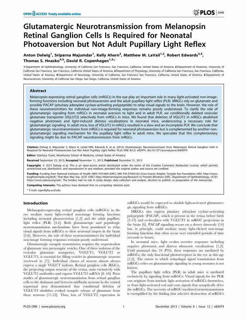
Glutamatergic Neurotransmission from Melanopsin Retinal Ganglion Cells Is Required for Neonatal Photoaversion but Not Adult Pupillary Light Reflex Anton Delwig 1 , Sriparna Majumdar 1 , Kelly Ahern 2 , Matthew M. LaVail 1,2 , Robert Edwards 3,4 , Thomas S. Hnasko 4,5 , David R. Copenhagen 1,3 * 1 Department of Ophthalmology, University of California San Francisco, San Francisco, California, United States of America, 2 Department of Anatomy, University of California San Francisco, San Francisco, California United States of America, 3 Department of Physiology, University of California San Francisco, San Francisco, California, United States of America, 4 Department of Neurology, University of California, San Francisco San Francisco, California, United States of America, 5 Department of Neurosciences, University of California San Diego, San Diego, California, United States of America Abstract Melanopsin-expressing retinal ganglion cells (mRGCs) in the eye play an important role in many light-activated non-image- forming functions including neonatal photoaversion and the adult pupillary light reflex (PLR). MRGCs rely on glutamate and possibly PACAP (pituitary adenylate cyclase-activating polypeptide) to relay visual signals to the brain. However, the role of these neurotransmitters for individual non-image-forming responses remains poorly understood. To clarify the role of glutamatergic signaling from mRGCs in neonatal aversion to light and in adult PLR, we conditionally deleted vesicular glutamate transporter (VGLUT2) selectively from mRGCs in mice. We found that deletion of VGLUT2 in mRGCs abolished negative phototaxis and light-induced distress vocalizations in neonatal mice, underscoring a necessary role for glutamatergic signaling. In adult mice, loss of VGLUT2 in mRGCs resulted in a slow and an incomplete PLR. We conclude that glutamatergic neurotransmission from mRGCs is required for neonatal photoaversion but is complemented by another non- glutamatergic signaling mechanism for the pupillary light reflex in adult mice. We speculate that this complementary signaling might be due to PACAP neurotransmission from mRGCs. Citation: Delwig A, Majumdar S, Ahern K, LaVail MM, Edwards R, et al. (2013) Glutamatergic Neurotransmission from Melanopsin Retinal Ganglion Cells Is Required for Neonatal Photoaversion but Not Adult Pupillary Light Reflex. PLoS ONE 8(12): e83974. doi:10.1371/journal.pone.0083974 Editor: Gianluca Tosini, Morehouse School of Medicine, United States of America Received September 23, 2013; Accepted November 11, 2013; Published December 31, 2013 Copyright: ß 2013 Delwig et al. This is an open-access article distributed under the terms of the Creative Commons Attribution License, which permits unrestricted use, distribution, and reproduction in any medium, provided the original author and source are credited. Funding: Funding from National Institutes of Health (NIH) EY01869 (DRC); NIH P30 EY002162 (Core Grant); Knights Templar Eye Foundation (AD), http://www. knightstemplar.org/ktef/; That Man May See, UCSF (DRC) http://thatmanmaysee.org/Research to Prevent Blindness (DRC, Department of Ophthalmology, UCSF) https://www.rpbusa.org/rpb/. The funders had no role in study design, data collection and analysis, decision to publish, or preparation of the manuscript. Competing Interests: The authors have declared that no competing interests exist. * E-mail: [email protected]Introduction Melanopsin-expressing retinal ganglion cells (mRGCs) in the eye mediate many light-evoked non-image forming functions including neonatal photoaversion [1,2] and the adult pupillary light reflex (PLR) [3,4]. Both glutamatergic and peptidergic neurotransmission mechanisms have been postulated to relay visual signals from mRGCs to their neuronal targets in the brain [5,6]. However, the role of these neurotransmitters for individual non-image forming responses remains poorly understood. Glutamatergic synaptic transmission requires the sequestration of glutamate into presynaptic vesicles. One of three isoforms of the vesicular glutamate transporter, VGLUT1, VGLUT2 or VGLUT3, is essential for filling vesicles in glutamatergic neurons (reviewed in [7]). Individual classes of neuron almost always express a single VGLUT isoform. Retinal ganglion cells (RGCs), the projecting output neurons of the retina, stain exclusively with VGLUT2 antibodies and express VGLUT2 mRNA [8–10]. Prior studies of glutamatergic neurotransmission from retinal ganglion cells to the thalamus and between midbrain neurons in the ventral tegmental area demonstrated that conditional deletion of VGLUT2 abolishes evoked synaptic release of glutamate from these neurons [11,12]. Thus, loss of VGLUT2 expression in mRGCs would be expected to abolish light-activated glutamater- gic signaling from mRGCs. MRGCs also express pituitary adenylate cyclase-activating polypeptide (PACAP), which is present in the retina before birth [5,13] and co-localizes with VGLUT2 in mRGC projections in the brain [6]. PACAP signaling occurs on a slower timescale [14] but, in principle, could mediate many light-elicited non-image forming functions that often occur over extended periods of time (seconds to hours). In neonatal mice, light evokes aversive responses including negative phototaxis and distress ultrasonic vocalizations [1,2]. Until postnatal day 10 (P10), these responses are mediated by mRGCs, the only functional photoreceptors in the eye at this age [1,2]. The extent to which retinofugal signal transmission from mRGCs relies on glutamatergic signaling in young neonates is not known. The pupillary light reflex (PLR) in adult mice is mediated exclusively by signaling from mRGCs. Visual signals for the PLR can originate from intrinsic light activation of mRGCs themselves, or from light-activated rod and cone signals that synaptically drive the mRGCs. The necessity of mRGC-mediated neurotransmission is exemplified by the finding that selective destruction of mRGCs PLOS ONE | www.plosone.org 1 December 2013 | Volume 8 | Issue 12 | e83974
Transcript
Glutamatergic Neurotransmission from MelanopsinRetinal Ganglion Cells Is Required for NeonatalPhotoaversion but Not Adult Pupillary Light ReflexAnton Delwig1, Sriparna Majumdar1, Kelly Ahern2, Matthew M. LaVail1,2, Robert Edwards3,4,
Thomas S. Hnasko4,5, David R. Copenhagen1,3*
1Department of Ophthalmology, University of California San Francisco, San Francisco, California, United States of America, 2Department of Anatomy, University of
California San Francisco, San Francisco, California United States of America, 3Department of Physiology, University of California San Francisco, San Francisco, California,
United States of America, 4Department of Neurology, University of California, San Francisco San Francisco, California, United States of America, 5Department of
Neurosciences, University of California San Diego, San Diego, California, United States of America
Abstract
Melanopsin-expressing retinal ganglion cells (mRGCs) in the eye play an important role in many light-activated non-image-forming functions including neonatal photoaversion and the adult pupillary light reflex (PLR). MRGCs rely on glutamate andpossibly PACAP (pituitary adenylate cyclase-activating polypeptide) to relay visual signals to the brain. However, the role ofthese neurotransmitters for individual non-image-forming responses remains poorly understood. To clarify the role ofglutamatergic signaling from mRGCs in neonatal aversion to light and in adult PLR, we conditionally deleted vesicularglutamate transporter (VGLUT2) selectively from mRGCs in mice. We found that deletion of VGLUT2 in mRGCs abolishednegative phototaxis and light-induced distress vocalizations in neonatal mice, underscoring a necessary role forglutamatergic signaling. In adult mice, loss of VGLUT2 in mRGCs resulted in a slow and an incomplete PLR. We conclude thatglutamatergic neurotransmission from mRGCs is required for neonatal photoaversion but is complemented by another non-glutamatergic signaling mechanism for the pupillary light reflex in adult mice. We speculate that this complementarysignaling might be due to PACAP neurotransmission from mRGCs.
Citation: Delwig A, Majumdar S, Ahern K, LaVail MM, Edwards R, et al. (2013) Glutamatergic Neurotransmission from Melanopsin Retinal Ganglion Cells IsRequired for Neonatal Photoaversion but Not Adult Pupillary Light Reflex. PLoS ONE 8(12): e83974. doi:10.1371/journal.pone.0083974
Editor: Gianluca Tosini, Morehouse School of Medicine, United States of America
Received September 23, 2013; Accepted November 11, 2013; Published December 31, 2013
Copyright: � 2013 Delwig et al. This is an open-access article distributed under the terms of the Creative Commons Attribution License, which permitsunrestricted use, distribution, and reproduction in any medium, provided the original author and source are credited.
Funding: Funding from National Institutes of Health (NIH) EY01869 (DRC); NIH P30 EY002162 (Core Grant); Knights Templar Eye Foundation (AD), http://www.knightstemplar.org/ktef/; That Man May See, UCSF (DRC) http://thatmanmaysee.org/Research to Prevent Blindness (DRC, Department of Ophthalmology, UCSF)https://www.rpbusa.org/rpb/. The funders had no role in study design, data collection and analysis, decision to publish, or preparation of the manuscript.
Competing Interests: The authors have declared that no competing interests exist.
quently, a recording trial began with a 1-min baseline in the dark,
a 1-min exposure to light, followed by an additional 1 min of
recording in the dark. Movement and vocal responses were
quantified as the amount of net movement (in kilo Pixels) and the
number of 62-kHz vocalizations during each 1-min interval.
Pupillary Light ResponsesAdult mice were dark adapted for at least 60 min. Under dim
red illumination, the dark-adapted, unanesthetized mice were
placed in a rodent restrainer device and positioned in front of an
infrared video camera. A blue (480 nm) light stimulus was
delivered to the right eye and pupillary constriction in the left
eye was monitored under infrared light. The infrared images were
recorded onto a laptop computer using BTV Pro (Ben Software,
http://www.bensoftware.com) at 30 frames per second. The
videos were analyzed in NIH Image. The pupil diameter was
manually measured once per two seconds (synchronized to the
onset of light). A trial consisted of 1 min in the dark, 1 min in the
light followed by 1 min in the dark. The data was plotted in
Matlab and GraphPad Prism. The age of mice ranged from P44 to
P266. The age had no discernible effect on the size of the PLR.
To confirm the ability of cKO mice to constrict their pupils,
5 ul of Pilocarpine HCl (1%; Bausch & Lomb) was applied to each
eye. After a few minutes, excess fluid was wiped with a paper
tissue. Recordings of pupil diameter were made in the darkness
20 min after the application of Pilocarpine. It should be noted that
Pilocarpine’s effect has a slow onset and is short in duration. After
40 min, pupils started to re-dilate.
Optokinetic ResponsesA virtual Optokinetic System was used to test visual acuity [20].
The apparatus consisted of four computer monitors around a
Glutamatergic Signaling from mRGCs
PLOS ONE | www.plosone.org 2 December 2013 | Volume 8 | Issue 12 | e83974
Glutamatergic Signaling from mRGCs
PLOS ONE | www.plosone.org 3 December 2013 | Volume 8 | Issue 12 | e83974
square testing arena (OptoMotry; Cerebral Mechanics). A sine
wave grating was drawn on a virtual cylinder projected in three-
dimensional coordinate space on the monitors, and the cylinder
was rotated at a constant speed (12u/s). The testing procedure
consisted of placing an unrestrained mouse onto a platform in the
center of the arena. A video camera provided real-time video
feedback from above, and the position of the head on each frame
was used to center the hub of the cylinder continually at the
mouse’s viewing position. On each trial, an experimenter judged
whether the mouse made tracking movements with its head and
neck to follow the drifting grating. The spatial frequency
threshold, the point at which animals no longer tracked, was
obtained by incrementally increasing the spatial frequency of the
grating at 100% contrast. Thresholds for each eye were measured
separately by reversing the rotation of the cylinder. Mice ranged in
age from P32 to P97.
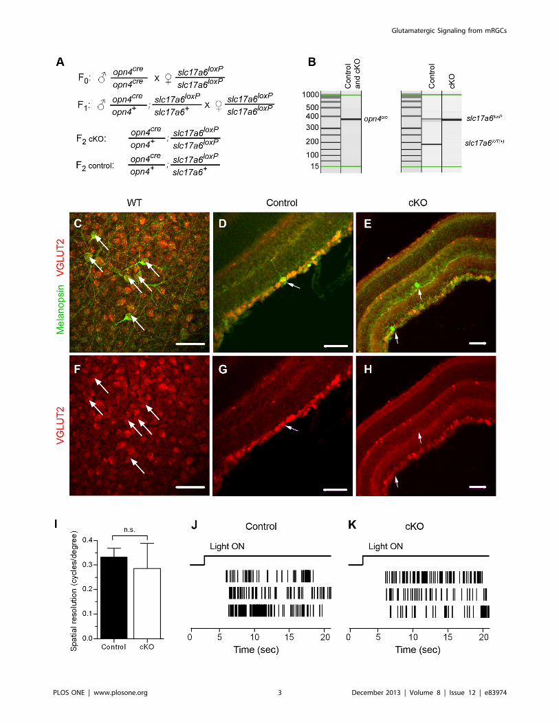
Figure 1. Conditional knockout of VGLUT2 from mRGCs. (A) Breeding scheme for obtaining conditional VGLUT2 knockouts (cKO) andcontrols. (B) Mice were genotyped by PCR with allele-specific primers. (C–H) VGLUT2 immunoreactivity is detected in melanopsin-immunoreactiveretinal ganglion cells in wild-type (WT) (C, F) and control mice (D, G) but not cKO mice (E, H). (C, F) flatmount retina; (D, E, G, H) retinal slices. Arrowspoint to retinal ganglion cells that are immunopositive for melanopsin. Scale bar: 50 um. (I) Mean optokinetic response, a measure of image-formingvision, was not different between control (n = 8 eyes) and cKO (n= 7 eyes) mice (P=0.4, Student’s t-test). (J, K) Typical action potential responses ofneonatal (P8) mRGCs to light in control (J) and cKO (J) retinas. These electrophysiological findings demonstrate that mRGCs in VGLUT2 cKO pupsretained their light responses.doi:10.1371/journal.pone.0083974.g001
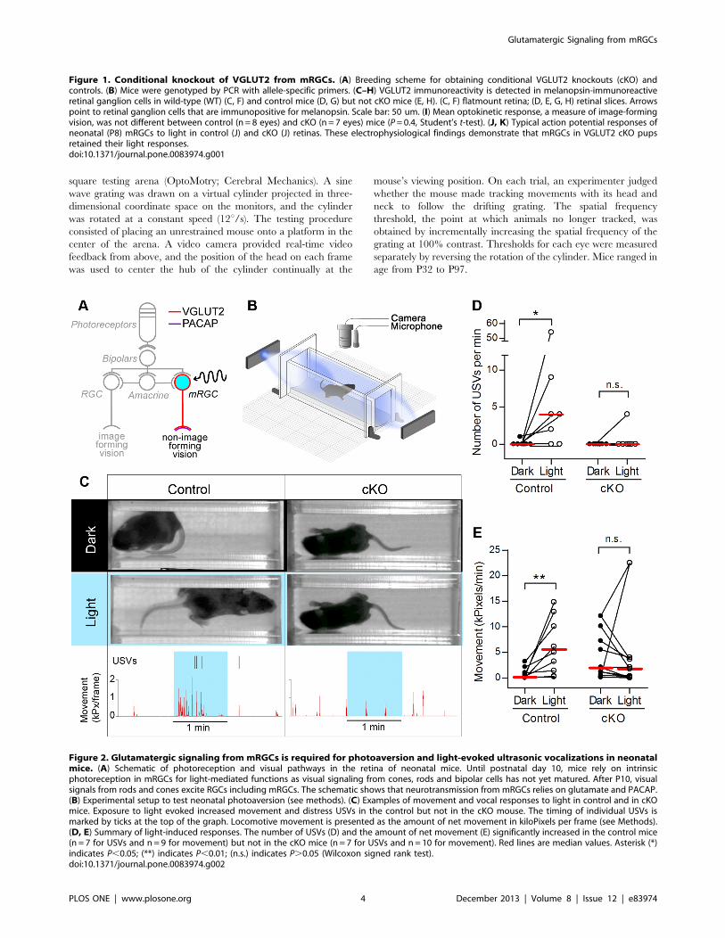
Figure 2. Glutamatergic signaling from mRGCs is required for photoaversion and light-evoked ultrasonic vocalizations in neonatalmice. (A) Schematic of photoreception and visual pathways in the retina of neonatal mice. Until postnatal day 10, mice rely on intrinsicphotoreception in mRGCs for light-mediated functions as visual signaling from cones, rods and bipolar cells has not yet matured. After P10, visualsignals from rods and cones excite RGCs including mRGCs. The schematic shows that neurotransmission from mRGCs relies on glutamate and PACAP.(B) Experimental setup to test neonatal photoaversion (see methods). (C) Examples of movement and vocal responses to light in control and in cKOmice. Exposure to light evoked increased movement and distress USVs in the control but not in the cKO mouse. The timing of individual USVs ismarked by ticks at the top of the graph. Locomotive movement is presented as the amount of net movement in kiloPixels per frame (see Methods).(D, E) Summary of light-induced responses. The number of USVs (D) and the amount of net movement (E) significantly increased in the control mice(n = 7 for USVs and n= 9 for movement) but not in the cKO mice (n = 7 for USVs and n=10 for movement). Red lines are median values. Asterisk (*)indicates P,0.05; (**) indicates P,0.01; (n.s.) indicates P.0.05 (Wilcoxon signed rank test).doi:10.1371/journal.pone.0083974.g002
Glutamatergic Signaling from mRGCs
PLOS ONE | www.plosone.org 4 December 2013 | Volume 8 | Issue 12 | e83974
ImmunohistochemistryAnimals were euthanized in a CO2 chamber. The enucleated
whole eyes were fixed in 4% PFA for 1 hour. To obtain retinal
slices, the eyes were sucrose protected, embedded in OCT, frozen
at 220uC and cut at 40 microns using a cryostat. Cut slices were
transferred onto glass slides. Wholemount retinas and retinal slices
were blocked in PBS with 10% normal goat serum (NGS), 10%
normal donkey serum (NDS) and 1% TX-100 for 1 hour at room
temperature. Subsequent primary and secondary stainings were
done in PBS with 1% NGS, 1% NDS and 0.1% TX-100. We used
the following primary antibodies: rabbit anti-melanopsin (UF006,
AB_1608077) at 1:1000 and guinea pig anti VGLUT2 (AB2251,
Millipore, http://antibodyregistry.org/AB_1587626) at 1:1000
dilution (overnight at +4uC). Donkey anti-rabbit Alexa488 at
1:1000 and goat anti-guinea pig Cy3 at 1:500 were used as
secondary antibodies. Images were acquired on a Pascal confocal
microscope (Zeiss) and subsequently processed in Photoshop
(Adobe). Mice ranged in age from P90 to P289. It should be
noted that VGLUTs are localized primarily in the axonal
terminals of glutamatergic neurons. It is therefore not unexpected
that VGLUT2 immunoreactivity was less than optimum in the
somas of mRGCs. However, in wild-type mice, which have two
copies of VGLUT2, VGLUT2 immunoreactivity is detectable in
the somas of mRGCs in the whole mount retina. However, in our
control mice, which have only one copy of VGLUT2 allele, the
VGLUT2 immunoreactivity is very poor. Therefore, we failed to
quantify the loss of VGLUT2 in somas of mRGCs in cKO mice as
compared to control mice. We further attempted to quantify the
loss of VGLUT2 in mRGCs by analyzing VGLUT2 immunore-
activity in their axonal terminals in the SCN (suprachiasmatic
nucleus), the major central target of mRGCs. Even though there
was a statistically significant decrease of overall VGLUT2 signal in
cKO mice (p = 0.006) the effect size was small (11% decrease)
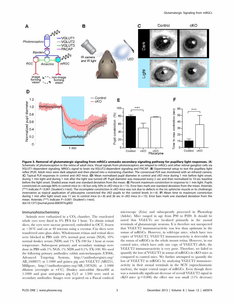
Figure 3. Removal of glutamatergic signaling from mRGCs unmasks secondary signaling pathway for pupillary light responses. (A)Schematic of photoreception in the retina of adult mice. Visual signals from photoreceptors are relayed to mRGCs and other retinal ganglion cells viaVGLUT1-dependent signaling. MRGCs signal to brain via VGLUT2-dependent signaling and PACAP. (B) Experimental setup to test the pupillary lightreflex (PLR). Adult mice were dark adapted and then placed into a restraining chamber. The consensual PLR was monitored with an infrared camera.(C) Typical PLR responses in control and cKO mice. (D) Mean normalized pupil diameter in control and cKO mice during 1 min before light onset,during 1 min light and during 1 min after the light was turned off. Pupil diameter was measured every 2 sec and then normalized to 10-sec baselinebefore the light onset. Shaded areas mark one standard deviation from the mean. (E) Percent maximum constriction in response to 1 min light. Pupilsconstricted on average 80% in control mice (n = 8) but only 50% in cKO mice (n = 15). Error bars mark one standard deviation from the mean. Asterisks(***) indicate P,0.001 (Student’s t-test). The incomplete constriction in cKO mice was not due to defects in the iris sphincter muscle or its cholinergicinnervation as topical application of pilocarpine constricted the cKO pupils to the control levels (n = 4). (F) Mean time to maximum constrictionduring 1 min after light onset was 11 sec in control mice (n = 8) and 36 sec in cKO mice (n = 15). Error bars mark one standard deviation from themean. Asterisks (***) indicate P,0.001 (Student’s t-test).doi:10.1371/journal.pone.0083974.g003
Glutamatergic Signaling from mRGCs
PLOS ONE | www.plosone.org 5 December 2013 | Volume 8 | Issue 12 | e83974
probably due to other, non retinal, glutamatergic VGLUT2-
expressing projections to the SCN. Given that the VGLUT2 loss
in cKO mice was sufficient to essentially abolish mRGC-
dependent neonatal responses to light, we think the degree of
VGLUT2 loss was significant. However, we cannot quantify the
extent of its loss in our mice.
Multielectrode Array Extracellular Recording from RetinaMultielectrode array (MEA-60, MCS) recordings of light-
evoked ganglion cell spiking were acquired and analyzed as
described previously [21,22]. Briefly, the MEA chambers consisted
of an array of 60 planar electrodes, each 10 mm in diameter, in
eight rows and spaced 100 mm apart for a total array size of 700
mm2. Acquired voltage signals were bandpass filtered at 0.1 Hz to
3 kHz and sampled at 50 kHz (MC_Rack, version 2.0; Multi-
Channel Systems). Offline, action potential waveforms from high-
pass filtered data (100 Hz lower cutoff) were detected by threshold
crossing. To isolate individual RGCs, action potential waveforms
were then clustered based on the first two principal components, as
described previously [22]. Cluster contours in principal compo-
nents space were either manually selected or derived from a k-
means algorithm (OfflineSorter, version 3.1; Plexon). The
algorithm eliminated outlier waveforms at a threshold of 1.3 times
the mean distance from the calculated cluster center. Light stimuli
were presented from a monitor (Dell Ultrascan P780; 100 Hz
vertical refresh) imaged onto the retinal surface at an approximate
intensity of 0.35 W/cm2. Mice ranged in age from P7 to P9.
Statistical AnalysesTests of statistical significance were determined by Student’s t-
test or Wilcoxon signed rank test (GraphPad Prism), with the
criteria of significance set at P,0.05.
Results
Selective Removal of Glutamate from Synaptic Vesicles inthe Melanopsin-expressing Cells
To selectively prevent glutamate sequestration into the synaptic
vesicles in the axons of melanopsin-expressing retinal ganglion
cells (mRGCs), we crossed mice with conditional VGLUT2 allele
(slc17a6loxP/slc17a6loxP [12]) to mice with cre recombinase in place
of the melanopsin gene (opn4cre/opn4cre [17]). Previous studies using
this same recombination approach demonstrated electrophysio-
logically its efficacy in eliminating synaptic transmission from
VGLUT2-expressing neurons [11,12]. Figure 1A describes the
breeding scheme we utilized for these present studies. For all
experiments, we compared control mice that possess one copy of
opn4cre gene and one copy of the floxed slc17a6 gene (opn4cre/+;
slc17a6loxP/+) with littermate conditional knockouts (cKO) that
possess one copy of opn4cre gene and two copies of the floxed
slc17a6 gene (opn4cre/+; slc17a6loxP/slc17a6loxP). Mice were geno-
typed by PCR with allele-specific primers [12,17] (Figure 1B). To
validate the removal of VGLUT2 from mRGCs, we analyzed
immunostained retinas. VGLUT2 co-localizes with melanopsin in
wild type (Figure 1C,F) and control (Figure 1D,G) but not in cKO
retinas (Figure 1E,H). Deletion of VGLUT2 in mRGCs did not
have a noticeable effect on the level of VGLUT2 expression in
other RGCs.
The cKO mice retained many normal visual functions. Loss of
glutamatergic signaling from mRGCs did not affect their intrinsic
photosensitivity as detected by the multielectrode array recordings
done at the same age range when photoaversive responses were
examined (P7 to P9; Figure 1J,K). Visual signaling from other
retinal ganglion cells was not discernibly affected as optokinetic
responses in control (n = 8 eyes in 4 mice) and cKO mice (n = 7
eyes in 4 mice) were not statistically different (P= 0.4) (Figure 1I).
There was no detectable difference in the average weight between
control and cKO mice (P= 0.25). Two out of sixteen cKO mice
had ataxia with a persistent head tilt. Veterinary inspection
revealed no ear infection. The rest of cKO mice and all control
mice (n = 15) had no noticeable anatomical or neurological
abnormalities. The two cKO mice with ataxia were included in
all tests except optokinetic responses.
Glutamatergic Signaling from mRGCs is Required forPhotoaversion and Light-evoked Ultrasonic Vocalizationsin Neonatal Mice
We previously showed that until P9, neonatal mice rely on
intrinsic photosensitivity of mRGCs to turn away from light
(negative phototaxis [1]) and to vocalize in response to light (light-
induced ultrasonic vocalizations [2]). However, it is not known if
these neonatal responses to light depend on glutamatergic or
peptidergic transmission from mRGCs. MRGCs are known to
express both VGLUT2 and PACAP [6] (Figure 2A). We therefore
asked if removal of glutamatergic transmission affects these
responses. Using the previously described testing setup [1,2]
(Figure 2B), we tested movement and vocal responses of P6–P9
control and cKO pups to light. Individual pups were dark adapted
and transferred under dim red light into the testing chamber and
allowed to acclimate in the dark until isolation-induced calls and
locomotor activity significantly diminished. We quantified light-
evoked behavioral responses by recording movement and 62-kHz
ultrasound vocalizations (USVs) during one min in the dark
followed by one min in the light (Figure 2C and Movie S1). During
the 1-min exposure to light, control mice showed a significant
increase in movement (n = 9) and the number of USVs (n = 7,
Figure 2C–E). In contrast, cKO mice did not exhibit an increase
in movement (n = 10) or the number of USVs (n = 7, Figure 2C–E,
Movie S2). These results demonstrate that VGLUT2 in mRGCs is
required for neonatal expression of negative phototaxis and light-
induced USVs.
Removal of Glutamatergic Signaling from mRGCsUnmasks Secondary Signaling Pathway for Pupillary LightResponses
In adult retina, light is absorbed by mRGCs and by
photoreceptors that signal via bipolar cells to both mRGCs and
conventional retinal ganglion cells (Figure 3A). To test the role of
VGLUT2-dependent glutamatergic transmission from mRGCs on
the pupillary light reflex (PLR), we measured light-induced
changes in the pupil diameter. Individual adult mice were placed
into a restraining chamber in darkness and allowed to acclimate
for 4–10 min. A bright blue LED directed at the right eye was
then turned on (power flux 35 mW/sq cm) for one min.
Consensual pupillary constriction in the infrared-illuminated left
eye was recorded with an infrared camera (Figure 3B and Movie
S3). Typical responses are illustrated in Figure 3C. A summary of
normalized responses is illustrated in Figure 3D. One min light
stimulation produced an average 80% constriction in controls
(n = 8), but only 50% constriction in cKO mice (n = 15) (Figure 3E
and Movie S4). The incomplete pupil constriction in cKO mice
was not due to the defect in the iris sphincter muscle or its
cholinergic innervation as the application of 1% Pilocarpine, a
muscarinic agonist, constricted the pupil to the control levels
(Figure 3E). Additionally, PLRs were noticeably slower in cKO
than in control mice (40 sec vs. 10 sec) (Figure 3F). This slower
and incomplete response in cKO mice is due to some other
Glutamatergic Signaling from mRGCs
PLOS ONE | www.plosone.org 6 December 2013 | Volume 8 | Issue 12 | e83974
secondary neurotransmission from mRGCs, which we speculate
may be PACAP (see Discussion).
We observed large variability of pupil sizes in cKO mice at
baseline in darkness. However, the mean pupil size in the control
and cKO mice were not statistically different (p = 0.07). We tried
various pharmacological manipulations of sympathetic and
parasympathetic innervation of the iris to determine the nature
of this variability but failed to determine its origin. These observed
variations of pupil size might be due to developmental compen-
sation at the level of innervation of the iris or central mechanisms
mediating pupil control.
Discussion
The role of glutamatergic signaling from melanopsin-expressing
retinal ganglion cells (mRGCs) remains largely unexplored. In this
study, we have shown that in neonatal mice, loss of VGLUT2-
dependent glutamatergic signaling from mRGCs abolishes nega-
tive phototaxis and light-induced distress vocalizations. We have
also shown that in adult mice, loss of VGLUT2-dependent
glutamatergic signaling from mRGCs reveals a slow and an
incomplete PLR. We conclude that glutamatergic neurotransmis-
sion from mRGCs is required for neonatal photoaversion but is
complemented by another non-glutamatergic signaling mecha-
nism for the pupillary light reflex in adult mice.
In neonatal mice, visual signaling from photoreceptors (rods and
cones) to bipolar cells and from bipolar cells to retinal ganglion
cells (RGC) does not emerge until P10 [1]. Until P10, these young
mice rely on the melanopsin photopigment for responses to light.
We previously showed that removal of melanopsin abolishes
locomotor and vocalization responses to light in neonates [1,2]. In
this present study, we show that conditional removal of VGLUT2
from mRGCs leads to the same loss of responses. These results
indicate that VGLUT2-dependent glutamatergic signaling from
mRGCs is responsible for both negative phototaxis and light-
evoked distress vocalizations. Our results also indicate that other
neurotransmitters including PACAP, which is co-expressed in
mRGCs before birth [23,5], are not capable of producing acute
behavioral responses to light in neonates.
In adults, loss of VGLUT2 expression in mRGCs reveals a
much slower and incomplete PLR, which we suggest is due to
PACAP neurotransmission from mRGCs. Previous studies have
established that mRGCs are the sole conduits of the light signal for
the PLR. Selective destruction of mRGCs leads to complete loss of
PLRs [4]. Therefore, the residual response in our conditional
VGLUT2 knockout mice is due to some other, VGLUT2-
independent signaling from mRGCs. VGLUT1 knockout mice,
which lack signaling from photoreceptors and bipolar cells and
thus only have mRGC-driven responses, have normal PLRs [3].
Previous studies have failed to identify the expression of other
vesicular glutamate transporters in RGCs [8–10]. Therefore, this
incomplete slow PLR response, unmasked by removal of
glutamatergic signaling, is due to complementary non-glutama-
tergic signaling from mRGCs. We suggest that this complemen-
tary signaling is carried out by PACAP. PACAP is present in
mRGCs that project to the olivary pretectal nucleus [24] and is
capable of signaling on the time scale of seconds [14]. Glutamate
appears to be the major signaling molecule for the PLR as the
removal of PACAP does not have noticeable effects [15].
However, in the absence of glutamatergic signaling studied here,
PACAP is capable of relaying light signal for the PLR, albeit
incomplete and at a much slower timescale. To fully elucidate the
role of PACAP, PLR responses need to be analyzed in VGLUT2
cKO mice with PACAP null allele.
Based solely on the PLR studies we cannot rule out the
possibility that the detected PLR in cKO mice is due to incomplete
penetrance of cre-dependent excision of VGLUT2 [25]. However,
essentially complete abolishment of neonatal responses to light in
the cKO mice, which mimics melanopsin-null phenotype, argues
against it. We also failed to detect any fast component of PLR in
cKO mice (Figure 3D), which we interpret as a lack of
glutamatergic signaling from mRGCs.
Our results indicate that mRGCs do not rely on the single
neurotransmitter for initiating all the behavioral and physiological
responses to light. Scherrer et al. [26] demonstrated that
and injury-induced heat hypersensitivity while peptidergic signal-
ing might have mediated other nociceptive functions carried by
the same afferents. Different brain targets of mRGCs might have
variable degree of dependence on glutamatergic or peptidergic
signaling. For neonatal photoaversive responses, mRGCs are
known to activate central amygdala (CeA) and posterior thalamic
region (Po), possibly via direct projections to these brain areas [2],
whereas for adult PLR, mRGCs relay the light signal via olivary
pretectal nucleus [24]. It is possible that for signaling to CeA and
Po, mRGCs rely exclusively on glutamate whereas for signaling to
olivary pretectal nucleus, mRGCs use both glutamate and
PACAP.
Supporting Information
Movie S1 Video and audio recording of negativephototaxis and light-induced 62-kHz USVs in a controlmouse pup. Ultrasonic calls are detected at 62-kHz and shifted
to audible frequency ranges by heterodyne circuitry. Video
recordings were done with an infrared camera. The first min of
recording shows the pup in darkness. The second min is during
light stimulation. The third segment shows the next min in
darkness.
(MOV)
Movie S2 Video and audio recording of negativephototaxis and light-induced 62-kHz USVs in a cKOmouse pup. Ultrasonic calls are detected at 62 kHz and shifted
to audible frequency ranges by heterodyne circuitry. Video
recordings were done with an infrared camera. The first min of
recording shows the pup in darkness. The second min is during
light stimulation. The third segment shows the next min in
darkness.
(MOV)
Movie S3 Video recording of pupillary light reflex in acontrol adult mouse. The first min of recording shows the
mouse in darkness. The second min is during light stimulation.
The third segment shows the next min in darkness.
(MOV)
Movie S4 Video recording of pupillary light reflex in acKO adult mouse. The first min of recording shows the mouse
in darkness. The second min is during light stimulation. The third
segment shows the next min in darkness.
(MOV)
Acknowledgments
The authors thank Derek Bredl for technical assistance, Suling Wang for
help with the illustrations, Howard Fields for a critical reading of the
manuscript, and Samar Hattar and Cliff Saper for the gift of opn-cre mice.
Glutamatergic Signaling from mRGCs
PLOS ONE | www.plosone.org 7 December 2013 | Volume 8 | Issue 12 | e83974
Author Contributions
Conceived and designed the experiments: AD DRC. Performed the
experiments: AD SM KA. Analyzed the data: AD KA SM DRC.
Contributed reagents/materials/analysis tools: AD SM KA MML TSH
RE DRC. Wrote the paper: AD DRC. Designed software used in analysis:
AD. Obtained mouse lines: DRC. Fabricated light stimulators: AD SM
DRC.
References
1. Johnson J, Wu V, Donovan M, Majumdar S, Renterı́a RC, et al. (2010)Melanopsin-dependent light avoidance in neonatal mice. Proc Natl Acad
Sci U S A 107: 17374–17378. doi:10.1073/pnas.1008533107.2. Delwig A, Logan AM, Copenhagen DR, Ahn AH (2012) Light Evokes
Melanopsin-Dependent Vocalization and Neural Activation Associated with
Aversive Experience in Neonatal Mice. PloS One 7: e43787. doi:10.1371/journal.pone.0043787.
3. Johnson J, Fremeau RT Jr, Duncan JL, Renterı́a RC, Yang H, et al. (2007)Vesicular glutamate transporter 1 is required for photoreceptor synaptic
signaling but not for intrinsic visual functions. J Neurosci Off J Soc Neurosci
27: 7245–7255. doi:10.1523/JNEUROSCI.0815-07.2007.4. Hatori M, Le H, Vollmers C, Keding SR, Tanaka N, et al. (2008) Inducible
ablation of melanopsin-expressing retinal ganglion cells reveals their central rolein non-image forming visual responses. PLoS One 3: e2451. doi:10.1371/
journal.pone.0002451.5. Fahrenkrug J, Nielsen HS, Hannibal J (2004) Expression of melanopsin during
development of the rat retina. Neuroreport 15: 781–784.
6. Engelund A, Fahrenkrug J, Harrison A, Hannibal J (2010) Vesicular glutamatetransporter 2 (VGLUT2) is co-stored with PACAP in projections from the rat
melanopsin-containing retinal ganglion cells. Cell Tissue Res 340: 243–255. Doi:10.1007/s00441-010-0950-3.
subsets of excitatory neurons and suggest novel roles for glutamate. TrendsNeurosci 27: 98–103. doi:10.1016/j.tins.2003.11.005.
8. Johnson J, Sherry DM, Liu X, Fremeau RT Jr, Seal RP, et al. (2004) Vesicularglutamate transporter 3 expression identifies glutamatergic amacrine cells in the