Evaluation of lenticular antioxidant and redox system componentsin the lenses of acetyl-L-carnitine treatment in BSO-inducedglutathione deprivation
R. Elanchezhian,1 M. Sakthivel,1 M. Isai,1 P. Geraldine,1 P.A. Thomas2
1Department of Animal Science, School of Life Sciences, Bharathidasan University, Tiruchirappalli, Tamil Nadu, India; 2Instituteof Ophthalmology, Joseph Eye Hospital, Tiruchirappalli, Tamil Nadu, India
Purpose: To investigate whether acetyl-L-carnitine (ALCAR) retards L-buthionine-(S,R)-sulfoximine (BSO)-inducedcataractogenesis in Wistar rat pups.Methods: On postpartum day 3, group I pups received intraperitoneal (ip) saline and group II and group III pups receivedi.p. injections of BSO once daily for three consecutive days. In addition, group III pups received ip ALCAR once dailyfrom postpartum days 3–15. Both eyes of each pup were examined up from postpartum day 16 to day 30. After sacrifice,extricated pup lenses were analyzed for antioxidant and redox system components.Results: There was dense lenticular opacification in all group II pups, minimal opacification in 40% of group III pups,and no opacification in 60% of group III pups and in all of group I pups. Group II lenses exhibited significantly lowervalues of antioxidant and redox system components and higher malondialdehyde concentrations than in group I or groupIII lenses.Conclusions: ALCAR prevents cataractogenesis in the BSO-induced cataract model, possibly by inhibiting depletingantioxidant enzyme and redox system components and inhibiting lipid peroxidation.
Oxidative stress is a common initiator of many age-related conditions and is probably the most importantmechanism in age-related cataractogenesis. Aging of the lensis associated with progressive changes in the physical andchemical properties of its structural proteins, the crystallins.Oxidative stress-induced changes include crystallin cross-linking, aggregation, loss of solubility, conformationalalterations, fragmentation, and enzyme inactivation. Highlyreactive species such as hydrogen peroxide (H2O2), singletoxygen, superoxide radicals, and hydroxyl radicals can begenerated in the eye through photochemical pathways [1] orFenton-type reactions [2]. The lens possesses severalprotective mechanisms to prevent or limit oxidative damage.Normal young lenses maintain optimal activity of antioxidantenzymes and high concentrations of ascorbate and glutathioneand hence minimize the alterations wrought by excessiveoxidation. If this balance of pro- and antioxidants is disturbed,aging occurs. Under the pathological condition, the oxidationof lenticular proteins may lead to senile cataract [3].
Glutathione is a major constituent of mammalian lensesand is mainly concentrated in the epithelium. Glutathionedecreases during the formation of most cataracts [4]. Reducedglutathione (GSH) is present in a high concentration in thelens [5,6]. GSH serves as an intracellularly-produced
Correspondence to: Dr. Pitchairaj Geraldine, Ph.D., BharathidasanUniversity, Animal Science, School of Life Sciences, Tiruchirrapalli,Tamil Nadu, 620024, India; Phone: +91-431-2407040; FAX:+91-431-2414969; email: [email protected]
antioxidant, which resists oxidative damage to cellularorganelles by recycling other antioxidants, scavenging freeradicals, and using H2O2 and hydroperoxides where itundergoes oxidation by glutathione peroxidase [7,8]. It alsopromotes the antioxidant properties of vitamin C and vitaminE by maintaining these nutrients in a reduced state [9]. Thesecond line of defense for the health of the lens is its contentof intrinsic repair enzymes that constantly dethiolate protein-thiol mixed disulfides (protein thiolation) or protein–proteindisulfides, which have been induced by oxidative stress. Thisprocess allows lenticular proteins to maintain their free thiolsagain, thus restoring lenticular proteins as well as the functionand activities of enzymes [10]. The role of GSH as anendogenous lenticular antioxidant results in the reduction oflenticular hydrogen peroxide [11] and dehydroascorbate[12].
L-buthionine-(S,R)-sulfoximine (BSO), an inhibitor ofGSH biosynthesis, can induce age-dependent cataracts in pre-weaning mice [13] and in early postnatal rats [14] and is thusa potential model for obtaining new information about the roleof GSH in maintaining transparency of the lens. In thepresence of reduced levels of GSH, newborn rats sufferextensive damage to the cytosolic proteins and membranelipids, leading to clouding of the lens [15-17]. BSO-inducedcataracts have been prevented or reduced in frequency in vivoby esters of GSH [12,14] and by lipoic acid [18] and ascorbate[12].
Acetyl-L-carnitine (ALCAR), a quaternary amine, is anaturally-occurring, short-chain derivative of L-carnitine,
Molecular Vision 2009; 15:1485-1491 <http://www.molvis.org/molvis/v15/a159>Received 12 May 2009 | Accepted 28 July 2009 | Published 31 July 2009
which is synthesized endogenously in the human brain, liver,and kidneys by the acetyl carnitine transferase enzyme orobtained from dietary sources [19]. ALCAR facilitates theuptake of acetyl-CoA into the mitochondria during fatty acidoxidation, enhances acetylcholine production, and stimulatessynthesis of protein and membrane phospholipids. It alsocounteracts oxidative stress by inhibiting increases in lipidhydroperoxidation [20]. ALCAR has been reported to preventselenite-induced cataractogenesis in a Wistar rat model bothin vitro and in vivo by maintaining lenticular antioxidant andredox system components [21,22] and lenticular calpainactivity [23] at near normal levels. In this study, an attempthas been made to determine the putative anticataractogeniceffect of ALCAR by preventing the depletion of glutathionein the BSO-induced cataract model. Certain key biochemicalparameters of antioxidant and redox system components andof lipid peroxidation have also been evaluated.
METHODSExperimental animal: Two-day-old rat pups (Wistar strain)were used in this study. The pups were housed with parentsin large spacious cages, and the parents were given food andwater ad libitum. The animal room was well ventilated, and aregular 12 h light and 12 h dark cycle was maintainedthroughout the experimental period. These animals were usedin accordance with institutional guidelines and with theAssociation for Research in Vision and OphthalmologyStatement for the Use of Animals in Research. The rat pupswere divided into three groups, each group comprising pupsfrom the same litter: Group I, which received only saline(control); Group II, which received BSO (cataract-untreated);Group III, which received BSO and ALCAR (cataract-treated).
Each rat pup in groups II and III received anintraperitoneal (ip) injection of BSO once daily for threeconsecutive days starting from postpartum day 2. In addition,pups in group III received ip injections of ALCAR (200 mg/kg bodyweight), which was administered half an hour beforethe BSO injection once a day until the pups opened their eyes.Morphological examination: When the rat pups first openedtheir eyes, a slit-lamp biomicroscopic examination wasperformed on each eye to detect opacification. Prior toperforming the examination, mydriasis was achieved by atopical ophthalmic solution, which was instilled every 30 minfor 2 h with the animals being kept in a dark room. After 2 h,the eyes were viewed by a slit-lamp biomicroscope (CarlZeiss, Jena, Germany) at 12X magnification. At the end of theexperimental period (postpartum day 30), each eye wasphotographed, and the degree of opacification was graded asfollows: 0=normal transparent lens; +=initial sign of nuclearopacity involving tiny scatters; ++=partial nuclear opacity;and +++=dense nuclear opacity.Biochemical evaluation of redox system components andantioxidant enzymes: Rat pups in all three groups were
anesthetized and then sacrificed by cervical dislocation onpostpartum day 30. The lenses were then excised. Both lensesof each individual rat were processed together to constitute asingle value. The lenses were homogenized in 50 mMphosphate buffer (pH 7.2; 1 ml/ 100 mg tissue) and centrifugedat 14,006x g for 15 min at 4 °C. The supernatant obtained wasused for the analysis of enzymatic and non-enzymaticparameters. To calculate the specific enzyme activity, proteinin each sample was estimated by the method of Bradford[24].Reduced glutathione: The GSH content was estimated by themethod of Moron et al. [25]. The lens homogenate wascentrifuged at 2,432x g for 15 min at 4 °C. To the resultingsupernatant, 0.5 ml of 10% trichloroacetic acid was added,and the mix was recentrifuged. The resulting protein-freesupernatant was allowed to react with 4 ml of 0.3 MNa2HPO4 (pH 8.0) and 0.5 ml of 0.04% (wt/vol) 5,5-dithiobis-2-nitrobenzoic acid. The absorbance of the resultingyellow color was read spectrophotometrically at 412 nm. Aparallel standard was also maintained. The results wereexpressed in μmoles/g wet weight.Glutathione reductase: This enzyme, which utilizesnicotinamide adenine dinucleotide phosphate (NADPH) toconvert oxidized glutathione to the reduced form, was assayedby the method of Stall et al. [26]. The change in absorbancewas read at 340 nm for 2 min at intervals of 30 s in anultraviolet (UV) spectrophotometer (Analytik Jena AG, Jena,Germany). The activity of glutathione reductase (GR) wasexpressed as nmoles of NADPH oxidized/min/mg protein.Glutathione S-transferase: The activity of glutathione S-transferase (GST) was determined by the method of Habig andJacoby [27]. The conjugation of GSH with 1-chloro-2,4-dinitrobenzene (CDNB), a hydrophilic substrate, wasobserved spectrophotometrically at 340 nm to measure theactivity of GST. One unit of GST was defined as the amountof enzyme required to conjugate 1 μmol of CDNB with GSHper min.Glutathione peroxidase: The activity of glutathioneperoxidase (GPx) was determined essentially as described byRotruck et al. [28]. The principle of this method is that the rateof glutathione oxidation by H2O2 as catalyzed by the GPxpresent in the supernatant is determined. The color thatdevelops is read against a reagent blank at 412 nm on aspectrophotometer. In the test, the enzyme activity wasexpressed as units/mg protein (one unit was the amount ofenzyme that converted 1 µmole of GSH to the oxidized formof glutathione [GSSH] in the presence of H2O2 per min).Catalase: Catalase (CAT) activity was determined by themethod of Sinha [29]. In this test, dichromatic acetic acid isreduced to chromic acetate when heated in the presence ofH2O2 with the formation of perchloric acid as an unstableintermediate. In the test, the green color developed was readat 590 nm against a blank on a spectrophotometer. The activity
of catalase was expressed as units/mg protein (one unit wasthe amount of enzyme that used 1 mmole of H2O2 per min).
Superoxide dismutase: Superoxide dismutase (SOD) activitywas determined by the method of Marklund and Marklund[30]. In this test, the degree of inhibition of pyrogallol auto-oxidation by the supernatant of the lens homogenate wasmeasured. The change in absorbance was read at 470 nmagainst the blank each min for 3 min on a spectrophotometer.The enzyme activity was expressed as units per mg protein.Determination of lipid peroxidation: The extent of lipidperoxidation was determined by the method of Ohkawa et al.[31]. The principle of this method is that malondialdehyde
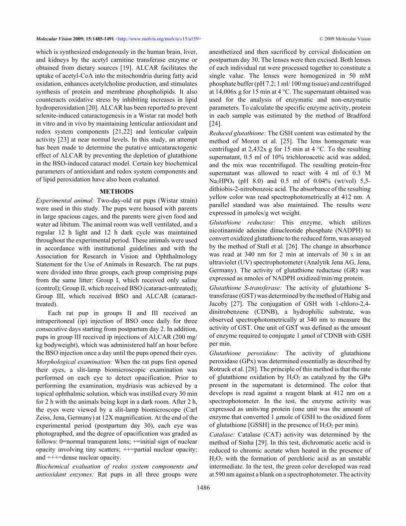
Figure 1. Slit-lamp appearance of the eye of a 30-day-old Wistar ratpup in group II. The eye exhibited dense opacification of the lens(Grade +++ opacification).
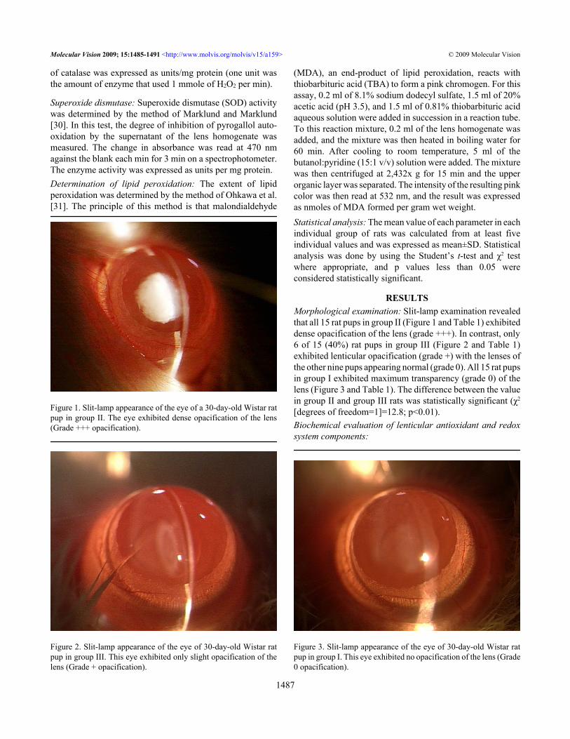
Figure 2. Slit-lamp appearance of the eye of 30-day-old Wistar ratpup in group III. This eye exhibited only slight opacification of thelens (Grade + opacification).
(MDA), an end-product of lipid peroxidation, reacts withthiobarbituric acid (TBA) to form a pink chromogen. For thisassay, 0.2 ml of 8.1% sodium dodecyl sulfate, 1.5 ml of 20%acetic acid (pH 3.5), and 1.5 ml of 0.81% thiobarbituric acidaqueous solution were added in succession in a reaction tube.To this reaction mixture, 0.2 ml of the lens homogenate wasadded, and the mixture was then heated in boiling water for60 min. After cooling to room temperature, 5 ml of thebutanol:pyridine (15:1 v/v) solution were added. The mixturewas then centrifuged at 2,432x g for 15 min and the upperorganic layer was separated. The intensity of the resulting pinkcolor was then read at 532 nm, and the result was expressedas nmoles of MDA formed per gram wet weight.Statistical analysis: The mean value of each parameter in eachindividual group of rats was calculated from at least fiveindividual values and was expressed as mean±SD. Statisticalanalysis was done by using the Student’s t-test and χ2 testwhere appropriate, and p values less than 0.05 wereconsidered statistically significant.
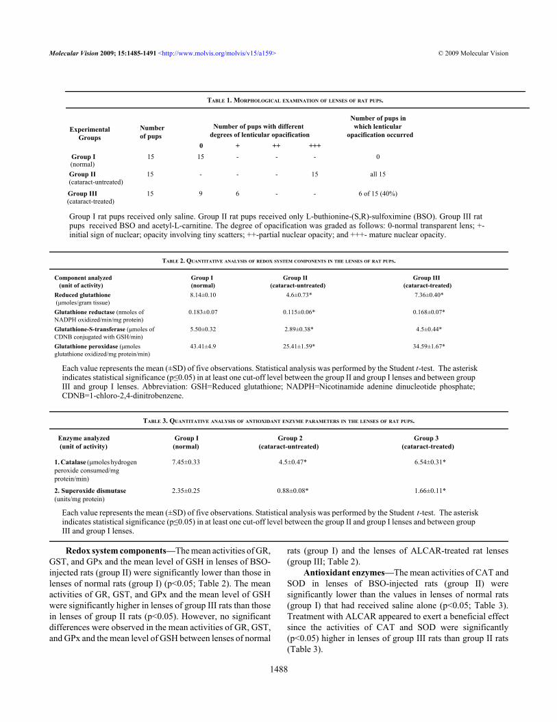
RESULTSMorphological examination: Slit-lamp examination revealedthat all 15 rat pups in group II (Figure 1 and Table 1) exhibiteddense opacification of the lens (grade +++). In contrast, only6 of 15 (40%) rat pups in group III (Figure 2 and Table 1)exhibited lenticular opacification (grade +) with the lenses ofthe other nine pups appearing normal (grade 0). All 15 rat pupsin group I exhibited maximum transparency (grade 0) of thelens (Figure 3 and Table 1). The difference between the valuein group II and group III rats was statistically significant (χ2
[degrees of freedom=1]=12.8; p<0.01).Biochemical evaluation of lenticular antioxidant and redoxsystem components:
Figure 3. Slit-lamp appearance of the eye of 30-day-old Wistar ratpup in group I. This eye exhibited no opacification of the lens (Grade0 opacification).
Redox system components—The mean activities of GR,GST, and GPx and the mean level of GSH in lenses of BSO-injected rats (group II) were significantly lower than those inlenses of normal rats (group I) (p<0.05; Table 2). The meanactivities of GR, GST, and GPx and the mean level of GSHwere significantly higher in lenses of group III rats than thosein lenses of group II rats (p<0.05). However, no significantdifferences were observed in the mean activities of GR, GST,and GPx and the mean level of GSH between lenses of normal
rats (group I) and the lenses of ALCAR-treated rat lenses(group III; Table 2).
Antioxidant enzymes—The mean activities of CAT andSOD in lenses of BSO-injected rats (group II) weresignificantly lower than the values in lenses of normal rats(group I) that had received saline alone (p<0.05; Table 3).Treatment with ALCAR appeared to exert a beneficial effectsince the activities of CAT and SOD were significantly(p<0.05) higher in lenses of group III rats than group II rats(Table 3).
ExperimentalGroups
Numberof pups
Number of pups with differentdegrees of lenticular opacification
Number of pups in which lenticularopacification occurred
0 + ++ +++Group I 15 15 - - - 0
Group II (cataract-untreated)
15 - - - 15 all 15
Group III (cataract-treated)
15 9 6 - - 6 of 15 (40%)
Group I rat pups received only saline. Group II rat pups received only L-buthionine-(S,R)-sulfoximine (BSO). Group III ratpups received BSO and acetyl-L-carnitine. The degree of opacification was graded as follows: 0-normal transparent lens; +-initial sign of nuclear; opacity involving tiny scatters; ++-partial nuclear opacity; and +++- mature nuclear opacity.
TABLE 2. QUANTITATIVE ANALYSIS OF REDOX SYSTEM COMPONENTS IN THE LENSES OF RAT PUPS.
Each value represents the mean (±SD) of five observations. Statistical analysis was performed by the Student t-test. The asteriskindicates statistical significance (p≤0.05) in at least one cut-off level between the group II and group I lenses and between groupIII and group I lenses. Abbreviation: GSH=Reduced glutathione; NADPH=Nicotinamide adenine dinucleotide phosphate;CDNB=1-chloro-2,4-dinitrobenzene.
TABLE 3. QUANTITATIVE ANALYSIS OF ANTIOXIDANT ENZYME PARAMETERS IN THE LENSES OF RAT PUPS.
Each value represents the mean (±SD) of five observations. Statistical analysis was performed by the Student t-test. The asteriskindicates statistical significance (p≤0.05) in at least one cut-off level between the group II and group I lenses and between groupIII and group I lenses.
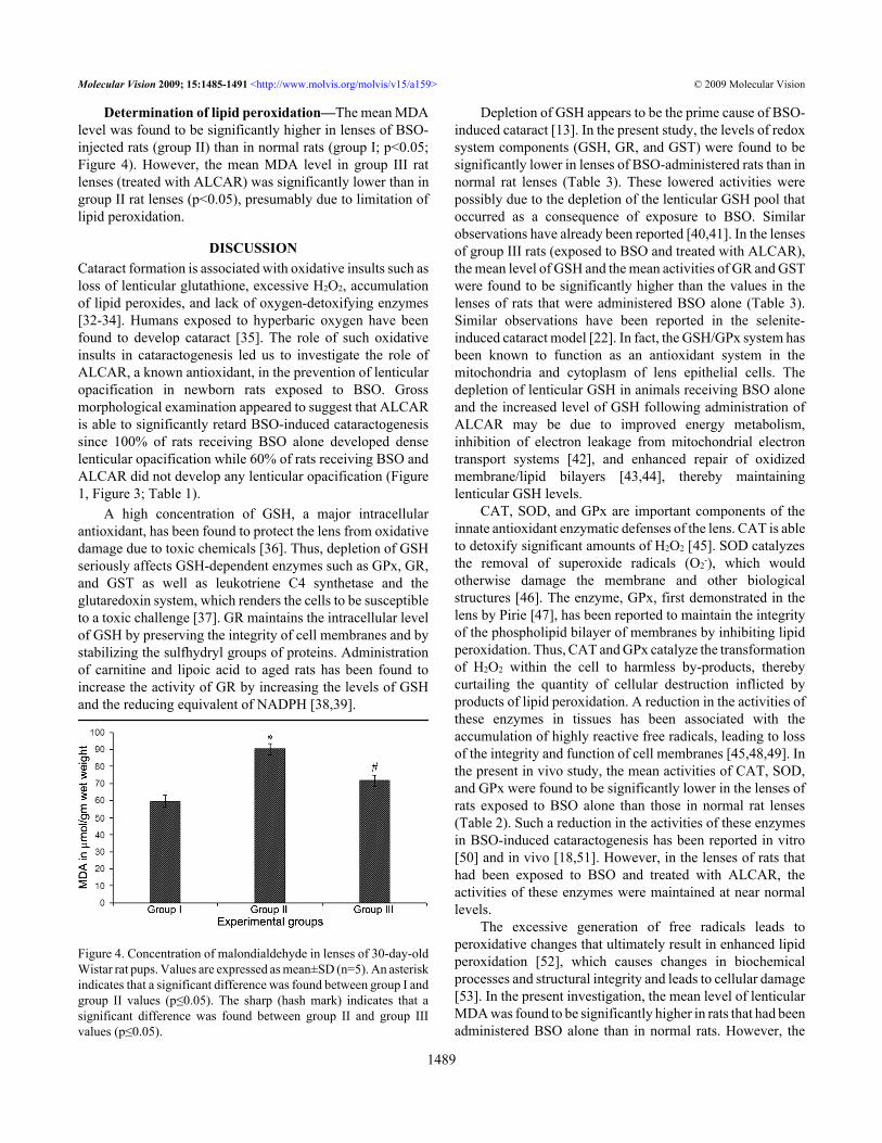
Determination of lipid peroxidation—The mean MDAlevel was found to be significantly higher in lenses of BSO-injected rats (group II) than in normal rats (group I; p<0.05;Figure 4). However, the mean MDA level in group III ratlenses (treated with ALCAR) was significantly lower than ingroup II rat lenses (p<0.05), presumably due to limitation oflipid peroxidation.
DISCUSSIONCataract formation is associated with oxidative insults such asloss of lenticular glutathione, excessive H2O2, accumulationof lipid peroxides, and lack of oxygen-detoxifying enzymes[32-34]. Humans exposed to hyperbaric oxygen have beenfound to develop cataract [35]. The role of such oxidativeinsults in cataractogenesis led us to investigate the role ofALCAR, a known antioxidant, in the prevention of lenticularopacification in newborn rats exposed to BSO. Grossmorphological examination appeared to suggest that ALCARis able to significantly retard BSO-induced cataractogenesissince 100% of rats receiving BSO alone developed denselenticular opacification while 60% of rats receiving BSO andALCAR did not develop any lenticular opacification (Figure1, Figure 3; Table 1).
A high concentration of GSH, a major intracellularantioxidant, has been found to protect the lens from oxidativedamage due to toxic chemicals [36]. Thus, depletion of GSHseriously affects GSH-dependent enzymes such as GPx, GR,and GST as well as leukotriene C4 synthetase and theglutaredoxin system, which renders the cells to be susceptibleto a toxic challenge [37]. GR maintains the intracellular levelof GSH by preserving the integrity of cell membranes and bystabilizing the sulfhydryl groups of proteins. Administrationof carnitine and lipoic acid to aged rats has been found toincrease the activity of GR by increasing the levels of GSHand the reducing equivalent of NADPH [38,39].
Figure 4. Concentration of malondialdehyde in lenses of 30-day-oldWistar rat pups. Values are expressed as mean±SD (n=5). An asteriskindicates that a significant difference was found between group I andgroup II values (p≤0.05). The sharp (hash mark) indicates that asignificant difference was found between group II and group IIIvalues (p≤0.05).
Depletion of GSH appears to be the prime cause of BSO-induced cataract [13]. In the present study, the levels of redoxsystem components (GSH, GR, and GST) were found to besignificantly lower in lenses of BSO-administered rats than innormal rat lenses (Table 3). These lowered activities werepossibly due to the depletion of the lenticular GSH pool thatoccurred as a consequence of exposure to BSO. Similarobservations have already been reported [40,41]. In the lensesof group III rats (exposed to BSO and treated with ALCAR),the mean level of GSH and the mean activities of GR and GSTwere found to be significantly higher than the values in thelenses of rats that were administered BSO alone (Table 3).Similar observations have been reported in the selenite-induced cataract model [22]. In fact, the GSH/GPx system hasbeen known to function as an antioxidant system in themitochondria and cytoplasm of lens epithelial cells. Thedepletion of lenticular GSH in animals receiving BSO aloneand the increased level of GSH following administration ofALCAR may be due to improved energy metabolism,inhibition of electron leakage from mitochondrial electrontransport systems [42], and enhanced repair of oxidizedmembrane/lipid bilayers [43,44], thereby maintaininglenticular GSH levels.
CAT, SOD, and GPx are important components of theinnate antioxidant enzymatic defenses of the lens. CAT is ableto detoxify significant amounts of H2O2 [45]. SOD catalyzesthe removal of superoxide radicals (O2
-), which wouldotherwise damage the membrane and other biologicalstructures [46]. The enzyme, GPx, first demonstrated in thelens by Pirie [47], has been reported to maintain the integrityof the phospholipid bilayer of membranes by inhibiting lipidperoxidation. Thus, CAT and GPx catalyze the transformationof H2O2 within the cell to harmless by-products, therebycurtailing the quantity of cellular destruction inflicted byproducts of lipid peroxidation. A reduction in the activities ofthese enzymes in tissues has been associated with theaccumulation of highly reactive free radicals, leading to lossof the integrity and function of cell membranes [45,48,49]. Inthe present in vivo study, the mean activities of CAT, SOD,and GPx were found to be significantly lower in the lenses ofrats exposed to BSO alone than those in normal rat lenses(Table 2). Such a reduction in the activities of these enzymesin BSO-induced cataractogenesis has been reported in vitro[50] and in vivo [18,51]. However, in the lenses of rats thathad been exposed to BSO and treated with ALCAR, theactivities of these enzymes were maintained at near normallevels.
The excessive generation of free radicals leads toperoxidative changes that ultimately result in enhanced lipidperoxidation [52], which causes changes in biochemicalprocesses and structural integrity and leads to cellular damage[53]. In the present investigation, the mean level of lenticularMDA was found to be significantly higher in rats that had beenadministered BSO alone than in normal rats. However, the
mean levels of MDA were significantly lower in lenses ofgroup III rats (BSO-administered and ALCAR-treated) thanin group II rat lenses (Figure 4). Thus, lenses of rats givenBSO alone showed a significant depletion of GSH andincreased membrane damage as indicated by the increasedlevels of MDA (Figure 4). However, ALCAR appeared toprevent the occurrence of such changes. Similar protectiveeffects of ALCAR have been previously reported in selenite-challenged rat lenses [22].
We have previously reported that ALCAR appears toprevent selenite-induced cataractogenesis [22]. The results ofthe present study add support to our hypothesis that ALCARcan also prevent cataractogenesis that is mediated byglutathione deprivation and induced by BSO. Thesepreventive effects of ALCAR are suggested by its ability tomaintain lenticular antioxidant and redox system componentsat near normal levels and to prevent excessive lipidperoxidation. The relevance of these results in the context ofhuman senile cataractogenesis requires further study.
ACKNOWLEDGMENTSThe authors thank the Department of Biotechnology (DBT),Government of India, for the financial assistance provided.Instrumentation facility provided by DST-FIST and UGC-SAP of the Department of Animal Science at BharathidasanUniversity (Tiruchirappalli, India) is also acknowledged.
REFERENCES1. Linetsky M, James HL, Ortwerth BJ. Spontaneous generation
of superoxide anion by human lens proteins and by calf lensproteins ascorbylated in vitro. Exp Eye Res 1999;69:239-48. [PMID: 10433859]
2. Fu S, Dean R, Southan M, Truscott R. The hydroxyl radical inlens nuclear cataractogenesis. J Biol Chem 1998;273:28603-9. [PMID: 9786852]
4. Laver NM, Robison WG Jr, Calvin HI, Fu SCJ. Early epitheliallesions in cataracts of GSH depleted mouse pups. Exp EyeRes 1993; 57:493-8. [PMID: 8282035]
5. Reddy VN. Metabolism of glutathione in the lens. Exp Eye Res1971; 11:310-28. [PMID: 4399363]
6. Hata N, Hockwin O. Enzymatic determination of reduced andoxidized glutathione in bovine lenses if different ages andtheir distribution in lens equator and nucleus. Ophthalmic Res1977; 9:256.
7. Meister A. On the antioxidant effects of ascorbic acid andglutathione. Biochem Pharmacol 1992; 44:1905-15. [PMID:1449510]
8. Anderson ME. Glutathione and glutathione deliverycompounds. Adv Pharmacol 1997; 38:65-78. [PMID:8895804]
9. May JM, Qu ZC, Whitesell RR, Cobb CE. Ascorbate recyclingin human erythrocytes: role of GSH in reducingdehydroascorbate. Free Radic Biol Med 1996; 20:543-51.[PMID: 8904295]
10. Lou MF. Thiol regulation in the lens. J Ocul Pharmacol Ther2000; 16:137-48. [PMID: 10803424]
11. Giblin FJ, Reddan JR, Schrimscher L, Dziedzic DC, Reddy VN.The relative roles of the glutathione redox cycle and catalasein the detoxification of H2O2 by cultured rabbit lens epithelialcells. Exp Eye Res 1990; 50:795-804. [PMID: 2373171]
12. Martensson J, Meister A. Glutathione deficiency decreasestissue ascorbate levels in newborn rats: Ascorbate sparesglutathione and protects. Proc Natl Acad Sci USA 1991;88:4656-60. [PMID: 2052548]
13. Calvin HI, Medvedovsky C, Worgul BV. Near-total glutathionedepletion and age-specific cataracts induced by buthioninesulfoximine in mice. Science 1986; 233:553-5. [PMID:3726547]
14. Martensson J, Steinherz R, Jain A, Meister A. Glutathione esterprevents buthionine sulfoximine-induced cataracts and lensepithelial cell damage. Proc Natl Acad Sci USA 1989;86:8727-31. [PMID: 2813421]
15. Spector A. The lens and oxidative stress. In: Seis H, editor.Oxidative stress: oxidants and antioxidants. London:Academic Press 1991; pp 529–58.
16. Jacques PF, Chylacjrk LT, Taylor A. Relationship betweennatural antioxidants and cataract formation. In: Frei B, editor.Natural antioxidants in human health and disease. San Diego:Academic Press; 1994. p. 515–33.
17. Taylor A, Nowel T. Oxidative stress and antioxidant functionin relation to the risk for cataract. Adv Pharmacol 1997;38:515-36. [PMID: 8895822]
18. Maitra I, Serbinova E, Trischler H, Packer L. Alpha-lipoic acidprevents buthionine sulfoximineinduced cataract formation innewborn rats. Free Radic Biol Med 1995; 18:823-9. [PMID:7750805]
19. Goa KL, Brogden RN. L–Carnitine. A preliminary review of itspharmacokinetics, and its therapeutic use in ischaemic cardiacdisease and primary and secondary carnitine deficiencies inrelationship to its role in fatty acid metabolism. Drugs 1987;34:1-24. [PMID: 3308409]
20. Yasui F, Matsugo S, Ishibashi M, Kajita T, Ezashi Y, OomuraY. Effects of chronic acetyl-L-carnitine treatment on brainlipid hydroperoxide level and passive avoidance learning insenescence-accelerated mice. Neurosci Lett 2002;334:177-80. [PMID: 12453624]
21. Geraldine P, Sneha BB, Elanchezhian R, Ramesh E, KalavathyCM, Kaliamurthy J, Thomas PA. Prevention of selenite-induced cataractogenesis by acetyl-L-carnitine: anexperimental study. Exp Eye Res 2006; 83:1340-9. [PMID:16962580]
22. Elanchezhian R, Ramesh E, Sakthivel M, Isai M, Geraldine P,Rajamohan M, Nelson Jesudasan C, Thomas PA. Acetyl-L-Carnitine prevents selenite-induced cataractogenesis in anexperimental animal model. Curr Eye Res 2007; 32:961-71.[PMID: 18027172]
23. Elanchezhian R, Sakthivel M, Geraldine P, Thomas PA. Theeffect of acetyl-l-carnitine on lenticular calpain activity inprevention of selenite-induced cataractogenesis. Exp Eye Res2009; 88:938-44. [PMID: 19150348]
24. Bradford MM. A rapid and sensitive method for the quantitationof microgram quantities of protein utilizing the principle ofprotein-dye binding. Anal Biochem 1976; 72:248-54. [PMID:942051]
25. Moron MS, Depierre JW, Mannervik B. Levels of glutathione,glutathione reductase and glutathione-S-transferase activitiesin rat lung and liver. Biochim Biophys Acta 1979;582:67-78. [PMID: 760819]
26. Staal GE, Visser J, Veeger C. Purification and properties ofglutathione reductase of human erythrocytes. BiochimBiophys Acta 1969; 185:39-48. [PMID: 5796111]
30. Marklund S, Marklund G. Involvement of the superoxide anionradical in the autoxidation of pyrogallol and a convenientassay for superoxide dismutase. Eur J Biochem 1974;47:469-74. [PMID: 4215654]
31. Ohkawa H, Ohishi N, Yagi K. Assay of lipid peroxide in animaltissue by thiobarbituric acid reaction. Anal Biochem 1979;95:351-8. [PMID: 36810]
32. Bhuyan KC, Bhuyan DK. Molecular mechanisms ofcataractogenesis: III. Toxic metabolites of oxygen asinitiators of lipid peroxidation and cataract. Curr Eye Res1984; 3:67-81. [PMID: 6317286]
33. Meister A. Glutathione deficiency produced by inhibition of itssynthesis, and its reversal; applications in research andtherapy. Pharmacol Ther 1991; 51:155-94. [PMID: 1784629]
34. Mitton KP, Dean PA, Dzialoszynski T, Xiong H, Sanford SE,Trevithick JR. Modelling cortical cataractogenesis: 13. Earlyeffects on lens ATP/ADP and glutathione in thestreptozotocin rat model of the diabetic cataract. Exp Eye Res1993; 56:187-98. [PMID: 8462652]
36. Hightower KR, McCready JP. Effect of selenite on epitheliumof cultured rabbit lens. Invest Ophthalmol Vis Sci 1991;32:406-9. [PMID: 1825204]
37. Kumaran S, Savitha S, Anusuya Devi M, Panneerselvam C.Lcarnitine and DL-alpha-lipoic acid reverse the age-relateddeficit in glutathione redox state in skeletal muscle and hearttissues. Mech Ageing Dev 2004; 125:507-12. [PMID:15246746]
38. Arivazhagan P, Ramanathan K, Panneerselvam C. Effect of DL-alphalipoic acid on glutathione metabolic enzymes in agedrats. Exp Gerontol 2001; 37:81-7. [PMID: 11738149]
39. Rani PJ, Panneerselvam C. Effect of L-carnitine on brain lipidperoxidation and antioxidant enzymes of old rats. J GerontolA Biol Sci Med Sci 2002; 57:B134-7. [PMID: 11909877]
40. Bhuyan KC, Bhuyan DK, Podos SM. Cataract induced byselenium in rats. II. Increased lipid peroxidation andimpairment of enzymatic defenses against oxidative damage.IRCS Med Sci 1981; 9:195-6.
41. Huang LL, Zhang CY, Hess JL, Bunce GE. Biochemicalchanges and cataract formation in lenses from rats receivingmultiple, low doses of sodium selenite. Exp Eye Res 1992;55:671-8. [PMID: 1478277]
42. Trush MA, Kensler TW. An overview of the relationshipbetween oxidative stress and chemical carcinogenesis. FreeRadic Biol Med 1991; 10:201-9. [PMID: 1864525]
43. Arduini A. Carnitine and its acyl esters as secondaryantioxidants? Am Heart J 1992; 123:1726-7. [PMID:1595568]
44. Liu J, Head E, Gharib AM, Yuan W, Ingersoll RT, Hagen TM,Cotman CW, Ames BN. Memory loss in old rats is associatedwith brain mitochondrial decay and RNA/DNA oxidation:partial reversal by feeding acetyl-L-carnitine and/or R-alpha-lipoic acid. Proc Natl Acad Sci USA 2002; 99:2356-61.[PMID: 11854529]
45. Cheng L, Kellogg EW 3rd, Packer L. Photoinactivation ofcatalase. Photochem Photobiol 1981; 34:125-9. [PMID:7291325]
46. Krishnakantha TP, Lokesh BR. Scavenging of superoxideanions by spice principles. Indian J Biochem Biophys 1993;30:133-4. [PMID: 8394839]
47. Pirie A. Glutathione peroxidase in lens and a source of hydrogenperoxide in aqueous humour. Biochem J 1965; 96:244-53.[PMID: 14343138]
48. Reddy AC, Lokesh BR. Studies on spice principle as anantioxidant in the inhibition of lipid peroxidation of rat livermicrosomes. Mol Cell Biochem 1992; 111:117-24. [PMID:1588934]
49. Sheela CG, Angusti K. Antiperoxide effects of S-allyl cysteinsulphoxide isolated from Allium sativum Linn and gugulipidin chlosterol diet fed rats. Indian J Exp Biol 1995;33:337-41. [PMID: 7558192]
50. Calvin HI, Banerjee U, Fu SC. Inhibition of mouse BSOcataracts by the protein-kinase inhibitor, H-7. InvestOphthalmol Vis Sci 1997; 38:5376.
51. Li ZR, Reiter RJ, Fujimori O, Oh CS, Duan YP.Cataractogenesis and lipid peroxidation in newborn ratstreated with buthionine sulfoximine: Preventive actions ofmelatonin. J Pineal Res 1997; 22:117-23. [PMID: 9213264]
52. Rikans LE, Hornbrook KR. Lipid peroxidation, antioxidantprotection and aging. Biochim Biophys Acta 1997;1362:116-27. [PMID: 9540842]
53. Bhatia AL, Jain M. Spinacia oleracea L. protects against gammaradiations: a study on glutathione and lipid peroxidation inmouse liver. Phytomedicine 2004; 11:607-15. [PMID:15636174]
The print version of this article was created on 28 July 2009. This reflects all typographical corrections and errata to the articlethrough that date. Details of any changes may be found in the online version of the article.