REVIEW ARTICLE Glycogen metabolism and the homeostatic regulation of sleep Jean-Marie Petit & Sophie Burlet-Godinot & Pierre J. Magistretti & Igor Allaman Received: 19 September 2014 /Accepted: 4 November 2014 /Published online: 16 November 2014 # The Author(s) 2014. This article is published with open access at Springerlink.com Abstract In 1995 Benington and Heller formulated an energy hypothesis of sleep centered on a key role of glycogen. It was postulated that a major function of sleep is to replenish glycogen stores in the brain that have been depleted during wakefulness which is associ- ated to an increased energy demand. Astrocytic glyco- gen depletion participates to an increase of extracellular adenosine release which influences sleep homeostasis. Here, we will review some evidence obtained by studies addressing the question of a key role played by glyco- gen metabolism in sleep regulation as proposed by this hypothesis or by an alternative hypothesis named “glycogenetic” hypothesis as well as the importance of the confounding effect of glucocorticoïds. Even though actual collected data argue in favor of a role of sleep in brain energy balance-homeostasis, they do not support a critical and direct involvement of glycogen metabolism on sleep regulation. For instance, glycogen levels during the sleep-wake cycle are driven by different physiological signals and therefore appear more as a marker-integrator of brain energy status than a direct regulator of sleep homeo- stasis. In support of this we provide evidence that blockade of glycogen mobilization does not induce more sleep episodes during the active period while locomotor activity is reduced. These observations do not invalidate the energy hypothesis of sleep but indicate that underlying cellular mechanisms are more complex than postulated by Benington and Heller. Keywords Glia . PTG . Glucocorticoïds . Energy metabolism . Noradrenaline . Brain While sleep has been investigated over decades, the funda- mental question of why we need to sleep is still unanswered. More specifically, the physiological function(s) fulfilled by sleep remain(s) elusive. Among the formulated hypotheses is the necessity of sleep to reestablish brain energy stores that are altered during wakefulness. The main postulate behind this theory is that energy stores are diminished during the meta- bolically active waking period and need to be restored during sleep. Based on this, Benington and Heller formulated almost 20 years ago an energy hypothesis of sleep proposing glyco- gen and adenosine (Ade) as key regulators of sleep homeo- stasis (Benington and Heller 1995). This model suggests that glycogen stores depletion occurs during waking leading to Ade formation in the extracellular space, which in turn acts as a promoting agent of sleepiness influencing sleep homeosta- sis. As glycogen is exclusively found in astrocytes, a glial cell type, in the central nervous system (CNS), this points to a major role of astrocyte functions (in particular with regard to brain energy homeostasis) in sleep regulation. Since the formulation of Benington and Heller hypothesis, numerous studies have been undertaken to validate, or not, the importance of glycogen store depletion and replenishment as a key regulator of sleep. This review will first introduce basic concepts of brain glycogen metabolism and of the structure and regulation of sleep. Then, major findings related to the J.<M. Petit (*) : S. Burlet-Godinot : P. J. Magistretti : I. Allaman Laboratory of Neuroenergetics and Cellular Dynamics, Brain Mind Institute, Ecole Polytechnique Fédérale de Lausanne (EPFL), Lausanne 1015, Switzerland e-mail: [email protected]P. J. Magistretti Division of Biological and Environmental Sciences and Engineering, KAUST, Thuwal, KSA, Saudi Arabia J.<M. Petit : P. J. Magistretti Center for Psychiatric Neuroscience, Department of Psychiatry, CHUV, 1008 Prilly, Switzerland Metab Brain Dis (2015) 30:263–279 DOI 10.1007/s11011-014-9629-x
Transcript
REVIEWARTICLE
Glycogen metabolism and the homeostatic regulation of sleep
Jean-Marie Petit & Sophie Burlet-Godinot &Pierre J. Magistretti & Igor Allaman
Received: 19 September 2014 /Accepted: 4 November 2014 /Published online: 16 November 2014# The Author(s) 2014. This article is published with open access at Springerlink.com
Abstract In 1995 Benington and Heller formulated anenergy hypothesis of sleep centered on a key role ofglycogen. It was postulated that a major function ofsleep is to replenish glycogen stores in the brain thathave been depleted during wakefulness which is associ-ated to an increased energy demand. Astrocytic glyco-gen depletion participates to an increase of extracellularadenosine release which influences sleep homeostasis.Here, we will review some evidence obtained by studiesaddressing the question of a key role played by glyco-gen metabolism in sleep regulation as proposed by thishypothesis or by an alternative hypothesis named“glycogenetic” hypothesis as well as the importance ofthe confounding effect of glucocorticoïds. Even thoughactual collected data argue in favor of a role of sleep inbrain energy balance-homeostasis, they do not support acritical and direct involvement of glycogen metabolismon sleep regulation. For instance, glycogen levels duringthe sleep-wake cycle are driven by different physiologicalsignals and therefore appear more as a marker-integrator ofbrain energy status than a direct regulator of sleep homeo-stasis. In support of this we provide evidence that blockadeof glycogen mobilization does not induce more sleep
episodes during the active period while locomotor activityis reduced. These observations do not invalidate the energyhypothesis of sleep but indicate that underlying cellularmechanisms are more complex than postulated byBenington and Heller.
While sleep has been investigated over decades, the funda-mental question of why we need to sleep is still unanswered.More specifically, the physiological function(s) fulfilled bysleep remain(s) elusive. Among the formulated hypotheses isthe necessity of sleep to reestablish brain energy stores that arealtered during wakefulness. The main postulate behind thistheory is that energy stores are diminished during the meta-bolically active waking period and need to be restored duringsleep. Based on this, Benington and Heller formulated almost20 years ago an energy hypothesis of sleep proposing glyco-gen and adenosine (Ade) as key regulators of sleep homeo-stasis (Benington and Heller 1995). This model suggests thatglycogen stores depletion occurs during waking leading toAde formation in the extracellular space, which in turn acts asa promoting agent of sleepiness influencing sleep homeosta-sis. As glycogen is exclusively found in astrocytes, a glial celltype, in the central nervous system (CNS), this points to amajor role of astrocyte functions (in particular with regard tobrain energy homeostasis) in sleep regulation.
Since the formulation of Benington and Heller hypothesis,numerous studies have been undertaken to validate, or not, theimportance of glycogen store depletion and replenishment as akey regulator of sleep. This review will first introduce basicconcepts of brain glycogen metabolism and of the structureand regulation of sleep. Then, major findings related to the
J.<M. Petit (*) : S. Burlet-Godinot : P. J. Magistretti : I. AllamanLaboratory of Neuroenergetics and Cellular Dynamics, Brain MindInstitute, Ecole Polytechnique Fédérale de Lausanne (EPFL),Lausanne 1015, Switzerlande-mail: [email protected]
P. J. MagistrettiDivision of Biological and Environmental Sciences and Engineering,KAUST, Thuwal, KSA, Saudi Arabia
J.<M. Petit : P. J. MagistrettiCenter for Psychiatric Neuroscience, Department of Psychiatry,CHUV, 1008 Prilly, Switzerland
involvement of glycogen in sleep will be discussed, focusingon:
a) Benington and Heller hypothesis (BHH)b) “Glycogenetic” hypothesisc) A synthesis of experimental datad) Effect of direct modulation of glycogen levels on sleepe) Confounding effects of glucocorticoïds (GCs)
Brain glycogen metabolism
The precise functions of brain glycogen are still not fullycharacterized. Nevertheless, it has been shown that glycogenreserves are consumed during failure of energy supply andstrong evidence suggests that glycogen mobilization is tightlycoupled to neuronal activity and could provide additionalenergy substrates for neurones during period of activity. Forinstance, glycogen accumulation is observed in conditions ofdecreased neuronal activity such as phenobarbital anaesthesia(Phelps 1972), hibernation (Swanson 1992) or during slowwave sleep (SWS) (Karnovsky et al. 1983). In contrast, cere-bral glycogen breakdown is observed following sensory stim-ulation (Dienel et al. 2007; Swanson et al. 1992). Moreover,the greatest accumulation of glycogen has been reported inareas of high synaptic density (Koizumi and Shiraishi 1970;Koizumi and Shiraishi 1971; Phelps 1972), further supportingthe idea that glycogen may be involved in neuronal activity.Experiments also demonstrated that glycogen is mobilized tosupport energy needs of axons upon action potential propaga-tion in an optic nerve preparation (Brown et al. 2003; Brownet al. 2004).
Most of our knowledge about glycogen metabolism origi-nates from studies conducted in peripheral tissues but arecommon for all tissues (see e.g. Bollen et al. 1998; Jensenand Richter 2012; Roach et al. 2012) (Fig. 1a). Uridine di-phosphate glucose (UDP-glucose) is the source of all glucosylresidues added to glycogen. UDP-glucose is formed from theconversion of glucose-6-phosphate to glucose-1-phosphate byphosphoglucomutase. The first step in the biogenesis of gly-cogen is the autocatalytic attachment of glucosyl units to asingle tyrosine residue of glycogenin. Elongation of this“primed” glycogenin is performed by the concerted action ofglycogen synthase (GS) and branching enzyme. Glycogensynthase elongates the glycogen chain whereas branchingenzyme produces new branches, creating a mature glycogengranule. In contrast to glycogen synthesis, glycogenolysis is aprocess that does not require energy. It is not a reverse reactionof glycogen synthesis, but implies the concerted action ofglycogen phosphorylase (GP) and the bi-functionaldebranching enzyme releasing glucose residue of glycogengranules as glucose-1-phosphate. Finally, glucose-1-
phosphate is converted by phosphoglucomutase to glucose-6-phosphate which is (in the brain) the final product of glyco-gen degradation. Glucose-6-phosphate can then re-enter gly-colysis or the pentose phosphate pathway (Fig. 1a).
Glycogen is a highly dynamic pool of glucose, the metab-olism of which is under complex regulation involving variousallosteric factors as well as covalent modifications and com-partmentalization of key enzymes (for reviews see Bollenet al. 1998; Brown 2004). In general, activation of glycogensynthesis results in the inhibition of the degradative pathway,and vice versa. Glycogen synthase and GP represent twocentral elements of this regulation. Glycogen synthase istightly controlled by the reversible phosphorylation of multi-ple serine residues by various protein kinases. Generally,phosphorylation is associated with an inactivation of the en-zyme (conversion of the active a-form to the b-form). LikeGS, GP is regulated by phosphorylation. Glycogen phosphor-ylase is converted from the inactive b-form to the active a-form through phosphorylation by phosphorylase kinase. Asdescribed, kinase-catalyzed phosphorylation of both enzymesleads to glycogenolysis (by activation of GP and inactivationof GS) whereas dephosphorylation favors glycogenesis.
Protein phosphatase-1 (PP-1) is the enzyme responsible fordephosphorylation of both GS and GP (Newgard et al. 2000).In order to be active, PP-1 must be bound to glycogen. This isaccomplished by specific glycogen-binding G-subunits,
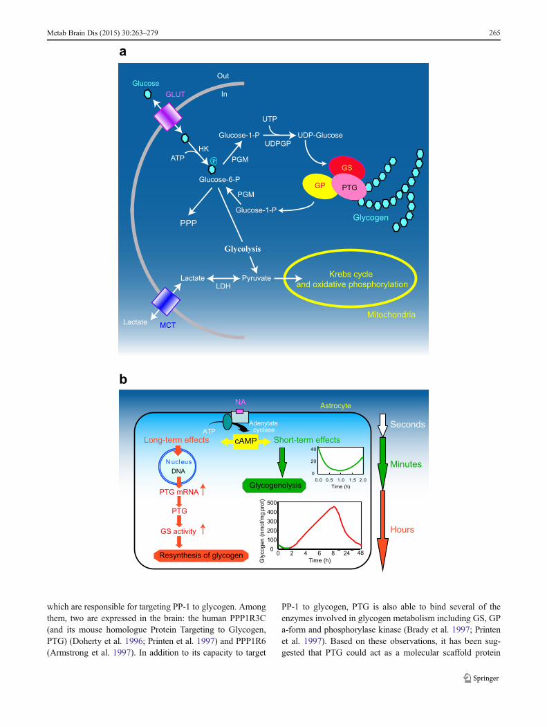
�Fig. 1 Gylcogenmetabolism in astrocytes. a Schematic representation ofglucose metabolism. Glucose enters cells trough glucose transporters(GLUT) and is phosphorylated by hexokinase (HK) to produceglucose-6-phosphate. Glucose 6-phosphate can be processed intodifferent metabolic pathways. It can be metabolized through the pentosephosphate pathway (PPP) or through glycolysis giving rise to pyruvateproduction. Pyruvate can enter mitochodria where it is metabolizedthrough the Krebs cycle and oxidative phosphorylation. Alternatively,pyruvate can be reduced to lactate by lactate dehydrogenase (LDH) andbe released in the extracellular space through monocarboxylatetransporters (MCT). Glucose-6-phosphate can also be used to storeglucosyl units as glycogen. As a first step glucose-6-phosphate isconverted to UDP-glucose through the action of phosphoglucomutase(PGM) and UDP-glucose pyrophosphorylase (UDPGP). UDP-glucose isthen incorporated into glycogen by glycogen synthase (GS). Proteintargeting to glycogen (PTG) is a specific glycogen-binding G-subunit,which is responsible for the targeting of protein phosphatase 1 toglycogen. Through this action, PTG promotes GS dephosphorylationand activation therefore favouring glycogen synthesis. In case of energyneeds, glycogen can be broken down by glycogen phosphorylase (GP) toproduce glucose-1-phosphate that is converted back to glucose-6-phosphate through the action of PGM. Adapted from (Allaman 2009)with permission. b Differential modulation of glycogen metabolism incultured astrocytes by noradrenaline (NA). Activation of cAMP-dependent intracellular signalling by NA results in a short term(seconds to minutes, in green) glycogenolysis and in a delayed (hours,in red) glycogen resynthesis. This long-term response requires inductionof gene expression and is accompanied by stimulation of mRNAexpression of protein targeting to glycogen (PTG), a member of theglycogen-targeting subunits of protein phosphatase 1, and by theactivation of glycogen synthase (see text for details). Adapted from(Magistretti 2006) with permission
264 Metab Brain Dis (2015) 30:263–279
which are responsible for targeting PP-1 to glycogen. Amongthem, two are expressed in the brain: the human PPP1R3C(and its mouse homologue Protein Targeting to Glycogen,PTG) (Doherty et al. 1996; Printen et al. 1997) and PPP1R6(Armstrong et al. 1997). In addition to its capacity to target
PP-1 to glycogen, PTG is also able to bind several of theenzymes involved in glycogen metabolism including GS, GPa-form and phosphorylase kinase (Brady et al. 1997; Printenet al. 1997). Based on these observations, it has been sug-gested that PTG could act as a molecular scaffold protein
Metab Brain Dis (2015) 30:263–279 265
assembling the different enzymes necessary for glycogenmetabolism (Brady et al. 1997; Mastick et al. 1998).
At the cellular level, glycogen is mainly located in astro-cytes in the CNS (Brown 2004; Cataldo and Broadwell 1986;Magistretti et al. 1993). The cellular localization of GS and GPcorrelates with the presence of glycogen. Both brain andmuscle isozymes of GP are located predominantly in astro-cytes (Ignacio et al. 1990; Pfeiffer-Guglielmi et al. 2003).
As in peripheral organs, brain glycogen levels are alsounder the tight control of numerous neuro-humoral factors.Thus, insulin and insulin-like growth factor I increase glyco-gen content of astrocytes in culture (Dringen and Hamprecht1992; Hamai et al. 1999). Moreover, various neuroactivesubstances possess glycogenolytic properties in vitro (forreview see Magistretti et al. 1993). In particular, a glycogen-olytic effect of potassium, vasoactive intestinal peptide (VIP),Ade and noradrenaline (NA) have been demonstrated inmouse cerebral cortical slices as well as in primary culturesof cortical astrocytes (Hof et al. 1988; Magistretti et al. 1981;Quach et al. 1978; Sorg and Magistretti 1991).
In addition to their rapid glycogenolytic effect, NA, VIPand Ade lead to massive glycogen resynthesis in culturedastrocytes, which takes place over several hours (Allamanet al. 2003; Sorg and Magistretti 1992). It was determinedthat this long-term dynamic regulation of glycogen in astro-cytes requires an activation of transcription and the synthesisof new protein(s). This process is mediated by cAMP-dependent mechanisms, involves the family of transcriptionfactors CCAAT/enhancer binding protein (C/EBP) and resultsin the induction of PTG expression and of GS activity(Allaman et al. 2000; Cardinaux and Magistretti 1996; Sorgand Magistretti 1992) (Fig. 1b). Interestingly, overexpressionof PTG in CHO cells (Printen et al. 1997), in cultured rathepatocytes (Berman et al. 1998) or in cultured mouse corticalastrocytes (unpublished data) leads to an increase in basalglycogen levels without the need of other stimuli. It wasfurther suggested that increased expression of PTG mightmaintain the cell in a glycogenic mode (Berman et al. 1998).This set of observations finally suggests that in astrocytes, theinduction of PTG alone might be sufficient to account for themassive glycogen resynthesis triggered by neuromodulatorssuch as NA, VIP or Ade.
Structure and regulation of sleep
Behavioral and neuronal correlates of sleep including loco-motor inactivity, specific body posture, high threshold ofsensory reactivity and decrease in global neuronal firing rate,are shared by numerous animal species from insects to mam-mals (Campbell and Tobler 1984). This has justified the use ofdrosophila and zebrafish as well as rodents as experimentalmodels in sleep studies (Cirelli and Tononi 2008; Zimmerman
et al. 2008). Using parallel recordings of electroencephalo-gram (EEG) and electromyogram (EMG), sleep in mammalsand birds can be divided in two main stages: SlowWave Sleep(SWS) also called Non-Rapid Eye Movement (NREM) sleepand Paradoxical Sleep (PS) equivalent to Rapid Eye Move-ment (REM) sleep. The three vigilance states (i.e. waking(W), SWS and PS) also display major differences in spectralcomponents of their EEG (Fig. 2). The EEG of SWS ischaracterized by oscillations of high amplitude and low fre-quencies. Power spectrum analysis of this EEG indicates alarge predominance of low frequencies, including sleep spin-dles (8–14 Hz), delta waves (1–4.5 Hz, also defined as SlowWave Activity (SWA)) and slow waves (<1 Hz), correspond-ing to a high level of synchronization of cortical cells dis-charge. More precisely, slow waves oscillations are composedby the alternation of synchronized cell firing periods (UPstates) and silent periods (DOWN states) (Fig. 2b). Whilethe muscle tone is lower in PS than during waking, the EEGsignal is apparently close to the waking one. EEG displaysoscillations of low amplitude and high frequencies. Powerspectrum of the PS EEG is shifted towards more rapid fre-quencies with a specific peak at 5-7 Hz (theta band). This peakis mainly due to the firing rate rhythm of hippocampal neuronsand is particularly apparent in rodents where hippocampusoccupies a dorsal location. During waking, oscillations of lowamplitude and rapid frequencies which correspond todesynchronization of cortical cells firing, are observed onthe EEG. Depending animal behavior, EEG power spectrumduring waking displays a relative increase in alpha (9-12 Hz),beta (12-30 Hz) and gamma (>30 Hz) frequency bands.
Alternation of SWS and PS (usually ended by a briefawakening) constitutes a sleep cycle whose duration is spe-cific of each animal species. Moreover, in several mammalssuch as human, SWS can be subdivided in light-SWS anddeep-SWS. Mammals display a great diversity in their totalsleep time per day as well as in the PS quantity (Siegel 2001).For example, the big brown bat sleeps almost 20 h/day with4 h in PS while a horse sleeps about 3 h/day with 0.5 h in PS.Such a difference in time spent asleep has been initially foundpositively correlated to metabolic rate and negatively corre-lated to brain size (Zepelin and Rechtschaffen 1974). Howev-er, more precise analysis found that sleep amount better re-flects ecological constraints in term of foraging or risk ofpredation (Capellini et al. 2008). The daily sleep distribution,which also varies greatly across mammals, is likely also underthe same ecological pressure and remains a major differencebetween man and other mammals classically used (rodents) insleep research.
The regulation of sleep, although not totally understood,can be considered as the interaction between circadian clockand homeostatic mechanisms (Borbely and Achermann1999). The molecular components (i.e. proteins) of circadianclock are present in each cell where their cyclic expressions
266 Metab Brain Dis (2015) 30:263–279
are regulated by successive positive and negative transcrip-tional feedback loops (see Ko and Takahashi 2006; Pandaet al. 2002). These cellular clocks are synchronized by thecentral clock of the body localized in the suprachiasmaticnucleus (SCN), a hypothalamic small nucleus situated justabove the optic chiasma from which it directly receives inputsencoding light information (Dibner et al. 2010). In basalconditions, sleep periods occur with a circadian rhythmicpattern which is abolished when SCN is destroyed (for reviewsee Moore 2007). Moreover, in humans and mice, polymor-phisms for clock genes are correlated to different sleep-wakecycle disturbances such as the familial advanced sleep-phasesyndrome (Per2 mutation) or familial delayed sleep phasesyndrome (Per3 mutations) (Archer et al. 2010; Chong et al.2012).
Sleep is also regulated via homeostatic mechanisms. In-deed, total sleep length increases following prolonged wake-fulness; a phenomenon also called “sleep rebound”. More-over, during SWS, an increase in SWA (Fig. 2a), correspond-ing to the power spectrum of the EEG delta band, is observedduring the first hours of the recovery period subsequent tosleep loss. This SWA increase is generally considered as themost reproducible index of the homeostatic sleep regulation(Borbely et al. 1981; Borbely et al. 1984). Interestingly, PSalso displays a homeostatic regulation which seems to be onlyreflected by an increase in length of its epochs without majorchange in the EEG power spectrum. During the last decade,experimental data in human and rodents showed that, during
SWS, the SWA is greater in cortical areas which were moreactivated during the previous wakefulness period even inabsence of sleep loss (Huber et al. 2004; Vyazovskiy et al.2000). More recently, using intra-cortical recordings,Vyazovskiy and coworkers reported that electrophysiolog-ical features of the SWS (e.g. slow waves between 0.1-1 Hz) are also expressed by an increasing number ofneurons in parallel to the waking duration (Vyazovskiyet al. 2011). These results indicate that the homeostaticregulation of SWA is a “local and use-dependent” phe-nomenon and challenge the classical view of sleep as theresult of interactions between distributed excitatory andinhibitory neuronal networks (mainly in diencephalon andin rhombencephalon) (Krueger and Tononi 2011;Rattenborg et al. 2012).
Benington and Heller hypothesis (BHH)
Sleep as a state of brain energy conservation
Although the energy cost of each sleep state at cellular or sub-cellular levels (synapses) is not known, the cerebral variationsof the energy consumption evaluated by glucose uptake mea-surements performed in human (Buchsbaum et al. 1989; Heisset al. 1985; Nofzinger et al. 2002), monkey (Kennedy et al.1982), cat (Ramm and Frost 1986) and rodents (Jay et al.1985; Vyazovskiy et al. 2008) clearly indicate that SWS is an
SWA (1-4.5Hz)
UP DOWN
Fig. 2 Sleep architecture. a Twenty four hour EEG power spectra inNREM sleep (SWS), REM sleep (PS) and waking in one representativeadult male rat (Wistar Kyoto strain). SlowWave Activity (SWA) frequen-cy spectrum (1–4.5 Hz) is highlighted in grey. b EEG signals andcorresponding cortical multiunit activity (raster plots below; each bar isa spike) representative of the three vigilance states. Note the fast irregular
pattern of cortical firing in waking and REM sleep, and regular occur-rence of generalized neuronal silence, corresponding to EEG slowwaves,in NREM sleep. UP (red) and DOWN (blue) states during NREM sleepare highlighted. Adapted from (Vyazovskiy and Faraguna 2014) withpermission
Metab Brain Dis (2015) 30:263–279 267
energy sparing stage relative to waking and PS.Measurementsof regional cerebral blood flow (Braun et al. 1997; Madsenet al. 1991) and metabolic rate (Katayose et al. 2009) duringsleep in human corroborate these results. These data led tohypothesize that one of the functions of sleep is to preservebrain energy levels, a fact that could be used to support thecellular mechanisms related to brain plasticity, especiallythose involved in off-line memory processes during sleep(Tononi and Cirelli 2006).
Sleep and glycogen
In a pioneering study, Karadzic and Mrsulja in 1969 showedthat 72 h of selective PS deprivation, in rats induce changes inglycogen content in different brain areas (Karadzic andMrsulja 1969). These results therefore suggested a direct linkbetween specific vigilance state (PS) and brain glycogencontent.
Glycogen is classically thought to supplement glucoseuptake by brain during physiological stimulation. A de-crease in glycogen levels was observed in primary sensorycortical areas following sensory stimulation (Dienel andCruz 2004; Swanson et al. 1992) and histochemistry ofthe GP (active a-form) has been used as metabolic markerof neuronal activation (Harley and Bielajew 1992; Woolfet al. 1985).
The BHH
Consistent with this view, Karnovsky and co-workers con-ducted a pivotal study where theymeasured glycogen levels inrat brain following different times spent awake or differenttimes spent asleep (Karnovsky et al. 1983). Their resultsshowed that glycogen levels were greatly increased (+70 %)during the first 15 min of a sleep period whereas they decreasemore slowly (−30 %) 20 min following an awakening. Forinstance, in vitro data indicate that neurotransmitters known tobe specifically released during waking such as NA, serotoninand histamine (reviewed in Jones 2005), induce a rapid gly-cogenolysis (Magistretti et al. 1993; Quach et al. 1978; Sorgand Magistretti 1991). In addition, brain glycogen levels arelower in the first half of the dark period, when the activity ofmice is maximal, and are higher during the rest period (Hutch-ins and Rogers 1970). Therefore, during activity period, re-petitive glycogen degradation should occur in response to thedifferent behavioral sequences and then, lead to a globalglycogen depletion at the cortical levels. Because glycogendepletion should correspond to a decrease in ATP/AMP ratio,Ade levels should increase resulting of 5′-AMP degradationby ectonucleotidases. Ade could act on different types ofreceptors (mainly A1 and A2A, A2B) present on neuronalcells. A1 receptor stimulation inhibits synaptic transmissionthrough an increase in K+conductance (gK+) whereas A2A
receptor stimulation activates adenylyl cyclase and exhibitsmore vasodilatation and metabolic effects (for review seeSheth et al. 2014).
In summary, Benington and Heller’s hypothesis (BHH)proposed that in response to local increase in neuronalactivity, the depletion of glycogen during waking inducesa decrease in the ATP/AMP ratio resulting in accumulationof extracellular Ade. Extracellular Ade decreases the neu-ronal firing rate through its action on A1 receptor. Finally,the inhibitory action of Ade triggers sleep. Sleep, in turn,allows replenishing glycogen stores which create the ben-eficial energetic conditions for subsequent waking period(Fig. 3).
As a direct correlate of BHH, prolonged wakefulnessshould induce more profound glycogen depletion comparedto undisturbed animals. Therefore, in order to obtain a directdemonstration of BHH different research groups assessedglycogen levels following acute total sleep deprivation (SD)in various species. One should note that the SD method usedin the subsequent described studies, if not otherwise specified,is the so-called “gentle” sleep deprivation method. This SDmethod consists on gently modifying the animal environment(i.e. changing a part of its bedding material, introducing newobject into the cage) when animal presents behavioral signs ofsleepiness (i.e. prolonged immobility or adopting sleepposture).
Testing the BHH
A first set of experiments was conducted by Heller’s group inwhich glycogen levels were measured in different brain re-gions following 6 h and 12 h periods of SD in young rats (Gipet al. 2002). In this study, animals were sacrificed usingmicrowave irradiation (Gip et al. 2002) to preserve glycogenfrom rapid degradation as induced by killing animals bydecapitation. Indeed, this method of sacrifice represents thebest available method to measure true physiological concen-trations of energy metabolites such as glycogen, lactate orglucose that display very rapid changes when ATP/AMP ratiofalls. Results of this study indicate that cerebellar levels ofglycogen were decreased after 6 h of SD whereas no changewas observed in cerebral cortex. Interestingly, a decrease inboth structures was only observed after 12 h of SD (Gip et al.2002). Similar results were also obtained by Kong and co-workers in rats also sacrificed with a microwave irradiator(Kong et al. 2002). These authors, who measured glycogenin the whole brain (minus cerebellum and brainstem),showed that glycogen levels were decreased by 38 % after12 h of SD.
The BHH has also been assessed in different inbred strainsof mice (C57BL/6j (B6), AKR/j (AK) and DBA/2j (D2)) thatdiffer in their homeostatic response in SWA to a 6 h of SDwith the most pronounced response in AKmice and the lowest
268 Metab Brain Dis (2015) 30:263–279
in D2 mice (Franken et al. 2003). Increased SWA response isthought to be correlated with more pronounced neuronalactivity during the previous waking period. According to theBHH, this should consequently lead a larger glycogen deple-tion in AK mice than in D2 mice. Similarly to experimentsperformed in rats by Heller’s group (see above), Franken andco-workers sacrificed the mice after 6 h of SD by microwaveirradiation and measured glycogen levels in the cerebral cor-tex, the brainstem and the cerebellum. The main results of thisstudy indicate that glycogen levels increased by 40 % in thecortex of B6 mice whereas they remained unchanged in AKandD2mice (Fig. 4). A straightforward conclusion from theseobservations is that mice that differ the most in their SWAresponse to SD did not exhibit differences in their glycogenlevels. This absence of correlation therefore does not supportBHH and suggests that factors other than homeostatic re-sponse in SWA may underlie strain differences in term ofglycogen response to SD. For instance, it was proposed bythe authors that differences in stress responses may governsuch metabolic response (see below). In addition to this, thesestudies demonstrated that cerebral glycogen regulation dis-plays some specific variations which depend on the brainregion studied. Indeed, in contrast to what was found at thecortical level, glycogen levels decreased by 20-38 % inbrainstem and cerebellum in AK and D2 mice though nochange was observed in B6 mice (Fig. 4).
Impact of SD on brain glycogen levels has also beenevaluated in the fruitfly (Drosophila melanogaster)(Zimmerman et al. 2004). In these experiments, Zimmermanand collaborators measured glycogen levels following 3 h or
6 h of rest deprivation (RD) (Zimmerman et al. 2004).While adecrease was observed in the whole head and body, 6 h of RDfailed to induce a significant decrease in glycogen levels in thebrain. Unexpectedly, it was also observed that brain glycogenlevels decreased after a shorter RD (3 h).
As a whole, these observations pinpoint the absence ofglycogen depletion in the cortex following a 6 h period ofSD (as observed in rats, mice and drosophila) and thereforefailed to validate BHH.
The “glycogenetic” hypothesis
Setting the context
Based on BHH, Magistretti and Borbèly later on proposed analternative hypothesis linking sleep and glycogen metabolismin the brain, that we will call the “glycogenetic” hypothesis.This hypothesis is essentially based on in vitro results obtain-ed in Magistretti’s laboratory on glycogen metabolism regu-lation using primary culture of astrocytes from postnatal mice.As opposed to BHH which only considers short term glyco-genolysis effects (in minutes) induced by the neurotransmit-ters released during waking, the “glycogenetic” hypothesisrelies on the observations that some of these same neurotrans-mitters (NA, VIP, Ade) also trigger long-term glycogen re-synthesis (in hours) (Sorg and Magistretti 1992) (see aboveand Fig. 1b). As previously mentioned, this resynthesis in-volved induction of transcription factors (Cardinaux and
EEG
EMG
EEG
EMG
SLEEP
WAKING
DABadministration
AND
Release of NA, VIPand Adenosine
Glycogenetic Hypothesis
Short term glycogenolysis
Triggering of long termresynthesis of glycogen by
transcriptional and translationalregulations
Replenishment of glycogen or prevention of glycogen depletionestablishing favorable energetic
conditions for sleep
EEG synchronisation
Benington and Heller’s Hypothesis
Release of NA, VIP, 5-HT, ...
Long term glycogenolysis
SDexperiments
Cummulative energetic deficit
Increase of adenosine release
A1 receptor stimulationincrease gK+ in neurons of
thalamic sensory relay nuclei
EEG synchronisation
Replenishment of glycogen
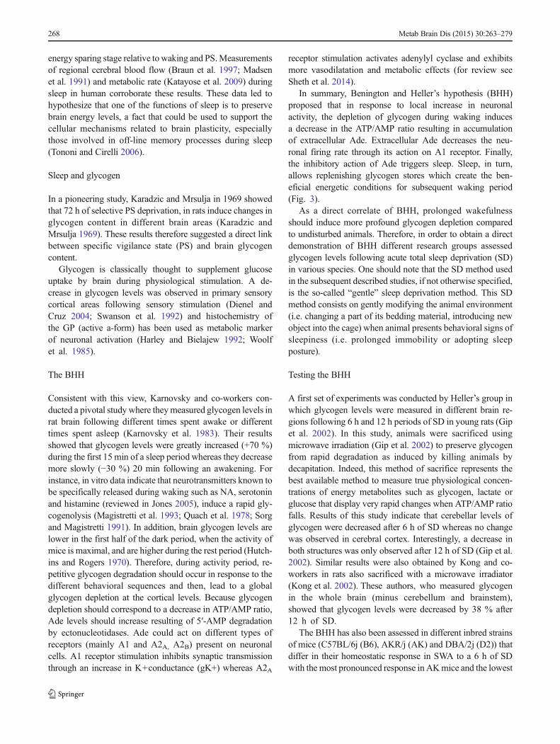
Fig. 3 Energy hypotheses ofsleep proposing glycogen as keyregulator of sleep homeostasis.Schematic views of Beningtonand Heller’s hypothesis (left side)and the “glycogenetic”hypothesis (right side) are shown(see text for detailed description).Red arrows indicate steps that arenot supported by experimentaldata. NA: noradrenaline; VIP:vasoactive intestinal peptide; 5-HT: 5-hydroxytryptamine orserotonin; SD: sleep deprivation;DAB: 1,4-dideoxy-1,4-imino-d-arabinitol; EEG:electroencephalogram; EMG:electromyogram
Metab Brain Dis (2015) 30:263–279 269
Magistretti 1996) as well as transcription and translation ofproteins involved in glycogen synthesis such as PTG whichset glycogen in a synthetic mode (Allaman et al. 2000;Allaman et al. 2003; Berman et al. 1998; Printen et al.1997). Hence, at the beginning of the waking period, onecan postulate that release of these aforementioned neurotrans-mitters induces a net consumption of glycogen but, in parallel,also triggers a “glycogenetic program” leading to a delayedresynthesis of glycogen that will occur during wakefulness.As a matter of fact, such a “rebound” in glycogen levels wasalso observed in vivo after a single hypoglycemic episode(Choi et al. 2003) and in mice injected with amphetamine(Hutchins and Rogers 1970). Such glycogen regulation is alsoconsistent with the observations made by Hutchins andRogers (1970) which indicate that, while brain glycogenlevels are low at the beginning of the active period, theyincrease before the end of this period (see Fig. 3 in Hutchinsand Rogers 1970).
The “glycogenetic” hypothesis
Actually, this alternative to BHH proposed that during wake-fulness, in addition to their glycogenolytic effects, NA, VIPand Ade also trigger glycogen synthesis through transcription-al and translational effects. Therefore, glycogen levels shouldbe maintained and even increased at the end of the wakeful-ness period (Fig. 3). Since the mechanisms which underlie thisassumption have been obtained from cortical cells and sinceEEG signal reflected cortical activity pattern, the cerebralcortex was mainly considered as “the site” for “glycogenetic”hypothesis. In addition, as a consequence of the“glycogenetic” hypothesis and in contrast to the BHH we alsoproposed that glycogen preservation could have a role in thetriggering of sleep and/or more particularly, in the occurrenceand/or length of the PS episodes. As a matter of fact, PS is anenergy consuming state that occurs following SWS episodesin which glucose consumption is low in the cortex. Thus,glycogen preserved during waking could be the cellularsource of glucose required to ensure the neuro-metaboliccoupling in response to increased neuronal activity duringPS episodes (Fig. 3).
Testing the “glycogenetic” hypothesis
In order to test this hypothesis, we first assessed the expressionin the cerebral cortex of important genes encoding proteinsrelated to glycogen metabolism such as GS, GP and PTG atfour time points across the sleep-wake cycle as well as the GSactivity following 6 h of SD in OF1 mice (Petit et al. 2002).Contrary to what was postulated, it was first observed that GSand GP mRNA levels were minimum during the active periodand maximum during the rest period and that both decreasedafter 6 h of SD. However, PTG mRNA levels reached amaximum at the end of the active period and were markedlyinduced by SD (+94 % vs control value). Since PTG mRNAinduction is known to promote GS activity and glycogensynthesis, one can logically deduce that a similar mechanismis engaged by 6 h of SD. Consistent with this, it was demon-strated that the active form of GS was significantly increasedfollowing SD in the cortex whereas the total form of GS (a+bforms) remained unchanged. Moreover, if ones assume thatsuch a glycogenetic mode is taking place at the cortical levelsthis could provide an explanation for the absence of glycogendepletion at the cortical level following 6 h of SD as describedin several studies (see above) (Franken et al. 2003; Gip et al.2002; Zimmerman et al. 2004).
Even though the “gentle” SD method used in the previous-ly described reports avoids stressful stimulations (acute in-tense noise or light, water or air puffs), plasma GCs levels areincreased by two to fourfold in such protocols (Gip et al. 2004;Petit et al. 2010; Tobler et al. 1983).
Fig. 4 Changes in brain glycogen after sleep deprivation vary withgenotype. Brain glycogen content in sleep-deprivedmice. Brain glycogenvaried with strain and structure (2-way ANOVAwith repeated measuresfor factor structure. Factor strain, F2,30=13.2, P<0.0001; structure,F2,60=71.9, P<0.0001; interaction, F4,60=1.6, P=0.20). Bars representmean values ±2SE. Top panels: *significant strain differences for eachbrain region (Tukey’s range test; P<0.05). Bottom panels: *significantdifferences from control (unpaired 2-sided t-tests; P<0.05). In the bottompanels, values were expressed as % difference from the mean values inthe control group (0 %). CTX, cerebral cortex; BS, brainstem; CB,cerebellum. Adapted from (Franken et al. 2003) with permission
270 Metab Brain Dis (2015) 30:263–279
In order to establish whether or not these metabolic chang-es were restricted to the “gentle” SDmethod, we also exploredthe impact of a pharmacological SD, which does not requireintervention and therefore limits possible stress confoundingeffects. To this end we used modafinil (MOD) which is a non-amphetamine vigilance–promoting drug classically used innarcoleptic patients (Wise et al. 2007). In mice, a singleinjection of this drug induces a continuous wakefulness during5–6 h followed by a SWA rebound in the following periodsimilar to those obtained after gentle SD (Kopp et al. 2002).Different glycogen metabolism indices including levels ofmRNA encoding GS, GP and PTG as well as glycogen levelswere measured in the cortex of mice. Whatever the methodused for SD (gentle or pharmacological), it was observed thatcortical PTG mRNA levels and GS activity were consistentlyincreased (Petit et al. 2010). In addition, we showed thatglycogen levels displayed no change in the cortex in bothSD methods used, indicating that glycogen regulation is driv-en by the prolongation of wakefulness and not by the “gentleSD” per se. Interestingly, PTG mRNA and glycogen levelswere normalized following 3 h of sleep recovery. Induction ofPTG mRNA levels was also reported in cDNA microarrayanalysis performed following SD experiments in mice (Maretet al. 2007; Mongrain et al. 2010). We should also notice thatan induction of glycogenin, is also regulated by “gentle” SD,suggesting that de novo granules formation could also partic-ipate to glycogen regulation after SD (Petit et al. 2010). Alongthe same line, a single dose of methylphenidate (Ritalin™)which is able to maintain B6 mice awake for 3–4 h, alsoinduced a significant increase in PTG mRNA levels (+50 %)as well as in glycogenin mRNA (+22%) in the anterior part ofthe cortex (unpublished data).
These results argue in favor of a glycogen synthesis occur-ring during waking in parallel to glycogenolysis induced byrepeated sensory-motor stimulations or pharmacological neu-ronal stimulations. This is likely achieved by the increase inPTG levels which is able to direct glycogen metabolismpreferentially towards synthesis.
Cortical glycogen turnover increases during prolongedwakefulness
One of the limitations of determination of glycogen levels atthe end of a SD, or following sleep recovery, is that it doesn’tallow the dynamic measurements of glycogen metabolismduring these periods. By measuring the incorporation of[1-13C]glucose into glycogen by Nuclear Magnetic Reso-nance (NMR) spectroscopy, Gruetter and co-workers deter-mined glycogen concentration and turnover in rat brainin vivo (Choi et al. 1999; Choi and Gruetter 2003; Lei et al.2007). This method has been used to determine glycogenconcentration and turnover in euglycemic rats maintainedawake during 5 h at the beginning of the rest period, compared
to undisturbed rats (Morgenthaler et al. 2009). Followingnormalization by the N-acetyl-aspartate turnover, that remainsconstant, it was demonstrated that glycogen cycles faster (2.9vs 5.3 h, p<0.05) in rats continuously maintained awake bysensory stimulations during the labeling period, revealing anincrease in glycogen turnover induced during prolongedwakefulness. In contrast, glycogen concentration remainedunchanged in the same animals (4.0 vs 3.9 μmol/g)(Morgenthaler et al. 2009). These results are in accordancewith those obtained in rodents and flies following 6 h of SDandwith our results showing an increase in GS activity in spiteof constant glycogenolysis evoked by prolonged wakefulness(Petit et al. 2010).
A synthesis of experimental data
Based on these experimental data, we can propose the follow-ing model of glycogen regulation throughout the sleep-wakecycle:
– When arousal occurs, the rise in wakefulness-related neuro-transmitters (monoamines, VIP and Ade) (e.g. seeTakahashi et al. 2010) triggers a rapid (within few minutes)glycogenolysis throughout the whole cortex. This phasecorresponds to the fall in glycogen concentration after 2–5 min of spontaneous awakening reported by Karnovskyand collaborators (Karnovsky et al. 1983). During the wak-ing period, glycogenolysis is also repeatedly triggered inspecific areas corresponding to sensory modalities solicitedby novel environmental conditions and in motor areas. Thisview is in accordance with an important neuromodulatoryrole of the cortical projections of the noradrenergic cellsfrom the locus coeruleus. These cells, in addition to a tonicmode of discharge during waking, display a phasic firing inresponse to salient sensory stimuli (Berridge andWaterhouse 2003; but see also Foote et al. 1983).
– At the same time, NA (and probably VIP and Ade)triggers the transcription and translation of PTG leadingto an increase in GS activity.
– Therefore, a balance between synthesis and degradationprogressively settles during the activity period and pre-vents glycogen decrease. This balance is also dependentof the stress levels since GCs favor glycogenolysis (seebelow). It appears that, this relative “steady state” cannotbe maintained when wakefulness duration is prolongedmore than 6–10 h in rodents as suggested by the resultsobtained after 12 h of SD (Gip et al. 2002; Kong et al.2002).
– When sleep emerges, the inhibition of the sensory inputsto the cortex together with the decrease in the monoam-inergic tone, reduces the cortical energy needs and
Metab Brain Dis (2015) 30:263–279 271
consequently decreases glycogen consumption. This rap-id unbalance in support of glycogen synthesis couldexplain the large increase in glycogen levels observedby Karnovsky and collaborators 15 min after sleep onset(Karnovsky et al. 1983).
– Finally, PTG levels and glycogen synthesis diminishduring sleep as indicated by glycogen levels mea-sured following 3 h of sleep recovery (Petit et al.2010).
Effect of direct modulation of glycogen levels on sleep
Because experimental data support the glycogen maintenanceproposed by the “glycogenetic” hypothesis, we wanted to testa physiological consequence of this observation which as-sumes that glycogen levels per se could regulate sleep. Toinvestigate this aspect we took advantage of pharmacologicalagents that block glycogen degradation (Treadway et al.2001).
1,4-dideoxy-1,4-D-arabinitol (DAB) as pharmacological tool
In order to normalize glycemia, a large number of moleculesinhibiting GP through different binding sites were designed(Oikonomakos and Somsak 2008). Among them, severalglucose analogs based on iminosugars such as Isofagomineor DAB act on the active form of GP (GP a-form), the rate-limiting enzyme of glycogen mobilization, by allosteric inhi-bition (Somsak et al. 2003). In vitro, DAB potently inhibitsGP in different preparations (Andersen andWestergaard 2002;Somsak et al. 2003) including brain tissue homogenates(IC50≈0.4 μM) (Walls et al. 2008). Moreover, DAB is effec-tive in blocking glycogen mobilization in astrocytes as shownby the full prevention of NA-induced glycogenolysis by thiscompound in astrocytes in culture (Walls et al. 2008). Impor-tantly, DAB was used to demonstrate the importance of brainglycogen mobilization for the establishment of memory for-mation (Gibbs et al. 2006; Newman et al. 2011; Suzuki et al.2011). The last set of results confirms DAB as a relevantpharmacological tool to investigate glycogen functionin vivo.
Testing the blockade of glycogen in spontaneous sleep-wakecycle
In order to assess more directly the role of glycogen on thesleep-wake architecture and /or on the occurrence and theduration of PS epochs, we used DAB to block brain glycogenmobilization in mice housed in basal conditions (light–darkcycle: 12:12), (Fig. 5). Mice received an intra-cerebro-ventricular (i.c.v) injection of either saline solution (control)
or DAB (0.5 M). Each injection was performed at the light/dark shift (i.e. zeitgeber time (ZT) 12) that corresponds to thebeginning of the active period in rodents.
We first verified the GP inhibitor efficiency in vivo bymeasuring cortical glycogen levels 3 h after i.c.v injection ofDAB. As expected, glycogen levels were significantly moreelevated (fivefold) in the DAB group as compared to controlgroup (Fig. 5a).
In a second set of experiments, using a similar protocol, weassessed a possible effect of glycogen mobilization blockadeon spontaneous locomotor activity (SLA) in mice. Injectionswere also performed at ZT12 and SLA was continuouslyrecorded during 10 h from ZT14 until the end of the darkperiod (ZT24). During this period, SLA was significantlydecreased by 38 % in DAB mice as compared to control(Fig. 5b). This last result suggests that an inhibition of glyco-genmobilization in the brain might induce an increase of sleepand/or quiet wake in mice.
To clarify this last point, we repeated the experiment withmice already implanted with EEG/EMG electrodes and wire-connected for sleep recordings. DAB or saline were injectedat ZT12 and classical vigilance states parameters were re-corded over a 10 h period (ZT14-ZT24). Results did notindicate any significant effect of DAB on SWS nor on PSparameters during this recording period (Fig. 5c). To investi-gate whether GP inhibition could alter sleep more qualitative-ly than quantitatively, we also performed a spectral analysisof the EEG during SWS epochs (by 2 h bin from ZT14 to ZT24 in control and DAB-injected mice). For this, we focusedour analysis on the SWA (power spectrum of the delta fre-quency band) which represents an index of sleep depth. Aslight but non-significant decrease in SWA was observedfollowing DAB injections indicating that the depth of theSWS was not altered by the glycogen blockade (data notshown).
These last results indicate that the large decrease in loco-motor activity observed after DAB injection is not due toincrease in sleep duration, even though an increase of quietwake/drowsiness could not be excluded. This could suggestthat blockade of glycogen mobilization might impact morespecifically brain areas involved in locomotion. Similarly,intraperitoneal injection of caffeine at a dose (200 mg/kg) thatinhibits GP, also depressed the locomotor activity in mice(Hutchins and Rogers 1970). Thus, and in contrast to the“second part” of our hypothesis where we proposed thatmaintaining glycogen levels during waking could regulatesleep, acute blockade of glycogen mobilization by DAB hasno effect on sleep parameters during the subsequent activeperiod. In particular, our results cast some doubt on the pos-sibility that glycogen could be used as energy source for PSepisodes since no change in both number or length of PSepochs was observed. Moreover, our DAB experiments con-stitute an additional argument that doesn’t support the BHH.
272 Metab Brain Dis (2015) 30:263–279
Indeed, according to this assumption, glycogen mobilizationblockade should create energy deficits more rapidly and,consequently, should increase length and/or number of sleepepisodes for activity period, a fact that is not verified experi-mentally (see above).
Based on this study with DAB, sleep occurrence andduration appear to be relatively independent to brain glycogencontent, at least on a relative short period (about 10–12 h) andin standard environment.
However, it cannot be excluded from this study that gly-cogen mobilization blockade affects other cellular processesin sub-cortical structures such as hippocampus where memory
mechanisms occur (for example the sharp waves ripples)during sleep stages (for review see Rasch and Born 2013).
Confounding effect of GCs
GCs affect cerebral metabolism
GCs are adrenal steroid hormones released in response tostressors. As previously mentioned, stress is a possible con-founding factor that accompanies most standard SD protocol
a
0
10
15
20
25
Gly
coge
n le
vels
(nm
ol/m
g pr
ot)
CTL
∗∗∗∗
5
30
35
DAB0
10
15
20
25
5
30
35
CTL DAB
∗∗∗
SLA
(arb
itrar
y un
it)
b
c
SWS
PS
CTL DAB CTL DAB CTL DAB
Duration (min) Nb of episodes Episode mean duration (min)
356.0 ± 30.9
18.1 ± 4.4
1.5 ± 0.2
1.9 ± 0.4
0.6 ± 0.1
225.9 ± 26.9
34.7 ± 5.2
2.1 ± 0.4
1.2 ± 0.1266.5 ± 28.3
22.1 ± 5.0 29.7 ± 6.0
311.3 ± 29.5 195.3 ± 27.4 207.8 ± 41.5
208.0 ± 43.0195.7 ± 29.3
0.6 ± 0.1
WAKE
Fig. 5 Effects of acute pharmacological inhibition of glycogen mobili-zation on locomotor activity and sleep parameters when added at darkonset. Experiments were performed on adult males mice (C57BL/6j fromJanvier, France) equipped with EEG and EMG electrodes (for furtherdetails see Petit et al. 2013) and implanted with a chronic cannula into thelateral ventricle. The mice were housed individually under a 12-h:12-hlight–dark cycle at 23 °C ambient temperature with food and water adlibitum. Twoweeks after the surgery, they were injectedwith 2μl of NaCl0.9 % (control [CTL] group, white bars) or 1,4-dideoxy-1,4-imino-D-arabinitol (DAB) at 0.5 M dissolved in NaCl 0.9 % (DAB group, graybars) at the beginning of the dark period (Zeitgeber Time 12, ZT12) undera light isoflurane anesthesia. After injections, mice were let 2 h in theircages for anesthesia recovery before recordings begin. a Brain glycogenlevels 3 h after i.c.v. administration of DAB. After the injection, theanimals were replaced in their cages and sacrificed 3 h later (ZT15).The brains were quickly removed, frozen and cut for site injectionverification and standard glycogen dosage (Petit et al. 2010). The valuesrepresent the mean of glycogen levels in nmol/mg prot (±SEM) measuredon brain slices from -200 μm posterior to +200 μm anterior compared tothe injection site in both group (CTL vs DAB). N=6. Statistics: Unpaired
t test with Welch’s correction,**** p value <0.0001 compared to CTL. bSpontaneous Locomotor Activity (SLA) after i.c.v injection of DAB.Each cage was equipped with passive infrared sensors on the top forSLA recordings. SLAwas recorded for 10-min interval during 10 h fromZT14 to ZT24. The same animals received NaCl 0.9 % and DAB onseparate days. The values represent mean SLA ± SEM over this 10 hperiod in both group (CTL vs DAB). N=12. Statistics: Paired t test, *** pvalue=0.0004 compared to CTL. c Quantitative parameters of the sleepwake cycle after i.c.v. injection of DAB. The EEG/EMG signals wererecorded and digitalized with an Embla A10 amplifier (Medcare, USA).The EEG was divided in 4-s epochs which were visually scored in one ofthe three states of vigilance (wakefulness (W), slow wave sleep (SWS) orparadoxical sleep (PS)) according to classical criteria (Tobler et al. 1997).Values represent the mean values (±SEM) of cumulative duration (min),number of episodes and episode mean duration (min) for each state ofvigilance for the 10 h of recording session from ZT14 to ZT24. N=6animals in each group. Statistics: One way ANOVA followed byBonferroni’s post-test. No statistical difference was observed betweenthe control and the DAB groups
Metab Brain Dis (2015) 30:263–279 273
(which can result in an increase of plasma corticosterone, aphysiological stress marker in rodents i.e. the main GCs in ratand mice).
An important role of GC in peripheral tissues is the regu-lation of glucose homeostasis (hence their name) via, amongothers, effects on glycogen metabolism (for review seeMcMahon et al. 1988). Although some direct effects of thesesteroid hormones on glycogen metabolism have been de-scribed, it is well recognized that they also act indirectly byinfluencing cells’ responsiveness to other hormones(Sapolsky et al. 2000). For example, it was demonstrated that,in perfused liver of adrenalectomized rats, stimulation ofglycogenolysis by adrenaline and glucagon was reduced com-pared to control animals (Exton et al. 1972).
GCs and sleep deprivation
GCs affect cerebral metabolism bymodulating glucose uptakeand utilization in cultured neurons and astrocytes (Allamanet al. 2004; Horner et al. 1990; Landgraf et al. 1978; Virginet al. 1991). Adrenalectomy is followed by decreased glucoselevels and increased glycolytic intermediates in the cortex(Plaschke et al. 1996). In most brain regions, GCs decreaselocal cerebral glucose utilization, whereas adrenalectomy in-creases it (e.g. see Doyle et al. 1994; Kadekaro et al. 1988).GCs also affect brain glycogen stores. In cultured cortical andhippocampal astrocytes, GCs decrease glycogen levels(Allaman et al. 2004; Tombaugh et al. 1992). Similarly, adre-nalectomy can either increase brain glycogen levels(Passonneau et al. 1971) or have no effect in the whole brain(Goldberg and O’Toole 1969) or in the cortex (Plaschke et al.1996).
Because GCs influence brain glycogen levels and are in-creased during SD, this raises the question of the contributionof GCs to changes of glycogen levels observed during SD aswell as for their role in the homeostatic regulation of sleep. Afirst observation suggesting a link between glycogen metabo-lism and GCs during SD has been obtained by Heller andcollaborators (Franken et al. 2003). Using three inbred micestrains, i.e. AK, D2, and B6, they showed that changes in brainglycogen after SD are strongly influenced by brain region andgenotype (see above and Fig. 4). Although corticosteronelevels after SD were not measured in this work, it was sug-gested that genotype differences in corticosterone inductionduring SDmight contribute to the glycogen increase observedin B6 mice and the decreases observed in AK and D2 mice,based on studies showing a decreased corticosterone responseto stress in B6 mice (e.g. see Roberts et al. 1992; Ryabininet al. 1999).
Heller’s group investigated the influence of GC during SDby measuring glycogen stores in different brain regions inintact and adrenalectomized (with corticosterone replacement;Adx) Long-Evans rats after 6 h of SD (Gip et al. 2004).
Following SD in control animals, glycogen levels de-creased in the cerebellum and hippocampus but not inthe cortex or brainstem. In contrast, glycogen levels inthe cortex of Adx rats increased by 43 % after SD,while other regions were unaffected. These results un-equivocally demonstrate that in the absence of a GCsurge (prevented by adrenalectomy), glycogen stores inall investigated brain regions were spared, implying adirect effect of GCs on cerebral glycogen levels regula-tion during SD. Moreover, this is in agreement with thenotion that elevated GCs secretion during SD causesbrain glycogenolysis and unbalances the synthesis/degradation ratio.
In addition to changes of glucose metabolism, the effect ofadrenalectomy observed in Heller’s study on glycogen levelsmay be explained by in vitro data that showing a suppressingaction of GC on the synthesis of glycogen induced by NA inastrocytes (Allaman et al. 2004). In Allaman et al. (2004) itwas observed that exposure of primary cultures of corticalastrocytes to dexamethasone (DEX), a synthetic GC, results inthe reduction of NA-induced glycogen resynthesis. DEX doesnot act through alteration of signal transduction mechanisms,as cAMP formation or PTG mRNA induced by noradrenergicstimulation was unchanged. In this context, it would be ofinterest to determine whether the suppressing action of GCson long-term glycogen regulation by NA is selective or alsoeffective for other neuromodulators such as VIP and Ade.These observations also support the view that glycogen me-tabolism during SD may be balanced between synthesis (e.g.induced by NA and Ade) and degradation (induced by GCsper se but also by the suppressive effects on NA-inducedglycogen synthesis) which may increase glycogen turnover.Such view is strengthened by the observations that prolongedwakefulness increases glycogen turnover in the cortex asshown by a NMR study (see above) (Morgenthaler et al.2009).
One should note that in the experiments by Heller’s andcollaborators on Adx Long-Evans rats, SD protocol induces amassive increase in GCs in intact animals (at least 8 foldincrease) (Gip et al. 2004). For comparison, the GCs fold-increase observed in AK and B6 mice are around 2 and 3,respectively (8 for D2, which is similar to the value for Long-Evans rats in Gip et al. 2004). Therefore, the high amplitude inGCs surge during SD as observed in Long-Evans rats mayalso amplify (override normal regulation) the GCs effects onglycogen levels. Interestingly, and as mentioned above, Petitet al (2010) compared the effects of wakefulness induced by“gentle” SD and or following injection of the wakefulness-promoting drug MOD on the expression of genes related toglucose and glycogen metabolism, GS activity as well as onglycogen levels in the cortex of the outbred mice strain OF1(Petit et al. 2010). In this study plasma corticosterone were notinfluenced byMOD whereas a moderate (2 fold) increase was
274 Metab Brain Dis (2015) 30:263–279
observed following “gentle” SD. Despite this differential ef-fect on GCs plasma levels, MOD and “gentle” SD exerted asimilar influence on gene expression including PTG, GSactivity and on glycogen levels. It could thus be concludedthat the mild stress induced by “gentle” SD was unlikelyinvolved in the effects of “gentle” SD on glycogen metabo-lism indices analyzed including glycogen content at the corti-cal level.
Are GCs modulators of sleep homeostasis?
An important question is therefore the role of GCs surge in thehomeostatic regulation of sleep. In human, plasma cortisol(and corticosterone in rodents) concentration follows a circa-dian rhythm independent of sleep. It peaks at the time ofawakening and slowly decreased during waking to reach aminimum when sleep begins (Van Cauter et al. 1991). Thisobservation suggests a role of GCs in the building of sleeppressure. Recently, Mongrain and collaborators provided animportant piece of work to answer this question (Mongrainet al. 2010). In this study, they aimed to elucidate the contri-bution of the corticosterone component of the stress responseto the sleep-wake associated changes in the electrophysiolog-ical correlates of sleep need. To this end, they took advantageof D2 mice which display high corticosterone increase fol-lowing SD to determine the contribution of this high cortico-sterone increase to blunted SWA rebound in Adx animals withcorticosterone replacement. These results clearly demonstrat-ed that while Adx successfully abolished the SD–inducedincrease in corticosterone secretion, Adx affected neither thebaseline dynamics of SWAnor the increase in SWA after sleepdeprivation as compared to in both intact controls and thesham-lesioned mice. Importantly, quantity and distribution ofsleep, similarly to SWA during SWS, remain unaffected. Thefact that Adx does not influence the SWA response to SD,argues against a major role of GCs in the homeostatic regula-tion of sleep. More importantly, whatever are the effects ofGCs in SD in the regulation of glycogen levels, GCs do notappear to participate in the homeostatic regulations of sleep asshown by Mongrain’s work. Therefore, such results (modula-tion or absence of modulation of glycogen levels by GCs asobserved in the different studies presented in this section) alsoquestioned the importance of glycogen store replenishment asa regulator of sleep homeostasis.
Concluding remarks
During the last decade, efforts were made to validate orinvalidate the original hypothesis made by Benington andHeller on the role played by glycogen metabolism in thehomeostatic regulation of sleep (part of these data have
already been reviewed by Scharf and collaborators (Scharfet al. 2008)) . When considering results obtained in the differ-ent studies reviewed here, one can conclude that, contrary towhat was initially postulated (i.e.glycogen reserves are mobi-lized during wakefulness), cortical glycogen levels remainrelatively stable throughout the sleep-wake cycle, except atthe beginning of rest and active periods, where glycogenlevels rise and fall respectively for few minutes in responseto rapid change in neuronal activity. More striking, glycogenreserves did not consistently decrease when wakefulness isprolonged.
Most results rather support the notion that, during wake-fulness, a parallel glycogen synthesis and degradation setsresulting in a glycogen turnover increase. This is in accor-dance with the “glycogenetic” hypothesis which postulatesthe occurrence of a glycogen synthesis during waking. How-ever, the assumption of the “glycogenetic” hypothesis thatglycogen synthesized during waking could favor sleep(SWS and /or PS), was not verified in the experiments inwhich glycogen mobilization was pharmacologically blockedby DAB (Fig. 5), at least when it was acutely administrated.
Hence, none of the actual results (including GCs effect onsleep homeostasis) fully support a direct correlation betweensleep and glycogen regulation (i.e. BHH or “glycogenetic”hypothesis) (Fig. 3). Nevertheless, a link between glycogenmetabolism and sleep mechanisms could not be totally ex-cluded. Indeed, experimental studies present several limita-tions that may impede the establishment of a direct role ofglycogen metabolism in sleep regulation:
– Modulation (i.e. no change or increase) of glycogenlevels was mainly assessed in the cortex (or whole brain)extracts of rodents whereas only few studies investigatedpossible changes in glycogen level in other brain areassuch as cerebellum or brainstem. Interestingly, in theselatter two brain areas a different regulation of glycogencontent after SD was observed, suggesting that associa-tion between sleep homeostasis and glycogen levels maybe stronger in these regions than in the cortex or wholebrain extracts. Therefore, studies focusing on differentand specialized brain structures might reveal importantlink between glycogen metabolism and sleep. In particu-lar, comparison of sleep-induced glycogen levels varia-tion between sleep-promoting areas (preoptic area, basalforebrain,…) and wake-promoting areas (posterior hypo-thalamus, locus coeruleus area,…), which may be sup-posed to display more pronounced variations during vig-ilance states, may bring interesting information to addressthis issue. Similarly, while glycogen-containing astro-cytes can be found in both grey and white matters, onlyone study specifically assessed glycogen levels changesin white matter during SD (Kong et al. 2002). It wasobserved that SD impacted glycogen levels in both white
Metab Brain Dis (2015) 30:263–279 275
and grey matters equally, indicating that white matterglycogen may also undergo specific regulation duringsleep which may deserve particular interest.
– Regulation of glycogen metabolism is likely betterreflected by turnover determinations rather than by staticmeasurements of glycogen levels, as indicated by thework of Morgenthaler and collaborators using NMRspectroscopy (Morgenthaler et al. 2009). Therefore, ef-forts to establish associations between brain glycogenturnover and sleep homeostasis would be of interest inorder to delineate the importance of glycogenmetabolismin this process.
– It is classically assumed that SD is a stress per se, henceunavoidably accompanying SD protocols. While GCs donot constitute the only stress marker, an increase in GCs isusually observed in classical SD procedures (even in“gentle” sleep deprivation such as in most of the studiesreviewed here (Gip et al. 2004; Petit et al. 2010)). Since,GCs affect glycogen regulation by dampening its synthe-sis, GCs have to be considered as a confounding factor inSD protocols. Fortunately, there are means to circumventthe GCs issue. For instance, pharmacological SD as usedby Petit and co-workers illustrates such a possible way toavoid increase in plasma GCs (Petit et al. 2010). More-over, the development of new non-stressful SD devicesrepresents another interesting alternative way to preventstress effects in metabolism-sleep interactions studies(e.g. see Leenaars et al. 2011; Petit et al. 2013).
– Mechanisms of glycogen regulation based on the“glycogenetic hypothesis” are supported by results ob-tained for 6 h SD. When wakefulness is more prolonged(such as in 12 h of SD), glycogen depletion is observed(Kong et al. 2002), suggesting that other mechanisms ofglycogen regulation take place.
To conclude, we should underline that thanks to the initialBHH, several experiments have been performed and contrib-uted to test it which have contributed a better understanding,albeit incomplete, of the relationship between glycogen regu-lation and sleep homeostasis. Even if the Benington andHeller’s assumption that sleep serves for glycogen replenish-ment is not supported by the experimental data, their hypoth-esis underlined the important role of astrocytic energy metab-olism in sleep regulation. Quite recently, such a role of astro-cytes related to Ade has been uncovered. For instance, Halassaand co-workers reported the loss of SWA rebound after SD inmice in which the vesicular release of ATP, the precursor ofAde, was suppressed specifically in astrocytes (Frank 2013;Halassa et al. 2009). In fact, a body of evidence indicates thatastrocytes-released ATP modulates neuronal transmission byacting on A1 receptors which ultimately leads to a SWAincrease (Blutstein and Haydon 2013). Interestingly, ATPrelease evoked by glutamate, potassium and adenosine in
cultured astrocytes was shown to be prevented by DAB (Hertzet al. 2014), providing a possible yet unsuspected link be-tween glycogen metabolism, ATP release and sleep regula-tion. In line with the importance of astrocytes in higher brainfunctions, the involvement of glycogen metabolism in mem-ory processes has recently been unveiled in rodents (Newmanet al. 2011; Suzuki et al. 2011).
Acknowledgments This work was supported by grants from SwissNational Science Foundation (FNRS) (no. 310030B-148169/1), fromthe NCCR Synapsy and from the Biaggi and Panacée Foundations toP.J.M.
Open Access This article is distributed under the terms of the CreativeCommons Attribution License which permits any use, distribution, andreproduction in any medium, provided the original author(s) and thesource are credited.
References
Allaman I (2009) Glial Glycogen Metabolism. In: Squire LR (ed)Encyclopedia of Neuroscience. Academic, Oxford, pp 811–818
Allaman I, Pellerin L, Magistretti PJ (2000) Protein targeting to glycogenmRNA expression is stimulated by noradrenaline in mouse corticalastrocytes. Glia 30:382–391
Allaman I, Lengacher S, Magistretti PJ, Pellerin L (2003) A2B receptoractivation promotes glycogen synthesis in astrocytes through modu-lation of gene expression. Am J Physiol Cell Physiol 284:C696–C704
Allaman I, Pellerin L, Magistretti PJ (2004) Glucocorticoids modulateneurotransmitter-induced glycogen metabolism in cultured corticalastrocytes. J Neurochem 88:900–908
Andersen B, Westergaard N (2002) The effect of glucose on the potencyof two distinct glycogen phosphorylase inhibitors. Biochem J 367:443–450
Archer SN, Carpen JD, Gibson M, Lim GH, Johnston JD, Skene DJ, vonSchantz M (2010) Polymorphism in the PER3 promoter associateswith diurnal preference and delayed sleep phase disorder. Sleep 33:695–701
Armstrong CG, Browne GJ, Cohen P, Cohen PT (1997) PPP1R6, a novelmember of the family of glycogen-targetting subunits of proteinphosphatase 1. FEBS Lett 418:210–214
Benington JH, Heller HC (1995) Restoration of brain energy metabolismas the function of sleep. Prog Neurobiol 45:347–360
Berman HK, O’Doherty RM, Anderson P, Newgard CB (1998)Overexpression of protein targeting to glycogen (PTG) in rat hepa-tocytes causes profound activation of glycogen synthesis indepen-dent of normal hormone- and substrate-mediated regulatory mech-anisms. J Biol Chem 273:26421–26425
Berridge CW, Waterhouse BD (2003) The locus coeruleus-noradrenergicsystem: modulation of behavioral state and state-dependent cogni-tive processes. Brain Res Brain Res Rev 42:33–84
Blutstein T, Haydon PG (2013) The Importance of astrocyte-derivedpurines in the modulation of sleep. Glia 61:129–139
Bollen M, Keppens S, Stalmans W (1998) Specific features of glycogenmetabolism in the liver. Biochem J 336:19–31
Borbely AA, Achermann P (1999) Sleep homeostasis and models ofsleep regulation. J Biol Rhythms 14:557–568
Borbely AA, Baumann F, Brandeis D, Strauch I, Lehmann D (1981)Sleep deprivation: effect on sleep stages and EEG power density inman. Electroencephalogr Clin Neurophysiol 51:483–495
276 Metab Brain Dis (2015) 30:263–279
Borbely AA, Tobler I, Hanagasioglu M (1984) Effect of sleepdeprivation on sleep and EEG power spectra in the rat. BehavBrain Res 14:171–182
Brady MJ, Printen JA, Mastick CC, Saltiel AR (1997) Role of proteintargeting to glycogen (PTG) in the regulation of proteinphosphatase-1 activity. J Biol Chem 272:20198–20204
Braun AR, Balkin TJ, Wesenten NJ, Carson RE, Varga M, Baldwin P,Selbie S, Belenky G, Herscovitch P (1997) Regional cerebral bloodflow throughout the sleep-wake cycle. An H2(15)O PET study.Brain 120(Pt 7):1173–1197
Brown AM (2004) Brain glycogen re-awakened. J Neurochem 89:537–552
Brown AM, Tekkok SB, Ransom BR (2003) Glycogen regulation andfunctional role in mouse white matter. J Physiol 549:501–512
Brown AM, Tekkok SB, Ransom BR (2004) Energy transfer fromastrocytes to axons: the role of CNS glycogen. Neurochem Int 45:529–536
BuchsbaumMS,Gillin JC,Wu J, Hazlett E, SicotteN,Dupont RM,BunneyWE Jr (1989) Regional cerebral glucose metabolic rate in human sleepassessed by positron emission tomography. Life Sci 45:1349–1356
Campbell SS, Tobler I (1984) Animal sleep: a review of sleep durationacross phylogeny. Neurosci Biobehav Rev 8:269–300
Capellini I, Barton RA, McNamara P, Preston BT, Nunn CL (2008)Phylogenetic analysis of the ecology and evolution of mammaliansleep. Evolution 62:1764–1776
Cardinaux JR, Magistretti PJ (1996) Vasoactive intestinal peptide,pituitary adenylate cyclase-activating peptide, and noradrenalineinduce the transcription factors CCAAT/enhancer binding pro-tein (C/EBP)-beta and C/EBP delta in mouse cortical astrocytes:involvement in cAMP-regulated glycogen metabolism. JNeurosci 16:919–929
Cataldo AM, Broadwell RD (1986) Cytochemical identification of cere-bral glycogen and glucose-6-phosphatase activity under normal andexperimental conditions: I. Neurons and Glia J Electron MicroscTech 3:413–437
Choi IY, Gruetter R (2003) In vivo 13C NMR assessment of brainglycogen concentration and turnover in the awake rat. NeurochemInt 43:317–322
Choi IY, Tkac I, Ugurbil K, Gruetter R (1999) Noninvasive measure-ments of [1-(13)C]glycogen concentrations and metabolism in ratbrain in vivo. J Neurochem 73:1300–1308
Choi IY, Seaquist ER, Gruetter R (2003) Effect of hypoglycemia on brainglycogen metabolism in vivo. J Neurosci Res 72:25–32
Chong SY, Ptacek LJ, Fu YH (2012) Genetic insights on sleep schedules:this time, it’s PERsonal. Trends Genet 28:598–605
Cirelli C, Tononi G (2008) Is sleep essential? PLoS Biol 6:e216Dibner C, Schibler U, Albrecht U (2010) The mammalian circadian
timing system: organization and coordination of central and periph-eral clocks. Annu Rev Physiol 72:517–549
Dienel GA, Cruz NF (2004) Nutrition during brain activation: does cell-to-cell lactate shuttling contribute significantly to sweet and sourfood for thought? Neurochem Int 45:321–351
Dienel GA, Ball KK, Cruz NF (2007) A glycogen phosphorylase inhib-itor selectively enhances local rates of glucose utilization in brainduring sensory stimulation of conscious rats: implications for gly-cogen turnover. J Neurochem 102:466–478
Doherty MJ, Young PR, Cohen PT (1996) Amino acid sequence of anovel protein phosphatase 1 binding protein (R5) which is related tothe liver- and muscle-specific glycogen binding subunits of proteinphosphatase 1. FEBS Lett 399:339–343
Doyle P, Guillaume-Gentil C, Rohner-Jeanrenaud F, Jeanrenaud B (1994)Effects of corticosterone administration on local cerebral glucoseutilization of rats. Brain Res 645:225–230
Dringen R, Hamprecht B (1992) Glucose, insulin, and insulin-like growthfactor I regulate the glycogen content of astroglia-rich primarycultures. J Neurochem 58:511–517
Exton JH, Friedmann N, Wong EH, Brineaux JP, Corbin JD, Park CR(1972) Interaction of glucocorticoids with glucagon and epinephrinein the control of gluconeogenesis and glycogenolysis in liver and oflipolysis in adipose tissue. J Biol Chem 247:3579–3588
Foote SL, Bloom FE, Aston-Jones G (1983) Nucleus locus ceruleus: newevidence of anatomical and physiological specificity. Physiol Rev63:844–914
Frank MG (2013) Astroglial regulation of sleep homeostasis. Curr OpinNeurobiol 23:812–818
Franken P, Gip P, Hagiwara G, Ruby NF, Heller HC (2003) Changes inbrain glycogen after sleep deprivation vary with genotype. Am JPhysiol Regul Integr Comp Physiol 285:R413–R419
Gibbs ME, Anderson DG, Hertz L (2006) Inhibition of glycogenolysis inastrocytes interrupts memory consolidation in young chickens. Glia54:214–222
Gip P, Hagiwara G, Ruby NF, Heller HC (2002) Sleep deprivationdecreases glycogen in the cerebellum but not in the cortex of youngrats. Am J Physiol Regul Integr Comp Physiol 283:R54–R59
Gip P, Hagiwara G, Sapolsky RM, Cao VH, Heller HC, Ruby NF (2004)Glucocorticoids influence brain glycogen levels during sleep depri-vation. Am J Physiol Regul Integr Comp Physiol 286:R1057–R1062
Goldberg ND, O’Toole AG (1969) The properties of glycogen synthetaseand regulation of glycogen biosynthesis in rat brain. J Biol Chem244:3053–3061
Halassa MM, Florian C, Fellin T, Munoz JR, Lee SY, Abel T, HaydonPG, Frank MG (2009) Astrocytic modulation of sleep homeostasisand cognitive consequences of sleep loss. Neuron 61:213–219
Hamai M, Minokoshi Y, Shimazu T (1999) L-Glutamate and insulinenhance glycogen synthesis in cultured astrocytes from the rat brainthrough different intracellular mechanisms. J Neurochem 73:400–407
Harley CA, Bielajew CH (1992) A comparison of glycogen phosphory-lase a and cytochrome oxidase histochemical staining in rat brain. JComp Neurol 322:377–389
Heiss WD, Pawlik G, Herholz K, Wagner R, Wienhard K (1985)Regional cerebral glucose metabolism in man during wakefulness,sleep, and dreaming. Brain Res 327:362–366
Hertz L, Xu J, Peng L (2014) Glycogenolysis and purinergic signaling.Adv Neurobiol 11:31–54
Hof PR, Pascale E, Magistretti PJ (1988) K+ at concentrations reached inthe extracellular space during neuronal activity promotes a Ca2+-dependent glycogen hydrolysis in mouse cerebral cortex. J Neurosci8:1922–1928
Horner HC, Packan DR, Sapolsky RM (1990) Glucocorticoids inhibitglucose transport in cultured hippocampal neurons and glia.Neuroendocrinology 52:57–64
Huber R, Ghilardi MF, Massimini M, Tononi G (2004) Local sleep andlearning. Nature 430:78–81
Hutchins DA, Rogers KJ (1970) Physiological and drug-inducedchanges in the glycogen content of mouse brain. Br JPharmacol 39:9–25
Ignacio PC, Baldwin BA, Vijayan VK, Tait RC, Gorin FA (1990) Brainisozyme of glycogen phosphorylase: immunohistological localiza-tion within the central nervous system. Brain Res 529:42–49
Jay TM, Jouvet M, Des Rosiers MH (1985) Local cerebral glucoseutilization in the free moving mouse: a comparison during twostages of the activity-rest cycle. Brain Res 342:297–306
Jensen TE, Richter EA (2012) Regulation of glucose and glycogenmetabolism during and after exercise. J Physiol 590:1069–1076
Jones BE (2005) From waking to sleeping: neuronal and chemical sub-strates. Trends Pharmacol Sci 26:578–586
KadekaroM, ItoM, Gross PM (1988) Local cerebral glucose utilization isincreased in acutely adrenalectomized rats. Neuroendocrinology 47:329–334
Karadzic V, Mrsulja B (1969) Deprivation of paradoxical sleep and brainglycogen. J Neurochem 16:29–34
Metab Brain Dis (2015) 30:263–279 277
Karnovsky ML, Reich P, Anchors JM, Burrows BL (1983) Changes inbrain glycogen during slow-wave sleep in the rat. J Neurochem 41:1498–1501
Katayose Y, Tasaki M, Ogata H, Nakata Y, Tokuyama K, Satoh M (2009)Metabolic rate and fuel utilization during sleep assessed by whole-body indirect calorimetry. Metabolism 58:920–926
Kennedy C, Gillin JC, Mendelson W, Suda S, Miyaoka M, Ito M,Nakamura RK, Storch FI, Pettigrew K, Mishkin M, Sokoloff L(1982) Local cerebral glucose utilization in non-rapid eyemovementsleep. Nature 297:325–327
Ko CH, Takahashi JS (2006) Molecular components of the mammaliancircadian clock. Hum Mol Genet 15(Spec No 2):R271–R277
Koizumi J, Shiraishi H (1970)Glycogen accumulation in astrocytes of thestriatum and pallium of the rabbit following administration of psy-chotropic drugs. J Electron Microsc (Tokyo) 19:182–187
Koizumi J, Shiraishi H (1971) Ultrastructural appearance of glycogen inthe hippocampus of the rabbit following administration of psycho-tropic drugs. Exp Brain Res 13:451–460
Kong J, Shepel PN, Holden CP, Mackiewicz M, Pack AI, Geiger JD(2002) Brain glycogen decreases with increased periods of wake-fulness: implications for homeostatic drive to sleep. J Neurosci 22:5581–5587
Kopp C, Petit JM, Magistretti P, Borbely AA, Tobler I (2002)Comparison of the effects of modafinil and sleep deprivation onsleep and cortical EEG spectra in mice. Neuropharmacology 43:110–118
Krueger JM, Tononi G (2011) Local use-dependent sleep; synthesis of thenew paradigm. Curr Top Med Chem 11:2490–2492
Landgraf R, Mitro A, Hess J (1978) Regional net uptake of 14C-glucoseby rat brain under the influence of corticosterone. Endocrinol Exp12:119–129
Leenaars CH, Dematteis M, Joosten RN, Eggels L, Sandberg H, SchirrisM, Feenstra MG, van Someren EJ (2011) A new automated methodfor rat sleep deprivation with minimal confounding effects on cor-ticosterone and locomotor activity. J Neurosci Methods 196:107–117
Lei H, Morgenthaler F, Yue T, Gruetter R (2007) Direct validation ofin vivo localized (13)C MRS measurements of brain glycogen.Magn Reson Med 57:243–248
Magistretti PJ, Morrison JH, Shoemaker WJ, Sapin V, Bloom FE (1981)Vasoactive intestinal polypeptide induces glycogenolysis in mousecortical slices: a possible regulatory mechanism for the local controlof energy metabolism. Proc Natl Acad Sci U S A 78:6535–6539
Magistretti PJ, Sorg O, Martin JL (1993) Regulation of glycogen metab-olism in astrocytes: physiological, pharmacological, and pathologi-cal aspects. In: Murphy S (ed) Astrocytes: pharmacology and func-tion. Academic, San Diego, pp 243–265
Maret S, Dorsaz S, Gurcel L, Pradervand S, Petit B, Pfister C,Hagenbuchle O, O’Hara BF, Franken P, Tafti M (2007) Homer1ais a core brain molecular correlate of sleep loss. Proc Natl Acad SciU S A 104:20090–20095
Mastick CC, Brady MJ, Printen JA, Ribon V, Saltiel AR (1998) Spatialdeterminants of specificity in insulin action. Mol Cell Biochem 182:65–71
McMahon M, Gerich J, Rizza R (1988) Effects of glucocorticoids oncarbohydrate metabolism. Diabetes Metab Rev 4:17–30
Mongrain V, Hernandez SA, Pradervand S, Dorsaz S, Curie T,Hagiwara G, Gip P, Heller HC, Franken P (2010) Separatingthe contribution of glucocorticoids and wakefulness to the mo-lecular and electrophysiological correlates of sleep homeostasis.Sleep 33:1147–1157
Moore RY (2007) Suprachiasmatic nucleus in sleep-wake regulation.Sleep Med 8(Suppl 3):27–33
Morgenthaler FD, Lanz BR, Petit JM, Frenkel H, Magistretti PJ, GruetterR (2009) Alteration of brain glycogen turnover in the conscious ratafter 5h of prolonged wakefulness. Neurochem Int 55:45–51
Newgard CB, Brady MJ, O’Doherty RM, Saltiel AR (2000) Organizingglucose disposal: emerging roles of the glycogen targeting subunitsof protein phosphatase-1. Diabetes 49:1967–1977
Newman LA, Korol DL, Gold PE (2011) Lactate produced by glycogen-olysis in astrocytes regulates memory processing. PLoS One 6:e28427
Nofzinger EA, Buysse DJ, Miewald JM, Meltzer CC, Price JC, SembratRC, Ombao H, Reynolds CF, Monk TH, Hall M, Kupfer DJ, MooreRY (2002) Human regional cerebral glucose metabolism duringnon-rapid eye movement sleep in relation to waking. Brain 125:1105–1115
Oikonomakos NG, Somsak L (2008) Advances in glycogen phosphory-lase inhibitor design. Curr Opin Investig Drugs 9:379–395
Panda S, Hogenesch JB, Kay SA (2002) Circadian rhythms from flies tohuman. Nature 417:329–335
Passonneau JV, Brunner EA, Molstad C, Passonneau R (1971) Theeffects of altered endocrine states and of ether anaesthesia on mousebrain. J Neurochem 18:2317–2328
Petit JM, Tobler I, Allaman I, Borbely AA, Magistretti PJ (2002) Sleepdeprivation modulates brain mRNAs encoding genes of glycogenmetabolism. Eur J Neurosci 16:1163–1167
Petit JM, Tobler I, Kopp C, Morgenthaler F, Borbely AA, Magistretti PJ(2010) Metabolic response of the cerebral cortex following gentlesleep deprivation and modafinil administration. Sleep 33:901–908
Petit JM, Gyger J, Burlet-Godinot S, Fiumelli H, Martin JL, MagistrettiPJ (2013) Genes Involved in the Astrocyte-Neuron Lactate Shuttle(ANLS) Are Specifically Regulated in Cortical AstrocytesFollowing Sleep Deprivation in Mice. Sleep 36:1445–1458
Pfeiffer-Guglielmi B, Fleckenstein B, Jung G, Hamprecht B (2003)Immunocytochemical localization of glycogen phosphorylase iso-zymes in rat nervous tissues by using isozyme-specific antibodies. JNeurochem 85:73–81
Phelps CH (1972) Barbiturate-induced glycogen accumulation in brain.An electron microscopic study. Brain Res 39:225–234
Plaschke K, Muller D, Hoyer S (1996) Effect of adrenalectomy andcorticosterone substitution on glucose and glycogen metabolism inrat brain. J Neural Transm 103:89–100
Printen JA, Brady MJ, Saltiel AR (1997) PTG, a protein phosphatase 1-binding protein with a role in glycogen metabolism. Science 275:1475–1478
Quach TT, Rose C, Schwartz JC (1978) [3H]Glycogen hydrolysis in brainslices: responses to neurotransmitters and modulation of noradren-aline receptors. J Neurochem 30:1335–1341
Ramm P, Frost BJ (1986) Cerebral and local cerebral metabolism in thecat during slow wave and REM sleep. Brain Res 365:112–124
Rasch B, Born J (2013) About sleep’s role in memory. Physiol Rev 93:681–766
Rattenborg NC, Lima SL, Lesku JA (2012) Sleep locally, act globally.Neuroscientist 18:533–546
Roach PJ, DePaoli-Roach AA, Hurley TD, Tagliabracci VS (2012)Glycogen and its metabolism: some new developments and oldthemes. Biochem J 441:763–787
Roberts AJ, Crabbe JC, Keith LD (1992) Genetic differences inhypothalamic-pituitary-adrenal axis responsiveness to acute ethanoland acute ethanol withdrawal. Brain Res 579:296–302
Ryabinin AE, Wang YM, Finn DA (1999) Different levels of Fos immu-noreactivity after repeated handling and injection stress in twoinbred strains of mice. Pharmacol Biochem Behav 63:143–151
Sapolsky RM, Romero LM, Munck AU (2000) How do glucocorticoidsinfluence stress responses? Integrating permissive, suppressive,stimulatory, and preparative actions. Endocr Rev 21:55–89
278 Metab Brain Dis (2015) 30:263–279
Scharf MT, Naidoo N, Zimmerman JE, Pack AI (2008) The energyhypothesis of sleep revisited. Prog Neurobiol 86:264–280
Sheth S, Brito R, Mukherjea D, Rybak LP, Ramkumar V (2014)Adenosine receptors: expression, function and regulation. Int JMol Sci 15:2024–2052
Siegel JM (2001) The REM sleep-memory consolidation hypothesis.Science 294:1058–1063
Somsak L, Nagya V, Hadady Z, Docsa T, Gergely P (2003) Glucoseanalog inhibitors of glycogen phosphorylases as potential antidia-betic agents: recent developments. Curr Pharm Des 9:1177–1189
Sorg O, Magistretti PJ (1991) Characterization of the glycogenolysiselicited by vasoactive intestinal peptide, noradrenaline and adeno-sine in primary cultures of mouse cerebral cortical astrocytes. BrainRes 563:227–233
Sorg O, Magistretti PJ (1992) Vasoactive intestinal peptide and noradren-aline exert long-term control on glycogen levels in astrocytes:blockade by protein synthesis inhibition. J Neurosci 12:4923–4931
Suzuki A, Stern SA, Bozdagi O, Huntley GW,Walker RH,Magistretti PJ,Alberini CM (2011) Astrocyte-neuron lactate transport is requiredfor long-term memory formation. Cell 144:810–823
Swanson RA (1992) Physiologic coupling of glial glycogen metabolismto neuronal activity in brain. Can J Physiol Pharmacol 70:S138–S144
Swanson RA, Morton MM, Sagar SM, Sharp FR (1992) Sensory stimu-lation induces local cerebral glycogenolysis: demonstration by au-toradiography. Neuroscience 51:451–461
Takahashi K, Kayama Y, Lin JS, Sakai K (2010) Locus coeruleus neuro-nal activity during the sleep-waking cycle in mice. Neuroscience169:1115–1126
Tobler I, Murison R, Ursin R, Ursin H, Borbely AA (1983) The effect ofsleep deprivation and recovery sleep on plasma corticosterone in therat. Neurosci Lett 35:297–300
Tobler I, Deboer T, FischerM (1997) Sleep and sleep regulation in normaland prion protein-deficient mice. J Neurosci 17:1869–1879
Tombaugh GC, Yang SH, Swanson RA, Sapolsky RM (1992)Glucocorticoids exacerbate hypoxic and hypoglycemic hippocam-pal injury in vitro: biochemical correlates and a role for astrocytes. JNeurochem 59:137–146
Tononi G, Cirelli C (2006) Sleep function and synaptic homeostasis.Sleep Med Rev 10:49–62
Treadway JL, Mendys P, Hoover DJ (2001) Glycogen phosphorylaseinhibitors for treatment of type 2 diabetes mellitus. Expert OpinInvestig Drugs 10:439–454
VanCauter E, Blackman JD, Roland D, Spire JP, Refetoff S, PolonskyKS(1991) Modulation of glucose regulation and insulin secretion bycircadian rhythmicity and sleep. J Clin Invest 88:934–942
Virgin CE Jr, Ha TP, Packan DR, Tombaugh GC, Yang SH, Horner HC,Sapolsky RM (1991) Glucocorticoids inhibit glucose transport andglutamate uptake in hippocampal astrocytes: implications for gluco-corticoid neurotoxicity. J Neurochem 57:1422–1428
Vyazovskiy VV, Faraguna U (2014) Sleep and Synaptic Homeostasis.Curr Top Behav Neurosci. doi:10.1007/7854_2014_301
Vyazovskiy V, Borbely AA, Tobler I (2000) Unilateral vibrissae stimula-tion during waking induces interhemispheric EEG asymmetry dur-ing subsequent sleep in the rat. J Sleep Res 9:367–371
Vyazovskiy VV, Cirelli C, Tononi G, Tobler I (2008) Cortical metabolicrates as measured by 2-deoxyglucose-uptake are increased afterwaking and decreased after sleep in mice. Brain Res Bull 75:591–597
Vyazovskiy VV, Olcese U, Hanlon EC, Nir Y, Cirelli C, Tononi G (2011)Local sleep in awake rats. Nature 472:443–447
Walls AB, SickmannHM, BrownA, Bouman SD, RansomB, SchousboeA, Waagepetersen HS (2008) Characterization of 1,4-dideoxy-1,4-imino-d-arabinitol (DAB) as an inhibitor of brain glycogen shuntactivity. J Neurochem 105:1462–1470
Wise MS, Arand DL, Auger RR, Brooks SN, Watson NF (2007)Treatment of narcolepsy and other hypersomnias of central origin.Sleep 30:1712–1727
Woolf CJ, Chong MS, Rashdi TA (1985) Mapping increased glycogenphosphorylase activity in dorsal root ganglia and in the spinal cordfollowing peripheral stimuli. J Comp Neurol 234:60–76
Zimmerman JE, Mackiewicz M, Galante RJ, Zhang L, Cater J, Zoh C,Rizzo W, Pack AI (2004) Glycogen in the brain of Drosophilamelanogaster: diurnal rhythm and the effect of rest deprivation. JNeurochem 88:32–40
Zimmerman JE, Naidoo N, Raizen DM, Pack AI (2008) Conservation ofsleep: insights from non-mammalian model systems. TrendsNeurosci 31:371–376