Ya Lin Tnay,a Edwin Kok Lee Yeow,a Liang Yang,c Lai Guan Ng,b Shunsuke Chiba*a
and Bengang Xing*ad
A novel theranostic divalent vancomycin analog using a planar
1,8-diazapyrene moiety as a rigid scaffold exhibits potent and
selective antibacterial activity against Gram (+) bacteria including
vancomycin-resistant strains, while having minimal influence on
Gram (�) bacteria and mammalian cells. Moreover, this theranostic
analog can be also applied for selective two-photon fluorescence
imaging of Gram (+) bacteria.
As the drugs of last defence, the glycopeptide based antibiotics,e.g. vancomycin (Van), have been extensively used in clinicsfor effective treatment of lethal bacterial infections caused bymethicillin-resistant Staphylococcus aureus (MRSA),1 mainly dueto their tight binding affinity to the C-terminal D-Ala–D-Ala motifpresent in bacterial cell wall precursors.1,2 However, recentemergence of virulent resistant species known as vancomycin-resistant Enterococci (VRE) and Staphylococcus,1,3 which canremodel their surface peptidoglycan sequence from D-Ala–D-Alato D-Ala–D-Lac and substantially decrease the binding affinity(B103 times loss) of the Van molecule,1,2,4 has raised seriousconcerns for human healthcare worldwide and thus urged moreefforts to develop alternative treatment strategies to combat theprevalence of antibiotic resistance.1,3
So far, some semisynthetic glycopeptide antibiotics, includingdalbavancin, oritavancin and telavancin etc., which incorporatelipophilic moieties in Van-like structures, have been utilized to treatinfections caused by multi-drug-resistant Gram (+) pathogens.1,5
The combination of inhibition of cell-wall synthesis and disrup-tion of membrane integrity has been considered as the majormechanism of action to sustain the potent antimicrobial activity.6
Alternative multivalent/polyvalent binding scaffolds based oncovalently linked Van dimers or oligomers have also beenproposed to enhance the potent activities against Van-resistantstrains,7 mostly attributed to their multivalent interactions thatcircumvent the low affinity between Van and the D-Ala–D-Lacpeptide in resistant bacteria.8 Despite the promising antibacterialactivities, the development of new antimicrobial drug analogs thatexhibit specific activity against VRE, while avoiding interferencewith the endogenous microbial population, is of high significancein clinical practice.9
Moreover, in line with the urgent demand for new antimicro-bial agents to combat the increasing resistance, early diagnosiswill also be a key to guarantee patients’ survival from resistantbacterial infections. The optical imaging technique based onfluorescent protein variants or small molecule probes offers rapidand direct analysis of a variety of cellular events with promisingsensitivity, and has become a powerful tool in studying bacterialinfections and monitoring the biological mechanisms in antibioticresistance.10 Despite its initial success, several technical barriers stillexist regarding nonspecific background signals, light bleaching,limited penetration and possible photodamage, especially for thosefluorescent probes requiring short wavelength UV or blue lightexcitation, which may hamper the biomedical applications of opticalimaging in living systems.11 Compared to the conventional opticalimaging modality, two-photon imaging exhibits promising advan-tages, including minimized auto-fluorescence background, reducedphoto-bleaching and phototoxicity of fluorescent dyes, as well aslarger penetration depth, and has thus received much attentionrecently in various bio-applications.12 Inspired by these uniqueproperties, the rational design of specific theranostic agents thatcan not only selectively inhibit, but can also be used for non-invasivetwo-photon imaging of VRE strains to facilitate antimicrobialstudies, will be highly desirable.
Herein, we demonstrate a unique and specific divalent bacterialrecognition analog using the glycopeptide vancomycin (Van) as an
a Division of Chemistry and Biological Chemistry, School of Physical &
Research (A*STAR), Biopolis, Singapore, 138648, Singaporec Singapore Centre for Environmental Life Sciences Engineering (SCELSE),
School of Biological Sciences, Nanyang Technological University, Singapore,
637551, Singapored Institute of Materials Research and Engineering (IMRE), Agency for Science,
Technology and Research (A*STAR), Singapore, 117602, Singapore
† Electronic supplementary information (ESI) available: Synthesis and character-ization of Van–DP conjugates and additional experimental details and figures. SeeDOI: 10.1039/c5cc10230h
Received 12th December 2015,Accepted 22nd February 2016
affinity ligand conjugated with a planar 1,8-diazapyrene (DP) linkagemoiety.13 The DP molecule was chosen as a bridging scaffold,mainly owing to its essential rigid structure and entropicallyenhanced affinity for multivalent Van interactions with bacterialsurface peptide precursors.14 In addition, the promising two-photon excitation property of DP is another attractive factorfor its effective use in imaging.13 Such a well-designed divalentVan–DP conjugate provides great advantages to selectively bind toVan-susceptible and VRE bacteria, with minimum interferencefrom Gram (�) Escherichia coli (E. coli), and can thus be used asa robust theranostic probe to stain and inactivate these Gram (+)bacteria over E. coli or mammalian cells. Considering the potentialmechanism of bacterial surface perturbation, we also incorporateda lipophilic chain into the divalent Van–DP structure, and theability of these probes to achieve selective bacterial imagingand inactivation will be systematically investigated.
Fig. 1 illustrates the synthetic process of the divalent Van–DPconjugates. First, the rigid DP linkage moiety 2a containing twomethyl groups was synthesized through the coupling reactionreported previously.13 After hydrolysis of the ethyl esters in 2a, theobtained compound (3a) was further reacted with Van (1) to affordmonovalent (4a) and divalent (5a) Van–DP analogs.10b The finalproducts were purified in 21% and 16% yields through reversed-phase HPLC and characterized by NMR and high resolution massspectrometry (HRMS). As a control, the DP structure with a lipophilicn-hexyl chain (2b) was also prepared and further conjugatedwith Van to afford monovalent (4b) and divalent products (5b).After purification of the Van–DP conjugates (4a, 4b, 5a and 5b),their spectroscopic properties were investigated (Fig. S7, ESI†).All the Van–DP conjugates showed similar absorption and emissionspectra to the original DP precursors,13 suggesting conjugationof Van has negligible influence on the photochemical propertiesof the DP moieties.
The antibacterial activities of the Van–DP conjugates wereinvestigated by standard minimum inhibitory concentration (MIC)assays (Table 1). In this study, six bacterial strains, including Gram(+) Bacillus subtilis (B. subtilis), Gram (+) Van-sensitive Enterococcusfaecium (E. faecium) and Enterococcus faecalis (E. faecalis), Gram (+)VRE (E. faecium (VanA genotype) and E. faecalis (VanB genotype)),and Gram (�) Escherichia coli (E. coli) DH5a were chosen to
prove our design. As shown in Table 1, similar to Van (1), bothmonovalent (4a) and divalent (5a) Van–DP derivatives showedeffective activities against all the Gram (+) Van-sensitive strains,namely B. subtilis, E. faecium and E. faecalis. Moreover, compared tofree Van (1), 4a exhibited slightly increased bacterial inhibitionagainst VRE, suggesting that the rigid and hydrophobic DP moietiesin the Van–DP analogs may supply additional affinity for the VREsurface and improve their interactions with peptide precursors incell-wall structures.5,6 More importantly, in contrast to Van or 4a, thedivalent Van–DP (5a) exhibited more promising activity against VRE.The in vitro binding constant analysis further confirmed that 5adisplayed higher affinity for Na,N0e-diacetyl–L-lysyl–D-alanyl–D-lactate,the sequence mimicking VRE surface peptide precursors, than themonovalent 4a and Van (1) (Table S1, ESI†).7,8 Moreover, the Van–DPanalog 5a was also found to show higher inhibition activity againstthe surface peptidoglycan biosynthesis in a VRE pathogen7d (Fig. S8,ESI†), suggesting that the DP-based divalent property of 5a would bethe major reason that resulted in the enhanced antimicrobialactivities against Gram (+) pathogens, especially for those withresistant functions.5,6 As a control, there was no significantantimicrobial activity detected when 4a and 5a were incubatedwith E. coli, clearly demonstrating the minimum influence ofVan–DPs (4a and 5a) towards Gram (�) strains.
Increasing the lipophilicity has become one prevalent strategyin numerous antibiotics to enhance bactericidal activities.5,6 Forcomparison, we also evaluated the bacterial inhibition effect withVan–DP derivatives containing a lipophilic chain (i.e. 4b and 5b).As shown in Table 1, in terms of bactericidal activity against Gram(+) pathogens, 4b or 5b exhibited comparable recognition to 4aor 5a. However, different from 5a, divalent Van–DP 5b was foundto have obvious activity against the E. coli DH5a strain. Moreover,further comparison was also provided by exploring the cytotoxicproperties of 5a and 5b against fibroblast NIH3T3 cells, a model ofhealthy mammalian cells, through standard MTT assays (Fig. S9,ESI†). There was no obvious toxic effect when cells were incubatedwith 5a. However, strong cytotoxicity was found when NIH3T3cells were incubated with 5b, most likely due to the possibility oflipophilic chain in 5b to perturb cell membrane structures.
To further investigate the selective recognition of divalentVan–DP derivatives toward different strains, imaging of livingbacteria was conducted by fluorescence microscopy. Initially,the divalent Van–DP derivatives (5a and 5b) or original DPmolecules (3a and 3b) were incubated with Gram (+) or Gram(�) bacterial strains at 37 1C for 1 h and bacterial imaging wasanalysed upon the excitation of the DP moiety. The bacterialstrains incubated with 3a or 3b did not show any significantfluorescence (Fig. S10, ESI†), implying the Van molecule isessential for bacterial staining. Conversely, as shown in Fig. S11(ESI†), after incubation with 5a or 5b, blue fluorescence wasobserved in both Van sensitive and resistant Gram (+) strains,suggesting the high binding affinity of divalent Van–DPs for thesurface of Gram (+) bacteria. In contrast, incubation of E. coli DH5awith 5b led to obvious fluorescence, whereas there was minimumfluorescence observed upon similar cell incubation with 5a.Furthermore, the divalent Van–DP derivatives were also appliedto evaluate the selective imaging of bacteria over mammalian cells.Fig. 1 Scheme for synthesis of Van–DP conjugates.
As a proof-of-concept, NIH3T3 cells were chosen and co-culturedwith a Gram (+) strain (i.e. VanB), and then incubated with 5a or 5bat 37 1C for 1 h (Fig. S12, ESI†). The imaging results revealed thegreat potential of 5a to selectively recognize Gram (+) bacteriaamong NIH3T3 cells, whereas 5b may stain both VanB and NIH3T3cells, demonstrating the good selectivity of 5a toward Gram (+)strains and that the lipophilic chains in 5b would attenuate itsselectivity among Gram (+), Gram (�) and mammalian cells.
In order to obtain insight into the different antibacterialproperties of 5a and 5b, scanning electron microscopy (SEM)was applied to verify the potential bacterial surface disruptionafter the antibacterial treatment. As shown in Fig. 2 and Fig. S13(ESI†), all the intact bacteria, including B. subtilis, VanB and E. coliDH5a, exhibited smooth morphology, indicating that the cell mem-branes have been well-maintained during sample processing. Incu-bation of B. subtilis and VanB with 5a or 5b would lead to a damagedor lysed surface, suggesting that the active mechanisms of 5a and 5bmay obviously influence Gram (+) cell-wall structures through theirhigher binding affinities with surface peptide precursors.7,8,14 Incontrast, incubation of E. coli DH5a with 5a would not change thesurface morphology, whereas significant collapse damage wasobserved for E. coli DH5a when treated with 5b, suggesting thatthe involvement of the lipophilic chain could be one key factor todisturb the bacteria surface, which therefore compromised theselectivity between Gram (+) and Gram (�) bacteria.
More importantly, the selective antibacterial activity of thedivalent Van–DP derivatives was further examined by using mixedbacterial cultures with Gram (+) and Gram (�) strains. Typically,Gram (+) S. aureus expressing green fluorescent protein (GFP) and
Gram (�) E. coli expressing red-fluorescent protein (RFP) werechosen to incubate with divalent Van–DP 5a or 5b (10 mM) at 37 1Cfor 24 h. The different antibacterial activities were monitoredby microscopic analysis (Fig. 3). The bacterial mixture withoutVan–DP (5a or 5b) treatment was used as a control. Both greenand red fluorescence was observed in the bacteria without probeincubation, while only red fluorescence was detected when themixed bacterial culture was incubated with 5a. These resultsdemonstrated that divalent Van–DP 5a could selectively targetGram (+) S. aureus over Gram (�) E. coli strains. However, afterincubation of the mixed bacterial culture with 5b, there wasalmost no bright fluorescence observed in the confocal image,suggesting that divalent Van–DP 5b containing a lipophilic chainwould damage the cell surface of both Gram (+) and Gram (�)bacteria and thus inactivate these two pathogens effectively.Unlike most antimicrobial studies, which evaluate the bacterialinhibition and imaging with a single bacterial species, here, oursystem based on bacterial mixture can prove the selective anti-bacterial properties of developed drug candidates. Such promis-ing antibacterial activity of 5a gives great potential to selectivelyinactivate Gram (+) pathogens including VRE without influencingGram (�) strains, which will thus afford broad treatment optionsto minimally perturb other endogenous microbial populationsduring antibacterial treatment.
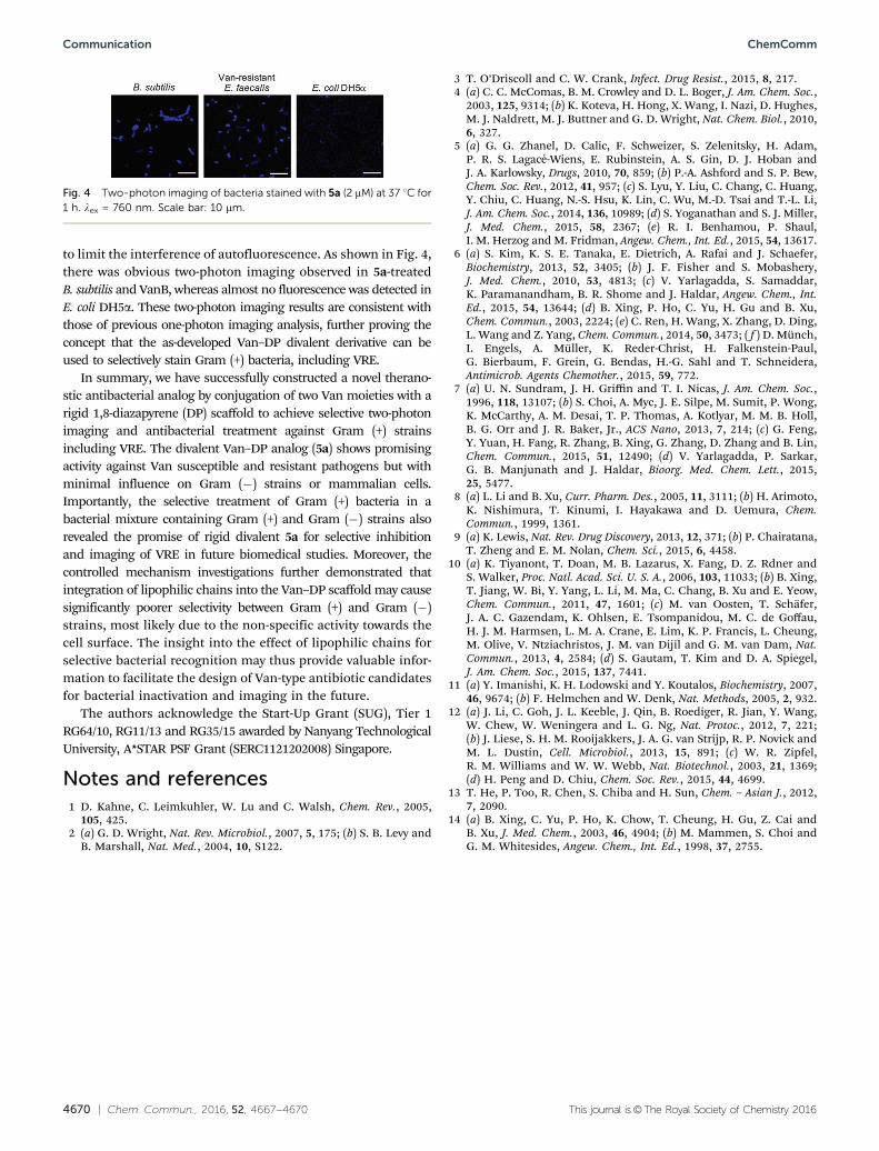
Finally, we examined the feasibility to further confirm theselective bacterial recognition through the two-photon imagingstrategy.13 Generally, bacteria B. subtilis, VanB or E. coli DH5awere incubated with 2 mM of divalent Van–DP (5a) at 37 1C for1 h. Two-photon microscopic imaging was performed uponexcitation of 5a at 760 nm, which was carefully chosen tominimize the bacterial photo-damage caused by UV light, and
Table 1 MIC values of Van, DP, and monovalent and divalent Van–DP analogs
MICa (mM)
B. subtilis Van-sensitive E. faecium Van-sensitive E. faecalis Van-resistant E. faecium Van-resistant E. faecalis E. coli DH5a
a The MIC is defined as the lowest concentration of the drug necessary to inhibit bacterial growth. Each bacterial strain was incubated in LB withdifferent concentrations of compounds in a 96-well plate for 18 h. The MIC values were determined from OD600 values in three separateexperiments. The OD600 values of the wells in the absence of bacteria were used as the control.
Fig. 2 SEM images of E. faecalis (VanB) and E. coli DH5a after treatmentwith 5a or 5b (10 mM) at 37 1C for 2 h. Scale bar: 1 mm.
Fig. 3 Fluorescence imaging of the mixture of GFP-expressing S. aureusand RFP-expressing E. coli treated with 5a or 5b (10 mM) at 37 1C for 24 h.Scale bar = 10 mm. (lex = 488 nm for GFP and lex = 543 nm for RFP, lem =515 � 30 nm for GFP and lem = 590 � 60 nm for RFP.)
to limit the interference of autofluorescence. As shown in Fig. 4,there was obvious two-photon imaging observed in 5a-treatedB. subtilis and VanB, whereas almost no fluorescence was detected inE. coli DH5a. These two-photon imaging results are consistent withthose of previous one-photon imaging analysis, further proving theconcept that the as-developed Van–DP divalent derivative can beused to selectively stain Gram (+) bacteria, including VRE.
In summary, we have successfully constructed a novel therano-stic antibacterial analog by conjugation of two Van moieties with arigid 1,8-diazapyrene (DP) scaffold to achieve selective two-photonimaging and antibacterial treatment against Gram (+) strainsincluding VRE. The divalent Van–DP analog (5a) shows promisingactivity against Van susceptible and resistant pathogens but withminimal influence on Gram (�) strains or mammalian cells.Importantly, the selective treatment of Gram (+) bacteria in abacterial mixture containing Gram (+) and Gram (�) strains alsorevealed the promise of rigid divalent 5a for selective inhibitionand imaging of VRE in future biomedical studies. Moreover, thecontrolled mechanism investigations further demonstrated thatintegration of lipophilic chains into the Van–DP scaffold may causesignificantly poorer selectivity between Gram (+) and Gram (�)strains, most likely due to the non-specific activity towards thecell surface. The insight into the effect of lipophilic chains forselective bacterial recognition may thus provide valuable infor-mation to facilitate the design of Van-type antibiotic candidatesfor bacterial inactivation and imaging in the future.
The authors acknowledge the Start-Up Grant (SUG), Tier 1RG64/10, RG11/13 and RG35/15 awarded by Nanyang TechnologicalUniversity, A*STAR PSF Grant (SERC1121202008) Singapore.
Notes and references1 D. Kahne, C. Leimkuhler, W. Lu and C. Walsh, Chem. Rev., 2005,
105, 425.2 (a) G. D. Wright, Nat. Rev. Microbiol., 2007, 5, 175; (b) S. B. Levy and
B. Marshall, Nat. Med., 2004, 10, S122.
3 T. O’Driscoll and C. W. Crank, Infect. Drug Resist., 2015, 8, 217.4 (a) C. C. McComas, B. M. Crowley and D. L. Boger, J. Am. Chem. Soc.,
2003, 125, 9314; (b) K. Koteva, H. Hong, X. Wang, I. Nazi, D. Hughes,M. J. Naldrett, M. J. Buttner and G. D. Wright, Nat. Chem. Biol., 2010,6, 327.
5 (a) G. G. Zhanel, D. Calic, F. Schweizer, S. Zelenitsky, H. Adam,P. R. S. Lagace-Wiens, E. Rubinstein, A. S. Gin, D. J. Hoban andJ. A. Karlowsky, Drugs, 2010, 70, 859; (b) P.-A. Ashford and S. P. Bew,Chem. Soc. Rev., 2012, 41, 957; (c) S. Lyu, Y. Liu, C. Chang, C. Huang,Y. Chiu, C. Huang, N.-S. Hsu, K. Lin, C. Wu, M.-D. Tsai and T.-L. Li,J. Am. Chem. Soc., 2014, 136, 10989; (d) S. Yoganathan and S. J. Miller,J. Med. Chem., 2015, 58, 2367; (e) R. I. Benhamou, P. Shaul,I. M. Herzog and M. Fridman, Angew. Chem., Int. Ed., 2015, 54, 13617.
6 (a) S. Kim, K. S. E. Tanaka, E. Dietrich, A. Rafai and J. Schaefer,Biochemistry, 2013, 52, 3405; (b) J. F. Fisher and S. Mobashery,J. Med. Chem., 2010, 53, 4813; (c) V. Yarlagadda, S. Samaddar,K. Paramanandham, B. R. Shome and J. Haldar, Angew. Chem., Int.Ed., 2015, 54, 13644; (d) B. Xing, P. Ho, C. Yu, H. Gu and B. Xu,Chem. Commun., 2003, 2224; (e) C. Ren, H. Wang, X. Zhang, D. Ding,L. Wang and Z. Yang, Chem. Commun., 2014, 50, 3473; ( f ) D. Munch,I. Engels, A. Muller, K. Reder-Christ, H. Falkenstein-Paul,G. Bierbaum, F. Grein, G. Bendas, H.-G. Sahl and T. Schneidera,Antimicrob. Agents Chemother., 2015, 59, 772.
7 (a) U. N. Sundram, J. H. Griffin and T. I. Nicas, J. Am. Chem. Soc.,1996, 118, 13107; (b) S. Choi, A. Myc, J. E. Silpe, M. Sumit, P. Wong,K. McCarthy, A. M. Desai, T. P. Thomas, A. Kotlyar, M. M. B. Holl,B. G. Orr and J. R. Baker, Jr., ACS Nano, 2013, 7, 214; (c) G. Feng,Y. Yuan, H. Fang, R. Zhang, B. Xing, G. Zhang, D. Zhang and B. Lin,Chem. Commun., 2015, 51, 12490; (d) V. Yarlagadda, P. Sarkar,G. B. Manjunath and J. Haldar, Bioorg. Med. Chem. Lett., 2015,25, 5477.
8 (a) L. Li and B. Xu, Curr. Pharm. Des., 2005, 11, 3111; (b) H. Arimoto,K. Nishimura, T. Kinumi, I. Hayakawa and D. Uemura, Chem.Commun., 1999, 1361.
9 (a) K. Lewis, Nat. Rev. Drug Discovery, 2013, 12, 371; (b) P. Chairatana,T. Zheng and E. M. Nolan, Chem. Sci., 2015, 6, 4458.
10 (a) K. Tiyanont, T. Doan, M. B. Lazarus, X. Fang, D. Z. Rdner andS. Walker, Proc. Natl. Acad. Sci. U. S. A., 2006, 103, 11033; (b) B. Xing,T. Jiang, W. Bi, Y. Yang, L. Li, M. Ma, C. Chang, B. Xu and E. Yeow,Chem. Commun., 2011, 47, 1601; (c) M. van Oosten, T. Schafer,J. A. C. Gazendam, K. Ohlsen, E. Tsompanidou, M. C. de Goffau,H. J. M. Harmsen, L. M. A. Crane, E. Lim, K. P. Francis, L. Cheung,M. Olive, V. Ntziachristos, J. M. van Dijil and G. M. van Dam, Nat.Commun., 2013, 4, 2584; (d) S. Gautam, T. Kim and D. A. Spiegel,J. Am. Chem. Soc., 2015, 137, 7441.
11 (a) Y. Imanishi, K. H. Lodowski and Y. Koutalos, Biochemistry, 2007,46, 9674; (b) F. Helmchen and W. Denk, Nat. Methods, 2005, 2, 932.
12 (a) J. Li, C. Goh, J. L. Keeble, J. Qin, B. Roediger, R. Jian, Y. Wang,W. Chew, W. Weningera and L. G. Ng, Nat. Protoc., 2012, 7, 221;(b) J. Liese, S. H. M. Rooijakkers, J. A. G. van Strijp, R. P. Novick andM. L. Dustin, Cell. Microbiol., 2013, 15, 891; (c) W. R. Zipfel,R. M. Williams and W. W. Webb, Nat. Biotechnol., 2003, 21, 1369;(d) H. Peng and D. Chiu, Chem. Soc. Rev., 2015, 44, 4699.
13 T. He, P. Too, R. Chen, S. Chiba and H. Sun, Chem. – Asian J., 2012,7, 2090.
14 (a) B. Xing, C. Yu, P. Ho, K. Chow, T. Cheung, H. Gu, Z. Cai andB. Xu, J. Med. Chem., 2003, 46, 4904; (b) M. Mammen, S. Choi andG. M. Whitesides, Angew. Chem., Int. Ed., 1998, 37, 2755.
Fig. 4 Two-photon imaging of bacteria stained with 5a (2 mM) at 37 1C for1 h. lex = 760 nm. Scale bar: 10 mm.