Biochem. J. (1991) 273, 767-775 (Printed in Great Britain) Further studies on the topography of human platelet glycoprotein Ilb Localization of monoclonal antibody epitopes and the putative glycoprotein Illa- and fibrinogen-binding regions Juan J. CALVETE,*t Juan ARIAS,*t Maria V. ALVAREZ,* Maria M. LOPEZ,* Agnes HENSCHENt and Jose GONZALEZ-RODRIGUEZ*$ *Instituto de Quimica Fisica, C.S.I.C., Serrano 119, 28006 Madrid, Spain, and tMax-Planck-Institut fur Biochemie, D-8033 Martinsried/Munchen, Federal Republic of Germany Glycoprotein Ilb (GPIIb) is a major glycoprotein of the human platelet plasma membrane, which together with glycoprotein IIIa (GPIIIa) forms a Ca2+-dependent heterodimer, GPIIb/IIIa, which serves as the major fibrinogen receptor in activated platelets. The precise localization of the epitopes for six anti-GPIIb monoclonal antibodies (M l-M6) has been determined by a combination of enzymic and chemical cleavage procedures, peptide isolation, N-terminal sequence analysis, peptide synthesis and enzyme immunoassay. The following localizations were found: MI, 81-16-36, ,f2-4-24; M2, a747-755; Ma2, a837-843; M3, a849-857; M4, al143-151; M5, a550-558; M6, a657-665. Besides considerations of the degree of exposure of these epitopes, several remarkable features are readily apparent. The earliest and main chymotryptic cleavage site of GPIIb in whole platelets is between acysteine-545 and aphenylalanine-551. The epitope for M3 was located within the same sequence (a842-857) as is the epitope for PMI-1 [Loftus, Plow, Frelinger, D'Souza, Dixon, Lacy, Sorge & Ginsberg (1987) Proc. Natl. Acad. Sci. U.S.A. 84, 7114-7118] in spite of the fact that the exposure of the latter in whole platelets is EDTA-dependent whereas that in the former is not. The epitope for M5 shares full homology with the 540-548 peptide stretch of the a-subunit of the vitronectin receptor, and this antibody cross-reacts with endothelial cells. The M6 epitope is located in the 25 kDa membrane-bound fragment of GPIIb, which is most resistant to chymotryptic digestion in whole platelets, contrary to what happens in isolated GPIIb in solution, where this epitope is destroyed at an early stage of chymotryptic digestion. This suggests that this region of GPIIb, somewhere between the epitope for M5 (a550-558) and the epitope for M2 (a747-755), may carry the surface of interaction of GPIIb with GPIIIa in the GPIIb/IIIa heterodimer. Finally, the sequence where the epitope for M6 has been located (a657-667) was the only one found to be hydropathically complementary to the y402-411 peptide of fibrinogen within the amino acid sequence of both GPIIb and GPIIIa. This complementariness, the EDTA- or thrombin-dependence of the exposure of the a657-665 stretch in whole platelets to M6 and the ability of this antibody to inhibit platelet aggregation led us to postulate that this peptide stretch is a putative binding site for fibrinogen in the platelet receptor. The overlap between the M6 epitope and the putative binding site for the y4O2-411 peptide sequence of fibrinogen would imply that the unmasking of the a658-667 peptide stretch could be one of the structural changes in GPIIb/IIIa required for the induction of the fibrinogen receptor in activated platelets. INTRODUCTION Glycoprotein lIb (GPIIb) is a major platelet plasma-membrane glycoprotein, which together with glycoprotein Illa (GPIIIa) forms a Ca 2+-dependent heterodimer, the GPIIb/IIIa complex, which serves as the inducible receptor for fibrinogen at the surface of activated platelets (Nurden et al., 1986; Marguerie et al., 1987; Phillips et al., 1988). GPIIb (136 kDa) is a bitopic two-chain glycoprotein joined by a single interchain disulphide bond (Calvete & Gonzailez-Rodriguez, 1986; Poncz et al., 1987; Usobiaga et al., 1987). The heavy chain GPIIba (114 kDa), also known as GPIIb H, is fully extracellular, whereas the light chain, GPIIb,f (23 kDa), also known as GPIIb L, carries the single transmembrane segment of GPIIb, as predicted from its cDNA- derived amino acid sequence (Poncz et al., 1987). The biochemical composition, amino acid sequence and covalent structure of GPIIb are known, but the tertiary structure and topology in the platelet membrane remain to be determined (Eirin et al., 1986; Calvete et al., 1989a,b; Lam et al., 1989). Anti-GPIIb monoclonal antibodies have been prepared in several laboratories (McEver et al., 1980; Varon & Karpatkin, 1983; Thorsen et al., 1985; Loftus et al., 1987; Calvete et al., 1989a); however, the precise localization of their epitopes is known for very few of them. The binding of some of these antibodies is EDTA-, thrombin- or RGD peptide-dependent, some are inhibitors of platelet aggregation and/or fibrinogen binding, some cross-react with the a-subunit of the vitronectin receptor in endothelial cells etc. The epitope for PMI-1 has been located between residues a842 and a858 (Loftus et al., 1987), the epitopes for MI, M3 and M4 were found within the sequences ,/4-24, a826-871 and al 15-285 respectively, and finally the epitopes for MS and M6 were located somewhere between residues oc550 and a700 (Calvete et al., 1989a). Recently considerable interest has been aroused in the local- ization of the fibrinogen-binding sites in both GPIIb and GPIIIa and some progress has already been made in determining these. RGD peptides become preferentially cross-linked to the peptide Vol. 273 Abbreviations used: GPIIb, glycoprotein Ilb; GPIIba and GPIIbfl, the a- and the fl-subunits respectively of GPIIb; CM-GPlIba., fully reduced and carboxymethylated ac-subunit of GPIIb; GPIIIa, glycoprotein Illa; Aac and y, fibrinogen Aa- and y-chains respectively. I To whom correspondence and reprint requests should be sent. 767
Transcript
Biochem. J. (1991) 273, 767-775 (Printed in Great Britain)
Further studies on the topography of human plateletglycoprotein IlbLocalization of monoclonal antibody epitopes and the putative glycoprotein Illa-and fibrinogen-binding regions
Juan J. CALVETE,*t Juan ARIAS,*t Maria V. ALVAREZ,* Maria M. LOPEZ,* Agnes HENSCHENtand Jose GONZALEZ-RODRIGUEZ*$*Instituto de Quimica Fisica, C.S.I.C., Serrano 119, 28006 Madrid, Spain, and tMax-Planck-Institut fur Biochemie,D-8033 Martinsried/Munchen, Federal Republic of Germany
Glycoprotein Ilb (GPIIb) is a major glycoprotein of the human platelet plasma membrane, which together withglycoprotein IIIa (GPIIIa) forms a Ca2+-dependent heterodimer, GPIIb/IIIa, which serves as the major fibrinogenreceptor in activated platelets. The precise localization of the epitopes for six anti-GPIIb monoclonal antibodies (M l-M6)has been determined by a combination of enzymic and chemical cleavage procedures, peptide isolation, N-terminalsequence analysis, peptide synthesis and enzyme immunoassay. The following localizations were found: MI, 81-16-36,,f2-4-24; M2, a747-755; Ma2, a837-843; M3, a849-857; M4, al143-151; M5, a550-558; M6, a657-665. Besidesconsiderations of the degree of exposure of these epitopes, several remarkable features are readily apparent. The earliestand main chymotryptic cleavage site of GPIIb in whole platelets is between acysteine-545 and aphenylalanine-551. Theepitope for M3 was located within the same sequence (a842-857) as is the epitope for PMI-1 [Loftus, Plow, Frelinger,D'Souza, Dixon, Lacy, Sorge & Ginsberg (1987) Proc. Natl. Acad. Sci. U.S.A. 84, 7114-7118] in spite of the fact that theexposure of the latter in whole platelets is EDTA-dependent whereas that in the former is not. The epitope for M5 sharesfull homology with the 540-548 peptide stretch of the a-subunit of the vitronectin receptor, and this antibody cross-reacts
with endothelial cells. The M6 epitope is located in the 25 kDa membrane-bound fragment of GPIIb, which is most
resistant to chymotryptic digestion in whole platelets, contrary to what happens in isolated GPIIb in solution, where thisepitope is destroyed at an early stage of chymotryptic digestion. This suggests that this region of GPIIb, somewherebetween the epitope for M5 (a550-558) and the epitope for M2 (a747-755), may carry the surface of interaction of GPIIbwith GPIIIa in the GPIIb/IIIa heterodimer. Finally, the sequence where the epitope for M6 has been located (a657-667)was the only one found to be hydropathically complementary to the y402-411 peptide of fibrinogen within the amino acidsequence of both GPIIb and GPIIIa. This complementariness, the EDTA- or thrombin-dependence of the exposure ofthe a657-665 stretch in whole platelets to M6 and the ability of this antibody to inhibit platelet aggregation led us to
postulate that this peptide stretch is a putative binding site for fibrinogen in the platelet receptor. The overlap betweenthe M6 epitope and the putative binding site for the y4O2-411 peptide sequence of fibrinogen would imply that theunmasking of the a658-667 peptide stretch could be one of the structural changes in GPIIb/IIIa required for the inductionof the fibrinogen receptor in activated platelets.
INTRODUCTION
Glycoprotein lIb (GPIIb) is a major platelet plasma-membraneglycoprotein, which together with glycoprotein Illa (GPIIIa)forms a Ca2+-dependent heterodimer, the GPIIb/IIIa complex,which serves as the inducible receptor for fibrinogen at thesurface of activated platelets (Nurden et al., 1986; Marguerieet al., 1987; Phillips et al., 1988). GPIIb (136 kDa) is a bitopictwo-chain glycoprotein joined by a single interchain disulphidebond (Calvete & Gonzailez-Rodriguez, 1986; Poncz et al., 1987;Usobiaga et al., 1987). The heavy chain GPIIba (114 kDa), alsoknown as GPIIb H, is fully extracellular, whereas the light chain,GPIIb,f (23 kDa), also known as GPIIb L, carries the singletransmembrane segment of GPIIb, as predicted from its cDNA-derived amino acid sequence (Poncz et al., 1987). The biochemicalcomposition, amino acid sequence and covalent structure of
GPIIb are known, but the tertiary structure and topology in the
platelet membrane remain to be determined (Eirin et al., 1986;Calvete et al., 1989a,b; Lam et al., 1989).
Anti-GPIIb monoclonal antibodies have been prepared inseveral laboratories (McEver et al., 1980; Varon & Karpatkin,1983; Thorsen et al., 1985; Loftus et al., 1987; Calvete et al.,1989a); however, the precise localization of their epitopes isknown for very few of them. The binding of some of theseantibodies is EDTA-, thrombin- or RGD peptide-dependent,some are inhibitors of platelet aggregation and/or fibrinogenbinding, some cross-react with the a-subunit of the vitronectinreceptor in endothelial cells etc. The epitope for PMI-1 has beenlocated between residues a842 and a858 (Loftus et al., 1987), theepitopes for MI, M3 and M4 were found within the sequences
,/4-24, a826-871 and al 15-285 respectively, and finally theepitopes for MS and M6 were located somewhere betweenresidues oc550 and a700 (Calvete et al., 1989a).
Recently considerable interest has been aroused in the local-ization of the fibrinogen-binding sites in both GPIIb and GPIIIaand some progress has already been made in determining these.RGD peptides become preferentially cross-linked to the peptide
Vol. 273
Abbreviations used: GPIIb, glycoprotein Ilb; GPIIba and GPIIbfl, the a- and the fl-subunits respectively of GPIIb; CM-GPlIba., fully reduced andcarboxymethylated ac-subunit of GPIIb; GPIIIa, glycoprotein Illa; Aac and y, fibrinogen Aa- and y-chains respectively.
I To whom correspondence and reprint requests should be sent.
767
J. J. Calvete and others
sequence 109-171 of GPIIIa, in thrombin-stimulated platelets,whereas peptides of the fibrinogen y-chain C-terminal type labelpredominantly GPIIb (Santoro & Lawing, 1987; D'Souza et al.,1988). A hydropathic complementarity approach has also beenused to predict the binding site for the Aa-chain of fibrinogen inthe platelet GPIIb/IIIa complex (Pasqualini et al., 1989).
In the present work, the localization of the epitopes for aset of monoclonal antibodies has been narrowed down, by acombination ofchemical and enzymic cleavage procedures, solid-state peptide synthesis and competitive enzyme immunoassay. Inaddition, we have applied the principle of complementaryhydropathy to predict the putative binding sites for fibrinogen inthe amino acid sequence of GPIIb.
aureus V8) and tosylphenylalanylchloromethane ('TPCK ')-treated trypsin were from Boehringer-Mannheim, Miles Labora-tories and Sigma Chemical Co., respectively. The other chemicalsand biochemicals were of analytical or chromatographic grade.Chromatographic columns and buffers, as well as the preparationof human platelets, platelet plasma membrane and the isolationof GPIIb and the fully reduced and carboxymethylated forms ofGPIIba (CM-GPIIba) and GPIIb,/ (CM-GPIIb,B) were as pre-viously described (Calvete & Gonzailez-Rodrfguez, 1986; Eirinet al., 1986).
Monoclonal antibody production and purificationMouse monoclonal antibodies anti-GPIIba (M2, M3, M4,
M5, M6 and Ma2) and anti-GPIIb,8 (MI) were prepared byusing either whole GPIIb or the isolated subunits according toimmunization and fusion protocols and screening assays de-scribed previously (Melero & Gonzalez-Rodriguez, 1984). Anti-bodies were purified from ascitic fluids after sequential 25 %- and50 %-satd.-(NH4)2SO4 precipitation. Finally, the 50 %-satd.-(NH4)2SO4 precipitates were subjected to affinity chromato-graphy on Protein A-Sepharose (Pharmacia) according to themanufacturer's instructions.
Solid-state peptide synthesisThe procedure described by Geysen et al. (1984) was followed,
using reagents supplied by Cambridge Research Biochemicalsand a polyethylene rod holder with the format and spacing of amicro-titre plate.
Enzyme immunoassay of the natural and synthetic peptidesThe criterion for epitope localization was based on the specific
recognition by the monoclonal antibodies of natural or syntheticpeptides. The assay of synthetic peptides coupled to polyethylenerods (see above) was done as described by Geysen et al. (1984).Anti-(mouse IgG) IgG coupled to horseradish peroxidase wasused as the second antibody, with either o-phenylenediaminedihydrochloride or 2,2'-azinobis-(3-ethylbenzthiazoline-6-sul-phonic acid) in the developing solution.The assay of natural peptides was done by a competitive
enzyme immunoassay which consisted in the incubation of thenatural peptide with the monoclonal antibody of interest for1 h at 37 °C, followed by the assay of the free antibody in thismixture by an ordinary enzyme immunoassay. Whole glyco-protein or some fragment derived from it was used to coat thewalls ofthe microplate wells. The second antibody and developingsolution were the same as those used above for the assay ofsynthetic peptides.
Other analytical methodsProtein assay was done by the procedure of Markwell et al.
(1978). Amino acid and amino sugar analyses were performedwith a Biotronik amino acid analyser, after sample hydrolysis at110 °C in 6 M-HCl for 24 h and in 4 M-HCI for 4 h respectively.N-Terminal sequence analyses were effected in a prototypeautomated spinning-cup Sequenator, as described previously(Edman & Henschen, 1975). SDS/PAGE was done according tothe procedure of Laemmli (1970). Immunoelectroblotting wascarried out after gel electrophoresis by transferring the proteinbands to nitrocellulose by a standard procedure (Towbin et al.,1979). The monoclonal antibodies were used, either as 50%-satd.-(NH4)2S04 precipitates or after purification by Protein A-Sepharose chromatography (see above). The second antibodywas a goat anti-(mouse IgG) IgG coupled to horseradish per-oxidase, with 4-chloro-1-naphthol in the developing solution.
Enzymic digestion of the CM-GPIIbc with endoproteinaseGlu-C
Isolated CM-GPIIba (5 mg/ml in 50 mM-sodium phosphatebuffer, pH 7.8) was digested with endoproteinase Glu-C at anenzyme/substrate ratio 1:25 (w/w) at 37 °C for 18 h. The reactionwas stopped by adding formic acid up to 30% (v/v) finalconcentration. The digestion products were separated by reverse-phase h.p.l.c., on a C18 (pore size 30 nm, particle size 10 ,um)Vydac column (25 cm x 0.4 cm) equilibrated in 0.1 % (v/v)trifluoroacetic acid in water (solution A) and 0.1 % (v/v)trifluoroacetic acid in acetonitrile (solution B) (90% solution A/10% solution B) and eluted at 1 ml/min, first isocratically for5 min, followed by a linear gradient up to 70 % of solution B in120 min.
Enzymic digestion of the 35 kDa N-terminal fragment ofCM-GPIIba with endoproteinase Lys-C
Isolated CM-GPIIba was cleaved with a 500-fold molar excessof CNBr over its theoretical methionine content in 70 % (v/v)formic acid, under N2 and in the dark. After 24 h at roomtemperature the mixture was diluted 10-fold with Milli Q waterand freeze-dried. The cleavage products were resuspended in70% formic acid and separated on a Sephacryl S200 column(130 cm x 1 cm), eluted with 10% formic acid as describedpreviously (Calvete et al., 1989b). The isolated 35 kDa CNBr-cleavage fragment, 1.75 mg/ml in 0.1 M-Tris/HCI buffer, pH 7.6,which contains the N-terminal peptide of GPIIba (al-285), wasfurther digested with endoproteinase Lys-C, at an enzyme/substrate ratio 1:25 (w/w) for 18 h at 37 'C. The reaction wasstopped by adding formic acid up to 30% (v/v) final con-centration, and the digestion products were separated by reverse-phase h.p.l.c. on a C18 Vydac column (25 cm x 0.4 cm),equilibrated in a mixture of 0.1 % trifluoroacetic acid in water(solution A) and 0.1 % trifluoroacetic acid in acetonitrile (solu-tion B) (95 solution A/5% solution B) and eluted at 1 ml/min,first isocratically for 5 min, followed by a linear gradient up to70% of solution B in 65 min.
Digestion of whole platelets with chymotrypsinPlatelets were washed in 150 mM-NaCl/10 mM-Tris/HCI
buffer, pH 7.4, containing either 1 mM-EDTA or 1 mM-CaCl2and 2,g of apyrase (Sigma grade V)/ml and resuspended at5 x 109 platelets/ml in the same buffer. The platelet suspensionwas incubated at 37 'C with 0.2 mg of chymotrypsin/ml.Samples were taken at 5 min, 15 min, 30 min, 1 h and 2 h, andthe digestion was stopped by the addition of phenylmethane-sulphonyl fluoride (25 mol/mol of chymotrypsin). Digestedplatelets were centrifuged at 10000 g (r8, 75 mm) for 10 min at
1991
768
Mapping of monoclonal antibody epitopes in platelet glycoprotein IIb
4 'C. The pellet was resuspended and sonicated in the samebuffer, and the particulate fraction obtained by ultracentri-fugation (160000 g, rav 65 mm) for 1 h at 4 'C.
RESULTS
Cleavage susceptibility patterns of the epitopes for monoclonalantibodies Ml, M2, MGC2, M3, M4, M5 and M6As a preliminary strategy to narrow down the location of the
epitopes for the set of anti-GPIIb antibodies studied here, wehave assessed the susceptibility of these epitopes to differentchemical and enzymic cleavage procedures by competitive en-zyme immunoassay. The cleavage susceptibility patterns for thedifferent antibodies are summarized in Table 1, from whichseveral guiding conclusions can be drawn. Whereas the epitopefor Ml is the only one remaining after tryptic digestion of GPIIb,the epitope for M6 is the only one destroyed by CNBr cleavageof GPIIb, and therefore the primary peptide fragments for theisolation of the epitopes for all the antibodies except for M6 canbe obtained after GPIIb cleavage by CNBr. From a considerationof the susceptibility of the epitopes to cleavage by the proteinasesused, it is probable that the epitopes for M2, M4, M5 and M6contain arginine, those for M3 and Ma2 include a lysine residuein their sequence, the epitope for MS contains some dicarboxylicamino acid and that for Ml is free from dicarboxylic acid,arginine and lysine residues. Finally, the susceptibility of theepitope for M6 to cleavage by CNBr suggests that there is somemethionine residue in its sequence.
This information was used subsequently to determine thecleavage procedures employed to isolate the peptide fragmentscontaining the epitopes for these antibodies, and to select thosepeptide sequences within them that were likely to contain anepitope with a certain cleavage susceptibility pattern. Furtherassessment of this prediction was done by a combination ofsolid-state peptide synthesis and enzyme immunoassay, as shownbelow.
Epitope for MlThe epitope for this anti-GPIIb,8 antibody had been found
previously .to be at the N-terminus of this subunit, between/JPro-4 and Met-24 (Calvete & Gonzailez-Rodriguez, 1986;Calvete et al., 1989a) by immunoelectroblotting. Now we haveconfirmed this result using competitive enzyme immunoassay.
Epitopes for M2, M3 and Ma2Previously we had located the epitope for M3 in the
C-terminal 19 kDa CNBr-cleavage product of CM-GPIIba
Table 2. Synthetic peptides designed from the amino acid sequence ofplatelet GPIIb
Peptides were synthesized by the procedure of Geysen et al. (1984)and the degree of overlapping of their sequences with those of theepitopes for a set of anti-GPIIb monoclonal antibodies was assessedby enzyme immunoassay (see Fig. 1).
The cleavage susceptibility was assessed by competitive enzymeimmunoassay, where the different mixtures of cleavage productswere made to compete for the different monoclonal antibodies withGPIIb or some fragments derived from it, adsorbed to the micro-titration plate. + or - mean antibody epitope conserved ordestroyed respectively after chemical or enzymic cleavage.
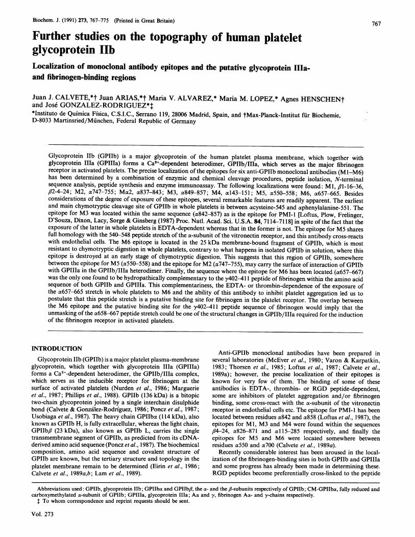
(a704-871) by immunoelectroblotting (Calvete et al., 1989a).Now, besides confirming this, we have located the epitopes forM2 and Ma2 in this polypeptide fragment, using competitiveenzyme immunoassay. To narrow down the localization of theseepitopes further, solid-state peptide synthesis was used to obtainthe overlapping peptide sequence which contained all the arginineand lysine residues of the C-terminal region of GPIIba. Thesepeptides were used in enzyme immunoassay to locate the threeepitopes (Table 2). In Fig. I we show that M2 recognizes thosesynthetic peptides whose sequences contain Arg-750, and thatM3 and Ma2 recognize those containing Lys-855 and Lys-836respectively, as expected from the predictions made from theircleavage susceptibility patterns.
Fig. 1. Localization of the epitopes for monoclonal antibodies M2, Mac2,M3, M4, MS and M6, using synthetic peptides and enzymeimmunoassay
The assessment of the degree of overlapping between the sequencesof the synthetic peptides listed in Table 2 and those of the epitopesfor the anti-GPIIb monoclonal antibodies was done by enzymeimmunoassay according to the procedure of Geysen et al. (1984).The antibodies were used at the following dilutions: M2 (1:2000),Ma2 (1:80000), M3 (1: 10000), M5 (1: 1000) and M6 (1:5000).
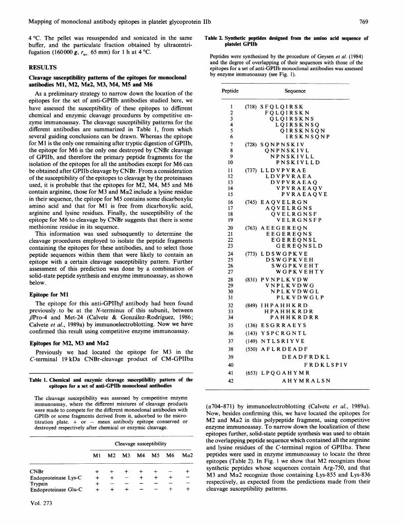
as efficiently as the whole 35 kDa CNBr-cleavage (Fig. 2b). TheN-terminal amino acid sequence and the amino acid analyses ofthis fraction led us to identify this peptide with the al25-164peptide stretch in the amino acid sequence of GPIIb. Whenpeptide synthesis combined with enzyme immunoassay was usedto narrow down the location of the M4 epitope it was foundbetween a143 and a l5l, as shown in Fig. 1 and Table 2. Ittherefore contains an arginine residue Arg-147, as expected.
Epitope for M5Given the fact that M5 cross-reacts with endothelial cells
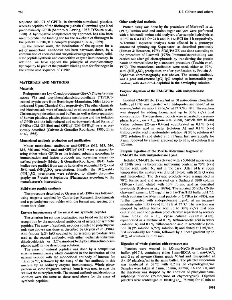
(Mufiiz et al., 1990), and therefore with the a-subunit of thevitronectin receptor in these cells, the epitope for MS should bewithin an amino acid sequence identical or similar in bothplatelet GPIIb and the a-subunit of the vitronectin receptor.Using immunoelectroblotting we had previously located the M5epitope within a 35 kDa membrane-bound chymotryptic productof digestion of GPIIb in whole platelets, which contained thepeptide stretch between residues a550 and a682 (Calvete et al.,1989a). The only sequence in this region common to the plateletGPIIb and the a-subunit of the vitronectin receptor is a550-565(Fig. 3). This sequence contains Arg-553 and several dicarboxylicamino acid residues, as expected from the cleavage susceptibilitypattern referred to above (Table 1). Peptide a550-580 wasisolated by reverse-phase h.p.l.c. as described previously (seeFig. 1 in Calvete et al., 1989b) and found, by competitive enzyme
Epitope for M4The N-terminal fragment (axl-285, 35 kDa), liberated by CNBr
cleavage of GPIIba, was isolated on Sephacryl S200 and furtherdigested with endoproteinase Lys-C. Among the cleavage prod-ucts separated by reverse-phase h.p.l.c. (Fig. 2a) is one (fraction5) that is capable of inhibiting the binding of M4 to GPIIb half
1.
1.0
0.8
0
C4 0.6
0.4
0.2
GPlba: (550) AFLRDEADFRDKLSPIVLSLNVSL PPTEAGM...
VNRa: (540)AYLRDESEFRDKLTPITIFMEYRLDYRTAADTTGL...Fig. 3. Comparison of the amino acid sequence c550-580 of platelet GPIIb
(Poncz et al., 1987) with the homologous sequence in the a-subunitof the vitronectin receptor (Fitzgerald et al., 1987)
100
QAov _-
a,
40
20
0
Elution time (min)
Fig. 2. Localization of the epitope for the monoclonal antibody M4 in the peptide sequence a125-164 of GPIIb
(a) Reverse-phase h.p.l.c. isolation of the products of digestion with endoproteinase Lys-C of the fully reduced and carboxymethylated 35 kDaCNBr-cleavage fragment of GPIIb (acl-285), prepared as described by Calvete et al. (1989b). A 175 ,sg portion of the CM-35 kDa CNBr-cleavagefragment in 100 ,u1 of 0.1 M-Tris/HCl buffer, pH 7.6, was digested with endoproteinase Lys-C at a peptide/enzyme ratio 25:1 (w/w) for 18 h at37 °C, and the reaction was stopped with formic acid at 30% (v/v) concentration. The digestion products were separated on a C18 (pore size 30 nm,particle size 10 ,um) Vydac column (25 cm x 0.4 cm) equilibrated in 0.1 % trifluoroacetic acid in water (solution A) and 0.1 % trifluoroacetic acidin acetonitrile (solution B) (95 % solution A/5% solution B), and eluted at 1 ml/min, first isocratically for 5 min, followed by a linear acetonitrilegradient from 5% to 70% solution B in 75 min. , A220; --, [acetonitrile]. (b) Competitive enzyme immunoassay of the CM-35 kDa CNBr-cleavage fragment of GPIIb (0), peptide fraction 5 (al125-164) from the column in (a) (CO.) and the rest of the column fractions (O), using I ,gof CM-GPIIbac adsorbed on each micro-titre plate well, and monoclonal antibody M4 at 1:10000 (v/v) dilution.
1991
M2 Mca2 M3 M4 M5 M6
I I I I .u_ . ". -74 r 4
770
Mapping of monoclonal antibody epitopes in platelet glycoprotein Ilb
immunoassay, to compete very effectively with GPIIb for bindingof M5. When peptide synthesis combined with enzyme immuno-assay was used to narrow down the location of this epitope, itwas found to be between a550 and a558 (Fig. I and Table 2).
Epitope for M6This epitope, which had also previously been located by
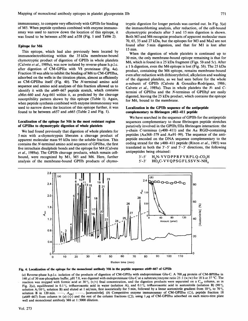
immunoelectroblotting within the 35 kDa membrane-boundchymotryptic product of digestion of GPIIb in whole platelets(Calvete et al., 1989a), was now isolated by reverse-phase h.p.l.c.after digestion of CM-GPIIa with V8 proteinase (Fig. 4a).Fraction 18 was able to inhibit the binding ofM6 to CM-GPIIba,adsorbed on the wells in the titration plates, almost as efficientlyas CM-GPIIba itself (Fig. 4b). The N-terminal amino acidsequence and amino acid analyses of this fraction allowed us toidentify it with the a649-667 peptide stretch, which containsaMet-660 and Arg-661 within it, as predicted by the cleavagesusceptibility pattern shown by this epitope (Table 1). Again,when peptide synthesis combined with enzyme immunoassay wasused to narrow down the location of this epitope further, it wasfound to be between a657 and a665 (Table 2 and Fig. 1).
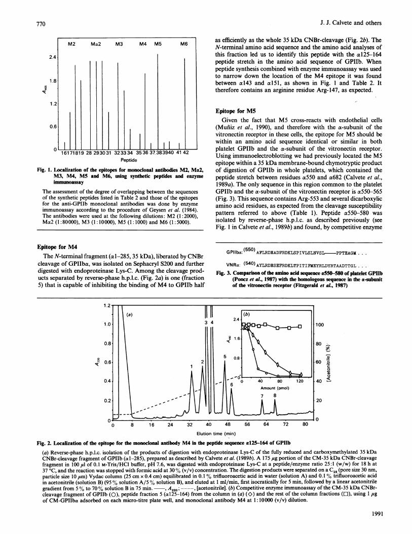
Localization of the epitope for M6 in the most resistant regionof GPIIba to chymotryptic digestion of whole plateletsWe had found previously that digestion of whole platelets for
5 min with a-chymotrypsin liberates a cleavage product ofapparent molecular mass 95 kDa into the soluble fraction. Thiscontains the N-terminal amino acid sequence of GPIIba, the firstfive intrachain disulphide bonds and the epitope for M4 (Calveteet al., 1989a). The GPIIb cleavage products, which remain cell-bound, were recognized by Ml, MS and M6. Here, furtheranalysis of the membrane-bound GPIIb products of chymo-
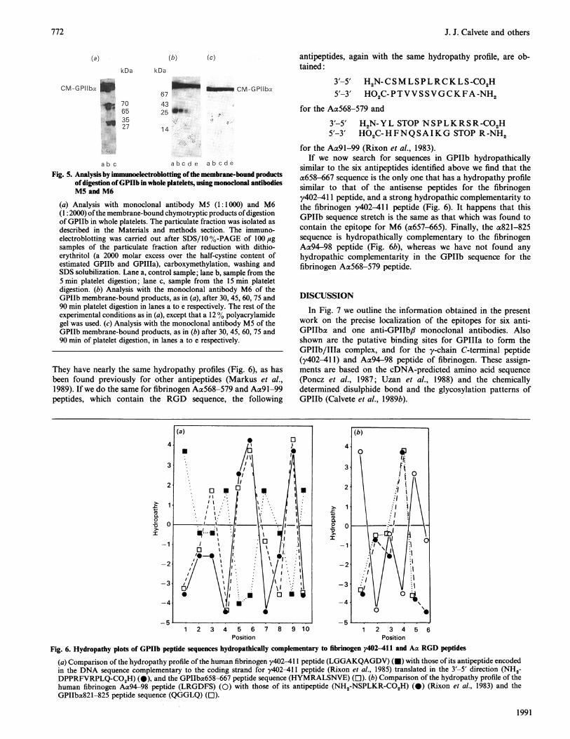
tryptic digestion for longer periods was carried out. In Fig. 5(a)the immunoblotting analysis, after reduction, of the cell-boundchymotryptic products after 5 and 15 min digestion is shown.Both MS and M6 recognize products of apparent molecular mass70, 65, 35 and 27 kDa, but the epitopes for M3 and Ma2 are notfound after 5 min digestion, and that for Ml is lost after15 min.When the digestion of whole platelets is continued up to
30 min, the only membrane-bound epitope remaining is that forM6, which is found in a 25 kDa fragment (Figs. Sb and 5c). Aftera 1 h digestion, even the M6 epitope is lost (Fig. Sb). The 25 kDaproduct, containing the M6 epitope, remains membrane-boundeven after reduction with dithioerythritol, alkylation and washingof the digested platelets, as we had seen before for the wholea-subunit of GPIIb (Calvete & Gonzalez-Rodriguez, 1986;Calvete et al., 1989a). Thus in whole platelets the N- and C-termini of GPIIba and the N-terminus of GPIIb,i are easilydigested, leaving the 25 kDa product, which contains the epitopefor M6, bound to the membrane.
Localization in the GPIIb sequence of the antipeptidecomplementary to fibrinogen y402-411 peptideWe have searched in the sequence of GPIIb for the antipeptide
sequences complementary to those fibrinogen peptide stretchesputatively involved in the GPIIb/IIIa fibrinogen interaction: they-chain C-terminus (y400-41 1) and the Aa RGD-containingpeptides (AaS68-579 and Aa9l-99). The sequence of the anti-peptide encoded on the DNA sequence complementary to thecoding strand for the y40-411 peptide (Rixon et al., 1985) wastranslated in both the 3'-5' and 5'-3' directions, the followingantipeptides being obtained:
Fig. 4: Localization of the epitope for the monoclonal antibody M6 in the peptide sequence ac649-667 of GPIIb
(a) Reverse-phase h.p.l.c. isolation of the products of digestion of CM-GPIIa with endoproteinase Glu-C. A 700 4tsg protein of CM-BPIIba in140 Izl of 50 mM-phosphate buffer, pH 7.8, was digested with endoproteinase Glu-C at a substrate/enzyme ratio 25: 1 (w/w) for 18 h at 37 °C. Thereaction was stopped with formic acid at 300% (v/v) final concentration, and the digestion products were separated on a C18 column, as inFig. 2(a), equilibrated in 0.1% trifluoroacetic acid in water (solution A), and 0.1% trifluoroacetic acid in acetonitrile (solution B) (90%solution A/100% solution B) and eluted at 1 ml/min, first isocratically for 5 min, followed by a linear acetonitrile gradient from 10% to 700%solution B in 120 min. , A220; -- , [acetonitrile]. (b) Competitive enzyme immunoassay of CM-GPIIba (0), peptide fraction 18(a649-667) from column in (a) (K>) and the rest of the column fractions (El), using 1 ,ug of CM-GPIIba adsorbed on each micro-titre platewell and monoclonal antibody M6 at 1: 5000 dilution.
Vol. 273
771
J. J. Calvete and others
(a) (b) (c)
kDa kDa
CM-GPilba _i_:* 70
653527
67CM-GPllbx674325 4lW
14
abc abcde abcdeFig. 5. Analysis by imnmunoelectroblotting ofthe membrane-bound products
ofdigestion ofGPIIb in whole platelets, using monoclonal antibodiesM5 and M6
(a) Analysis with monoclonal antibody M5 (1: 1000) and M6(1: 2000) ofthe membrane-bound chymotryptic products ofdigestionof GPIIb in whole platelets. The particulate fraction was isolated asdescribed in the Materials and methods section. The immuno-electroblotting was carried out after SDS/10 %-PAGE of 100lgsamples of the particulate fraction after reduction with dithio-erythritol (a 2000 molar excess over the half-cystine content ofestimated GPIIb and GPIIIa), carboxymethylation, washing andSDS solubilization. Lane a, control sample; lane b, sample from the5 min platelet digestion; lane c, sample from the 15 min plateletdigestion. (b) Analysis with the monoclonal antibody M6 of theGPIIb membrane-bound products, as in (a), after 30, 45, 60, 75 and90 min platelet digestion in lanes a to e respectively. The rest of theexperimental conditions as in (a), except that a 12% polyacrylamidegel was used. (c) Analysis with the monoclonal antibody M5 of theGPIIb membrane-bound products, as in (b) after 30, 45, 60, 75 and90 min of platelet digestion, in lanes a to e respectively.
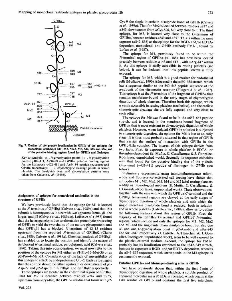
They have nearly the same hydropathy profiles (Fig. 6), as hasbeen found previously for other antipeptides (Markus et al.,1989). If we do the same for fibrinogen Aa568-579 and Aa9l-99peptides, which contain the RGD sequence, the following
antipeptides, again with the same hydropathy profile, are ob-tained:
for the Aa9l-99 (Rixon et al., 1983).If we now search for sequences in GPIIb hydropathically
similar to the six antipeptides identified above we find that thea658-667 sequence is the only one that has a hydropathy profilesimilar to that of the antisense peptides for the fibrinogeny402-411 peptide, and a strong hydropathic complementarity tothe fibrinogen y402-411 peptide (Fig. 6). It happens that thisGPIIb sequence stretch is the same as that which was found tocontain the epitope for M6 (a657-665). Finally, the a821-825sequence is hydropathically complementary to the fibrinogenAa94-98 peptide (Fig. 6b), whereas we have not found anyhydropathic complementarity in the GPIIb sequence for thefibrinogen Aa568-579 peptide.
DISCUSSION
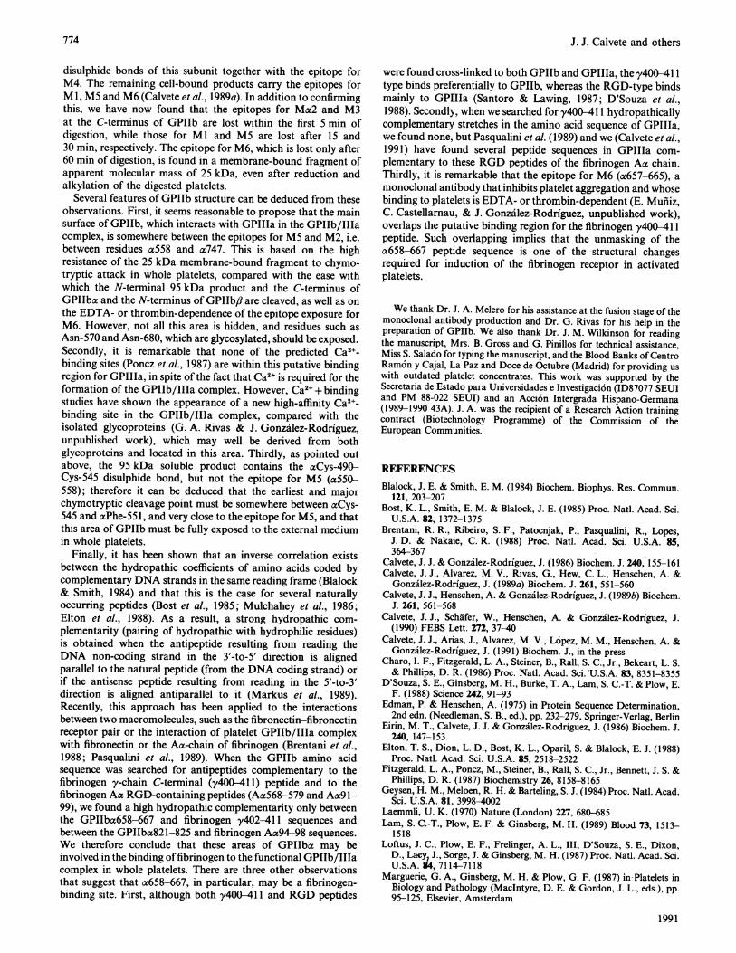
In Fig. 7 we outline the information obtained in the presentwork on the precise localization of the epitopes for six anti-GPIIba and one anti-GPIIb,l monoclonal antibodies. Alsoshown are the putative binding sites for GPIIIa to form theGPIIb/IIIa complex, and for the y-chain C-terminal peptide(y4O2-41 1) and Aa94-98 peptide of fibrinogen. These assign-ments are based on the cDNA-predicted amino acid sequence(Poncz et al., 1987; Uzan et al., 1988) and the chemicallydetermined disulphide bond and the glycosylation patterns ofGPIIb (Calvete et al., 1989b).
4-
0-0
I
-c0'a
1 2 3 4 5 6 7 8 9 10 1 2 3 4 5 6Position Position
Fig. 6. Hydropathy plots of GPIIb peptide sequences hydropathically complementary to fibrinogen y4O24ll and Aa RGD peptides(a) Comparison of the hydropathy profile of the human fibrinogen y402-411 peptide (LGGAKQAGDV) (-) with those of its antipeptide encodedin the DNA sequence complementary to the coding strand for y4O2-411 peptide (Rixon et al., 1985) translated in the 3'-5' direction (NH2-DPPRFVRPLQ-CO2H) (0), and the GPIIbo658-667 peptide sequence (HYMRALSNVE) (l). (b) Comparison of the hydropathy profile of thehuman fibrinogen Aoc94-98 peptide (LRGDFS) (0) with those of its antipeptide (NH2-NSPLKR-CO2H) (0) (Rixon et al., 1983) and theGPIIba821-825 peptide sequence (QGGLQ) ([1).
1991
772
Mapping of monoclonal antibody epitopes in platelet glycoprotein Ilb
GPIlbac
GPila
Ax94-98
Mx2
M3
M6 .658 y402-411....667
Platelet membrane
Fig. 7. Outline of the precise localization in GPIIb of the epitopes formonoclonal antibodies MI, M2, Mac2, M3, M4, M5 and M6, andof the putative binding regions found for GPIIIa and fibrinogen
Key to symbols: O-, N-glycosylation points; 0-, O-glycosylationpoints; y4O2-41 1, Aa94-98 and GPIIIa, putative binding regionsfor the fibrinogen y402-411 and Aa94-98 peptide sequences andGPIIIa respectively; - , chymotryptic cleavage points in wholeplatelets. The disulphide bond and glycosylation patterns weretaken from Calvete et al. (1989b).
Assignment of epitopes for monoclonal antibodies in thestructure of GPIIbWe have previously found that the epitope for Ml is located
at the N-terminus of GPIIb,3 (Calvete et al., 1989a) and that thissubunit is heterogeneous in size with two apparent forms, /11, thelarger, and /82 (Calvete et al., 1989a,b). Loftus et al. (1987) foundthat the heterogeneity is due to alternative proteolytic processingof GPIIb to yield the two chains of the mature glycoprotein, andthat GPIIb/8l has a blocked N-terminus of 12-15 residuesupstream from the reported N-terminus of GPIIb,/2 (Charoet al., 1986; Calvete et al., 1989a). Chemical analysis of GPIIb/Jlhas enabled us to locate the position and identify the nature ofits blocked N-terminal residue, pyroglutamic acid (Calvete et al.,1990). Taking this into consideration, we must now refer to theposition of the epitope for Ml either as 81-Pro-16-Met-36 or as/32-Pro-4-Met-24. Consideration of the lack of susceptibility ofthis epitope to attack by endoproteinase Glu-C leads us to suggestthat the epitope should be either upstream or downstream of/,1-Asp-22 and /32-Asp-10 in GPIIb/3l and GPIIbp32 respectively.
Three epitopes are located in the C-terminal region of GPIIbax.That for M2 is localized between residues a747 and a755,upstream from aCys-826, the GPIIba residue that forms with /32-
Cys-9 the single interchain disulphide bond of GPIIb (Calveteet al., 1989a). That for Ma2 is located between residues a837 anda843, downstream from aCys-826, but very close to it. The thirdepitope, for M3, is located very close to the C-terminus ofGPIIba, between residues a849 and a857. This is within the samesegment (a842-858) as the epitope for the RGD- and/or EDTA-dependent monoclonal anti-GPIIb antibody PMI-1, found byLoftus et al. (1987).The epitope for M4, previously found to be within the
N-terminal region of GPIIba (al-185), has now been locatedprecisely between residues a143 and al5l, with aArg-147 withinit. As this epitope is easily accessible in resting platelets (seebelow), it can be deduced that this peptide stretch is fullyexposed.The epitope for M5, which is a good marker for endothelial
cells (Mufiiz et al., 1990), is located in the a550-558 stretch, whichhas a sequence similar to the 540-548 peptide sequence of thea-subunit of the vitronectin receptor (Fitzgerald et al., 1987).This epitope is at the N-terminus of the fragment of GPIIba thatremains membrane-bound in the early stages of chymotrypticdigestion of whole platelets. Therefore both this epitope, whichis easily accessible in resting platelets (see below), and the earliestchymotryptic cleavage site are fully exposed and very close toeach other.The epitope for M6 was found to be in the a657-665 peptide
stretch, and is located in the membrane-bound fragment ofGPIIba that is most resistant to chymotryptic digestion of wholeplatelets. However, when isolated GPIIb in solution is subjectedto chymotryptic digestion, the epitope for M6 is lost at an earlystage. It is thus most probably situated in that region of GPIIbthat carries the surface of interaction with GPIIIa in theGPIIb/IIIa complex. The interest of this epitope derives fromtwo facts. First, its exposure in whole platelets is EDTA- orthrombin-dependent (E. Muniiz, C. Castellarnau & J. Gonzailez-Rodriguez, unpublished work). Secondly its sequence coincideswith that found for the putative binding site of the y-chainC-terminal (y402-41 1) peptide of fibrinogen in GPIIb (seebelow).
Preliminary experiments using immunofluorescence micro-scopy and fluorescence-activated cell sorting have shown thatantibodies Ml, M2, Mca2, M3, M4 and M5 label resting plateletsreadily in physiological medium (E. Mufiiz, C. Castellarnau &J. Gonzalez-Rodriguez, unpublished work). These observations,together with the ease with which the GPIIba C-terminal and theGPIIb,3 N-terminal regions are cleaved at the early stages ofchymotryptic digestion of whole platelets and with which thesingle interchain disulphide bond is reduced, both in solutionand in whole platelets (Calvete et al., 1989a), allow us to outlinethe following features about this region of GPIIb. First, themajority of the GPIIba C-terminal and GPIIb,8 N-terminalregions, which include not only the epitopes for Ml, M2, Ma2and M3 and the single interchain disulphide bond, but also oneN- and one O-glycosylation point at fl2-Asn-60 and aSer-845and/or -847 respectively (J. Calvete, A. Henschen & J. Gon-zalez-Rodriguez, unpublished work), seem to be well exposed tothe platelet external medium. Second, the epitope for PMI-1probably has its localization restricted to the a842-849 stretch,because its exposure is RGD- and/or EDTA-dependent, whereasthe a849-857 sequence, which corresponds to the M3 epitope, ispermanently exposed.
Putative GPIIIa- and fibrinogen-binding sites in GPIIbWe have previously shown that, within the first 5 min of
chymotryptic digestion of whole platelets, a soluble product ofapparent molecular mass 95 kDa is liberated, which begins at the17th residue of GPIIb and contains the first five interchain
Vol. 273
773
J. J. Calvete and others
disulphide bonds of this subunit together with the epitope forM4. The remaining cell-bound products carry the epitopes forM1, M5 and M6 (Calvete et al., 1989a). In addition to confirmingthis, we have now found that the epitopes for Ma2 and M3at the C-terminus of GPIIb are lost within the first 5 min ofdigestion, while those for Ml and M5 are lost after 15 and30 min, respectively. The epitope for M6, which is lost only after60 min of digestion, is found in a membrane-bound fragment ofapparent molecular mass of 25 kDa, even after reduction andalkylation of the digested platelets.
Several features of GPIIb structure can be deduced from theseobservations. First, it seems reasonable to propose that the mainsurface of GPIIb, which interacts with GPIIIa in the GPIIb/IIIacomplex, is somewhere between the epitopes for M5 and M2, i.e.between residues a558 and a747. This is based on the highresistance of the 25 kDa membrane-bound fragment to chymo-tryptic attack in whole platelets, compared with the ease withwhich the N-terminal 95 kDa product and the C-terminus ofGPIhba and the N-terminus of GPIIb# are cleaved, as well as onthe EDTA- or thrombin-dependence of the epitope exposure forM6. However, not all this area is hidden, and residues such asAsn-570 and Asn-680, which are glycosylated, should be exposed.Secondly, it is remarkable that none of the predicted Ca2+-binding sites (Poncz et al., 1987) are within this putative bindingregion for GPIIIa, in spite of the fact that Ca2+ is required for theformation of the GPIIb/IIIa complex. However, Ca2+ + bindingstudies have shown the appearance of a new high-affinity Ca2+-binding site in the GPIIb/IIIa complex, compared with theisolated glycoproteins (G. A. Rivas & J. Gonzalez-Rodriguez,unpublished work), which may well be derived from bothglycoproteins and located in this area. Thirdly, as pointed outabove, the 95 kDa soluble product contains the aCys-490-Cys-545 disulphide bond, but not the epitope for M5 (ac550-558); therefore it can be deduced that the earliest and majorchymotryptic cleavage point must be somewhere between aCys-545 and aPhe-551, and very close to the epitope for M5, and thatthis area of GPIIb must be fully exposed to the external mediumin whole platelets.
Finally, it has been shown that an inverse correlation existsbetween the hydropathic coefficients of amino acids coded bycomplementary DNA strands in the same reading frame (Blalock& Smith, 1984) and that this is the case for several naturallyoccurring peptides (Bost et al., 1985; Mulchahey et al., 1986;Elton et al., 1988). As a result, a strong hydropathic com-plementarity (pairing of hydropathic with hydrophilic residues)is obtained when the antipeptide resulting from reading theDNA non-coding strand in the 3'-to-5' direction is alignedparallel to the natural peptide (from the DNA coding strand) orif the antisense peptide resulting from reading in the 5'-to-3'direction is aligned antiparallel to it (Markus et al., 1989).Recently, this approach has been applied to the interactionsbetween two macromolecules, such as the fibronectin-fibronectinreceptor pair or the interaction of platelet GPIIb/IIIa complexwith fibronectin or the Aa-chain of fibrinogen (Brentani et al.,1988; Pasqualini et al., 1989). When the GPIIb amino acidsequence was searched for antipeptides complementary to thefibrinogen y-chain C-terminal (y400-41 1) peptide and to thefibrinogen Aa RGD-containing peptides (Aa568-579 and Aa91-99), we found a high hydropathic complementarity only betweenthe GPIIba658-667 and fibrinogen y402-411 sequences andbetween the GPIIba821-825 and fibrinogen Aa94-98 sequences.We therefore conclude that these areas of GPIIba may beinvolved in the binding offibrinogen to the functional GPIIb/IIIacomplex in whole platelets. There are three other observationsthat suggest that a658-667, in particular, may be a fibrinogen-binding site. First, although both y400-411 and RGD peptides
were found cross-linked to both GPIIb and GPIIIa, the y40-411type binds preferentially to GPIIb, whereas the RGD-type bindsmainly to GPIIIa (Santoro & Lawing, 1987; D'Souza et al.,1988). Secondly, when we searched fory4 411 hydropathicallycomplementary stretches in the amino acid sequence of GPIIIa,we found none, but Pasqualini et al. (1989) and we (Calvete et al.,1991) have found several peptide sequences in GPIIIa com-plementary to these RGD peptides of the fibrinogen Aa chain.Thirdly, it is remarkable that the epitope for M6 (a657-665), amonoclonal antibody that inhibits platelet aggregation and whosebinding to platelets is EDTA- or thrombin-dependent (E. Mufiiz,C. Castellarnau, & J. Gonzalez-Rodriguez, unpublished work),overlaps the putative binding region for the fibrinogen y400-411peptide. Such overlapping implies that the unmasking of thea658-667 peptide sequence is one of the structural changesrequired for induction of the fibrinogen receptor in activatedplatelets.
We thank Dr. J. A. Melero for his assistance at the fusion stage of themonoclonal antibody production and Dr. G. Rivas for his help in thepreparation of GPIIb. We also thank Dr. J. M. Wilkinson for readingthe manuscript, Mrs. B. Gross and G. Pinillos for technical assistance,Miss S. Salado for typing the manuscript, and the Blood Banks of CentroRamon y Cajal, La Paz and Doce de Octubre (Madrid) for providing uswith outdated platelet concentrates. This work was supported by theSecretaria de Estado para Universidades e Investigaci6n (ID87077 SEUIand PM 88-022 SEUI) and an Acci6n Intergrada Hispano-Germana(1989-1990 43A). J. A. was the recipient of a Research Action trainingcontract (Biotechnology Programme) of the Commission of theEuropean Communities.
REFERENCES
Blalock, J. E. & Smith, E. M. (1984) Biochem. Biophys. Res. Commun.121, 203-207
Bost, K. L., Smith, E. M. & Blalock, J. E. (1985) Proc. Natl. Acad. Sci.U.S.A. 82, 1372-1375
Brentani, R. R., Ribeiro, S. F., Patocnjak, P., Pasqualini, R., Lopes,J. D. & Nakaie, C. R. (1988) Proc. Natl. Acad. Sci. U.S.A. 85,364-367
Calvete, J. J. & Gonzilez-Rodriguez, J. (1986) Biochem. J. 240, 155-161Calvete, J. J., Alvarez, M. V., Rivas, G., Hew, C. L., Henschen, A. &
Gonzilez-Rodriguez, J. (1989a) Biochem. J. 261, 551-560Calvete, J. J., Henschen, A. & Gonzalez-Rodriguez, J. (1989b) Biochem.
J. 261, 561-568Calvete, J. J., Schdfer, W., Henschen, A. & Gonzalez-Rodriguez, J.
(1990) FEBS Lett. 272, 37-40Calvete, J. J., Arias, J., Alvarez, M. V., L6pez, M. M., Henschen, A. &
Gonzilez-Rodriguez, J. (1991) Biochem. J., in the pressCharo, I. F., Fitzgerald, L. A., Steiner, B., Rall, S. C., Jr., Bekeart, L. S.& Phillips, D. R. (1986) Proc. Natl. Acad. Sci.'U.S.A. 83, 8351-8355
D'Souza, S. E., Ginsberg, M. H., Burke, T. A., Lam, S. C.-T. & Plow, E.F. (1988) Science 242, 91-93
Edman, P. & Henschen, A. (1975) in Protein Sequence Determination,2nd edn. (Needleman, S. B., ed.), pp. 232-279, Springer-Verlag, Berlin
Eirin, M. T., Calvete, J. J. & Gonzilez-Rodriguez, J. (1986) Biochem. J.240, 147-153
Elton, T. S., Dion, L. D., Bost, K. L., Oparil, S. &'Blalock,'E. J. (1988)Proc. Natl. Acad. Sci. U.S.A. 85, 2518-2522
Fitzgerald, L. A., Poncz, M., Steiner, B., Rall, S. C., Jr., Bennett, J. S. &Phillips, D. R. (1987) Biochemistry 26, 8158-8165
Geysen, H. M., Meloen, R. H. & Barteling, S. J. (1984) Proc. Natl. Acad.Sci. U.S.A. 81, 3998-4002
Laemmli, U. K. (1970) Nature (London) 227, 680-685Lam, S. C.-T., Plow, E. F. & Ginsberg, M. H. (1989) Blood 73, 1513-
1518Loftus, J. C., Plow, E. F., Frelinger, A. L., III, D'Souza, S. E., Dixon,
D., Lacy, J., Sorge, J. & Ginsberg, M. H. (1987) Proc. Natl. Acad. Sci.U.S.A. 84, 7114-7118
Marguerie, G. A., Ginsberg, M . & Plow, G. F. (1987) in Platelets inBiology and Pathology (MacIntyre, D. E. & Gordon, J. L., eds.), pp.95-125, Elsevier, Amsterdam
1991
774
Mapping of monoclonal antibody epitopes in platelet glycoprotein Ilb
Markus, G., Tritsch, G. L. & Parthasarathy, R. (1989) Arch. Biochem.Biophys. 272, 433-439
Markwell, M. A. K., Haas, S. M., Bieber, L. L. & Tolbert, N. E. (1978)Anal. Biochem. 87, 206-210
McEver, R. P., Baenziger, N. L. & Majerus, P. W. (1980) J. Clin. Invest.66, 1311-1318
Melero, J. A. & Gonzilez-Rodriguez, J. (1984) Eur. J. Biochem. 141,421-427
Mulchahey, J. J., Neill, J. D., Dion, L. D., Bost, K. L. & Balalock, E. J.(1986) Proc. Natl. Acad. Sci. U.S.A. 83, 9714-9718
Mufniz, E., Castellarnau, C., Ribera, A., Madoz, P. & Gonzalez-Rodriguez, J. (1990) Blood 75, 318-319
Nurden, A. T., George, J. N. & Phillips, D. R. (1986) in Biochemistryof Platelets (Phillips, D. R. & Shuman, M. A., eds.) pp. 159-224,Academic Press, New York
Pasqualini, R., Chamone, D. F. & Brentani, R. R. (1989) J. Biol. Chem.264, 14566-14570
Phillips, D. R., Charo, I. F., Parise, L. V. & Fitzgerald, L. A. (1988)Blood 71, 831-843
Received 21 May 1990/6 August 1990; accepted 9 August 1990
Poncz, M., Eisman, R., Heindenreich, R., Silver, S. M., Vilaire, G.,Surrey, S., Schwartz, E. & Bennett, J. S. (1987) J. Biol. Chem. 262,8476-8482
Rixon, M. W., Chan, W. Y., Davie, E. W. & Chung, D. W. (1983)Biochemistry 22, 3237-3244
Rixon, M. W., Chung, D. W. & Davie, E. W. (1985) Biochemistry 24,2077-2086
Santoro, S. A. & Lawing, W. J. (1987) Cell 48, 867-873Thorsen, L. I., Guadernack, G., Brosstad, F., Olsen, T. M. & Solum,
N. 0. (1985) Thromb. Haemostasis 54, 182Towbin, H., Staehelin, T. & Gordon, J. (1979) Proc. Natl. Acad. Sci.
U.S.A. 76, 4350-4354Usobiaga, P., Calvete, J. J., Saiz, J. L., Eirin, M. T. & Gonzdlez-
Rodriguez, J. (1987) Eur. J. Biophys. 14, 211-218Uzan, G., Frachet, P., Lajmanovich, A., Prandini, M. H., Dernarier, D.,
Duperray, A., Loftus, J., Ginsberg, M., Plow, E. & Marguerie, G.(1988) Eur. J. Biochem. 171, 87-93
Varon, D. & Karpatkin, S. (1983) Proc. Natl. Acad. Sci. U.S.A. 80,6992-6995