Glycosyldiselenides as lectin ligands detectable byNMR in biofluids†
Ignacio Perez-Victoria,‡ Omar Boutureira,‡ Tim D. W. Claridge* andBenjamin G. Davis*
The ability of glycosyldiselenides to act as lectin ligands and their
selective detection in plasma by 77Se NMR is reported.
Glycosyldisulfides are potentially interesting non-hydrolysableoligosaccharide mimetics that have attracted the attentionof the Chemical Glycobiology community.1 The synthesis ofglycosyl disulfides has been motivated not only to make effectiveglycosyl donors2 or cytotoxic agents3 but also to exploit theirsynthetic flexibility in the development of site-selective proteinmodification methods.4 Additionally, the reversible formation ofdisulfide linkages has made possible the discovery of new lectinbinders through the generation and screening of dynamiclibraries prepared in the presence of protein receptors.1,5 It hasbeen suggested that hits found in these libraries have potentialas chemical platforms for lectin inhibitor design.6 Despite theflexibility and topological differences among O-glycosides andS-glycosides,7 experimental evidence has shown that bothS-glycosides and glycosyldisulfides bind to lectins similarly tothe corresponding O-glycosides.5c,6,8 Therefore, a step forwardin the field would be the replacement of the ‘untraceable’ O andS atoms by 77Se as a label atom which possesses similar chemicalproperties but also an NMR-active nucleus.9 This proxy atommight ultimately work as a privileged spectroscopic handle thatwould report key structural information with minimal stericconstraints. As such, oxygen substitution in a glycosidic linkageby Se is compatible, and the binding of Se-glycosides to lectinshas been recently detected by STD and 77Se NMR methods.10
Together this suggests that the respective glycosyldiselenides,
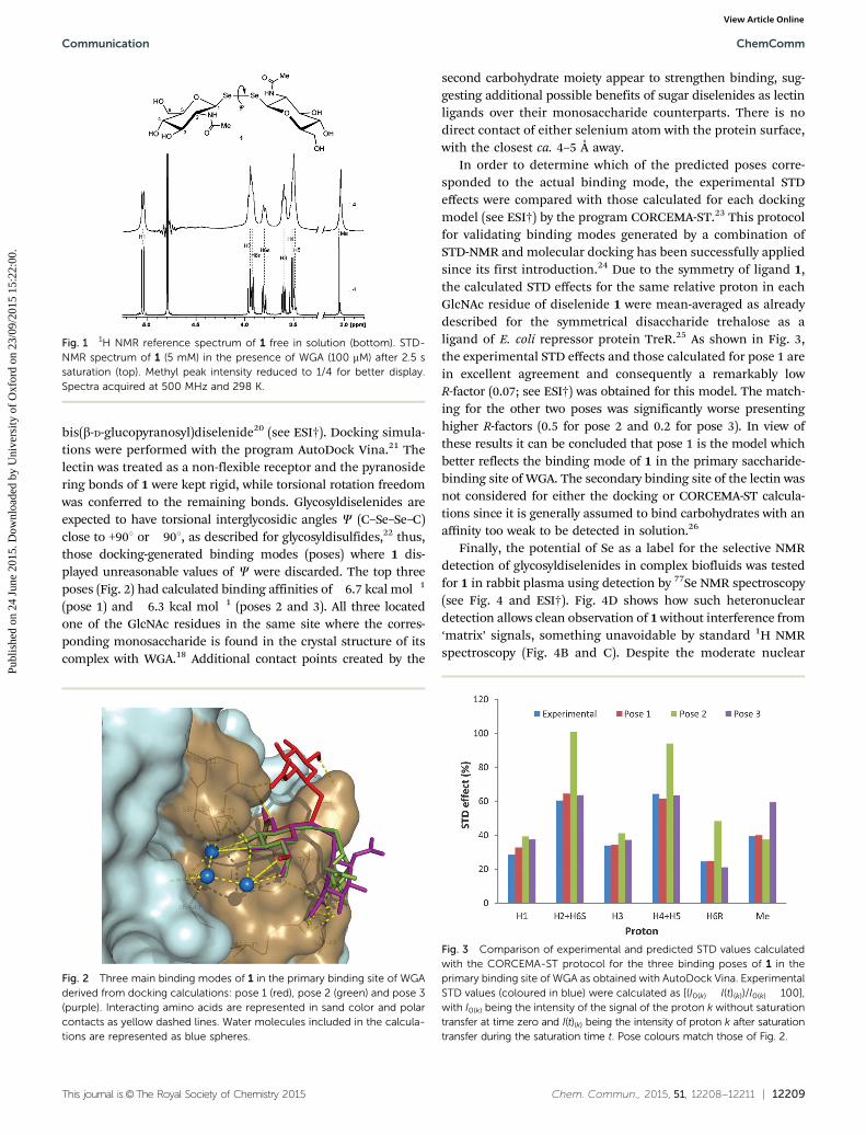
whose conformational flexibility can resemble that of glycosyl-disulfides, would also act as lectin ligands,6 with sufficientsensitivity for detection even in complex biological environments,such as blood. Given the emerging importance of O-GlcNAcylationin biology,11 we chose here to address this question by probingfor the first time the binding of glycosyldiselenides to lectins bySTD-NMR and molecular docking studies, as well as demon-strating the advantage of Se for the selective detection of suchcarbohydrates in complex biological fluids by 77Se NMRspectroscopy. The interaction of the widely-employed, GlcNAc-binding protein wheat-germ agglutinin (WGA) with modelligand probe bis(b-D-GlcNAc)diselenide 112 is reported and itsbinding mode determined by molecular docking, STD-NMRand CORCEMA-ST calculations. Among the NMR methodsemployed to identify and characterize the binding of ligands toproteins, the saturation transfer difference (STD) experiment13 isvery robust and has been widely employed to characterizecarbohydrate–protein interactions.14 When surveyed for bindingto WGA in vitro, 1 showed a clear STD effect (Fig. 1). Additionalexperimental evidence of binding was also demonstrated by boththe line broadening of the ligand resonances after addition ofthe receptor and from 1D transferred NOESY (Tr-NOESY) experi-ments (see ESI†). The equilibrium binding constant of theWGA : 1 complex was further determined by classical titrationof the ligand into the protein, following the changes in thelinewidths of the N-acetyl resonance.15 Using this method adissociation constant (KD) of 1.6 mM was obtained (see ESI†),similar to that determined for GlcNAc (KD = 2.2 mM) by otherclassical methods,15b suggesting representative binding is main-tained in the diselenide, at least with WGA.
Next, the theoretical prediction of the binding mode wasapproached by molecular docking,16 as described for the inter-action of WGA with GlcNAc and a number of its derivatives.17
The most recently reported WGA�b-GlcNAc X-ray crystallographicstructure18 was chosen and the receptor was prepared for thedocking study as already described.19 Ligand 1 was modelledstarting from the crystal structure of b-GlcNAc in complexwith the lectin18 and the crystal structure of the peracetylated
Department of Chemistry, Chemistry Research Laboratory, University of Oxford,
† Electronic supplementary information (ESI) available: Details on sample pre-paration, NMR experiments, molecular docking simulations and CORCEMA-STcalculations. See DOI: 10.1039/c5cc03952e‡ Present address: Fundacion MEDINA, Centro de Excelencia en Investigacion deMedicamentos Innovadores en Andalucıa, Avda. del Conocimiento 3, ParqueTecnologico de Ciencias de la Salud, E-18160 Armilla, Granada, Spain (I.P.-V.) andDepartment of Chemistry, University of Cambridge, Lensfield Road, CambridgeCB2 1EW, UK (O.B.).
bis(b-D-glucopyranosyl)diselenide20 (see ESI†). Docking simula-tions were performed with the program AutoDock Vina.21 Thelectin was treated as a non-flexible receptor and the pyranosidering bonds of 1 were kept rigid, while torsional rotation freedomwas conferred to the remaining bonds. Glycosyldiselenides areexpected to have torsional interglycosidic angles C (C–Se–Se–C)close to +901 or �901, as described for glycosyldisulfides,22 thus,those docking-generated binding modes (poses) where 1 dis-played unreasonable values of C were discarded. The top threeposes (Fig. 2) had calculated binding affinities of�6.7 kcal mol�1
(pose 1) and �6.3 kcal mol�1 (poses 2 and 3). All three locatedone of the GlcNAc residues in the same site where the corres-ponding monosaccharide is found in the crystal structure of itscomplex with WGA.18 Additional contact points created by the
second carbohydrate moiety appear to strengthen binding, sug-gesting additional possible benefits of sugar diselenides as lectinligands over their monosaccharide counterparts. There is nodirect contact of either selenium atom with the protein surface,with the closest ca. 4–5 Å away.
In order to determine which of the predicted poses corre-sponded to the actual binding mode, the experimental STDeffects were compared with those calculated for each dockingmodel (see ESI†) by the program CORCEMA-ST.23 This protocolfor validating binding modes generated by a combination ofSTD-NMR and molecular docking has been successfully appliedsince its first introduction.24 Due to the symmetry of ligand 1,the calculated STD effects for the same relative proton in eachGlcNAc residue of diselenide 1 were mean-averaged as alreadydescribed for the symmetrical disaccharide trehalose as aligand of E. coli repressor protein TreR.25 As shown in Fig. 3,the experimental STD effects and those calculated for pose 1 arein excellent agreement and consequently a remarkably lowR-factor (0.07; see ESI†) was obtained for this model. The match-ing for the other two poses was significantly worse presentinghigher R-factors (0.5 for pose 2 and 0.2 for pose 3). In view ofthese results it can be concluded that pose 1 is the model whichbetter reflects the binding mode of 1 in the primary saccharide-binding site of WGA. The secondary binding site of the lectin wasnot considered for either the docking or CORCEMA-ST calcula-tions since it is generally assumed to bind carbohydrates with anaffinity too weak to be detected in solution.26
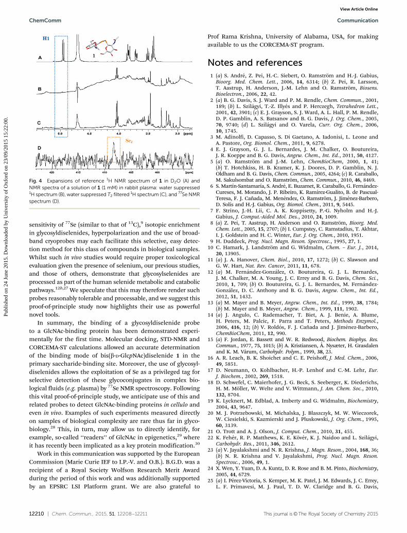
Finally, the potential of Se as a label for the selective NMRdetection of glycosyldiselenides in complex biofluids was testedfor 1 in rabbit plasma using detection by 77Se NMR spectroscopy(see Fig. 4 and ESI†). Fig. 4D shows how such heteronucleardetection allows clean observation of 1 without interference from‘matrix’ signals, something unavoidable by standard 1H NMRspectroscopy (Fig. 4B and C). Despite the moderate nuclear
Fig. 1 1H NMR reference spectrum of 1 free in solution (bottom). STD-NMR spectrum of 1 (5 mM) in the presence of WGA (100 mM) after 2.5 ssaturation (top). Methyl peak intensity reduced to 1/4 for better display.Spectra acquired at 500 MHz and 298 K.
Fig. 2 Three main binding modes of 1 in the primary binding site of WGAderived from docking calculations: pose 1 (red), pose 2 (green) and pose 3(purple). Interacting amino acids are represented in sand color and polarcontacts as yellow dashed lines. Water molecules included in the calcula-tions are represented as blue spheres.
Fig. 3 Comparison of experimental and predicted STD values calculatedwith the CORCEMA-ST protocol for the three binding poses of 1 in theprimary binding site of WGA as obtained with AutoDock Vina. ExperimentalSTD values (coloured in blue) were calculated as [(I0(k) � I(t)(k))/I0(k) � 100],with I0(k) being the intensity of the signal of the proton k without saturationtransfer at time zero and I(t)(k) being the intensity of proton k after saturationtransfer during the saturation time t. Pose colours match those of Fig. 2.
sensitivity of 77Se (similar to that of 13C),9 isotopic enrichmentin glycosyldiselenides, hyperpolarization and the use of broad-band cryoprobes may each facilitate this selective, easy detec-tion method for this class of compounds in biological samples.Whilst such in vivo studies would require proper toxicologicalevaluation given the presence of selenium, our previous studies,and those of others, demonstrate that glycosylselenides areprocessed as part of the human selenide metabolic and catabolicpathways.12b,27 We speculate that this may therefore render suchprobes reasonably tolerable and processable, and we suggest thisproof-of-principle study now highlights their use as powerfulnovel tools.
In summary, the binding of a glycosyldiselenide probeto a GlcNAc-binding protein has been demonstrated experi-mentally for the first time. Molecular docking, STD-NMR andCORCEMA-ST calculations allowed an accurate determinationof the binding mode of bis(b-D-GlcpNAc)diselenide 1 in theprimary saccharide-binding site. Moreover, the use of glycosyl-diselenides allows the exploitation of Se as a privileged tag forselective detection of these glycoconjugates in complex bio-logical fluids (e.g. plasma) by 77Se NMR spectroscopy. Followingthis vital proof-of-principle study, we anticipate use of this andrelated probes to detect GlcNAc-binding proteins in cellulo andeven in vivo. Examples of such experiments measured directlyon samples of biological complexity are rare thus far in glyco-biology.28 This, in turn, may allow us to directly identify, forexample, so-called ‘‘readers’’ of GlcNAc in epigenetics,29 whereit has recently been implicated as a key protein modification.30
Work in this communication was supported by the EuropeanCommission (Marie Curie IEF to I.P.-V. and O.B.). B.G.D. was arecipient of a Royal Society Wolfson Research Merit Awardduring the period of this work and was additionally supportedby an EPSRC LSI Platform grant. We are also grateful to
Prof Rama Krishna, University of Alabama, USA, for makingavailable to us the CORCEMA-ST program.
Notes and references1 (a) S. Andre, Z. Pei, H.-C. Siebert, O. Ramstrom and H.-J. Gabius,
Bioorg. Med. Chem. Lett., 2006, 14, 6314; (b) Z. Pei, R. Larsson,T. Aastrup, H. Anderson, J.-M. Lehn and O. Ramstrom, Biosens.Bioelectron., 2006, 22, 42.
2 (a) B. G. Davis, S. J. Ward and P. M. Rendle, Chem. Commun., 2001,189; (b) L. Szilagyi, T.-Z. Illyes and P. Herczegh, Tetrahedron Lett.,2001, 42, 3901; (c) E. J. Grayson, S. J. Ward, A. L. Hall, P. M. Rendle,D. P. Gamblin, A. S. Batsanov and B. G. Davis, J. Org. Chem., 2005,70, 9740; (d) L. Szilagyi and O. Varela, Curr. Org. Chem., 2006,10, 1745.
3 M. Adinolfi, D. Capasso, S. Di Gaetano, A. Iadonisi, L. Leone andA. Pastore, Org. Biomol. Chem., 2011, 9, 6278.
4 E. J. Grayson, G. J. L. Bernardes, J. M. Chalker, O. Boutureira,J. R. Koeppe and B. G. Davis, Angew. Chem., Int. Ed., 2011, 50, 4127.
5 (a) O. Ramstrom and J.-M. Lehn, ChemBioChem, 2000, 1, 41;(b) T. Hotchkiss, H. B. Kramer, K. J. Doores, D. P. Gamblin, N. J.Oldham and B. G. Davis, Chem. Commun., 2005, 4264; (c) R. Caraballo,M. Sakulsombat and O. Ramstrom, Chem. Commun., 2010, 46, 8469.
6 S. Martın-Santamarıa, S. Andre, E. Buzamet, R. Caraballo, G. Fernandez-Cureses, M. Morando, J. P. Ribeiro, K. Ramırez-Gualito, B. de Pascual-Teresa, F. J. Canada, M. Menendez, O. Ramstrom, J. Jimenez-Barbero,D. Solıs and H.-J. Gabius, Org. Biomol. Chem., 2011, 9, 5445.
7 F. Strino, J.-H. Lii, C. A. K. Koppisetty, P.-G. Nyholm and H.-J.Gabius, J. Comput.-Aided Mol. Des., 2010, 24, 1009.
8 (a) Z. Pei, T. Aastrup, H. Anderson and O. Ramstrom, Bioorg. Med.Chem. Lett., 2005, 15, 2707; (b) I. Cumpstey, C. Ramstadius, T. Akhtar,I. J. Goldstein and H. C. Winter, Eur. J. Org. Chem., 2010, 1951.
9 H. Duddeck, Prog. Nucl. Magn. Reson. Spectrosc., 1995, 27, 1.10 C. Hamark, J. Landstrom and G. Widmalm, Chem. – Eur. J., 2014,
20, 13905.11 (a) J. A. Hanover, Chem. Biol., 2010, 17, 1272; (b) C. Slawson and
G. W. Hart, Nat. Rev. Cancer, 2011, 11, 678.12 (a) M. Fernandez-Gonzalez, O. Boutureira, G. J. L. Bernardes,
J. M. Chalker, M. A. Young, J. C. Errey and B. G. Davis, Chem. Sci.,2010, 1, 709; (b) O. Boutureira, G. J. L. Bernardes, M. Fernandez-Gonzalez, D. C. Anthony and B. G. Davis, Angew. Chem., Int. Ed.,2012, 51, 1432.
13 (a) M. Mayer and B. Meyer, Angew. Chem., Int. Ed., 1999, 38, 1784;(b) M. Mayer and B. Meyer, Angew. Chem., 1999, 111, 1902.
14 (a) J. Angulo, C. Rademacher, T. Biet, A. J. Benie, A. Blume,H. Peters, M. Palcic, F. Parra and T. Peters, Methods Enzymol.,2006, 416, 12; (b) V. Roldos, F. J. Canada and J. Jimenez-Barbero,ChemBioChem, 2011, 12, 990.
15 (a) F. Jordan, E. Bassett and W. R. Redwood, Biochem. Biophys. Res.Commun., 1977, 75, 1015; (b) A. Kristiansen, Å. Nysæter, H. Grasdalenand K. M. Vårum, Carbohydr. Polym., 1999, 38, 23.
16 A. R. Leach, B. K. Shoichet and C. E. Peishoff, J. Med. Chem., 2006,49, 5851.
17 D. Neumann, O. Kohlbacher, H.-P. Lenhof and C.-M. Lehr, Eur.J. Biochem., 2002, 269, 1518.
18 D. Schwefel, C. Maierhofer, J. G. Beck, S. Seeberger, K. Diederichs,H. M. Moller, W. Welte and V. Wittmann, J. Am. Chem. Soc., 2010,132, 8704.
19 K. Lycknert, M. Edblad, A. Imberty and G. Widmalm, Biochemistry,2004, 43, 9647.
20 M. J. Potrzebowski, M. Michalska, J. Blaszczyk, M. W. Wieczorek,W. Ciesielski, S. Kazmierski and J. Pluskowski, J. Org. Chem., 1995,60, 3139.
21 O. Trott and A. J. Olson, J. Comput. Chem., 2010, 31, 455.22 K. Feher, R. P. Matthews, K. E. Kover, K. J. Naidoo and L. Szilagyi,
Carbohydr. Res., 2011, 346, 2612.23 (a) V. Jayalakshmi and N. R. Krishna, J. Magn. Reson., 2004, 168, 36;
(b) N. R. Krishna and V. Jayalakshmi, Prog. Nucl. Magn. Reson.Spectrosc., 2006, 49, 1.
24 X. Wen, Y. Yuan, D. A. Kuntz, D. R. Rose and B. M. Pinto, Biochemistry,2005, 44, 6729.
25 (a) I. Perez-Victoria, S. Kemper, M. K. Patel, J. M. Edwards, J. C. Errey,L. F. Primavesi, M. J. Paul, T. D. W. Claridge and B. G. Davis,
Fig. 4 Expansions of reference 1H NMR spectrum of 1 in D2O (A) andNMR spectra of a solution of 1 (1 mM) in rabbit plasma: water suppressed1H spectrum (B), water suppressed T2 filtered 1H spectrum (C), and 77Se NMRspectrum (D).
Chem. Commun., 2009, 5862; (b) S. Kemper, M. K. Patel, J. C. Errey,B. G. Davis, J. A. Jones and T. D. W. Claridge, J. Magn. Reson., 2010, 203, 1.
26 G. Bains, R. T. Lee, Y. C. Lee and E. Freire, Biochemistry, 1992,31, 12624.
27 (a) C. W. Nogueira and J. B. T. Rocha, Arch. Toxicol., 2011, 85, 1313;(b) A. P. Fernandes and V. Gandin, Biochim. Biophys. Acta, Gen. Subj.,2015, 1850, 1642.
28 S. Mari, D. Serrano-Gomez, F. J. Canada, A. L. Corbı and J. Jimenez-Barbero, Angew. Chem., Int. Ed., 2005, 44, 296.
29 J. A. Hanover, M. W. Krause and D. C. Love, Nat. Rev. Mol. Cell Biol.,2012, 13, 312.
30 (a) K. Sakabe, Z. Wang and G. W. Hart, Proc. Natl. Acad. Sci. U. S. A.,2010, 107, 19915; (b) B. A. Lewis and J. A. Hanover, J. Biol. Chem.,2014, 289, 34440.